Human Molecular Genetics, 1995, Vol. 4, No. 5 801-806
Sex-specific meiotic recombination in the
Prader-Willi/Angelman syndrome
imprinted region
Wendy P.Robinson* and Marc Lalande
1Department of Medical Genetics, University of British Columbia, Vancouver, BC, Canada and Institute of Medical Genetics, University of Zurich, Switzerland and 1Genetics Division, Children's Hospital, Department of Pediatrics, Harvard Medical School
and Howard Hughes Medical Institute, Boston, MA, USA
Received December 15, 1994, Revised and Accepted February 13, 1995
Meiotic recombination is a specifically timed and regulated process which does not occur randomly throughout
the genome, but tends to be clustered in 'hotspots'. There is extensive evidence that recombination rate is
influenced by chromatin conformation and that events are primarily initiated at gene promoter regions. In an
effort to determine the pattern of chromatin condensation and recombination at meiosis in an Imprinted
region, fine scale genetic mapping in the approximately 4 Mb Prader-Willi/Angelman syndrome deletion
region was undertaken. The results indicate that the male-female recombination ratio can vary significantly
over short regions. A male recombination hotspot is localized to between the 3' end of GABRA5 and D15S156,
which is adjacent to but outside the putative AS/PWS imprinted regions. In addition, a region of relatively
high recombination in females is observed between D15S128 and D15S97, which spans a domain of paternal
allele-specific transcription implicated in the Prader-Willi syndrome. It is inferred that the inactivation and
relative condensation of this latter region on the maternal chromosome occurs as a post-meiotic modification.
INTRODUCTION
In the human genome, I Mb of DNA is equivalent, on average,
to I cM (l% recombination) (I). Recombination does not
occur randomly, however, but tends to cluster in 'hotspots',
regions with relatively high recombination rates, separated by
stretches of diminished recombination. The human female
genetic map is about 50% longer than the male map; however,
this is not a generalized reduction and recombination is higher
in male meiosis for some specific intervals (2). There is
also a striking sexual dimorphism in observed cytogenetic
chromosome length, with human female pachytene
chromo-somes being approximately 50% longer than those in males
(3). It thus seems likely that the 50% higher recombination in
human females versus males is also a reflection of the more
condensed state of male chromosomes.
A link between chromatin conformation and meiotic
recomb-ination in humans is also suggested by the observation that
the recombinationally inactive sex chromosomes in male
spermatocytes are highly methylated, condensed and
transcrip-tionally inactive, whereas the X chromosomes in female
oocytes, which do participate in recombination, are euchromatic
and transcriptionally active (4). This has led to the suggestion
that the specific heterochromatization of the sex chromosomes
during meiosis in males is a means to prevent unwanted
recombination (5). Further evidence for an influence of
chro-matin structure on recombination is supported by the virtual
absence of any crossing-over in constitutive heterochromatin,
which is both highly condensed and devoid of transcribed
genes. In addition, hotspots for meiotic recombination in yeast
and mice (which may be strain or sex-specific) have repeatedly
been mapped to the sites of gene promoters or enhancers
(6-11). In yeast it appears that all meiotic recombination, general
and site-specific, may be initiated by double-strand breaks at
promoter regions (11). Evidence suggests that it is an open
chromatin conformation, and not gene transcription, that is
necessary for recombination to occur at these hotspots
(5,10,12,13).
Parental imprinting is the gamete-specific modification that
distinguishes the maternal and paternal chromosomes in higher
eukaryotes and may result in differential expression of genes
depending on parent of origin (14). It has been hypothesized
that male-female differences in recombinatory activity may
be related to differences in gene activity during meiosis of the
two sexes and specifically that regions activated in the
imprinting process are potential sites for recombination (15).
It has also been suggested that the process of homologous
pairing and recombination may play a role in the modification
of chromatin structure associated with imprinting (16). Other
evidence suggests that the full imprint, as reflected by
804 Human Molecular Genetics, 1995, Vol. 4, No. 5
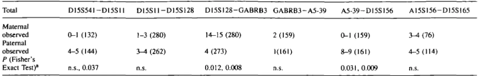
Table 1. A comparison of male to female recombination frequency. Number of observed recombinants and total informative meioses are indicated for each interval Total D15S541-D15S11 D15S11-D15S128 D15S128-GABRB3 GABRB3-A5-39 A5-39-D15S156 A15S156-D15S165 Maternal observed Paternal observed P (Fisher's Exact Test)0 0-1 (132) 4-5 (144) n.s., 0.037 1-3 (280) 3-4 (262) n.s. 14-15 (280) 4 (273) 0.012,0.008 2 (159) 1(161) n.s. 0-1(159) 3-4(76) 8-9(161) 4-5(114) 0.031,0.009 n.s.
T w o probabilities are given when the number of recombinants are variable: the first corresponds to the minimum possible difference and the second to maximum difference.
paternal early/maternal late replicating domain from SNRPN
to GABRA5 (44).
DISCUSSION
The 3 . 5 ^ Mb segment from D15S543 to D15S156 shows a
sex-averaged genetic distance of approximately 10 cM,
exceeding the 1 cM per 1 Mb average in the human genome.
As recombination is known in other organisms to be specifically
initiated at the sites of gene promoters, the rate of recombination
may be influenced by gene density. This could indicate that
the PWS/AS region contains a greater number of genes than
the human genome average of one gene per 40-50 kb (45).
As yet, only 13 genes have been identified within the common
PWS/AS deletion region including SNRPN, PAR-1, PAR-5,
IPW, PAR-7, PAR-4, PAR-2, OP2, HPEVE6A, GABRA3,
GABRA5, GABRG3, and P (23,24,33-36). Since gene
map-ping efforts have concentrated primarily in two regions; around
SNRPN and around the GABA receptor genes, there are likely
to be many more genes in this region as yet unmapped. A
possible correlation of recombination with gene density is,
however, confounded by the sex specificity of the observed
recombination. Clearly, there are secondary factors influencing
recombination rate, which may be due, in part, to the packaging
of the DNA into chromatin.
We have identified three domains of sex-specific meiotic
recombination within the PWS/AS imprinted region. Whether
or not sex-specific recombination is a characteristic of
imprinted regions, the recombination data yield interesting
information about chromatin state during meiosis. Comparison
with chromatin state inferred from methylation, replication
and gene expression in somatic cells can potentially indicate
which regions are actively altered by the imprinting process
after fertilization.
The region proximal to D15S11 shows an excess of paternal
versus maternal recombination. No imprinted genes have been
identified here, although the D15S9 locus (in the vicinity of
D15S543) shows hypomethylation at several sites on the
paternal chromosome (37). It is therefore most likely that this
region is relatively decondensed on the male chromosome
both during meiosis and in the mature embryo, with the
reverse true for the female chromosome. The broad region
(approximately 1400 kb) between D15S128 and D15S97/
GABRB3 exhibits relatively high female recombination and
is therefore presumed to be decondensed during maternal
meiosis. In contrast, the maternally inherited chromosome in
somatic tissues shows lack of transcription of the four known
imprinted genes within the PWS critical region (23,24) and
Table 2. Pairwise comparison of sex ratio between intervals (assumes equal number of informative meioses for each sex)
Maternal Paternal D15S54I-DI5S128 D15S128-A5-39 A5-39-D15S165 2-3 16-17 4 8 5 13
X2 = 12.66, df = 2, p <0.002 (minimizing differences between cells)
X2 = 14.78, df = 2, p <0.000l (maximizing differences between cells)
Intervals from Table 1 were lumped in pairs due to minimum sample size requirements.
is also late-replicating (26), suggesting a highly condensed
chromatin structure. Therefore it is hypothesized that it is not
until after fertilization that the maternal copy of this region is
actively converted to a more condensed 'imprinted' state.
Deletion of a putative 'imprinting control locus' located
proximal to SNRPN appears to result in abnormal inactivation
of the paternal PWS region (23). A lack of the maternal copy
of this 'control locus' has no phenotypic effect (23) and thus
this PWS 'imprinting control locus' seems not to be involved
in this post-zygotic inactivation of the maternal chromosome.
The most striking sex-specific deviations in recombination
rates are observed in the less than 500 kb region just distal to
GABRA5. This male hotspot of recombination is separated
from the highest region of maternal recombination by the
GABRB3 and GABRA5 gene cluster. It is intriguing that the
sharpest transition from high female/low male recombination
to very high male/very low female recombination would occur
in the vicinity of a domain which is also characterized by
sharp boundaries in transition of parent specific early/late
timing of replication during the S phase (44). It appears that
the basic genetic processes of recombination and replication
are under remarkably fine-tuned control within this segment
of the chromosome. Transcription, replication and
recombina-tion are not completely unrelated processes. They all involve
very specifically timed and programmed events which are
commonly initiated at the promoter regions of genes. This
presumably involves a preferred ability of the involved factors
to bind to, uncoil, and denature DNA at these sites. Although
this region has not been directly implicated in PWS or AS, it
has been shown that biparental inheritance of chromosome 15
is somehow necessary to establish the parent-of-origin specific
replication timing observed within the GABRB3 and GABRA5
cluster (44). It is possible that an altered chromatin state distal
to GABRA5 serves as a parent-of-origin 'tag' involved in
the establishment of the replication imprint of the proximal
adjacent region.
Human Molecular Genetics, 1995, Vol. 4, No. 5 805
Although it is intriguing that the PWS/AS region displays
sex-specific patterns of recombination, it is unlikely that that
all regions snowing sex-specific differences in recombination
are associated with genomic imprinting. There are numerous
male-female differences in genetic maps which may be due
to fundamental differences in how chromatin is organized in
oocytes versus spermatocytes, or due to differences in
expres-sion of genes involved in oogenesis and spermatogenesis. If,
however, significant differences in sex-specific recombination
are found to be one characteristic of regions showing
imprinting, this may provide a strategy for searching for
imprinted genes. Strong evidence suggests that there is at least
one maternally imprinted gene on chromosome 7 associated
with the Silver—Russell syndrome (46). The search for a
putative growth factor gene on chromosome 7 could possibly
be aided by screening regions showing the greatest
sex-dependent deviations in recombination rate.
In addition, an awareness of regions subject to significant
differences in male and female recombination rates are
import-ant for linkage studies. Recently, Fink et al. (47) reported
linkage of autosomal spastic paraplegia to the PWS/AS region
on 15q. Positive lod scores at zero recombination were observed
for D15S128 and D15S156 with negative lod scores for
D15S122 and D15S165. It is likely that this gene could be
more accurately localized within this region by accounting for
the strong sex-dependent nature of recombination, rather than
assuming sex-averaged recombination for all intervals. A
fine-scale analysis of recombination in other defined regions should
prove useful for providing more accurate data for linkage
studies, furthering our knowledge of the mechanism of
recomb-ination and gleaning useful information concerning the genomic
organization during meiosis.
MATERIALS AND METHODS
Based on the publicly available on-line CEPH mapping data (version I), parental haplotypes for markers from D15S11 to D15S165 were constructed. Segregation of these haplotypes was tracked within each family to identify crossover events. For most CEPH families (excepting the eight reference pedigrees), marker information was only available for D15SI1, DI5S97 and GABRB3. There is no observed recombination between D15S97 and GABRB3, and virtually all families were informative for one or both these markers. By typing for two polymorphisms (D15S541 and D15S542) proximal to DI5SI1 in a subset of CEPH families, additional crossovers between D15S541 and GABRB3 could be identified (these data have been submitted to the CEPH data base). The eight CEPH reference pedigrees had also been typed for additional markers within the deletion region distal to GABRB3 including DI5S2I9 and D15SI65. In addition, pedigrees which had already been typed for multiple proximal chromosome 15 markers for a previous study were used (32). A description of these pedigrees was given previously. In total, 49 crossovers were localized to this region (26 paternal and 23 maternal) from the original typing data. Since true double crossovers (i.e. excluding gene conversion events) are unlikely to occur over short distances, due to chiasma interference, it is unlikely that we have missed any crossover events using these markers.
Microsatellite polymorphisms were typed by the PCR using standard techniques. PCR amplification was performed on a Perkin Elmer Thermocyler with 30-32 cycles of I min at 94°C denaturation, 1 min at 55-57°C annealing and 1-2 min at 72°C extension temperatures. 0.5-3 ml of reaction was mixed with an equal volume of urea loading buffer (42% urea, 0.1 % xylene cyanol, 0.1% Bromphenol blue and 0.1% of 0.5 M EDTA) and directly loaded on to a 0.4 mm thick 6% polyacrylamide/50% urea gel. Visualization of bands was done by silver staining of the gels.
Information on all primers is available from the Genome Data Base; see also refs 33, 40 and 43). Microsatellite loci used are indicated in Figure 1. Only a few of these markers have been included in published genetic maps and this information is presented also in Figure 1. Recombination estimates
for D15S541 to D15S1I are based on the present. Physical map data and PWS/AS critical region information were inferred from several sources (23,28,40,43,48). Individuals showing a recombinant haplotype are indicated on Figure 1 by either the CEPH identification number or, in the case of previously published families (32), by a letter designation.
ACKNOWLEDGMENTS
We would like to thank Drs Huda Zoghbi and Stephen Wood for CEPH DNA samples, Drs Sue Christian and David Ledbetter for the D15S541, DI5S542 and D15S543 PCR primers, Dr Albert Schinzel for support, Dr R.Spiegel for DNA from some non-CEPH families, and Drs Carolyn Brown, and Joe McDermott for critical comments on the manuscript. This research was supported by British Columbia Health Research Foundation Grant #196(94-1) (WPR); Swiss National Foundation Grant 32.37798.93 (WPR); and NIH grant R01 NS30628 (ML). ML is an assistant investigator of the Howard Hughes Medical Institute.
REFERENCES
1. Donis-Keller, H., Green, P., Helms, C , Cartinhour, S., Weiffenbach, B., Stephens, K., Keith, T.P., Bowden, D.W., Smith, D.R., Lander, E.S., Botstein, D., Akots, G., Rediker, K.S., Gravius, T, Brown, V.A., Rising, M.B., Parker, C , Powers, J.A., Watt, D.E., Kauffman, E.R., Bricker, A., Phipps, P., Muller-Kahle, H., Fulton, T.R., Ng, S., Schumm, J.W., Braman, J.C., Knowlton, R.G., Barker, D.F., Crooks, S.M., Lincoln, S.E., Daly, MJ., and Abrahamson, J. (1987) A genetic linkage map of the human genome. Cell 51, 319-337.
2. NIH/Ceph Collaborative mapping Group (1992) A comprehensive genetic linkage map of the human genome. Science 258, 67-86.
3. Wallace, B.M.K. and Hulten, M.A. (1985) Meiotic chromosome pairing in the normal female. Ann. Hum. Genet. 49, 215—226.
4. Handel, M.A. and Hunt, P.A. (1992) Sex-chromosome pairing and activity during mammalian meiosis. Bioessays 12, 817-822.
5. McKee, B.D. and Handel, M.A. (1993) Sex chromosomes, recombination, and chromatin conformation. Chmmosoma 102, 71-80.
6. Nicholas, A., Treco, D., Schultes, N.P., and Szostak, J.W. (1989) An initiation site for meiotic recombination gene conversion in the yeast
Saccharomyces cerevisae. Nature 338, 35—39.
7. Sun, H., Treco, D., Schultes, N.P., and Szostak, J.W. (1989) Double-strand breaks at an initiation site for meiotic gene conversion. Nature
338, 87-90.
8 Stapleton, A. and Petes, T.D. (1991) The Tn3 p"-lactamase gene acts as a hotspot for meiotic recombination in yeast. Genetics 127, 39-51. 9. Shenkar, R., Shen, M., and Arnheim, N. (1991) DNase I-hypersensitivity
sites and transcription binding motifs within the mouse Ep* meiotic recombination hotspot. Mol Cell. Biol. 11, 1813-1819.
10. Wu, T-C. and Lichten, M. (1994) Meiosis-induced double-strand break sites determined by yeast chromatin structure. Science 263, 515-517. 11. Hsher-Lindahl, K. (1991) His and Her recombinational hotspots. Trends
Genet. 7, 273-276.
12. Esposito, M.S., Ramirez, R.M., and Bruschi, C.V. (1993) Recombinators, recombinases and recombination of genes of yeasts. Curr. Genet. 13. Thon, G., Cohen, A., and Klar, A.J. (1994) Three additional linkage
groups that repress transcription and meiotic recombination in the mating type region of Schizosaccharomyces pombe. Genetics 138, 29—38. 14. Barlow, D.P. (1994) Imprinting: a gametes point of view. Trends Genet.
10, 194-199.
15. Thomas, BJ. and Rothstein, R. (1991) Sex, maps, and imprinting. Cell 64, 1-4.
16. Hulten, M.A. and Hall, J.C. (1989) Proposed meiotic mechanism of genomic imprinting. Chromosomes Today 10, 157-162.
17. Ledbetter, D.H., Riccardi, V.M., Airhart, S.D., Strobel, RJ., Keenan, S.B., and Crawford, J.D. (1981) Deletions of chromosome 15 as a cause of the Prader-Willi syndrome. N. Engl. J. Med 304, 325-329.
18. Nicholls, R.D. (1994) New insights reveal complex mechanisms in genomic imprinting. Am. J. Hum. Genet. 54, 733—740.
19. Nicholls, R.D., Knoll, J.H M., Butler, M.G., Karam, S., and Lalande, M. (1989) Genetic imprinting suggested by maternal heterodisomy in non-deletion Prader-Willi syndrome. Nature 342, 281-285.
20. Robinson, W.P., Bottani, A., Yagang, X., BaJakrishman, J., Binkert, F., Machler, M., Prader, A., and Schinzel, A. (1991) Molecular, cytogenetic,
806 Human Molecular Genetics, 1995, Vol. 4, No. 5
and clinical investigations of Prader-Willi syndrome patients. Am. J.
Hum. Genet. 49, 1219-1234.
21. Knoll, J.H.M., Nicholls, R.D., Magenis, R.E., Graham, J M., Lalande, M., and Latt, S.A. (1989) Angelman and Prader-Willi syndromes share a common chromosome 15 deletion but differ in parental origin of the deletion. Am. J. Med. Genet. 32, 285-290.
22. Malcolm, S., Clayton-Smith, J., Nichols, M., Robb, S., Webb, T, Armour, J.A.L., Jeffreys, AJ., and Pembrey, M.E. (1991) Uniparental paternal disomy in Angelman's syndrome. Lancet 337, 694-697.
23. Sutcliffe, J.S., Nakao, M., ChrisUan, S., Orstavik, K.H., Tommerup, N., Ledbetter, D.H., and Beudet, A.L. (1994) Seletions of a differentially methylated CpG island at the SNRPN gene define a putative imprinting control region. Nature Genet. 8, 52-59.
24. Wevrick, R., Kerns, J.A., and Francke, U. (1994) Identification of a novel paternally expressed gene in Prader-Willi syndrome. Hum. Mol. Genet. 3, 1877-1882.
25. Kitsberg, D., Selig, S., Brandeis, M., Simon, I., Keshet, I., Dnscoll, DJ., Nicholls, R.D., and Cedar, H. (1993) Allele-specific replication timing of imprinted gene regions. Nature 364, 459—463.
26. Knoll, J.M., Cheng, S-D., and Lalande, M. (1994) Allele specificity of replication timing in the Angelman/Prader-Willi syndrome imprinted chromosomal region. Nature Genet. 6, 41—46.
27. Buiting, K., Dittrich, B., Grop\ S., Greger, V, Lalande, M., Robinson, W.P., Mutirangura, A., Ledbetter, D., and Horsthemke, B. (1993) Molecular definition of the Prader—Willi syndrome chromosome region and orientation of the SNRPN gene. Hum. Mol. Genet. 2, 1991-1994. 28. Buxton, J.L., Chan, C-TJ., Gilbert, H., Clayton-Smith, J., Burn, J.,
Pembrey, M., and Malcolm, S. (1994) Angelman syndrome associated with a maternal I5ql 1-13 deletion of less than 200 kb. Hum. Mol. Genet. 3, 1409-1413.
29. Meijers-Heijboer, E.J., Sandkuijl L.A., Brunner, H.G., Smeets, HJ.M., Hoogeboom, A.J.M, Deelan, W.H., van Hemel, J.O., Nelen, M.R , Smeets, D.F.C.M., Niermeijer, M.F., and Halley, D.JJJ. (1992) Linkage analysis with 15i)ll-ql3 markers shows genetic imprinting in familial Angelman syndrome. J. Med. Genet. 29, 853-857.
30. Clayton-Smith, J., Webb, T., Pembrey, M.E., Nichols, M., and Malcolm, S. (1992) Maternal origin of deletion I 5 q l l - I 3 in 25/25 cases of Angelman syndrome. Hum. Genet. 88, 376-378.
31. Wagstaff, J., Knoll, J.H.M., Glatt, K.A., Shugart, Y.Y., Sommer, A., and Lalande, M. (1992) Maternal but not paternal transmission of 15qll-13-linked nondeletion Angelman syndrome leads to phenotypic expression.
Nature Genet. 1, 291-294.
32. Robinson, W.P., Spiegel, R., and Schinzel, A.A. (1993) Deletion breakpoints associated with the Prader-Willi and Angelman syndromes ( 1 5 q l l - q l 3 ) are not sites of high homologous recombination. Hum.
Genet. 91, 181-184.
33. Malcolm, S. and Donlon, T.A. (1994) Report of the second international workshop on human chromosome 15 mapping 1994. Cytogenet. Cell
Genet. 67, 2-14.
34. Ozcelik, T, Sanjines, E., Robinson, W., Donlon, T., Lalande, M., Leff, S., Schinzel, A., and Francke, U. (1992) Small nuclear ribonucleoprotein N (SNRPN), the first expressed gene in the Prader-Willi syndrome critical region. Nature Genet. 2, 265-269.
35. Nakao, M., Sutcliffe, J.S., Durtschi, B., Mutirangura, A., Ledbetter, D.H., and Beaudet, A.L. (1994) Imprinting analysis of three genes in the Prader—Willi/Angelman region: SNRPN, E6-associated protein, and PAR-2 (D15S255E). Hum. Mol. Genet. 3, 309-315.
36. Greger, V., Knoll, J.H.M , Woolf, E., Glatt, K., Tyndale, R.F., DeLorey, T, Olsen, R., Tobin, AJ., Sikela, J.M., Nakatsu, Y., Brilliant, M.H., Whiting, PJ., and Lalande, M. The y-aminobutyric acid receptor y3 subunit gene (GABRG3) is tightly linked to the rx5 subunit gene (GABRA5) on human chromosome 1 5 q l l - q l 3 and is transcribed in the same orientation. Genomics, in press.
37. Driscoll, DJ., Waters, M.F., Williams, C.A., Zori, R.T., Glenn, C.C., Avidano, K.M., and Nichols, R.D. (1992) A DNA methylation imprint, determined by the sex of the parent, distinguishes the Angelman and Prader-Willi syndromes. Genomics 13, 917-924.
38. Dittrich, B., Buiting, K., and Horsthemke, B. (1994) Characterization of a methylation imprint in the Prader-Willi syndrome chromosome region.
Med. Genet. 1, 83.
39. Glenn, C.C., Porter, K.A., Jong, M.T.C., Nicholls, R.D., and Driscoll, DJ. (1993) Functional imprinting and epigenetic modification of the human SNRPN gene. Hum. Mol. Genet. 2, 2001-2005.
40. Mutirangura, A., Jayakumar, A., Sutcliffe, J.S., Nakao, M.. McKinney, MJ., Beaudet, A.L., Chinault, A.C., and Ledbetter, D.H.
(1993) A complete YAC contig of the Prader-Willi/Angelman chromosome region (I5ql I —q 13) and refined localization of the SNRPN gene. Genomics 18, 546-552.
41. Sinnett, D., Wagstaff, J., Glatt, K., Woolf, E., Kirkness, E.J., and Lalande, M. (1993) High-resolution mapping of the 7-aminobutyric acid receptor subunit P3 and a5 gene cluster on chromosome 15, and localization of breakpoints in two Angelman syndrome patients. Am. J. Hum. Genet. 52,
1216-1229
42 Beckman, J.S., Tomfohrde, J., Barnes, R.I., Williams, M., Broux, O., Richard, 1., Weissenback, J., and Bowcock, A.M. (1993) A linkage map of human chromosome 15 with an average resolution of 2 cM and containing 55 polymorphic microsatellites. Hum. Mol. Genet. 2, 2019— 2030.
43. Glatt, K., Sinnett, D., and Lalande, M. (1994) The human 7-aminobutync acid receptor subunit [33 and cc5 gene cluster in chromosome 15ql 1 —ql3 is rich in highly polymorphic (CA)n repeats. Genomics 19, 157-160. 44. LaSalle, J.M. and Lalande, M. Domain organization of allele-specific
replication within the GABRB3 gene cluster requires a biparcntal chromosome 15qll—q!3 contribution. Nature Genet., in press. 45. Fields, C , Adams, M.D., White, O., and Venter, J.C. (1994) How many
genes in the human genome? Nature Genet. 7, 345-346.
46. Kotzot, D., Schmitt, S., Bemasconi, F., Robinson, W.P., Lurie, I.W., Ilyana, H.t Mehes, K., Hamel, B.CJ., Otten, BJ., Hergersberg, M.,
Werder, E., Schoenle, E., and Schinzel, A. Uniparental disomy 7 in Silver-Russell syndrome and primordial growth retardation. Hum. Mol.
Genet., in press.
47. Fink, J.K., Wu, C-t.B., Jones, S.M., Sharp, G.B., Lange, B.M., Lesicki, A., Reinglass, T., Varvil, T., Otterud, B., Leppert, M. (1995) Autosomal dominant familial spastic paraplegia: Tight linkage to chromosome 15q.
Am. J. Hum. Genet. 56, 188-192.