Nephrol Dial Transplant (2010) 25: 862–867 doi: 10.1093/ndt/gfp577
Advance Access publication 5 November 2009
Original Articles
Is intra-operative blood flow predictive for early failure of
radiocephalic arteriovenous fistula?
François Saucy
1, Erik Haesler
2, Claude Haller
1, Sébastien Déglise
1, Daniel Teta
3and Jean-Marc Corpataux
11
Department of Thoracic and Vascular Surgery, Lausanne University Hospital, Lausanne, Switzerland,2Department of Vascular Medicine, Lausanne University Hospital, Lausanne, Switzerland and3Department of Nephrology, Lausanne University Hospital, Lausanne, Switzerland
Correspondence and offprint requests to: François Saucy; Email: Francois.saucy@chuv.ch
Abstract
Background. For over 50 years, radiocephalic wrist arte-riovenous fistulae (RCAVF) have been the primary and best vascular access for haemodialysis. Nevertheless, early failure due to thrombosis or non-maturation is a major complication resulting in their abandonment. This pro-spective study was designed to investigate the predictive value of intra-operative blood flow on early failure of pri-mary RCAVF before the first effective dialysis.
Methods. We enrolled patients undergoing creation of primary RCAVF for haemodialysis based on the pre-operative ultrasound vascular mapping discussed in a multidisciplinary approach. Intra-operative blood flow measurement was systematically performed once the anas-tomosis had been completed using a transit-time ultrasonic flowmeter. During the follow-up, blood flow was esti-mated by colour flow ultrasound at various intervals. Any events related to the RCAVF were recorded. Results. Autogenous RCAVFs (n = 58) in 58 patients were constructed and followed up for an average of 30 days. Thrombosis and non-maturation occurred in eight (14%) and four (7%) patients, respectively. The intra-operative blood flow in functioning RCAVFs was significantly high-er compared to non-functioning RCAVFs (230 vs 98mL/ min; P = 0.007), as well as 1 week (753 vs 228 mL/min; P = 0.0008) and 4 weeks (915 vs 245 mL/min, P < 0.0001) later. Blood flow volume measurements with a cut-off value of 120 mL/min had a sensitivity of 67%, specificity of 75% and positive predictive value of 91%. Conclusions. Blood flow <120 mL has a good predictive value for early failure in RCAVF. During the procedure, this cut-off value may be used to select appropriately which RCAVF should be investigated in the operation the-atre in order to correct in real time any abnormality.
Keywords: autogenous fistula; haemodialysis; maturation; thrombosis; vascular access flow
Introduction
For over 50 years, radiocephalic arteriovenous f istula (RCAVF) has demonstrated unquestionable superiority over all other types of vascular access for haemodialysis [1]. The main benefits of arteriovenous fistula (AVF) are better long-term patency and lower complication rate. Nev-ertheless, failure within the first 4 months after surgery oc-curs in 20% to 50% of AVF [1–11]. Early failure of AVF is defined as occlusion or insufficient development to sup-port haemodialysis within the first 3months [12]. Matura-tion of an AVF depends on flow volume and diameter of the vein used for cannulation. In this regard, Robbin et al. showed that the likelihood of sufficient growth was 95% if flow volume was 500mL/min or greater in association with a diameter of 4 mm [13]. In most successful AVF, these flow and size parameters are generally met within the first few weeks after construction [12].
Several pre-operative factors have been shown to predict the risk for early AVF failure. The main predictors are the diameters of the artery and vein. Pre-operative ultrasound (US) mapping is a useful tool for the accurate measure-ment of vessel diameter as a basis for planning operative strategy. Female gender and diabetes may also be risk fac-tors for early AVF failure. In the scoring system proposed by Lok et al. to predict failure to mature of AVF, the four main clinical predictors were age over 65, peripheral or coronary artery disease and white race [14]. During surgi-cal exploration, the operator should take into account the quality and size of the artery and the vein when deciding whether or not to create the fistula at the site chosen during pre-operative workup.
Few studies have evaluated intra-operative factors to predict early failure immediately after construction of an AVF. Two previous studies evaluated the predictive value of intra-operative blood flow volume. Wong et al. were un-able to demonstrate a link between intra-operative flow volume and the risk of early occlusion [10]. On the other
© The Author 2009. Published by Oxford University Press on behalf of ERA-EDTA. All rights reserved. For Permissions, please e-mail: journals.permissions@oxfordjournals.org
hand, Johnson et al. showed that the failure risk was sig-nificantly higher if flow volume was less than 170mL/min [15]. However, none of these studies were specifically aimed to assess the value of intra-operative blood flow measurements for the outcome of RCAVF. The purpose of this study was thus to investigate the value of intra-operative blood flow measurements in the prediction for early failure and/or adequate function of RCAVF in a cohort of patients about to start a chronic haemodialy-sis program in our institution.
Materials and methods
Between January 2005 and December 2007, we performed 120 first-time AVF including 67 RCAV, i.e. 56% of the total number. The study cohort included a total of 58 patients that benefited from a first-time RCAVF; nine patients were not included due to their previous vascular access by central venous catheter. The criteria for inclusion were indication for ini-tial haemodialysis requiring first-time AVF and complete follow-up data up to the time of the first functioning dialysis. Follow-up was discontin-ued at the time of the first functioning haemodialysis but never exceeded 8weeks. The functioning RCAVF was defined as the ability to use the fistula for dialysis with two needles and maintain a dialysis machine blood flow rate adequate for optimal dialysis (≥300mL/min) according to the Dialysis Consortium Study (DAC) criteria [16]. During the fol-low-up, the occlusions and the RCAVF unable to reach DAC's criteria were defined as non-functioning. In accordance with our department's fol-low-up policy for patients undergoing AVF for haemodialysis, all cases were reviewed weekly at multidisciplinary staff meetings attended by the nephrologist, angiologist, radiologist, vascular surgeon and dialysis nurse. All patients underwent a pre-operative vascular mapping by means of duplex ultrasound.
Pre-operative physical examination
The upper limb, shoulder and neck were examined to detect any sign of previous trauma, muscle atrophy, venous hypertension related to proximal vein obstruction or chronic ischaemia. Pulses were palpated, and an Allen test was performed. Auscultation was focused on the neck as well as the subclavian and axillary spaces.
Pre-operative duplex ultrasound examination
Duplex US was performed by experienced clinicians from the vascular medicine department using 5 to 12MHz linear transducers (Envisor and HDI 5000, Philips Medical Systems Switzerland, Gland, Switzerland; Vingmed System V, GE Medical Systems Switzerland, Glattbrugg, Swit-zerland). The arterial tree was assessed from the clavicle down to the wrist. Vessel diameters and anatomical variations were noted. Spectral Doppler waveforms were obtained at different levels. Presence of arterial plaque was recorded regardless of whether resulting narrowing was hae-modynamically significant or not. The extent of diffuse vessel wall calci-fication was subjectively evaluated based on the echogenicity of vessel walls and graded as follows: (i) no sign of diffuse calcification; (ii) mild calcification causing increased echogenicity of vessel walls and shadowing as observed in the transverse plane; (iii) moderate calcification causing not only shadowing but also Doppler attenuation resulting in incomplete luminal filling using colour mode; and (iv) severe calcification with high echogenicity, marked shadowing and Doppler attenuation causing colour to be nearly absent or to fill only very short segments of the artery, show-ing a patchy pattern.
The anteroposterior diameters of the superficial veins and distal part of the brachial veins were measured in the transverse plane after inflation of a pressure cuff positioned as on the arm to 80–90mmHg to interrupt venous return and induce maximal distal vein distension. Thrombosis of draining veins was detected by B-mode imaging and compression. Dopp-ler waveforms of the internal jugular and subclavian or axillary veins were analysed. Proximal venous obstruction was considered as highly un-likely in patients presenting the following three criteria: Doppler waves in both internal jugular and subclavian veins exhibiting clear respiratory and cardiac phasicity with the maximal velocity curve crossing the zero line;
absence of clinical signs such as distended veins and visible collaterals especially around the neck, the shoulder and along the trunk; and normal vein compressibility based on subjective assessment using the ultrasound probe in B-mode.
After completing the US examination, findings were used to map the anatomical features of vessels including not only course and diameter but also the location of any abnormalities (plaque, stenosis and occlusions), degree of wall calcification and characteristics of proximal venous flow. Using this map, the clinician proposed the most suitable site for AVF con-struction. The preferred minimal vein and artery diameters for RCAVF creation were 2.5 and 2.0mm, respectively [9]. However, these size pre-ferences were waived in some cases, e.g. young patients for whom slightly smaller vessels were sometimes used and patients with a high degree of arterial wall calcification for whom the artery size cut-off value was in-creased to 3.0mm.
Surgical technique
All procedures were performed under local or regional anaesthesia using a same standardized technique for creation of AVF, i.e. the minimal touch technique with limited dissection of the cephalic vein and radial artery. The artery was clamped using a coronary artery clamp, and an end-to-side anastomosis was made using a continuous 7.0 polypropylene suture. Su-turing was visualized through magnifying loupes (×3.5). At the operator's discretion, papaverine was used after clamp removal if there were signs of vasospasm. Systemic or local heparinization was not used. The patients were operated on by a single vascular surgeon or senior resident super-vised by this surgeon.
Intra-operative measurement of blood flow volume
Intra-operative measurement of blood flow volume was systematically performed immediately after the construction of the AVF. In the case of vasospasm requiring the use of papaverine, the measurement was delayed for 5min. Measurements were performed by placing a 3–4mm handheld flowprobe (MediStim, Oslo, Norway) around the draining vein 2 cm downstream from the anastomosis. Direct readings in mL/min were re-corded after stabilization for 30 s. At least three readings were made until consistent values were obtained usually within the first 5 min. Systolic and diastolic pressure was recorded concomitantly with flow volume measurements. All data were consigned to a dedicated study form imme-diately after the procedure.
Post-operative ultrasound
Post-operative US was performed at each follow-up examination at 1 week, 1month and just prior to the first RCAVF cannulation. Blood flow was assessed by measuring mean flow velocity and luminal diameter in separate segments of brachial, ulnar, pre-anastomotic and post-anastomot-ic radial arteries. Spectral Doppler and mean velocity were obtained with a Doppler angle at 60° and a sample size covering the entire lumen. Ac-cess blood flow was then calculated by adding pre-anastomotic radial to post-anastomotic radial flow, in case of retrograde flow in the latter. If post-anastomotic radial flow was antegrade, it was subtracted from panastomotic flow. During Doppler sonography, blood pressure was re-corded several times on the controlateral arm by means of an automated oscillometric manometer.
After access blood flow determination, feeding arteries and draining veins as well as the anastomosis were checked for the presence of steno-ses. First, a colour Doppler scan of arteries from the clavicle down to the anastomosis was performed while analysing any flow disturbance by spectral Doppler to obtain peak systolic velocity. The same was done for the veins, from the anastomosis up to the clavicle and internal jugular vein. Then, a precise B-mode image of each stenosis detected by colour Doppler was obtained, and minimal luminal diameter was measured. Study results were consigned to a dedicated form indicating access flow, mean systolic and diastolic blood pressures and a vascular map of the entire limb showing flow direction, vessel diameters and any stenosis with its peak systolic velocity and minimal luminal diameter.
Statistical analysis
Statistical analysis was performed using Stata version 9.2 (StataCorp, Col-lege Station, TX, USA). Results were expressed either as number of pa-tients (percentage) or as mean (± standard deviation). Between-group
comparisons were performed using the Wilcoxon test for quantitative data and Fisher's exact test for qualitative data. Threshold values were screened using the roctg function of Stata with 25mL/min blood flow volume in-tervals. Specificity, sensitivity and predictive values were computed to-gether with the respective 95% confidence intervals using the diatg function of Stata. A P value less than 0.05 was considered as statistically significant.
Results
Patient data including demographic characteristics, surgi-cal findings, vessel diameters and comorbid conditions are presented in Table 1. The mean age of patients was 63 ± 14.3years with 38% over the age of 70years.
The mean duration of follow-up was 30days (range, 15 to 45days; median, 21days). During follow-up, one patient (1.7%) died at home from causes unrelated to the RCAVF, and 12 patients (20.6%) presented events related to the RCAVF, i.e. either failure to mature in four cases or perma-nent occlusion in eight cases. All eight occlusions occurred within the first post-operative week (mean delay, 3.3days; range, 1-6 days). In all cases, alternative vascular access sites were created in more proximal locations including ip-silateral brachiocephalic AVF in 63% and placement of a prosthetic loop on the forearm in 37%. Placement of a per-manent dialysis catheter was necessary in four of the eight patients (50%) with inadequate RCAVF.
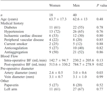
Table 2 compares patients who presented no events with patients who presented early failure due to occlusion or failure to mature. Female gender and low intra-operative flow volume were significantly associated with early fail-ure. To gain further insight on the impact of female gender, the overall data were compared by gender (Table 3). Find-ings showed that the artery diameter was significantly
smaller in women than men: 2.6±0.5 vs 3.0±0.6mm, re-spectively (P = 0.03). The post-operative blood flow after 4 weeks was also statistically higher in men (P = 0.02). Vessel wall calcifications were identified in three patients (16%) in the non-functioning RCAVF and six (15%) in the functioning RCAVF group.
Flow measurements in all patent RCAVF at 1 month af-ter the creation demonstrated a mean flow volume of 733± 296mL/min, i.e. a mean increase of 261% in comparison with intra-operative findings. Figure 1 shows that during the f irst post-operative month, the non-functioning RCAVF flow volume only increased by 142% from 101± 87 to 245±412mL/min, whereas blood flow of functioning RCAVF demonstrated an increase of 294% from 232 ± 194 to 915 ± 353mL/min. The increase of blood flow developed mainly during the first post-operative week. Indeed, in the functioning RCAVF, the blood flow increased by 224% from 232 ± 194 to 753 ± 269mL/min, and only by 125% from Weeks 1 to 4. These kinetics also occurred in the non-functioning RCAVF with an increase of 21% during the first post-operative week and only 7% from Weeks 1 to 4. Further analysis of flow volume data showed that the most sensitive cut-off value to discriminate functioning and non-functioning RCAVF was 120 mL/min. Nine of the 12 non-functioning RCAVF (75%) presented flow vo-lumes less than 120 mL/min as compared to only 15 of the 46 (32%) functioning RCAVF. A flow volume greater than 120mL/min exhibited good sensitivity and specificity, i.e. 67.4% (range, 51.9–80.5%) and 75% (range, 42.8– 94.5%), respectively. The positive predictive value of this parameter was 91.2%, i.e. excellent (range, 76.3–98.1%). The negative predictive value was, however, only 37.5% (range; 18.8–59.4%).
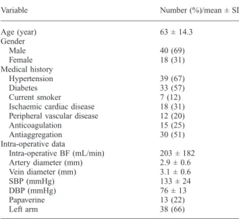
Table 1.Demographic features, comorbidities, surgical findings and vessel diameters in 58 patients who underwent first-time RCAVF
Variable Number (%)/mean ± SD
Age (year) 63 ± 14.3 Gender Male 40 (69) Female 18 (31) Medical history Hypertension 39 (67) Diabetes 33 (57) Current smoker 7 (12)
Ischaemic cardiac disease 18 (31) Peripheral vascular disease 12 (20) Anticoagulation 15 (25) Antiaggregation 30 (51) Intra-operative data Intra-operative BF (mL/min) 203 ± 182 Artery diameter (mm) 2.9 ± 0.6 Vein diameter (mm) 3.1 ± 0.6 SBP (mmHg) 133 ± 24 DBP (mmHg) 76 ± 13 Papaverine 13 (22) Left arm 38 (66)
BF, blood flow; SBP, systolic blood pressure; DBP, diastolic blood pressure. Continuous variables are presented as mean ± standard deviation (SD) and discrete variables as number with (percentage).
Table 2.Comparison of demographic features, surgical findings, vessel diameters and comorbid conditions in 58 patients with non-functioning (n = 12) and functioning (n = 46) RCAVF
Variable Non-functioning RCAVF Functioning RCAVF P value n 12 46 Age (years) 57.5 ± 16.5 64.4 ± 13.5 0.12 Female gender 7 (58) 11 (24) 0.03 Medical history Hypertension 10 (83) 29 (63) 0.3 Diabetes 10 (83) 23 (50) 0.06 Ischaemic cardiac disease 3 (25) 12 (26) 0.94 Peripheral vascular disease 3 (25) 9 (19) 0.68 Anticoagulation 4 (33) 11 (23) 0.51 Antiaggregation 8 (26) 22 (47) 0.25 Current smoker 1 (8) 6 (13) 0.1 Pre-operative data Artery diameter (mm) 2.8 ± 0.6 2.9 ± 0.6 0.58 Vein diameter (mm) 2.9 ± 0.6 3.2 ± 0.8 0.21 Arterial calcification 5 (18) 12 (46) 0.83 Intra-operative data BF (mL/min) 98 ± 65 230 ± 194 0.007 SBP (mmHg) 134 ± 25 137 ± 25 0.82 DBP (mmHg) 75 ± 13 78 ± 13 0.86 Other Papaverine 3 (25) 10 (22) 1.00 Left arm 6 (50) 32 (70) 0.31 Local anaesthesia 13 (72) 32 (69) 0.51 BF, blood flow; SBP, systolic blood pressure; DBP, diastolic blood pressure.
Discussion
Our findings demonstrate that the intra-operative measure-ment of blood flow immediately after construction of a RCAVF was useful to predict early AVF failure. Statistical analysis identified a cut-off value of 120mL/min for dis-criminating functioning and non-functioning RCAVF. Johnson et al. reported that a high intra-operative flow vol-ume defined as 320mL/min or greater was associated with a lower number of surgical revisions and longer access sur-vival regardless of gender, race or presence of diabetes. The same authors reported that an intra-operative flow rate of less than 170mL/min was correlated with a 56% risk for AVF failure within the first 50days after construction [15]. In their 50-patient series, Won et al. also showed that an intra-operative flow rate of less than 160mL/min was pre-dictive of early failure of RCAVF [17]. A recent study in-cluding a cohort of 109 patients undergoing vascular
access surgery for first-time haemodialysis showed that an intra-operative flow rate greater than 200mL/min was associated with better middle-term outcome in terms of re-quirement for revision and early patency rate [18].
In this study, follow-up was limited to the period imme-diately after construction of the RCAVF up to the first hae-modialysis. The goal was to eliminate the effect of puncture trauma associated with repeated puncture for ve-nous access. This trauma may cause stenosis and/or hae-morrhagic complications that are probably detrimental to AVF patency. Previous authors reported longer follow-up than ours but could not rule out the confounding effect of haemodialysis-related injury on their results. Moreover, the period prior to dialysis is crucial for AVF development that appears to be correlated with flow volume. Immedi-ately after AVF construction, flow volume increases rapid-ly, reaching a maximum within 4 to 12weeks [10,19–22]. Between 40% and 60% of the total increase in flow vol-ume occurs within 24 h after creation of the AVF [10,19–22]. For most fistulas located on the forearm, max-imum flow volume is reached within 4 weeks after con-struction [10,19]. The rate of the flow volume increase depends partly on the diameter of the vein lumen. In an earlier report, our group studied the size of the draining vein and observed that the lumen increased 86% after 1 week and 179% after 12weeks in comparison with the lu-men of the contralateral vein [22]. These results confirmed a previous study involving AVF located on the forearm that showed a 56% increase within the first 24h after con-struction to 123% at 12weeks in comparison with controls [10]. A fistula can be considered as mature or suitable to support haemodialysis when it allows insertion of two nee-dles and can provide sufficient blood flow, i.e. at least 350–450mL/min. Adequate flow volume is at least of 500mL/min for an AVF [23]. There is currently no con-sensus as to criteria for predicting the outcome of AVF de-velopment. Some authors wait as long as 3 to 4 months before declaring an AVF non-functioning [16,24].
Several studies have attempted to identify predictors of AVF development. Pre-operative vascular mapping that we have been using routinely for many years has been shown to maximize the opportunities for autogenous fistulas and lower the need for prosthetic fistulas [9,25]. However, a reliable correlation has not been established between pre-operative evidence of adequate artery and vein diameter and AVF development [10,26,27]. A recent study [28] in-dicated that measurement of vein compliance was a good predictor of AVF development, but this finding was not confirmed in two subsequent studies [21,29]. Post-opera-tive measurement of blood flow volume by duplex US has not been consistently shown to be useful in predicting AVF development [30,31]. Our data showed that the increase in flow volume during the first month after creation was 261% for functioning RCAVF as compared to only 142% for non-functioning RCAVF. This finding suggests that there is a good correlation between rapid flow volume increase in the first post-operative month and maturation of the RCAVF. Intra-operative and post-operative measure-ments showed that mean blood flow volume was signifi-cantly higher in functioning than non-functioning RCAVF. The intra-operative measurement of arterial dilation in
re-Table 3.Comparison of demographic features, comorbidities, surgical findings and vessel diameters in men (n = 40) and women (n = 18)
Women Men P value
n 18 40
Age (years) 63.7 ± 17.3 62.6 ± 13 0.48 Medical history
Diabetes 11 (61) 22 (55) 0.78 Hypertension 13 (72) 26 (65) 0.76 Ischaemic cardiac disease 6 (33) 12 (30) 0.80 Peripheral vascular disease 4 (22) 8 (20) 0.85 Current smoker 2 (25) 5 (12) 0.88 Anticoagulation 5 (27) 10 (40) 0.82 Antiaggregation 9 (50) 21 (52) 0.86 Blood Flow Intra-operative BF (mL/min) 142.7 ± 94.7 230.2 ± 205.6 0.13 Post-operative BF (mL/min) 513.6 ± 330.2 784.7 ± 278.9 0.02 Pre-operative diameter Artery diameter (mm) 2.6 ± 0.5 3.0 ± 0.6 0.03 Vein diameter (mm) 3.1 ± 0.7 3.1 ± 1.0 0.99 Other Papaverin 5 (27) 8 (20) 0.52 Left arm 11 (61) 27 (67) 0.77 BF, blood flow. Results are expressed as mean ± standard deviation or as number of subjects and (percentage). Statistical analysis by Wilcoxon test or Fisher's exact test.
Day 0 Week 1 Week 4 0 500 1000 1500 p<0.05 p<0.05 p<0.05
Blood flow (mL/min)
Fig. 1.Blood flow (in mL/min) in functioning (black box) and non-functioning RCAVF (white box).
sponse to reactive hyperaemia has also been identified as a predictor for AVF development with a sensitivity of 95%, a specificity of 61% and a positive predictive value of 87% [29]. In comparison, our results indicated that blood flow volume measurements with a cut-off point of 120mL/ min had a sensitivity of 67%, specificity of 75% and pos-itive predictive value of 91%.
In agreement with previous studies [32,33], our data also showed that failure of an RCAVF to mature was frequently associated with females. Comparison of vessel diameters according to gender in our cohort demonstrated that arter-ies were significantly smaller in women than in men. However, it should be underlined that our findings are in contradiction with two recent studies showing no signifi-cant gender difference in the pre-operative diameters of veins and arteries [32,34].
Failure of an AVF to mature can have many aetiologies [35]. The most frequent cause is neointimal hyperplasia typically occurring in the juxta-anastomotic vein. Hyper-plasia develops during the first post-operative month. In our study, all AVF occlusion occurred during the first post-operative week. Possible explanations include a defect in the surgical technique, poor vein quality or insufficient vessel diameter that was incorrectly estimated during US mapping. Pre-operative measurements should be made to detect possible sources of insufficient flow volume. US mapping could also decrease the functional maturation rate by the increase in performing complex procedures which were often secondary ones and by the use of smaller vein only detected by US but invisible during upper limb inspection [36]. Our vascular access improvement pro-gram provides for increasing the rate of autogenous vascu-lar access and decreasing early failure. Pre-operative US mapping, early referral to vascular surgeon and surveil-lance program are some of the measures to achieve these goals. Despite the implementation of the Disease Out-comes Quality Initiative (DOQI) guidelines in our institu-tion, 12 (20%) RCAVF failed to mature or occluded. The intra-operative surgical assessment of the vessels is the last possibility to choose the right strategy. To detect technical defects and identify poor artery or vein quality, Lin et al. proposed performing fistulography during the same proce-dure. The same authors pointed out that if a technical de-fect requiring surgical correction was not found, careful post-operative surveillance was necessary to allow early detection and prompt treatment of possible problems. They also proposed immediate construction of another fistula in diabetic patients presenting intra-operative flow volume less than 200mL/min [18]. Finally, Berman et al. recommended that RCAVF with an intra-operative flow rate of <120mL/min should either be immediately aban-doned for another site or, at a minimum, evaluated for an immediate revision [37]. In view of these results, we intend to find out whether intra-operative haemodynamic and anatomical criteria may directly impact on the deci-sion-making process in patients awaiting a functional AVF. Our study may have a direct implication for practical clinical issue. Indeed, the intra-operative blood flow mea-surement should be carried out after each procedure to get a start value. A blood flow <120mL/min has a good pre-dictive value of early failure. This implies that we should
aim for intra-operative investigations in order to look for a possible reason for this low blood flow. For instance, a fis-tulography with potentially a subsequent interventional procedure to re-establish a higher blood flow may be pro-posed. This hypothesis will be tested in a further study in our institution. In contrast, an intra-operative blood flow >120ml/min may be interpreted as a safe result in terms of future RCAVF maturation. In conclusion, the findings of this study indicated that the intra-operative measure-ment of blood flow is a useful tool to predict the outcome of maturation in first-time RCAVF. A simple transit-time device can be used to obtain immediate readings that can be used to adapt operative strategy and thus improve out-come. These findings should be associated with blood flow value measurement just before the first puncture which is obviously an important criterion for making a decision. Whether this can be used to modify the intra-operative strategy, thus reducing the failure-to-mature rate, needs to be investigated.
Acknowledgements. The authors thank Dr Pedro Marques-Vidal of the Institute of Social and Preventive Medicine, Lausanne for his support in biostatistics.
Conflict of interest statement. None declared.
References
1. Ascher E, Gade P, Hingorani A et al. Changes in the practice of an-gioaccess surgery: impact of dialysis outcome and quality initiative recommendations. J Vasc Surg 2000; 31 (1 Pt 1): 84–92
2. Gibson KD, Caps MT, Kohler TR et al. Assessment of a policy to reduce placement of prosthetic hemodialysis access. Kidney Int 2001; 59: 2335–2345
3. Golledge J, Smith CJ, Emery J et al. Outcome of primary radiocepha-lic fistula for haemodialysis. Br J Surg 1999; 86: 211–216 4. Hakaim AG, Nalbandian M, Scott T. Superior maturation and
pa-tency of primary brachiocephalic and transposed basilic vein arte-riovenous fistulae in patients with diabetes. J Vasc Surg 1998; 27: 154–157
5. Hodges TC, Fillinger MF, Zwolak RM et al. Longitudinal compari-son of dialysis access methods: risk factors for failure. J Vasc Surg 1997; 26: 1009–1019
6. Miller PE, Tolwani A, Luscy CP et al. Predictors of adequacy of ar-teriovenous fistulas in hemodialysis patients. Kidney Int 1999; 56: 275–280
7. Rocco MV, Bleyer AJ, Burkart JM. Utilization of inpatient and out-patient resources for the management of hemodialysis access compli-cations. Am J Kidney Dis 1996; 28: 250–256
8. Sedlacek M, Teodorescu V, Falk A et al. Hemodialysis access place-ment with preoperative noninvasive vascular mapping: comparison between patients with and without diabetes. Am J Kidney Dis 2001; 38: 560–564
9. Silva MB Jr, Hobson RW 2nd, Pappas PJ et al. A strategy for increas-ing use of autogenous hemodialysis access procedures: impact of pre-operative noninvasive evaluation. J Vasc Surg 1998; 27: 302–307; discussion 7-8
10. Wong V, Ward R, Taylor J et al. Factors associated with early failure of arteriovenous fistulae for haemodialysis access. Eur J Vasc Endo-vasc Surg 1996; 12: 207–213
11. Huber TS, Carter JW, Carter RL et al. Patency of autogenous and polytetrafluoroethylene upper extremity arteriovenous hemodialysis accesses: a systematic review. J Vasc Surg 2003; 38: 1005–1011 12. Asif A, Roy-Chaudhury P, Beathard GA. Early arteriovenous fistula
failure: a logical proposal for when and how to intervene. Clin J Am Soc Nephrol 2006; 1: 332–339
13. Robbin ML, Chamberlain NE, Lockhart ME et al. Hemodialysis ar-teriovenous fistula maturity: US evaluation. Radiology 2002; 225: 59–64
14. Lok CE, Allon M, Moist L et al. Risk equation determining un-successful cannulation events and failure to maturation in arterio-venous fistulas (REDUCE FTM I). J Am Soc Nephrol 2006; 17: 3204–3212
15. Johnson CP, Zhu YR, Matt C et al. Prognostic value of intraoperative blood flow measurements in vascular access surgery. Surgery 1998; 124: 729–737; discussion 37-8
16. Dember LM, Kaufman JS, Beck GJ et al. Design of the Dialysis Access Consortium (DAC) Clopidogrel Prevention of Early AV Fistula Thrombosis Trial. Clin Trials (London, England) 2005; 2: 413–422
17. Won T, Jang JW, Lee S et al. Effects of intraoperative blood flow on the early patency of radiocephalic fistulas. Ann Vasc Surg 2000; 14: 468–472
18. Lin CH, Chua CH, Chiang SS et al. Correlation of intraoperative blood flow measurement with autogenous arteriovenous fistula out-come. J Vasc Surg 2008; 48: 167–172
19. Lomonte C, Casucci F, Antonelli M et al. Is there a place for duplex screening of the brachial artery in the maturation of arteriovenous fistulas? Sem Dial 2005; 18: 243–246
20. Remuzzi A, Ene-Iordache B, Mosconi L et al. Radial artery wall shear stress evaluation in patients with arteriovenous fistula for he-modialysis access. Biorheology 2003; 40: 423–430
21. Yerdel MA, Kesenci M, Yazicioglu KM et al. Effect of haemody-namic variables on surgically created arteriovenous fistula flow. Ne-phrol Dial Transplant 1997; 12: 1684–1688
22. Corpataux JM, Haesler E, Silacci P et al. Low-pressure environment and remodelling of the forearm vein in Brescia-Cimino haemodialy-sis access. Nephrol Dial Transplant 2002; 17: 1057–1062 23. Dixon BS, Novak L, Fangman J. Hemodialysis vascular access
sur-vival: upper-arm native arteriovenous fistula. Am J Kidney Dis 2002; 39: 92–101
24. Beathard GA, Arnold P, Jackson J et al. Aggressive treatment of early fistula failure. Kidney Int 2003; 64: 1487–1494
25. Allon M, Robbin ML. Increasing arteriovenous fistulas in hemo-dialysis patients: problems and solutions. Kidney Int 2002; 62: 1109–1124
26. Lockhart ME, Robbin ML, Allon M. Preoperative sonographic radial artery evaluation and correlation with subsequent radiocephalic fistu-la outcome. J Ultrasound Med 2004; 23: 161–168; quiz 9-71 27. Mendes RR, Farber MA, Marston WA et al. Prediction of wrist
arte-riovenous fistula maturation with preoperative vein mapping with ul-trasonography. J Vasc Surg 2002; 36: 460–463
28. van der Linden J, Lameris TW, van den Meiracker AH et al. Forearm venous distensibility predicts successful arteriovenous fistula. Am J Kidney Dis 2006; 47: 1013–1019
29. Malovrh M. Non-invasive evaluation of vessels by duplex sonogra-phy prior to construction of arteriovenous fistulas for haemodialysis. Nephrol Dial Transplant 1998; 13: 125–129
30. Lin SL, Chen HS, Huang CH et al. Predicting the outcome of hemo-dialysis arteriovenous fistulae using duplex ultrasonography. J For-mos Med Assoc 1997; 96: 864–868Taiwan yi zhi
31. Robbin ML, Oser RF, Allon M et al. Hemodialysis access graft ste-nosis: US detection. Radiology 1998; 208: 655–661
32. Miller CD, Robbin ML, Allon M. Gender differences in outcomes of arteriovenous fistulas in hemodialysis patients. Kidney Int 2003; 63: 346–352
33. Prischl FC, Kirchgatterer A, Brandstatter E et al. Parameters of prog-nostic relevance to the patency of vascular access in hemodialysis patients. J Am Soc Nephrol 1995; 6: 1613–1618
34. Caplin N, Sedlacek M, Teodorescu V et al. Venous access: women are equal. Am J Kidney Dis 2003; 41: 429–432
35. Dixon BS. Why don't fistulas mature? Kidney Int 2006; 70: 1413–1422
36. Patel ST, Hughes J, Mills JL Sr. Failure of arteriovenous fistula mat-uration: an unintended consequence of exceeding dialysis outcome quality Initiative guidelines for hemodialysis access. J Vasc Surg 2003; 38: 439–445; discussion 45
37. Berman SS, Mendoza B, Westerband A et al. Predicting arteriove-nous fistula maturation with intraoperative blood flow measurements. J Vasc Access 2008; 9: 241–247
Received for publication: 23.1.09; Accepted in revised form: 6.10.09
Nephrol Dial Transplant (2010) 25: 867–873 doi: 10.1093/ndt/gfp565
Advance Access publication 4 November 2009
Daily online haemodiafiltration promotes catch-up growth in children
on chronic dialysis
Michel Fischbach, Joelle Terzic, Soraya Menouer, Céline Dheu, Laure Seuge and Ariane Zalosczic
Nephrology Dialysis Transplantation Children's Unit, University Hospital Hautepierre, Avenue Molière, 67098 Strasbourg, France Correspondence and offprint requests to: Michel Fischbach; E-mail: Michel.Fischbach@chru-strasbourg.fr
Abstract
Background. In children, growth can be used as a measur-able parameter of adequate nutrition and dialysis dose. De-spite daily administration of recombinant human growth hormone (rhGH), growth retardation remains a frequent problem in children on chronic dialysis. Therefore, we per-formed an observational prospective non-randomized study
of children on in-centre daily on line haemodiafiltration (D-OL-HDF) dialysis with the aim of promoting growth. Patients and methods. Mean age at the start of the study was 8 years and 3 months, and all children had been receiv-ing rhGH treatment for >12 months before enrolment. Mean follow-up time on D-OL-HDF was 20.5 ± 8 months (range, 11–39 months). Renal residual function was either <3 mL/
© The Author 2009. Published by Oxford University Press on behalf of ERA-EDTA. All rights reserved. For Permissions, please e-mail: journals.permissions@oxfordjournals.org