HAL Id: cea-01792056
https://hal-cea.archives-ouvertes.fr/cea-01792056
Submitted on 3 Mar 2020HAL is a multi-disciplinary open access archive for the deposit and dissemination of sci-entific research documents, whether they are pub-lished or not. The documents may come from teaching and research institutions in France or abroad, or from public or private research centers.
L’archive ouverte pluridisciplinaire HAL, est destinée au dépôt et à la diffusion de documents scientifiques de niveau recherche, publiés ou non, émanant des établissements d’enseignement et de recherche français ou étrangers, des laboratoires publics ou privés.
Encapsulated nanodiamonds in smart microgels toward
self-assembled diamond nanoarrays
Hugues Girard, Pierre Benayoun, Candice Blin, Adeline Trouvé, Celine
Gesset, Jean-Charles Arnault, Philippe Bergonzo
To cite this version:
Hugues Girard, Pierre Benayoun, Candice Blin, Adeline Trouvé, Celine Gesset, et al.. Encapsulated nanodiamonds in smart microgels toward self-assembled diamond nanoarrays. Diamond and Related Materials, Elsevier, 2013, 33, pp.32-37. �10.1016/j.diamond.2012.12.007�. �cea-01792056�
Encapsulated nanodiamonds in smart microgels toward self-assembled diamond nanoarrays.
By Hugues A. Girard,*a Pierre Benayoun a, Candice Blin a, Adeline Trouvé a, Céline Gesset a,
Jean-Charles Arnault a and Philippe Bergonzo a
a CEA LIST, Diamond Sensors Laboratory, CEA-Tech, F-91191 Gif-sur-Yvette, France
E-mail: [email protected]
Keywords: nanodiamonds, pNIPAM, smart microgels, self assembly, 2D arrays
Abstract:
We report on the successful attempt to synthesize thermoresponsive nanodiamonds hybrid
microgels through a straightforward method. Resulting microgels exhibit smart properties, as
the reversible collapsing of the structures when raising the temperature, with embedded
nanodiamonds. Thanks to the colloidal and smart properties of the microgels, nanoscaled 2D
self-assembled arrays of nanodiamonds with varying inter-particle distances were realized,
without any lithographic processes, as an illustration of the promising properties of these
novel diamond-based microgels. Such assemblies can constitute a first step toward a
bottom-up elaboration of innovative diamond-based devices.
1. Introduction
Among “hard” nanoparticles, nanodiamonds (NDs) have become during the last decade a very
promising tool for biological and technological applications, as they combine most of the
physical and chemical outstanding properties of the bulk material, at the nanometric scale. For
of high quantum yield which originate from color centers, as Nitrogen-Vacancy centers[1]
emitting in far-red/near infrared, perfectly adapted for biological labeling [2]. Such N-V
centers in nanodiamonds are also widely used as single photon emitters for quantum
application, where the small size of the individual diamond crystals helps to increase the
photon collection efficiency [3]. They have already been incorporated into photonic [4] and
plasmonic nanostructures, or used as the apex of an AFM tip toward scanning probe
magnetometry [5]. If their diamond cores exhibit the chemical and mechanical inertness of the
bulk material, their surface can be widely functionalized through the carbon-related groups
present on their surface: carboxylic acids, alcohols or alkenes provided by wet chemistry [6],
annealing [7,8] or plasma treatments [9,10] act as starting points toward amidation,
silanization or carbon-carbon couplings. Numerous chemical moities have thus already been
successfully conjugated with NDs, toward biological uses as nucleic acids[11,12], protein
[6,13], genes [11,12], chemotherapeutic drugs [14–18] or toward catalysis purposes[19].
Besides their use as luminescent probe with versatile surface chemistry, increasing interest
has also been recently devoted to NDs in the form of thin layers. This research field has
remarkably been boosted over the last years, notably because NDs can serve as seeds to
promote the growth of diamond layers by Chemical Vapor Deposition (CVD) [20]. This
so-called “nanoseeding” relies on the deposition of NDs as monolayers onto suitable substrates
(silicon, quartz, metals), with ideally densities above 1011 particles.cm-2. Several approaches
have been reported in the literature, like the dispersion by spin-coating of NDs suspended in
adapted solvents[21], the self-adhesion of NDs assisted by ultrasonic agitation[20,22], or the
electrostatic grafting of NDs onto a substrate driven by their surface chemistry[23,24]. By this
mean, ultrathin polycrystalline diamond layers with thickness below 100 nm can be grown
onto flat or even 3D substrates [25]. In some cases, the CVD growth is even not necessary, as
layers, as their versatile surface chemistry, their chemical resilience, their biocompatibility or
their optical properties, without inherent drawbacks of CVD grown layers, technologically
limited in terms of size and processing temperature. For instance, Chevallier et al. reported on
functionalized NDs multilayer deposited onto Surface Acoustic Wave (SAW) transducer
toward enhanced sensing properties[26]. For biological purpose, highly biocompatible NDs
thin layers were integrated with therapeutic molecules to realize localized drug-delivery
devices[27], or used to promote the formation of functional neuronal networks [28]. In
electrochemical field, Foord and coworkers reported on NDs deposited onto electrodes toward
improvement of Pt particles electrodeposition [29] or enhanced catalytic properties through
redox reactions occurring at the ND surface [30]. NDs layers can also be used as hard mask
for etching processes, when randomly dispersed onto a substrate [31].
All these applications developed from NDs thin layers rely on well-mastered techniques to
disperse them onto a substrate. Depending on the targeted properties of the film, NDs can be
now deposited as monolayer or multilayers up to micrometric thickness. However, if very
high densities can be achieved, the spatial ordering of the deposition still remains
uncontrolled. Formation of NDs arrays still requires lithographic and etching post-processes.
To our knowledge, no ordering of NDs has been yet reported in the literature, while
self-assembly into 2D or even 3D arrays is known to spontaneously occur for some “hard” or
“soft” nanoparticles. So-called “colloidal lithography”, based on the self-assembly of colloids
enables simple, fast and cost efficient production of 2D-ordered arrays, without the use of
complex equipment. For instance, long range order nanocrystal superlattices or colloid
crystals were synthesized from colloid suspensions of metallic [32] or oxides [33]
nanoparticles and various polymeric beads [34], mainly toward photonic, plasmonic or
Among the polymeric beads able to self-assemble themselves in 2D arrays[35,36], microgels
exhibit serious advantages and have been thoroughly investigated during the last decade [37–
41]. They are able to undergo conformational changes in response to external stimuli as
temperature[42], pH[43] or ionic strenght[44], which enable their self-assemblies in both
close-packed or periodic loosely packed arrays [35,45], with tunable separation distances, key
parameter for photonic, nanofuidic devices[46], biosensors or biomedical applications[47].
Furthermore, it has been shown that hybrids systems can be designed, by combining
microgels and nanoparticles[48]. Resulting composite material exhibits the swelling
properties of the polymer matrix and the optical, magnetic or catalytic properties of the
inorganic particle. In the last decade, such composite structures have been investigated and
hybrid systems hosting, for instance, magnetic iron oxide[49], gold[50,51], silica
nanoparticles[52] and quantum dots[53] have already been reported in the literature[54]. By
this mean, 2D-organized arrays were synthesized, for instance with hybrid particles
containing up to 20% wt% of ZnS, in which interparticles distance can be controlled[55], or
3D nanocrystal with gold hybrids with tunable spacings between gold cores ranging from 50
to 500 nm.
There is no example of hybrid smart microgels hosting NDs in the literature. Barras et al.
recently reported on surface functionalization of NDs with pNIPAM[56], but apparently the
study was led to validate a new grafting route using dopamine derivatives and no microgels
were shown. Encapsulation of NDs in smart microgels could be very promising for applied or
basic researches, and may offer new opportunities for NDs-based devices. Using the self
assembly properties of the microgels, it would give access to controlled spatial deposition of
NDs onto substrates, not accessible so far without cost and time consuming lithographic
processes. In this study, we choose to work with pNIPAM microgels, based on the monomer
the most widely studied water-swellable microgel systems[58]. We thus propose to report on
the synthesis of pNIPAM/NDs hybrid system, fully characterized by DLS, Zeta
measurements, and SEM analysis. Furthermore, as an illustration of the new possibilities
given by the pNIPAM encapsulation, and thanks to the colloidal properties of the microgels,
driven self assemblies into close-packed or loosely-packed 2D arrays of pNIPAM/NDs
hybrids were realized, exhibiting 2D organization on large scale of NDs.
2. Experimental
Materials & Chemicals
High Pressure High Temperature (HPHT) nanodiamonds (NDs) were purchased from Van
Moppes (Syndia® SYP 0-0.02: ND30 and Syndia® SYP 0-0.1: ND100). The NIPAM
N-isopropylacrylamide (NIPAM, 97%), the Potassium Persulfate (KPS, 99.99%), the Sodium
Hydroxide (NaOH, >97%), the N,N-methylenebisacrylamide (BIS, >99.5%) were purchased
from Sigma-Aldrich, and used as-received. Ultrapure water with a resistivity of 18.2 MΩ.cm
(25°C) was used in all experiments.
Preparation of microgels
The microgel preparation was based on the procedure described by Pelton and Chibante[57].
In this work, N-isopropylacrylamide (NIPAM) was used as the main monomer,
N,N’-methylene-bis-acrylamide as a cross-linker (BIS) and potassium persulfate (KPS) as an
initiator. The polymerization experiments were carried out in a reaction vessel equipped with
a magnetic stirrer. 0.681 g (6.10-3 mol) of NIPAM and 0.094 g (6.10-4 mol) of BIS were
introduced in 60 mL of degassed ultrapure water. For synthesis with nanodiamonds, NDs
were directly introduced in the 60 mL of water at a low concentration (see table 1). To
remove the oxygen, the solution was vigorously stirred for 20 min in the presence of nitrogen
flow. Then the temperature was raised to 70°C and 0.6 mL of KPS solution at 0.1M (6.10-5
speed was lowered to prevent the flocculation, and the reaction was left to proceed for 3 h. All
the microgel particles were then washed by several cycles of centrifugation–redispersion
using ultrapure water.
Preparation of 2D microspheres arrays
2D microspheres arrays were realized by dip-coating deposition (KSV NIMA) onto 2x1 cm²
silicon substrate, at a vertical speed of ca. 15 µm/sec, using a suspension of ND30 hybrid
microgel (50 mg/mL). The deposition process was performed in a thermostatic bath.
Plasma exposure
MPCVD parameters were tuned with a pressure of 40 mBar, a CH4/H2 ratio of 1/99. The
microwave power was adjusted to 1.1 kW in order to reach a temperature of 750 °C after 10
min measured in situ using an optical pyrometer (IRCON MODLINE 3 bi-chromatic).
Characterization
Hydrodynamic diameters and zeta potentials of the particles were measured using dynamic
light scattering (DLS) by a Malvern NanoZS at 173° angle. All experiments were performed
with the manufacturer calibration procedures. The average value of at least five measurements
was taken at a given condition. For temperature trends, samples were equilibrated for 2
minutes before measurement.
Electron microscopy imaging was performed using an in-lens Field Emission Scanning
Electron Microscope ZEISS Supra-40, operating with an acceleration voltage of 1 kV.
3. Results and discussion
3.1 Synthesis of NDs hybrids microgels
High Pressure High Temperature (HPHT) NDs were used in this study, exhibiting a mean
[23], exhibits a negative zeta potential of -45 mV at neutral pH and room temperature. NDs
suspended in a colloidal solution were introduced directly in the aqueous phase of the reaction
mixture, at a concentration of 50 µg/mL, before the addition of the other reagents, without any
further modification. After synthesis, the resulting microgels were separated and thoroughly
washed by successive centrifugation/redispersion cycles in pure water. Diluted suspensions
were then characterized by DLS measurements as shown on Fig. 1. For comparison, Fig. 1
also reports DLS measurements of ND30 colloidal suspension and “pure” microspheres
dispersed in water.
Fig.1 Hydrodynamic diameter distribution of as received ND30, “pure” microgels and ND30
hybrid microgels, recorded at 20°C.
As-received ND30 suspension exhibits hydrodynamic diameters centered on 30 nm. The
width of the size distribution peak and its associated polydispersity index (0.2) reveals that the
suspension is characterized by a scattered size distribution, coming from the production
method (milling). By contrast, “pure” pNIPAM microgels exhibit a narrower size distribution
peak, with a very low polydispersity index below 0.05, reflecting a quasi-monodisperse
diameter than “pure” microgels. From 531 nm for pNIPAM microgels, the hydrodynamic
diameter decreases to 255 nm when NDs were added to the synthesis. Note that besides a
chemical effect leading to a size reduction of the hybrid objects, a convoluted effect of the
scattering of both diamond core and polymeric shell may also affect DLS measurements.
However, the polydispersity index remains very low, still below 0.05. The fact that the
polydispersity index remains at a very low value for hybrid microgels highlights the
efficiency of the cleaning process. An inefficient rinsing, resulting in free NDs among the
microspheres, would have led to a higher polydispersity index. Furthermore, an adsorption of
NDs at the surface of the microgels is not consistent with the present observations, as it would
have, at the opposite, induced an increase of their size. Introduction of NDs in the reaction
mixture is however clearly at the origin of a severe modification of the pNIPAM microgels
structure, illustrated by the diameter decrease.
Fig.2 Evolution of the hydrodynamic diameters of « pure » pNIPAM microgels and ND30
hybrid microgels according to the temperature
pNIPAM microgels are known to undergo morphological modification according to an
temperature trends between 20 and 55 °C were thus realized. The variation of the
hydrodynamic diameters with respect to the temperature is shown in the fig. 2. Temperature
trends of “pure” and ND30 hybrid microgels are compared. For “pure” microgels, diameters
of 525 and 315 nm were recorded at 20 and 55°C respectively. From these minimum and
maximum diameters, a swelling ratio can be calculated, which is defined as the ratio between
the particle volume in collapsed and swollen state:
Rh(55°C) is the hydrodynamic radius at 55°C, and Rh(20°C) is the hydrodynamic radius in the
fully swollen state at 20°C.
“Pure” pNIPAM microspheres exhibit a swelling ratio of 0.22. For synthesis in which NDs
were added, temperature trend shows a decrease of the diameters from 259 to 154 nm at 20
and 55°C respectively. The corresponding swelling ratio exhibits a value of 0.21, very close
to the value obtained from “pure” microgels. At the same time, the transition between the
swollen and the collapsed states occurs roughly in the same temperature range, around 32°C.
Therefore, if the addition of NDs in the reaction mixture induces a size modification of the
resulting microgels, it does not affect significantly their swelling properties. A collapsing of
the microsphere with increased temperature still occurs, and in a reversible manner. Smart
properties of pNIPAM thus remain unaffected even when NDs are introduced in the reaction
Fig.3 SEM pictures of ND30 hybrid microgels deposited on a Si substrate
SEM characterizations were performed to check the structure of the microspheres. Fig. 3
shows the SEM pictures of microgels resulting from the addition of ND30. Considering only
the general morphology, the addition of NDs in the reaction medium does not affect the
spherical shape of the pNIPAM structures, which consist of independent and quasi
monodispersed objects. Compared to DLS measurements, the diameter undergoes a decrease,
with a mean value of 210 nm from the SEM pictures, which is attributed to the vacuum
environment of the SEM chamber. On the magnification, brighter spots can be revealed
within the microspheres, which can be attributed to NDs. It is important to note that these
brighter spots are only colocalized within microspheres, and not everywhere on the substrate.
This point supports that, at the deposition of the solution drop, NDs were attached to the
microspheres and not only left in the suspension among them. Obviously, at this stage, one
could not assert that all microspheres are filled with NDs, as brighter spots assigned to NDs
are not observable in all microspheres. However, HPHT nanodiamonds used in this study are
polydispersed, with NDs of sizes ranging from few nanometers to tens of nanometers. One
could not exclude that the smallest particles are hidden by the polymeric shell. More insights
polymer removal. Nevertheless, these SEM pictures confirm that hybrid objects were
synthesized, consisting in NDs/pNIPAM systems.
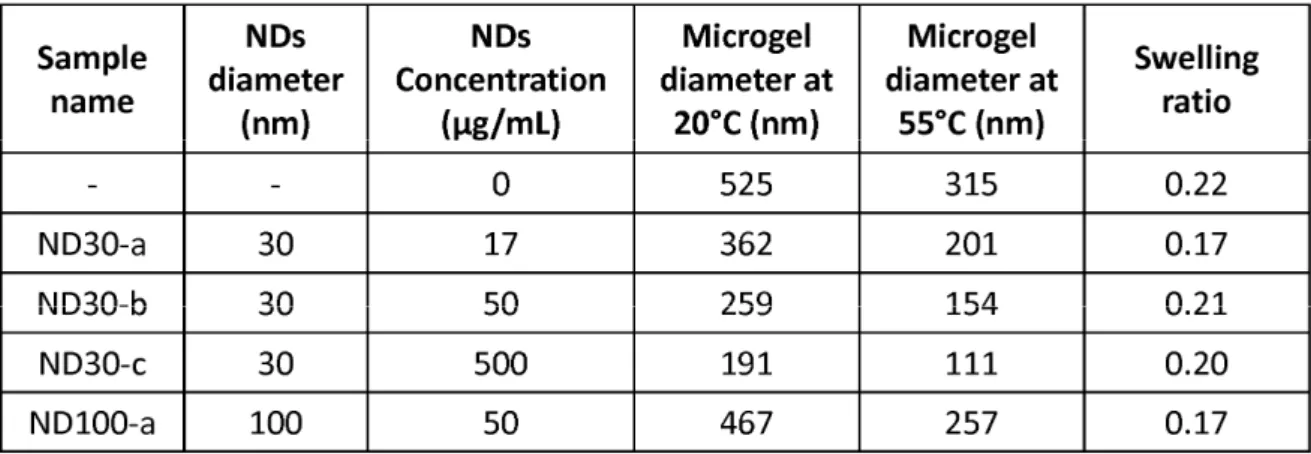
Table 1: Maximal, minimal diameters and swelling ratios of microgels according to the
diameters and the concentration of the NDs introduced during the synthesis.
Fig.4 Estimated surface area of NDs introduced in each synthesis according to the diameter of
the resulting microsphere in swollen state.
As NDs are introduced in the reaction mixture through the aqueous phase, without any
hybrid object have to be resolved. To gain further insights on the interactions between the
NDs and the monomers, additional synthesis were performed. The effect of three different
concentrations (from 17 to 500 µg/mL) and two different mean diameters (30 and 100 nm) of
NDs were studied. Concentrations, diameters at 20°C and 55°C and swelling ratios of the
resulting microgels are reported in the table 1.
Remarkable information concerns the evolution of the size of the microgels with respect to
the NDs initial concentration. If the swelling ratio is kept almost constant, both swollen and
collapsed states of the microgels undergo a strong decrease of their diameter for higher NDs
concentration. Besides, when the mean diameter of the NDs introduced in the reaction
mixture is raised from 30 to 100 nm, at similar concentration, the diameter of the resulting
microgels is also increased. Fig. 4 reports the diameter (at 20°C) of each microgel according
to the developed surface area of the NDs introduced in the synthesis. These surface areas were
calculated using a very simple approach, considering a spherical particle, their mean diameter
and their concentration in the solution. Note that the polydispersity of the NDs was taking into
account and represented by the error bars. An interesting point concerns the 100 nm
nanoparticles. With an initial concentration of 50 µg/mL in the aqueous phase, calculations
show that they finally develop a surface area lower than any 30 nm particles, whatever their
concentration. At the same time, resulting microgels exhibit a larger diameter than those
loaded with 30 nm, which is in agreement with the trend observed for the different
concentration of the 30 nm NDs. Thus, it confirms that the size of the resulting hybrid
microspheres depends on the total surface area of the NDs introduced in the reaction mixture,
and not only on their size.
As a first approach, it can be suggested that polymerization starts from the surface of the NDs,
as in core-shell structure. A proposed mechanism could be based on a seed precipitation
formation of pNIPAM spheres around a silica core, through the addition of C=C bonds on the
surface of the silica cores[59]. Here, no specific functionalization was applied to NDs, but
they natively carry on their surface carboxylic groups as well as a mix of sp2 and sp3 carbons
in aliphatic structures[60]. The contribution these C=C bonds may play in the polymerization
can be significant, as well as the interaction between the carboxylic groups and the NIPAM
monomer. Another experimental observation comes in support of this hypothesis, which
concerns the light opalescence during the synthesis. The appearance of the opalescence
somehow reveals when the microgels start to grow in the suspension. Here, depending on the
total NDs surface area introduced in the aqueous phase, a delay of the opalescence appearance
has been observed, which can reach 4h for the maximal concentration of NDs, i.e. the largest
surface area, compared to few minutes for 100 nm NDs, i.e. the lower surface area. Therefore,
a preliminary step of adsorption/grafting of the reagents on the NDs seems to occur, before
the growth of the microsphere, directly linked to the total surface area of NDs introduced in
the reaction mixture.
3.2 Self assembled layers of NDs hybrid microgels
Beyond the use of the hybrid microgels as “isolated objects” combining the smart properties
of the pNIPAM microgels and the assets of NDs toward biological or optical applications, our
novel hybrid systems can also be employed to build self-assembled layers of NDs on a
surface. Fig. 5a shows a typical deposit of ND30 hybrid microgels onto a silicon substrate by
dip-coating, from a diluted suspension of hybrid microgels in water, stabilized at 45°C. These
hexagonal ordered periodic loosely packed arrays result from a self-assembly of the
microspheres at the air/liquid interface, occurring at the dewetting of the substrate. Such
assemblies are driven by repulsive forces, resulting from electrostatic and steric interaction
water surface[35]. In our case, electrostatic interactions can be tuned through the zeta
potential of the particles, by playing on the temperature, while steric interactions are mainly
driven by the swelling or deswelling of the pNIPAM soft shell. Note that such ordered
monolayers have been easily obtained on large areas of several hundreds of square
micrometers. From the magnification (insert of fig. 5a), a mean separation distance between
two microspheres of c.a. 500 nm has been calculated with an average diameter of each
microspheres of 330 nm. The same sample was then exposed to H2/CH4 CVD plasma at 750
°C for few minutes, in order to burn all the polymeric phases. Indeed, we already showed that
such CVD treatment is efficient to remove polymeric layers on NDs [21,23]. Note that in
these conditions, NDs are preserved from any etching, a weak growth (few nm) can even
occur. As shown on Fig. 5b, after such plasma treatment, the organized microspheres have
given the place to organized NDs (bright spots), surrounded by a dark disk, which can be
reasonably attributed to a mark left by the microspheres on the silicon substrate when burned
away. The hexagonal order of the periodic loosely packed arrays is retained, with a similar
separation distance between the objects. From the magnification (insert of Fig. 5b), it appears
that almost all microspheres contained at least one ND, which confirms their encapsulation in
pNIPAM during the synthesis. Furthermore, the removal of the polymer matrix reveals the
scattered size distribution of the encapsulated NDs, ranging from few nanometers to tens of
nanometers, in agreement with DLS measurements (see table 2). This explains the difficulty
we had to observe NDs in all microspheres (see fig. 3), the smallest being hidden by the
Figure 5 SEM pictures of periodic loosely packed arrays of ND30 hybrid microgels (a)
before and (b) after CVD plasma. Scale bare : 1µm
The deposition parameters during the dip-coating, as the temperature, the concentration of the
suspension, or the dewetting velocity are key parameters to control the architecture of the
self-assembled monolayer. For instance, Fig. 6 exhibits close packed (Fig. 6a) and periodic loosely
packed (Fig. 6b) arrays obtained from the same suspension of ND30 hybrid microgels. To
achieve these close packed arrays, temperature of the suspension during the deposition was
decreased to 20°C, against 50°C for periodic loosely packed, reducing the overall surface
charge of each particle and enhancing their swelling, and the suspension was 20 times more
concentrated. Zeta potential measurements reported a strong decrease of the absolute value of
the particles charge when reducing the temperature, from -32 mV at 50°C to -18 mV at 20°C,
thus minimizing the electrostatic repulsion forces between the particles. Consequently, close
packed arrays were realized with a separation distance between centers of two microspheres
reduced from c.a. 600 nm to c.a. 335 nm. At the same time, all microspheres lie in contact to
each other, and the diameter of each microsphere has been reduced from c.a. 260 nm to c.a.
159 nm for loosely packed and closed packed arrays respectively. It can also be noticed that
for loosely packed arrays, a decrease of 5°C of the suspension temperature during the
compare Fig. 5a and Fig. 6b. This illustrates the versatility of the method to control the
separation distance between the microspheres organized in 2D arrays.
Figure 6: SEM pictures of (a) close packed (b) periodic loosely packed arrays of ND30
hybrid microgels. Scale bare : 1µm
3. Conclusions
In this study, we report on the synthesis of nanodiamonds hybrid microgels, characterized by
DLS and SEM. These new hybrid microgels exhibit similar smart properties than “pure”
pNIPAM microspheres, as the reversible collapsing of the structures when raising the
temperature, with almost equivalent swelling ratios and transition temperatures. Reaction
mechanisms at the origin of the loading the NDs is still under debate, but a correlation
between the surface areas of the NDs introduced in the reaction media and the size of the
resulting microspheres suggests a precipitation polymerization starting from the surface of the
NDs, through a prior adsorption of the reagents on the NDs surface.
Such hybrid microspheres are very promising for different fields of applications. The
encapsulation open up the doors to cost-limited nanoscaled NDs organization on a substrate
with a great flexibility in term of dimensions, toward useful technological or optical
applications. For instance, such assemblies can constitute a first step toward a bottom-up
elaboration of diamond-based devices, from simple mask lithography to 2D or 3D systems
toward catalysis, biological or optical uses. The synthesis of these novel ND-based hybrid
objects is thus very promising for the realization of innovative diamond-based devices.
Acknowledgements
The author would like to thank the financial support from the French Atomic Commission.
References
[1] S.-J. Yu, M.-W. Kang, H.-C. Chang, K.-M. Chen, Y.-C. Yu, J. Am. Chem. Soc. 127 (2005) 17604–17605.
[2] Y.-R. Chang, H.-Y. Lee, K. Chen, C.-C. Chang, D.-S. Tsai, C.-C. Fu, T.-S. Lim, Y.-K. Tzeng, C.-Y. Fang, C.-C. Han, H.-C. Chang, W. Fann, Nat Nano 3 (2008) 284–288.
[3] A.D. Greentree, I. Aharonovich, S. Castelletto, M.W. Doherty, L.P. McGuinness, D.A. Simpson, Opt. Photon. News 21 (2010) 20–25.
[4] L.A. Stewart, Y. Zhai, J.M. Dawes, M.J. Steel, J.R. Rabeau, M.J. Withford, Optics Express 17 (2009) 18044.
[5] L.P. McGuinness, Y. Yan, A. Stacey, D.A. Simpson, L.T. Hall, D. Maclaurin, S. Prawer, P. Mulvaney, J. Wrachtrup, F. Caruso, R.E. Scholten, L.C.L. Hollenberg, NATURE NANOTECHNOLOGY 6 (2011) 358–363.
[6] L.-C.L. Huang, H.-C. Chang, Langmuir 20 (2004) 5879–5884.
[7] Y. Liu, Z. Gu, J.L. Margrave, V.N. Khabashesku, Chemistry of Materials 16 (2004) 3924–3930.
[8] T. Petit, J.-C. Arnault, H.A. Girard, M. Sennour, P. Bergonzo, Physical Review B 84 (2011) 233407.
[9] H.A. Girard, J.C. Arnault, S. Perruchas, S. Saada, T. Gacoin, J.-P. Boilot, P. Bergonzo, Diamond Relat. Mater. 19 (2010) 1117–1123.
[10] H.A. Girard, T. Petit, S. Perruchas, T. Gacoin, C. Gesset, J.C. Arnault, P. Bergonzo, Physical Chemistry Chemical Physics : PCCP 13 (2011) 11517–11523.
[11] A. Alhaddad, M.-P. Adam, J. Botsoa, G. Dantelle, S. Perruchas, T. Gacoin, C. Mansuy, S. Lavielle, C. Malvy, F. Treussart, J.-R. Bertrand, Small (2011) n/a–n/a.
[12] M. Chen, X.Q. Zhang, H.B. Man, R. Lam, E.K. Chow, D. Ho, The Journal of Physical Chemistry Letters 1 (2010) 3167–3171.
[13] S.A. Dahoumane, M.N. Nguyen, A. Thorel, J.-P. Boudou, M.M. Chehimi, C. Mangeney, Langmuir 25 (2009) 9633–9638.
[14] H. Huang, E. Pierstorff, E. Osawa, D. Ho, Nano Letters 7 (2007) 3305–3314.
[15] E.K. Chow, X.-Q. Zhang, M. Chen, R. Lam, E. Robinson, H. Huang, D. Schaffer, E. Osawa, A. Goga, D. Ho, Science Translational Medicine 3 (2011) 73ra21.
[16] X.-Q. Zhang, M. Chen, R. Lam, X. Xu, E. Osawa, D. Ho, ACS Nano 3 (2009) 2609– 16.
[17] M. Chen, E.D. Pierstorff, R. Lam, S.-Y. Li, H. Huang, E. Osawa, D. Ho, ACS Nano 3 (2009) 2016–2022.
[18] K.-K.L. and W.-W.Z. and C.-C.W. and Y.-C.C. and C.-L.C. and Y.-S.L. and C.C. and J.-I. Chao, Nanotechnology 21 (2010) 315106.
[19] K. Goldberg, A. Krueger, T. Meinhardt, W. Kroutil, B. Mautner, A. Liese, Tetrahedron: Asymmetry 19 (2008) 1171–1173.
[20] O.A. Williams, O. Douh�ret, M. Daenen, K. Haenen, E. Osawa, M. Takahashi, Chem. Phys. Lett. 445 (2007) 255–258.
[21] E. Scorsone, S. Saada, J.C. Arnault, P. Bergonzo, J. Appl. Phys. 106 (2009) 14908.
[22] O. Shenderova, S. Hens, G. McGuire, Diamond and Related Materials 19 (2010) 260– 267.
[23] H.A. Girard, S. Perruchas, C. Gesset, M. Chaigneau, L. Vieille, J.-C. Arnault, P. Bergonzo, J.-P. Boilot, T. Gacoin, ACS Applied Materials & Interfaces 1 (2009) 2738– 2746.
[24] J. Hees, A. Kriele, O. a. Williams, Chemical Physics Letters 509 (2011) 12–15.
[25] H.A. Girard, E. Scorsone, S. Saada, C. Gesset, J.C. Arnault, S. Perruchas, L. Rousseau, S. David, V. Pichot, D. Spitzer, P. Bergonzo, Diamond and Related Materials 23 (2012) 83–87.
[26] E. Chevallier, E. Scorsone, H.A. Girard, V. Pichot, D. Spitzer, P. Bergonzo, Sensors and Actuators B: Chemical 151 (2010) 191–197.
[27] H. Huang, E. Pierstorff, E. Osawa, D. Ho, ACS Nano 2 (2008) 203–212.
[28] A. Thalhammer, R.J. Edgington, L.A. Cingolani, R. Schoepfer, R.B. Jackman, Biomaterials 31 (2010) 2097–2104.
[29] J. Hu, X. Lu, J.S. Foord, Electrochemistry Communications 12 (2010) 676–679.
[30] K.B. Holt, C. Ziegler, D.J. Caruana, J. Zang, E.J. Millan-Barrios, J. Hu, J.S. Foord, Phys. Chem. Chem. Phys. 10 (2008) 303–310.
[31] C. Lu, Y. Li, S. Tian, W. Li, J. Li, C. Gu, Microelectronic Engineering 88 (2011) 2319–2321.
[32] X.M. Lin, H.M. Jaeger, C.M. Sorensen, K.J. Klabunde, The Journal of Physical Chemistry B 105 (2001) 3353–3357.
[33] O.D. Velev, T.A. Jede, R.F. Lobo, A.M. Lenhoff, Nature 389 (1997) 447–448.
[34] P.M. Tessier, O.D. Velev, A.T. Kalambur, J.F. Rabolt, A.M. Lenhoff, E.W. Kaler, Journal of the American Chemical Society 122 (2000) 9554–9555.
[35] M. Horecha, V. Senkovskyy, A. Synytska, M. Stamm, A.I. Chervanyov, A. Kiriy, Soft Matter 6 (2010) 5980–5992.
[36] Y. Lu, M. Drechsler, Langmuir 25 (2009) 13100–13105.
[37] M. Ballauff, Y. Lu, Polymer 48 (2007) 1815–1823.
[38] R. Pelton, Advances in Colloid and Interface Science 85 (2000) 1–33.
[39] D. Duracher, A. Elaïssari, F. Mallet, C. Pichot, Langmuir 16 (2000) 9002–9008.
[40] R.F.S. Freitas, E.L. Cussler, Chemical Engineering Science 42 (1987) 97–103.
[41] M.J. Serpe, C.D. Jones, L.A. Lyon, Langmuir 19 (2003) 8759–8764.
[42] S. Pankasem, J.K. Thomas, M.J. Snowden, B. Vincent, Langmuir 10 (1994) 3023– 3026.
[43] T. Hoare, R. Pelton, Langmuir 22 (2006) 7342–7350.
[44] M. Shibayama, F. Ikkai, S. Inamoto, S. Nomura, C.C. Han, The Journal of Chemical Physics 105 (1996) 4358–4366.
[45] S.B. Quint, C. Pacholski, Soft Matter 7 (2011) 3735–3738.
[46] L. Lu, A. Eychmüller, Accounts of Chemical Research 41 (2008) 244–253.
[47] K. Wu, L. Shi, W. Zhang, Y. An, X.-X. Zhu, Journal of Applied Polymer Science 102 (2006) 3144–3148.
[48] M. Karg, T. Hellweg, Journal of Materials Chemistry 19 (2009) 8714.
[49] N. Shamim, L. Hong, K. Hidajat, M.S. Uddin, Colloids and Surfaces. B, Biointerfaces 55 (2007) 51–8.
[50] M. Das, N. Sanson, D. Fava, E. Kumacheva, Langmuir 23 (2006) 196–201.
[51] H.H. Park, T.R. Lee, Journal of Nanoparticle Research 13 (2010) 2909–2918.
[52] K. Zhang, J. Ma, B. Zhang, S. Zhao, Y. Li, Y. Xu, W. Yu, J. Wang, Materials Letters 61 (2007) 949–952.
[53] O. Tagit, N. Tomczak, A. Jafarpour, D. Jańczewski, M.Y. Han, G.J. Vancso, J.L. Herek, Nanotechnology 22 (2011) 265701.
[54] A.Z. Pich, H.-J.P. Adler, Polymer International 56 (2007) 291–307.
[55] A. Pich, J. Hain, Y. Lu, V. Boyko, Y. Prots, H.-J. Adler, Macromolecules 38 (2005) 6610–6619.
[56] A. Barras, J. Lyskawa, S. Szunerits, P. Woisel, R. Boukherroub, Langmuir (2011).
[57] R.H. Pelton, P. Chibante, Colloids and Surfaces 20 (1986) 247–256.
[58] B.R. Saunders, B. Vincent, Advances in Colloid and Interface Science 80 (1999) 1–25.
[59] Y.H. Deng, W.L. Yang, C.C. Wang, S.K. Fu, Advanced Materials 15 (2003) 1729– 1732.
[60] M. Chaigneau, H.A. Girard, J.-C. Arnault, R. Ossikovski, MRS Proceedings 1282 (2011).