PB Scho¨ttle SF Fucentese J Romero
Clinical and radiological outcome of medial
patellofemoral ligament reconstruction with
a semitendinosus autograft for patella
instability
Received: 8 October 2004 Accepted: 31 January 2005 Published online: 15 June 2005 Ó Springer-Verlag 2005
Abstract Background: Recurrent patellar instability is a common problem after dislocation. The med-ial patellofemoral ligament (MPFL) contributes 40–80% of the total medial restraining forces. This study assessed the clinical and radiological outcome after a follow-up of 4 years after linear MPFL reconstruction using an ipsilateral Semitendinosus tendon autograft. Study design and methods: 15 knees in 12 patients were examined with a mean of 47 months after linear reconstruction of the MPFL at a mean age of 30 years. 3 knees underwent previous surgery. 3 patients had mild trochlear dysplasia grade I or II, according to the clas-sification of Dejour. If preoperative tibial tuberosity-trochlear groove distance (TTTG) was more than 15 mm, patients underwent addi-tional medialisation of the tibial tuberosity (n=8) creating a similar postoperative situation for all pa-tients. All patients were available for a postoperative evaluation, which consisted of a subjective question-naire, the Kujala score, and the recording of potential patellar re-dislocation and apprehension. Patellar height and tilt was measured on plain radiographs. Postoperative
CT scans were performed in patients with an additional tibial tuberosity-transfer. Results: Postoperatively, one patient reported on recurrent bilateral redislocation. Physical examination however revealed no findings. Three knees presented with persistent patellar apprehension. Thirteen knees had improved sub-jectively after surgery. The mean Kujala score improved significantly from 55.0 to 85.7 points. The patel-lar tilt decreased significantly from 11.3° to 9.2°. Four knees had patella alta preoperatively, but only two at the latest follow-up visit. Previous surgery or additional trochlear dys-plasia had no influence on the clini-cal outcome. Conclusion: MPFL reconstruction improves clinical symptoms, reduces the patellar tilt substantially, and may correct pa-tella alta. Additional mild trochlear dysplasia did not compromise the outcome; however, this fact needs further attention in a larger study group.
Keywords Patella instability Æ Medial patellofemoral ligament Æ TTTG Æ Reconstruction Æ Tibial tuberosity DOI 10.1007/s00167-005-0659-0 P. Scho¨ttle Æ S. Fucentese J. Romero (&) Orthopedic Department,
University Hospital Balgrist Zurich, Forchstr. 340, CH, 8008 Zurich, Switzerland
E-mail: jose.romero@balgrist.ch Tel.: +41-1-3861275
Introduction
Persistent instability is common after patellar disloca-tion [23,29,32,34], particularly if the medial soft tissues become lax. A number of abnormalities such as effusion, retraction of the distal part of the vastus medialis obli-quus (VMO) muscle, bone bruises of the lateral femoral condyle or medial patellar facette, or increased T2 weighted signal in the patellotibial ligament near the tibial insertion on MR images [36] have been associated with an increased redislocation rate [39]. Several factors such as trochlear dysplasia, a lateralised tibial tuberos-ity, patella alta, or a weak quadriceps muscle have been made responsible for persisting patellar instability. Most operation techniques intend to realign the extensor mechanism, reducing the lateralisation of the patella when the quadriceps is loaded [5, 12,13, 24–26,29, 31,
41]. However, those procedures do not respect the underlying pathologic condition of patellar instability. Distal realignment procedures like the medial transfer of the tibial tuberosity have been shown to be of limited clinical success [1]. Proximal realignment procedures depend on quadriceps contraction to maintain the pa-tella in the trochlear groove. In contrast, intact passive stabilizers, like the medial patellofemoral ligament seem to play an important role regardless of the malalign-ment. Cadaver studies have proven the importance of the medial soft tissue in resisting lateral patellar motion [10,12,19,29]. The medial patellofemoral ligament as a primary stabilizer of the patella contributes 40–80% of the total medial restraining force [7, 19,29].
Reconstruction of the medial patellofemoral ligament has been advocated by several authors for patellofemoral
instability as it seems more effective than proximal or distal realignment procedures [20, 28, 35, 38, 39]. This retrospective study analyses the results of recon-struction of the medial patellofemoral ligament using a Semitendinosus autograft.
Materials and methods Indication
The indication for surgery included patients with two or more patellar dislocations or one dislocation and a persistent apprehension sign. In the clinical examination, all patients had an increased lateralisation of the patella combined with a medial tenderness due to the insuffi-cient medial patellofemoral ligament. All patients underwent reconstruction of the medial patellofemoral ligament using an ipsilateral Semitendinosus autograft. Patients who presented with a TTTG more than 15 mm in the axial scan [3,28] underwent medialisation of the tibial tuberosity (Fig.1) for creating a postoperative normal TTTG in all patients.
Surgical technique
The proximal one-third aspect of the patella is exposed through a 1–2 cm long incision (Fig.2, A) and a bony groove is performed. Two suture anchors carrying No. 3 non-absorbable braided suture are placed into that groove. The adductor tubercle is exposed through a 2 cm long incision (Fig.2, B) and a guide wire is placed across the epicondyles. A hole of 6 mm diameter and 2.5 cm depth is drilled over the guide wire. Then, the
Fig. 1 The tibial tuberosity-trochlear groove distance (TT-TG): distance (arrow) between the deepest point of the trochlear groove (TG) and the most prominent point of the tibial tuberosity (TT) in a perpendicular reference to a tangent to the posterior condyles
Fig. 2 The incisions used for the MPFL reconstruction at a left knee: A incision over the superomedial patellar border; B incision over the adductor tubercle; C incision over the pes anserinus to harvest the Semitendinosus-graft
Semitendinosus tendon is harvested through an addi-tional 2–3 cm long oblique incision (Fig. 2, C). The anserine bursa is incised and the tendon of the Semi-tendinosus muscle harvested with the tendon stripper. Muscle fibres are removed from the proximal aspect of the harvested tendon and the proximal end is tubed using 4–0 absorbable non-braided sutures. Krackow stitches with No. 0 absorbable-braided sutures are used for both ends of the tendon. The graft is then folded in half, and its centre portion is sutured to the anchors at the patella. The two free endings of the Semitendinosus graft are pulled into the drill hole at the adductor tubercle by removing the guide wire through the medial epicondyle. Tensioning of the graft is performed with the patella aligned in the trochlear groove throughout entire range of motion of the knee. An absorbable soft tissue interference screw is then inserted into the drill hole at the adductor tubercle (Fig. 3). Subcutaneous tissues and skin are approximated in the normal fashion.
Postoperatively, the patient is placed in a knee immobiliser and weight bearing is allowed no more than half-body weight for 6 weeks. In the first two weeks, passive range of motion up to 60° is performed with the physiotherapist. After two weeks leg raising and quad-riceps setting exercises are instituted, and range of
motion is increased up to 90°. After four weeks, range of motion is increased with no restriction. Full activity is permitted after 6 months if full range of motion and normal quadriceps strength is achieved. Postoperative regime did not alter, if an osteotomy of the tibial tuberosity was performed.
Patients and methods
Between January 1998 and November 2001, recon-struction of the medial patellofemoral ligament for patellar instability was performed in 15 knees in 12 pa-tients (8 female, 4 male) by the senior author. The average age at surgery was 30.1 years (range, 19 to 36 years). Eight operations were performed on the left knee and 7 on the right knee (3 bilateral). Preoperatively 2 patients reported on recurrent subluxation after initial dislocation, while all other patients had recurrent full dislocation. Four patients had had traumatic disloca-tions, while 11 suffered of patellar instability without initial trauma. Three patients (B.R., B.S., P.M.-left) had had previous surgery, such as lateral release, medial transfer of the tibial tuberosity and shortening of the vastus medialis obliquus muscle.
Preoperative physical examination consisted of the apprehension sign, retropatellar crepitus and pain on patellar compression. Complete history of dislocation and subluxation was recorded. The Kujala knee score was recorded pre- and postoperatively [33]. In addition, a subjective questionnaire [20] was completed postop-eratively. Radiographic examination consisted of a straight lateral and axial view in all patients. CT scan was performed in all cases preoperatively, and postop-eratively only in those patients, who had undergone additional transfer of the tibial tuberosity.
The trochlear bump, the trochlear depth and the crossing sign as qualitative and quantitative signs for trochlear dysplasia as well as the Caton index were measured on the straight lateral x-rays [8, 9, 17, 18]. Patellar tilt was measured on axial views.
The TTTG was measured on the computed tomog-raphy images as described by Goutallier (Fig.1). Pre-and postoperative data obtained from the radiographs were compared using the Man-Whitney-U test. The significance level was set at 0.05. The mean follow up was 47.5 months (range, 24 to 70 months).
Results
The subjective assessment revealed, that 13 of 15 knees had improved after surgery. 8 knees (53%) had excellent results, 5 (33%) had a good and 2 (13%) had a fair result. The fair results were in both knees of a patient who was on treatment for schizoid psychosis. Overall
Fig. 3 Attachment of the Semitendinosus graft by two sutures anchors at the superomedial border of the patella and tendon to bone tunnel fixation by an interference screw at the adductor tubercle
86% of the patients believed that the procedure had improved their knee. The objective findings of the knees did not correlate with the patients described discomfort. Preoperatively, 14 of 15 knees had a positive appre-hension sign, which remained positive in 3 knees of 2 patients postoperatively, including the above-mentioned patient (N.G.). That particular patient complained of severe pain during normal activities of daily living, but felt that her knees had improved since the operation. No patient had an abnormal medial tenderness, medial patellar glide or pain with patellar compression against the trochlear groove (crepitus).
The mean Kujala score improved statistically signif-icant from 53.3 points (range, 31 to 76 points) to 85.7 points (range, 55 to 100 points) at latest follow-up (p<0.001).
The mean patellar tilt deceased statistically significant from 11.4° (range, 4° to 18°) preoperatively to 8.5° (range, 2° to 16°) at latest follow up (p=0.03). The mean preoperative Caton ratio averaged 1.14 (range 0.92 to 1.3) with 4 cases of patella alta (ratio>1.2), while at the most recent follow up it averaged 1.04 (range 0.8 to 1.26) with 2 remaining cases of patella alta (p=0.258).
Eight knees with preoperative TTTG of more than 15 mm had a medial transfer of the tibial tuberosity. In those patients (marked with * in Table1) the mean preoperative TTTG decreased from 19.1 mm (range, 16 to 24 mm) preoperatively to 11.8 mm (range, 9 to 14 mm) postoperatively (p<0.001). The other patients (n=7) had a mean preoperative TTTG of 13.4 mm (range, 12 to 14). There was no significant difference in any outcome parameter between the patients with or without transfer of the tibial tuberosity.
Three patients had a trochlear dysplasia (marked with # in Table 1) according to the criteria of Dejour [18]. They also showed no difference with respect to clinical and radiological outcome.
Discussion
Non operative treatment of first time dislocation has a redislocation rate of 14 to 44% [11, 15, 16, 30]. Distal realignment procedures and the lateral retinacular release for the treatment of instability have a success rate of only 65% [1, 4, 6]. The role of the medial patellofe-moral ligament as the primary ligamentous restraint against lateral patellar displacement has been described by several authors [7, 12, 13, 19, 29]. Cadaver studies have proved that the MPFL acts as restraint against lateral forces [7,19, 29]. A tear of the medial patellofe-moral ligament has been found in 8 of 10 cases of experimentally performed cadaveric patellar dislocations [7], and in 15 of 16 cases in an in vivo study [39]. In a cadaver study Davis and Fithian have shown that an insufficient medial patellofemoral ligament is a critical factor for an asymptomatic patella to become unstable if any predisposition is present [16]. The reconstruction of the medial patellofemoral ligament, which restores nor-mal length and stiffness of the medial soft tissue, must be the aim of an evident procedure. As a consequence, Hautamaa proved the repair of the medial patellofe-moral ligament alone to be sufficient restoring lateral mobility within normal values [29]. Success rates of procedures which restore the medial restraints using different types of grafts and techniques ranges between 83% and 93% [2,20,21,27,37]. To our knowledge, this is the first outcome report of a minimum 2 year clinical and radiological follow up after reconstruction of the medial patellofemoral ligament, using Semitendinosus autograft only. The technique which reconstructs the torn or elongated medial patellofemoral ligament with a Semitendinosus graft was first described by Erasmas [22] and treats the primary pathoanatomical lesion of patellar dislocation. The subjective assessment, the Kujala score and objective signs improved in all patients. Only one
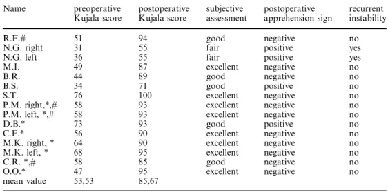
Table 1 pre- and postoperative Kujala score, postoperative subjective assessment, postoperative apprehension sign, and postoperative patellar instability
Patients marked with (*) had an additional transfer of the tibial tuberosity, patients marked wi-th (#) had an additional troch-lear dysplasia Name preoperative Kujala score postoperative Kujala score subjective assessment postoperative apprehension sign recurrent instability R.F.# 51 94 good negative no
N.G. right 31 55 fair positive yes N.G. left 36 55 fair positive yes M.I. 49 87 excellent negative no
B.R. 44 89 good negative no
B.S. 34 71 good positive no
S.T. 76 100 excellent negative no P.M. right,*,# 58 93 excellent negative no P.M. left, *,# 58 93 excellent negative no
D.B.* 73 93 good positive no
C.F.* 56 90 excellent negative no M.K. right, * 64 90 excellent negative no M.K. left, * 68 95 excellent negative no C.R. *,# 58 85 good negative no O.O.* 47 95 excellent negative no mean value 53,53 85,67
patient reported on recurrent instability postoperatively, but the objective findings did not correlate with the pa-tient’s described discomfort (N.G.). A persistent appre-hension sign was found in the same patient. No other patient suffered from a persistent apprehension sign or a dislocation postoperatively. The mean Caton index did not change postoperatively. There was no postoperative patella baja, since the patellotibial ligament is not reconstructed which might act as additional tibial rein and pull the patella distally. A favorable effect on patellar tracking was also noticed, since the patellar tilt was sig-nificantly improved by the dorsal pull of the medial border of the patella through the graft. In patients with a TTTG more than 15 mm a transfer of the tibial tuber-osity was performed in addition although it has been reported that a distal realignment procedure is unneces-sary when proximal realignment is carried out [14,40]. However, Gomes et al. have reported that the best results in MPFL reconstruction are obtained in patients with a normal Q angle [21]. We performed the tibial tuberosity transfer to obtain a normal TTTG in all patients post-operatively and to avoid the influence of a pathologic value onto postoperative data. No negative effect was seen from an additional tibial tuberosity transfer. The
lack of bony constraint in case of trochlear dysplasia can put the ligament at risk for repeated failure, because of structural deficiency, increasing or supporting the native medial tether [2,21,35,37]. Trochlear dysplasia in the 3 patients of the present series was not pronounced enough to have an effect on the clinical outcome. None of these 3 patients complained of retropatellar pain after recon-struction, although an exaggerated tightening of the medial patellofemoral ligament reconstruction may result in a medial retropatellar hyperpression syndrome in the dysplastic trochlear groove. Using the technique already described by Gomes [21, 27], which consists of three small incisions on the medial side (Fig. 2), a favorable cosmetic result is achieved.
In conclusion, linear reconstruction of the medial patellofemoral ligament, using a Semitendinosus auto-graft is successful. Patellar stability can reliably be re-stored without creating further complications such as retropatellar pain or hyperpression. The study does not answer the question whether patients with a TTTG more than 15 mm or with major trochlear dysplasia will benefit from isolated reconstruction of the medial pa-tellofemoral ligament, or if an additional procedure of an underlying bony malalignment may be necessary.
References
1. Aglietti P, Buzzi R, De Biase P, Giron F (1994) Surgical treatment of recurrent dislocation of the patella. Clin Orthop 308:8–17
2. Avikainen VJ, Nikku RK, Seppanen-Lehmonen TK (1993) Adductor mag-nus tenodesis for patellar dislocation. Technique and preliminary results. Clin Orthop 297:12–16
3. Beaconsfield T, Pintore E, Maffulli N, Petri GJ (1994) Radiological measure-ments in patellofemoral disorders. A review. Clin Orthop 308:18–28 4. Betz RR, Magill JT III, Lonergan RP
(1987) The percutaneous lateral reti-nacular release. Am J Sports Med 15:477–482
5. Blauth W, Mann M (1977) [Medial-and simultaneous anterior-transfer of the tibial tuberosity (author’s transl)]. Z Orthop Ihre Grenzgeb 115:252–255 6. Brown DE, Alexander AH, Lichtman DM (1984) The Elmslie-Trillat proce-dure: evaluation in patellar dislocation and subluxation. Am J Sports Med 12:104–109
7. Burks RT, Desio SM, Bachus KN, Ty-son L, Springer K (1998) Biomechanical evaluation of lateral patellar disloca-tions. Am J Knee Surg 11:24–31 8. Caton J, Deschamps G, Chambat P,
Lerat JL, Dejour H (1982) Les rotules basses. A propos de 128 observations. Rev Chir Orthop Reparatrice Appar Mot 68:317–325
9. Caton J, Deschamps G, Chambat P, Lerat JL, Dejour H (1982) [Patella inf-era. Apropos of 128 cases]. Rev Chir Orthop Reparatrice Appar Mot 68:317– 325
10. Cerullo G, Puddu G, Conteduca, Fer-retti A, Mariani PP (1988) Evaluation of the results of extensor mechanism reconstruction. Am J Sports Med 16:93–96
11. Cofield RH, Bryan RS (1977) Acute dislocation of the patella: results of conservative treatment. J Trauma 17:526–531
12. Conlan T, Garth WP Jr, Lemons JE (1993) Evaluation of the medial soft-tissue restraints of the extensor mecha-nism of the knee. J Bone Joint Surg Am 75:682–693
13. Cox JS (1982) Evaluation of the Roux-Elmslie-Trillat procedure for knee extensor realignment. Am J Sports Med 10:303–310
14. Crosby EB, Insall J (1976) Recurrent dislocation of the patella. Relation of treatment to osteoarthritis. J Bone Joint Surg Am 58:9–13
15. Davies AP, Costa ML, Shepstone L, Glasgow MM, Donell S, Donnell ST (2000) The sulcus angle and malalign-ment of the extensor mechanism of the knee. J Bone Joint Surg Br 82:1162– 1166
16. Davis DK, Fithian DC (2002) Tech-niques of medial retinacular repair and reconstruction. Clin Orthop 402:38–52 17. Dejour H, Walch G, Neyret P, Adeleine
P (1990) [Dysplasia of the femoral trochlea]. Rev Chir Orthop Reparatrice Appar Mot 76:45–54
18. Dejour H, Walch G, Nove-Josserand L, Guier C (1994) Factors off patellar instability: an anatomic radiographic study. Knee Surg Sports Traumatol Arthrosc 2:19–26
19. Desio SM, Burks RT, Bachus KN (1998) Soft tissue restraints to lateral patellar translation in the human knee. Am J Sports Med 26:59–65
20. Drez D Jr, Edwards TB, Williams CS (2001) Results of medial patellofemoral ligament reconstruction in the treatment of patellar dislocation. Arthroscopy 17:298–306
21. Ellera Gomes JL, Stigler Marczyk LR, Cesar de Cesar P, Jungblut CF (2004) Medial patellofemoral ligament recon-struction with semitendinosus autograft for chronic patellar instability: a follow-up study. Arthroscopy 20:147–151 22. Erasmas PJ (1998) Reconstruction of
the medial patellofemoral ligament in recurrent traumatic patellar dislocation. Arthroscopy 14:S42 (suppl., abstract) 23. Fithian DC, Mishra DK, Balen PF,
Stone ML, Daniel DM (1995) Instru-mented measurement of patellar mobil-ity. Am J Sports Med 23:607–615 24. Fithian DC, Paxton EW, and Cohen
AB (2004) Indications in the treatment of patellar instability. J Knee Surg 17:47–56
25. Garth WP Jr, DiChristina DG, Holt G (2000) Delayed proximal repair and distal realignment after patellar dislo-cation. Clin Orthop 377:132–144 26. Goldwaith JE (1904) Slipping or
recur-rent dislocation of the patella. Boston Med Surg J :169–174
27. Gomes JL (1992) Medial patellofemoral ligament reconstruction for recurrent dislocation of the patella: a preliminary report. Arthroscopy 8:335–340
28. Goutallier D, Bernageau J, Lecudonnec B (1978) [The measurement of the tibial tuberosity. Patella groove distanced technique and results (author’s transl)]. Rev Chir Orthop Reparatrice Appar Mot 64:423–428
29. Hautamaa PV, Fithian DC, Kaufman KR, Daniel DM, Pohlmeyer AM (1998) Medial soft tissue restraints in lateral patellar instability and repair. Clin Orthop 349:174–182
30. Hawkins RJ, Bell RH, Anisette G (1986) Acute patellar dislocations. The natural history. Am J Sports Med 14:117–120
31. Hughston JC (1972) Reconstruction of the extensor mechanism for subluxating patella. Am J Sports Med 1:6–13 32. Kolowich PA, Paulos LE, Rosenberg
TD, Farnsworth S (1990) Lateral re-lease of the patella: indications and contraindications. Am J Sports Med 18:359–365
33. Kujala UM, Jaakkola LH, Koskinen SK, Taimela S, Hurme M, Nelimarkka O (1993) Scoring of patellofemoral dis-orders. Arthroscopy 9:159–163 34. Kujala UM, Osterman K, Kvist M,
Aalto T, Friberg O (1986) Factors pre-disposing to patellar chondropathy and patellar apicitis in athletes. Int Orthop 10:195–200
35. Muneta T, Sekiya I, Tsuchiya M, Shinomiya K (1999) A technique for reconstruction of the medial patellofe-moral ligament. Clin Orthop 339:151– 155
36. Nomura E (1999) Classification of le-sions of the medial patello-femoral lig-ament in patellar dislocation. Int Orthop 23:260–263
37. Nomura E, Horiuchi Y, Kihara M (2000) A mid-term follow-up of medial patellofemoral ligament reconstruction using an artificial ligament for recurrent patellar dislocation. Knee 7:211–215 38. Nomura E, Horiuchi Y, Kihara M
(2000) Medial patellofemoral ligament restraint in lateral patellar translation and reconstruction. Knee 7:121–127 39. Sallay PI, Poggi J, Speer KP, Garrett
WE (1996) Acute dislocation of the patella. A correlative pathoanatomic study. Am J Sports Med 24:52–60 40. Scuderi G, Cuomo F, Scott WN (1988)
Lateral release and proximal realign-ment for patellar subluxation and dis-location. A long-term follow-up. J Bone Joint Surg Am 70:856–861
41. Trillat A, Dejour H, Couette A (1964) [Diagnosis and treatment of recurrent dislocations of the patella]. Rev Chir Orthop Reparatrice Appar Mot 50:813– 824