. . . .
. . . .
Jagged1 intracellular domain-mediated inhibition
of Notch1 signalling regulates cardiac homeostasis
in the postnatal heart
Me´lanie Metrich
1, April Bezdek Pomey
1, Corinne Berthonneche
2, Alexandre Sarre
2,
Mohamed Nemir
1, and Thierry Pedrazzini
1,2*
1
Experimental Cardiology Unit, Department of Medicine, University of Lausanne Medical School, Rue du Bugnon 27, CH-1011 Lausanne, Switzerland; and2
Cardiovascular Assessment Facility, University of Lausanne, Lausanne, Switzerland
Received 22 October 2014; revised 22 July 2015; accepted 23 July 2015; online publish-ahead-of-print 6 August 2015 Time for primary review: 41 days
Aims Notch1 signalling in the heart is mainly activated via expression of Jagged1 on the surface of cardiomyocytes. Notch
con-trols cardiomyocyte proliferation and differentiation in the developing heart and regulates cardiac remodelling in the stressed adult heart. Besides canonical Notch receptor activation in signal-receiving cells, Notch ligands can also activate Notch receptor-independent responses in signal-sending cells via release of their intracellular domain. We evaluated therefore the importance of Jagged1 (J1) intracellular domain (ICD)-mediated pathways in the postnatal heart.
Methods and results
In cardiomyocytes, Jagged1 releases J1ICD, which then translocates into the nucleus and down-regulates Notch tran-scriptional activity. To study the importance of J1ICD in cardiac homeostasis, we generated transgenic mice expressing a tamoxifen-inducible form of J1ICD, specifically in cardiomyocytes. Using this model, we demonstrate that J1ICD-mediated Notch inhibition diminishes proliferation in the neonatal cardiomyocyte population and promotes matur-ation. In the neonatal heart, a response via Wnt and Akt pathway activation is elicited as an attempt to compensate for the deficit in cardiomyocyte number resulting from J1ICD activation. In the stressed adult heart, J1ICD activation results in a dramatic reduction of the number of Notch signalling cardiomyocytes, blunts the hypertrophic response, and reduces the number of apoptotic cardiomyocytes. Consistently, this occurs concomitantly with a significant down-regulation of the phosphorylation of the Akt effectors ribosomal S6 protein (S6) and eukaryotic initiation factor 4E binding protein1 (4EBP1) controlling protein synthesis.
Conclusions Altogether, these data demonstrate the importance of J1ICD in the modulation of physiological and pathological
hypertrophy, and reveal the existence of a novel pathway regulating cardiac homeostasis.
-Keywords Cardiac hypertrophy † Cardiomyocyte differentiation † Notch signalling † Jagged1-intracelullar domain
1. Introduction
The Notch pathway is an evolutionarily conserved communication sys-tem between adjacent cells expressing Notch receptors (Notch1 – 4) and membrane-bound ligands of the Jagged (Jagged 1,2) or Delta-like
(Dll1,3,4) family.1Notch signalling is crucial for cardiac morphogenesis
during development. In particular, Notch regulates myocyte
prolifer-ation in the trabeculate myocardium and valve formprolifer-ation.2
Further-more, Notch controls proliferation and differentiation both in
committed cardiac progenitor cells and in immature cardiomyocytes.3–6
Importantly, the Notch pathway is also essential for the maintenance
of structural and functional integrity of the postnatal heart.7–11In the
adult heart, Jagged1-mediated Notch signalling is activated upon dam-age and limits the extent of the hypertrophic response in stressed
car-diomyocytes.7,11 On the contrary, pharmacological and genetic
inhibition of Notch signalling exacerbates cardiac hypertrophy
follow-ing increased workload.7Accordingly, transgenic (TG) mice
overex-pressing Jagged1 on the surface of cardiomyocytes demonstrated a limited hypertrophic and fibrotic response to pressure overload, and
cardiac precursor expansion.11Along the same line, forced activation
*Corresponding author. Tel:+41 21 314 0765; fax: +41 21 314 5978, E-mail: [email protected]
&The Author 2015. Published by Oxford University Press on behalf of the European Society of Cardiology.
This is an Open Access article distributed under the terms of the Creative Commons Attribution Non-Commercial License (http://creativecommons.org/licenses/by-nc/4.0/), which permits non-commercial re-use, distribution, and reproduction in any medium, provided the original work is properly cited. For commercial re-use, please contact [email protected]
of the Notch pathway in the infarcted heart following induced cardiomyocyte-specific Notch1 expression or intramyocardial delivery of a Notch1 pseudoligand increased survival, improved cardiac
func-tion, and minimized fibrosis.8,9
Canonical Notch signalling is initiated in the signal-receiving cell by interaction of Notch receptors with their ligands. This event triggers the sequential cleavage of the Notch receptor by A Disintegrin and Metalloproteinase (ADAM) and a g-secretase complex, and then the release of the Notch intracellular domain (NICD), which enters the nu-cleus, interacts with RBP-Jk and other co-activators to activate target
gene expression.1Prototypical target genes of Notch are
transcription-al repressors of the Hes and Hey family. Importantly, endocytosis of the ligand in the signal-sending cell is required for induction of Notch
re-ceptor proteolysis.12Indeed, ubiquitylation-dependent internalization
of the ligand is thought to exert a pulling force on the receptor to ex-pose its cleavage sites and thereby promote enzymatic cleavage and downstream activation. For this reason, truncated forms of Jagged and Delta, lacking ubiquitylation-sensitive intracellular domains, are
unable to activate Notch signalling.13,14
Interestingly, Notch ligands can also be cleaved in signal-sending cells by the ADAM metalloprotease and the g-secretase complex following
ligand – receptor interaction.15–18This process induces the release of
an intracellular fragment of the ligand, which then translocates into the nucleus. This raises therefore the interesting possibility that ligand activation could produce cellular responses within signal-sending cells. In this context, the Jagged1 intracellular domain (J1ICD) has been
shown to regulate cell transformation and proliferation.15,19Similarly,
the nuclear translocation of X-Serrate-1ICD in Xenopus was shown
to inhibit primary neurogenesis.20Finally, Delta-like 1 ICD production
was demonstrated to block proliferation, cell motility, and neuronal
mi-gration,21,22independently of its activity as a Notch receptor ligand.23,24
Given the crucial role of Notch signalling in cardiac homeostasis, we evaluated therefore the importance of Jagged1-mediated pathways in postnatal cardiomyocyte differentiation and growth. We demonstrated that Jagged1 is indeed cleaved in cardiomyocytes and produces a J1ICD fragment, which can be detected in the nucleus of responding cells. To investigate Jagged1-mediated effects in the heart, we therefore gener-ated TG mice carrying a cardiomyocyte-specific and inducible form of J1ICD. In the neonatal heart, J1ICD accelerates the maturation of car-diomyocytes, in part via inhibition of the Notch signalling pathway. In the adult heart, J1ICD production modulates hypertrophic remodelling during the response to pressure overload. Altogether, these data sug-gest an important role for Notch ligand-mediated signalling in the de-veloping and the adult heart.
2. Methods
For detailed methods, see Supplementary material online.
2.1 Transgenic (TG) mER-J1ICD-mER mice
The rat J1ICD was isolated from the pTOPO-J1ICD plasmid (kindly pro-vided by Matthew J. LaVoie, Harvard Medical School, Boston, USA), and fused at its C- and N-terminal ends to amino acids 281 – 599 of the mutated (G525R) mouse oestrogen receptor (mER; a kind gift from Michael Reth, Max-Plank Institute, Freiburg, Germany). The resultant fragment was sub-cloned downstream of the a-myosin heavy chain (a-MHC) promoter. The a-MHC-mER-J1ICD-mER construct was linearized and injected into the male pronucleus of fertilized B6D2F1 embryos. The resulting TG mice were backcrossed into the C57/BL6 background strain. Nuclear transloca-tion of the transgene was induced by intraperitoneal (i.p.) injectransloca-tion of
20 mg/kg of tamoxifen (Sigma, Saint Louis, MO). In some experiments, mice were injected with MG132 i.p. at 7 mg/kg.
2.2 Animal experiments
Mice were housed under standard conditions. Ten-week-old mice were anaesthetized by i.p. injection of ketamine/xylazine/acepromazine (65/15/ 2 mg/kg body weight). Mice were placed on a warm pad for maintenance of body temperature. Transaortic constriction (TAC) was created using a 7.0-silk suture tied twice around the aorta between the right innominate artery and the left common carotid and a predetermined-gauge needle size (25 gauge). The needle was then gently retracted leaving a restriction of the same calibre among different mice. For animals undergoing a sham operation, the ligature was placed in an identical location but was not tied. Animals were injected daily with 20 mg/kg of tamoxifen i.p. (Sigma) or vehicle (95% ethanol and sunflower oil only). Adult mice were sacrificed
by CO2inhalation and subsequent cervical dislocation. Neonatal mice
were sacrificed by rapid decapitation. Animal experiments were approved by the Government Veterinary Office (Lausanne, Switzerland) and performed according to the guidelines from Directive 2010/63/EU of the European Parliament.
2.3 Echocardiography
Transthoracic echocardiography was performed using a 30 MHz probe and the Vevo 770 Ultrasound machine (Visualsonics, Toronto, ON, Canada) under mild anaesthesia with 1 – 1.5% isoflurane. Diastolic and systolic in-ternal ventricular septum (IVSd and IVSs), diastolic and systolic left ventricular free posterior wall thickness (LVPWd and LVPWs), and left ven-tricular internal end-diastolic and end-systolic chamber (LVIDd and LVIDs) dimensions were measured three times on M-mode images. LV fractional shortening (%FS) and ejection fraction (%EF) were also calculated. The right ventricle internal diameter and wall thickness were also measured on parasternal short-axis view, using papillary muscles as reference points.
2.4 Statistical analysis
In this study, data are expressed as mean + SEM. One-way ANOVA with Benferroni’s multiple comparison test was used to assess significance of differences between experimental groups. Two-sided Student’s t-test was used when only two groups are compared. P values of ,0.05 were con-sidered as significant. Analysis was performed using the GraphPad Prism version 5.04 (GraphPad Software, Inc.).
3. Results
3.1 J1ICD inhibits canonical Notch
activation in cardiomyocytes
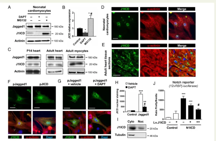
Evidence suggested that membrane-bound Notch ligands released intracellular fragments within signal-sending cells to activate Notch receptor-independent cellular responses. Since Jagged1 was demon-strated to be the predominant form expressed in the heart, we exam-ined whether J1ICD could be produced in primary cardiomyocytes. Jagged1 and J1ICD were immunoprecipitated from neonatal cardio-myocyte lysates using antibodies directed against the C-terminal end of the protein. We indeed identified a 20 kDa fragment corresponding
to the expected size for J1ICD (Figure1A and B). This fragment
accumu-lated following proteasome inhibition using MG132. Furthermore, J1ICD release was blocked by the g-secretase inhibitor, N-[N-(3,5-Difluorophenacetyl)-L-alanyl]-S-phenylglycine t-butyl ester (DAPT), demonstrating the involvement of g-secretase-mediated Jagged1 cleav-age in J1ICD production. J1ICD was also produced in the postnatal and
adult heart as well as in isolated adult cardiomyocytes (Figure1C). We
were expressed. In untransfected cardiomyocytes, antibodies directed against J1ICD, which recognize both full-length Jagged1 and J1ICD, marked the cytoplasm as well as the perinuclear and nuclear regions
(Figure1D). To further analyse subcellular localization, we took advantage
of TG mice overexpressing Jagged1 specifically in cardiomyocytes,
which were recently established in our laboratory (Figure1E).11Analysis
of heart sections stained with antibodies against J1ICD confirmed the presence of the fragment at the membrane, in the cytoplasm, and in the perinuclear region. Then, we transfected cardiomyocytes in vitro
with either a Jagged1 or a J1ICD encoding plasmid (Figure1F). Following
Jagged1 or J1ICD overexpression, J1ICD was readily detected in all three cellular compartments. Furthermore, nuclear localization was more apparent in cells transfected with J1ICD, supporting translocation of this fragment into the nucleus. Importantly, nuclear staining is markedly diminished in Jagged1-overexpressing cardiomyocytes treated with DAPT, indicating that J1ICD production and translocation were
dependent on g-secretase-mediated cleavage of full-length Jagged1
(Figure1G and H ). Using purified cytoplasmic/membrane and nuclear
fractions from neonatal cardiomyocytes and immunoblotting against J1ICD, we further demonstrated the presence of J1ICD in both
com-partments (Figure1I). We next evaluated the effects of J1ICD on
Notch signalling in isolated cardiomyocytes. Notch transcriptional activity was measured using a RBP-Jk-responsive luciferase (12xRBP-Jk-Luc) reporter gene. Therefore, neonatal cardiomyo-cytes were transfected with the 12xRBP-Jk-Luc reporter, which was induced by forced Notch1 intracellular domain (N1ICD) ex-pression. Cardiomyocytes were then infected with increasing amounts of a lentiviral vector encoding J1ICD (Lv.J1ICD). Lv.J1ICD had no impact on basal luciferase activity. In contrast, Lv.J1ICD sig-nificantly decreased N1ICD-induced transcriptional activity in a
dose-dependent manner (Figure1J), indicating that J1ICD produced
an important inhibitory effect on Notch signalling.
Figure 1 Jagged1 cleavage in cardiomyocytes produces J1ICD. (A) Neonatal cardiomyocytes were treated with either DAPT (10 mM) or MG132 (10 mM) for 12 h. Immunoprecipitated full-length Jagged1 and J1ICD proteins were detected by western blotting. Anti-a-actinin immunoblot was
per-formed as a loading control. (B) J1ICD levels were quantified by densitometry. *P≤ 0.05 vs. vehicle,#P≤ 0.05 vs. DAPT + MG132. (C)
Immunopre-cipitated Jagged1 and J1ICD in postnatal hearts of P14 and adult mice, and isolated adult mice cardiomyocytes. (D) Neonatal cardiomyocytes or (E) adult heart sections from Jagged1-overexpressing mice were stained with anti-Jagged1 (green) and anti-a-sarcomeric-actinin antibodies (red). Nuclei were stained with DAPI (blue). Scale bar, 20 mm. (F ) Cardiomyocytes were transfected with either pcDNA-Jagged1 or pcDNA-J1ICD vector and stained as in D. Images in D – F are representative of four-independent experiments. (G) Confocal images of Jagged1-expressing cells treated with either vehicle or DAPT (10 mM) for 12 h. (H ) Nuclear J1ICD staining obtained in G was quantified and normalized to the nucleus area. Bar graphs are mean + SEM
from three experiments. **P≤ 0.01; ***P ≤ 0.001 vs. control;#P≤ 0.001 vs. Jagged1 + DAPT. (I) J1ICD immunoprecipitation in cytoplasmic and
nu-clear fractions of neonatal myocytes. (J ) Neonatal cardiomyocytes were transfected with either pcDNA (control) or pcDNA-NICD together with a Notch reporter (12xRBPJk-luciferase) and an internal control (pCMV-Renilla). Cells were then infected for 24 h with different amounts of a J1ICD-expressing lentiviral vector (Lv.J1ICD) or a control vector as indicated. Notch transcriptional activity was determined by quantification of
3.2 TG mice with inducible
cardiomyocyte-specific J1ICD expression
To investigate the effects of Jagged1 signalling on postnatal cardiac growth and function, we generated an inducible form of J1ICD, in which
J1ICD was fused to two mutant oestrogen receptor hormone-binding domains to generate the mER-J1ICD-mER fragment. The mER se-quences in the construct confer tamoxifen-mediated inducibility in car-diomyocytes and prevent potential detrimental effects of transgene expression during development. The suitability of the system was
Figure 2TG mice with cardiac-specific expression and inducible activation of J1ICD. (A) HEK 293 cells transfected with pEF1a-mER-J1ICD-mER were treated with 4-OH-tamoxifen (1 mM) or DMSO. Cytoplasmic and nuclear fractions were separated and assessed for J1ICD expression by western blot. Anti-a-tubulin and anti-histone H1 immunoblots were performed as loading controls. (B) Schematic representation of the a-MHC:mER-J1ICD-mER cDNA used to generate TGJ1ICD mice (top). In the absence of tamoxifen, the mER-J1ICD-mER fusion protein forms a complex with HSP proteins and is sequestered in the cytosol. Tamoxifen injections release mER-J1ICD-mER and induce its nuclear translocation (bottom). (C ) Quantitative RT-PCR analysis of the transgene expression. Results were normalized to endogenous Gapdh and Jagged1 expression and were expressed as mean + SEM
(n ¼ 10 – 15 mice per group; *P≤ 0.01). (D) Anti-Jagged1 immunoblot performed on whole heart extracts from WT or TGJ1ICD mice. (E) RT-PCR
specific for the transgene and Gapdh gene were performed on different organs of TGJ1ICD mice to show the cardiac-specific expression of the trans-gene. (F and G) Heart sections from WT or TGJ1ICD mice, treated with vehicle or tamoxifen for 48 h, were immunostained with anti-Esr1 antibody, which recognizes the mER motif of the transgene (green) and anti-a-sarcomeric-actinin antibody (red). Nuclei were stained with DAPI (blue). Scale bar, 10 mm (F ) and 20 mm (G). Images in F and G are representative of at least five mice per group.
then tested in transfected HEK 293 cells. As expected, the mER-J1ICD-mER fusion protein was found primarily in the cytoplasm in vehicle-treated cells, whereas it was predominantly detected in the
nuclear fraction in cells treated with tamoxifen (Figure2A). We next
generated TG mice expressing mER-J1ICD-mER under control of the
cardiomyocyte-specific a-MHC promoter (herein TGJ1ICD;
Figure2B). Three different TG lines were initially produced.
Character-ization demonstrated no obvious phenotypic difference between these lines. We present therefore data obtained using one particular line. Sig-nificant cardiac expression of the transgene was readily detected by
RT-PCR and western blot in TG mice (Figure2C and D), and its
expres-sion was restricted to the heart (Figure2E). Similarly, immunostaining of
heart sections from TGJ1ICD mice showed that J1ICD was seques-tered in the cytoplasm of cardiomyocytes in the absence of tamoxifen, whereas tamoxifen administration allowed the TG protein to enter the
nucleus (Figure2F). Of note, all cardiomyocytes demonstrated nuclear
staining after tamoxifen treatment (Figure2G).
3.3 J1ICD accelerates cardiomyocyte
maturation in vitro via inhibition of Notch
signalling
We first evaluated the importance of J1ICD on neonatal cardiomyo-cyte proliferation and maturation. Ventricular cardiomyocardiomyo-cytes from neonatal wild-type (WT) and TGJ1ICD mice were therefore isolated and cultured in the presence of tamoxifen. We first analysed the effects of tamoxifen-induced J1ICD activation on Notch signalling. Immunos-taining against the activated form of Notch1 (N1ICD) was used to detect a-actinin-positive cardiomyocytes actively signalling via the Notch1 receptor (see Supplementary material online, Figure S1A). Quantitative analysis showed a smaller percentage of N1ICD-positive cardiomyocytes in the population derived from TGJ1ICD TG mice following tamoxifen treatment (see Supplementary material online, Figure S1B), confirming that nuclear translocation of J1ICD inhibited Notch signalling. Inhibition of Notch signalling was also demonstrated by the significant down-regulation of Notch target gene expression, Hes1 and Hey2, in TGJ1ICD cardiomyocytes compared with WT cul-tures (see Supplementary material online, Figure S1C). Down-regulation of the Notch pathway has been associated with differentiation and mat-uration in cardiomyocytes. Interestingly, there was a greater propor-tion of cardiomyocytes with striated a-actinin in TGJ1ICD vs. WT cultures (see Supplementary material online, Figure S1A and B). This ob-servation is consistent with J1ICD activation promoting cardiomyocyte differentiation via Notch signalling inhibition. Finally, we determined the number of proliferating neonatal cardiomyocytes after incorporation of the nucleoside analogue, 5-bromo-2-deoxyuridine (BrdU). Fewer BrdU-positive cardiomyocytes were present in TGJ1ICD cultures (see Supplementary material online, Figure S1D and E). Overall, these data indicated that J1ICD has inhibitory effects on Notch signalling, leading to decreased myocyte proliferation and accelerated cardio-myocyte maturation.
3.4 Accelerated cardiomyocyte maturation
in neonatal TGJ1ICD mice following J1ICD
activation
To assess whether J1ICD had an impact on cardiomyocyte maturation in vivo, neonatal WT and TGJ1ICD mice received daily injections of tam-oxifen from birth to postnatal day 14 (P14), i.e. during the time period
in which cardiomyocytes gradually exit from the cell cycle.25,26As
observed in isolated cells, Notch signalling was down-regulated in
car-diomyocytes in the neonatal TG myocardium (Figure3A and B). Indeed,
the number of N1ICD-positive cardiomyocytes was significantly lower in the heart of TGJ1ICD vs. WT mice at P7 and P14, respectively. Im-portantly, the expression of the Notch target genes, such as Hey1, Hey2, and Hes1, was significantly decreased in TGJ1ICD hearts when compared with WT. Jagged1 expression was also down-regulated in TG hearts, whereas expression of the Notch1 receptor was similar
to what observed in WT (Figure3C). Interestingly, the inhibitory effects
of J1ICD activation on Notch signalling were particularly evident at P7. Previous studies suggested that NICD could be regulated via targeted
ligand intracellular domain-mediated degradation.27,28To investigate
whether the negative regulation exerted by J1ICD on Notch signalling occurred via a similar mechanism, anti-J1ICD immunoprecipitation was performed from WT and TGJ1ICD heart lysates, and the presence of J1ICD and N1ICD in immunoprecipitates was revealed by
immuno-blotting (Figure3D). N1ICD co-immunoprecipitated with J1ICD in
WT hearts, indicating that J1ICD physically interacts with N1ICD. The amounts of N1ICD were significantly reduced in extract from TGJ1ICD mouse hearts, consistent with J1ICD-mediated N1ICD deg-radation. Indeed, N1ICD was protected from degradation in TG hearts in the presence of the ubiquitin proteasome inhibitor, MG132.
We then followed cardiac growth in WT and TGJ1ICD mice follow-ing tamoxifen administration, and found that the heart in TGJ1ICD mice was characterized by a significantly different growth curve than that
observed in WT animals (Figure4). Specifically, the TG hearts
demon-strated a slight increase in ventricular mass at P7, but a decreased
weight and dimensions at P14 (Figure4A and B). Importantly,
cardio-myocyte apoptosis was not increased in 2-week-old TGJ1ICD mice, as detected using a TUNEL assay, excluding that cell death participates in the reduction of cardiac mass in TG hearts (data not shown). Cardi-omyocytes from TGJ1ICD mice were found significantly larger at P4
and P7 but smaller at P14 (Figure4C). Since cardiomyocyte
differenti-ation and maturdifferenti-ation are usually inversely correlated with proliferdifferenti-ation, we evaluated cardiomyocyte mitosis in neonatal WT and TGJ1ICD hearts. At P4, the number of PH3-positive cardiomyocytes was
re-duced in TG hearts when compared with WT (Figure4D and E). These
findings were confirmed using BrdU incorporation to detect
proliferat-ing cardiomyocytes (Figure4F). When a single dose of BrdU was
admi-nistrated, the number of BrdU-positive cardiomyocytes was found lower at P4 in TG vs. WT hearts. These data are consistent with J1ICD-activated cardiomyocytes having an increased propensity to exit the cell cycle and to mature during the first days after birth. In agreement with this, the decreased ratio between expression of the fe-tal (b-MHC) and adult (a-MHC) isoforms of the MHC in TG hearts at P4 demonstrated early maturation (see Supplementary material online, Figure S2B).
3.5 Compensatory induction of the Wnt
and AKT signalling pathways in the neonatal
heart upon J1ICD activation
Although J1ICD-mediated pathways reduced cardiomyocyte prolifer-ation during the first week of age, TGJ1ICD mice eventually demon-strated a compensatory increase in cardiomyocyte proliferation. Indeed, cardiomyocyte mitosis decreased rapidly after birth in WT hearts as assessed by the number of PH3-positive cells and by BrdU
in-corporation (Figure 4E and F ). In contrast, the J1ICD-activated
proliferative activity at P7. Sustained cardiomyocyte proliferation in the TG group was also confirmed by a significant up-regulation of cyc-lin B1 and cyccyc-lin D1 mRNA expression during the recovery period (see Supplementary material online, Figure S2A). Finally, the b- to a-MHC ratio was increased at P14, attesting the more immature phenotype of proliferating cardiomyocytes (see Supplementary material online, Figure S2B). These findings suggested that cardio-myocytes with reduced Notch activity secondary to transgene activa-tion could activate compensatory signalling pathways to stimulate myocyte division.
The Wnt pathway has been implicated in cardiogenesis29and is
known to interact with the Notch pathway to regulate cell fate decision
during cardiac differentiation.30–32In particular, canonical Wnts,
in-cluding Wnt3A, Wnt6, Wnt8, and b-catenin signalling, negatively
influ-ence cardiac specification and terminal cardiac differentiation.33–38On
the other hand, the non-canonical Wnt pathway is known to promote
cardiac differentiation.39–41We thus measured the expression of
dif-ferent Wnt ligands and Wnt regulators, specific of both canonical and non-canonical Wnt signalling, in neonatal WT and TGJ1ICD
mice. Expression levels were determined at P7 during the peak of
the compensatory proliferative response (Figure5A). Wnt3A and
Wnt8 levels were undetectable, and Wnt6 was not significantly chan-ged. However, the Wnt inhibitors, such as Dkk1, Axin2, and Wif1, were significantly down-regulated in TG at P7, consistent with an activation of canonical Wnt signalling in TG hearts during the proliferative phase. To confirm activation of the Wnt pathway at P7, we determined the amounts of activated (dephosphorylated) and total b-catenin in WT
vs. TGJ1ICD heart extracts by immunoblotting (Figure5B and C ).
The ratio of active to total b-catenin was increased in TGJ1ICD hearts, supporting a compensatory response via the Wnt pathway in neonatal TG mice. Finally, the expression level of the Wnt inhibitors Sfrp1 and Sfrp2 was unchanged. Moreover, Wnt11, a component of the
non-canonical Wnt pathway, was down-regulated (Figure 5A). This is
coherent with the immature state of TG cardiomyocytes around P14. Overall, these data suggest that cardiac canonical and non-canonical Wnt pathways were regulated in a way to counterbalance J1ICD-mediated Notch signalling inhibition. Interestingly, the expres-sion of Wnt ligands and Wnt inhibitors as well as the active/total
Figure 3 Notch activation is decreased in neonatal TGJ1ICD mice. WT and TGJ1ICD neonatal mice treated daily with tamoxifen were sacrificed at postnatal days P0, P4, P7, and P14. (A) Confocal images of heart sections immunostained against N1ICD (green), laminin (red), and a-sarcomeric-actinin (white). Nuclei were stained with DAPI (blue). Scale bar, 20 mm. (B) The number of NICD-positive cardiomyocytes at P7 and P14 was quantified on images depicted in A, and expressed as % of total DAPI-positive cardiomyocytes (n ¼ 4 – 6 mice per group). (C ) RNA expression of Notch target genes was analysed by quantitative RT-PCR. Results, normalized to Gapdh, represent fold change relative to the WT group at P0 and are expressed as mean +
SEM (n ¼ 4 mice per group and per time point; *P≤ 0.05). (D) Tamoxifen-treated mice were injected or not with MG132 from P9 to P14. J1ICD
im-munoprecipitation was performed using anti-Jagged1 antibody on whole heart extracts. Immunoprecipitates were immunoblotted with anti-N1ICD, anti-Jagged1 to detect J1ICD, and anti-tubulin antibodies. A quantification of N1ICD band intensity relative to tubulin is shown on the right panel
b-catenin ratio were not altered in TGJ1ICD mice at P4 (see Supple-mentary material online, Figure S3), suggesting that positive regulation of the Wnt pathway observed at P7 occurred secondary to J1ICD-mediated Notch signalling inhibition.
The mTOR/Akt signalling pathway also plays an important role in
physiological proliferation and growth in the postnatal heart.42We
measured therefore the activity of this pathway 1 week after birth and observed a significant increase of Akt phosphorylation at Thr308 and Ser473 in TG hearts, which demonstrated full activation of Akt at this particular time point. Consistently, phosphorylation of Akt downstream targets, namely ribosomal S6 protein (S6) and eukaryotic initiation factor 4E binding protein1 (4EBP1), was increased in TGJ1ICD
mice (Figure5D and E).
3.6 Attenuated cardiac hypertrophic
response to pressure overload in adult
TGJ1ICD mice following J1ICD activation
To evaluate the impact of J1ICD activation in the adult heart, WT and TGJ1ICD mice received daily injections of tamoxifen during 1 week. However, echocardiographic analysis of cardiac dimensions and func-tion did not show any differences between WT and TGJ1ICD mice (see Supplementary material online, Table S1). Although Notchsignalling can be readily detected in isolated adult cardiomyocytes and in the adult WT heart, Notch signalling was unaffected by J1ICD activation under normal conditions (see Supplementary material on-line, Figure S4). These findings are not surprising, given the fact that the Notch pathway is minimally activated in the unstressed adult heart. We therefore subjected the adult heart to pressure overload using the
TAC model (Figure6). In this case, the number of N1ICD-positive cells
increased in the WT heart under stress, indicating that the Notch path-way was induced. In contrast, stimulated Notch activity was blunted in TGJ1ICD mice, as assessed by the lower number of N1ICD-positive cells in the stressed TG heart following tamoxifen-induced J1ICD
acti-vation (Figure6A and B). We then evaluated the hypertrophic response
in the stressed WT and TGJ1ICD hearts. Compared with WT mice, the development of cardiac hypertrophy was significantly attenuated in TGJ1ICD mice in response to pressure overload as assessed by a
de-creased LV mass to tibia length ratio (Figure6C; see Supplementary
ma-terial online, Table S1). Echocardiographic analysis also showed reduced septal and ventricular wall thicknesses, and suggested
im-proved function in TGJ1ICD mice after TAC (Table1). Importantly,
lim-ited hypertrophy in TGJ1ICD hearts after TAC was associated with a
significant reduction in cardiomyocyte size (Figure6A and D). Finally,
the number of apoptotic cells was also reduced (Figure6E). Cardiac
markers of stress were, however, not different in the two genotypes
Figure 4 Altered cardiomyocyte maturation in neonatal TGJ1ICD mice following J1ICD activation. TGJ1ICD neonatal mice treated daily with tam-oxifen were sacrificed at postnatal days P0, P2, P4, P7, and P14. (A) The ventricular weight to tibia length ratio of WT and TGJ1ICD mice. (B) Heart sections of 2-week-old mice (top) were used to measure cardiac dimensions (bottom). LVPW: left ventricular posterior wall; IVS: interventricular sep-tum; RV: right ventricle. Scale bar, 0.5 mm. (C ) Cross-sectional areas of≥1000 myocytes per group were measured on laminin-stained heart sections. B and C; n ¼ 6 mice per group. *P≤ 0.05, ***P ≤ 0.01 vs. WT. (D) Heart sections were immunostained against PH3 (green), a-sarcomeric-actinin (red), and laminin (white). Nuclei were stained with DAPI (blue). Arrows indicate PH3-positive cardiomyocytes. Scale bar, 20 mm. (E) Quantification of
PH3-positive cardiomyocytes (n ¼ 4 – 8 mice per group; *P≤ 0.05 vs. P0,#P≤ 0.05 vs. WT). (F) Mice received BrdU at P4 and P7, and were analysed
after TAC (see Supplementary material online, Figure S5). Quantifica-tion of collagen content by Masson trichrome staining, and measure-ment of fibrosis-associated gene expression, showed that TGJ1ICD
mice developed a normal fibrotic response after TAC (Figure 6F
and G). Altogether, these results indicated that J1ICD activation limited cardiomyocyte hypertrophy and apoptosis induced by pressure over-load in adult TGJ1ICD mice without affecting fibrosis.
The Akt pathway promotes cardiomyocyte proliferation and survival
in the postnatal heart.43Furthermore, Akt also regulates the
develop-ment of cardiac hypertrophy in adulthood through activation of mTOR
and its effector proteins, S6 and 4EBP1.44–47Therefore, the Akt
path-way has been implicated in the stimulation of cardiomyocyte growth and survival during the response of the heart to stress. Indeed, pressure overload in WT mice induced phosphorylation of Akt, mTOR, S6, and 4EBP1, all components of the mTOR complex 1 (mTORC1) pathway
controlling protein synthesis (Figure 7). Consistent with J1ICD
activation having a negative action on cardiac hypertrophy, phosphoryl-ation of S6 and 4EBP1 was abolished in TGJ1ICD mice after TAC. Inter-estingly, this occurred concomitantly with a phosphorylation of Akt at T308. To identify at which level the pathway was regulated, we measured the expression of a number of regulators, among them Phosphatase and Tensin homolog (PTEN), phosphoinositide-dependent protein kinase-1 (PDK1), Proviral Integration Site 1 (Pim-1), Tuberous Sclerosis Complex (TSC)1, and TSC2. However, none of them demonstrated different ex-pression between the two genotypes (data not shown), indicating that the inhibitory effects on S6 and 4EBP-1 phosphorylation occurred down-stream mTORC1. Finally, the Wnt pathway is also required for adaptive
cardiac remodelling.30,48,49Accordingly, we observed a significant
up-regulation of the Wnt ligand expression accompanied with a down-regulation of the expression of the Wnt inhibitors Sfrp1 and Wif1, and Axin2, a negative regulator of canonical Wnt, in both genotypes after TAC (see Supplementary material online, Figure S6A). This was
Figure 5J1ICD-mediated Notch inhibition affects AKT and Wnt signalling pathways. WT and TGJ1ICD neonatal mice were treated daily with tam-oxifen until postnatal day P7. (A) Quantitative RT-PCR analysis of Wnt ligands and Wnt inhibitors. Results, normalized to Gapdh, represent fold change relative to WT. (B) The active (dephosphorylated) and the total form of b-catenin were analysed by western blot. An immunoblot anti-a-actinin is shown as a loading control. (C ) The active-b-catenin immunoblot was quantified and normalized to total b-catenin. (D) Western blot analysis of AKT and its downstream targets. (E) Bands intensities were quantified by densitometry and the ratios of the phosphorylated to total signals were normalized to the
correlated by an increase in the active form of b-catenin (see Supplementary material online, Figure S6B and C). Altogether, these data indicated a similar activation of the Wnt pathway in the stressed heart in both genotypes. However, the Wnt pathway was not differen-tially activated in WT vs. TGJ1ICD mice.
4. Discussion
Notch signalling has been appreciated primarily as a one-way commu-nication system between two adjacent cells. One cell expresses a Notch ligand such as Jagged1, which then sends a signal to a receiving
Figure 6 Attenuated hypertrophic response to pressure overload in adult TGJ1ICD hearts. Adult WT and TGJ1ICD littermates were subjected to TAC or sham operation and were treated with tamoxifen for 1 week. (A) Heart sections were immunostained against NICD (green), laminin (red), and a-sarcomeric-actinin (white). Nuclei were stained with DAPI (blue). Scale bar, 20 mm. (B) The number of NICD-positive cardiomyocytes was quantified. **P≤ 0.01, ***P ≤ 0.001 vs. control or vs. indicated values. (C) LV mass normalized to tibia length in TAC or sham operated WT and TGJ1ICD mice. (D)
Cross-sectional areas of≥3000 myocytes per group were measured on laminin-stained heart sections. (E) Number of TUNEL-positive cells per heart
sections. (F ) Top, Masson Trichrome staining. Scale bars, 2 mm. Bottom, the fibrotic area was quantified and expressed as the percentage of the total section area. (G) Quantitative RT-PCR analysis of fibrosis markers. Results, normalized to Gapdh, represent fold change relative to WT sham. (B – G)
cell expressing a Notch receptor.1In some instances, however, Notch
ligand-mediated events have been described.15–18In accordance with
these previous findings, we present the first evidence supporting a functional role for the intracellular domain of the Notch ligand Jagged1 in cardiomyocytes during physiological and pathological growth.
We demonstrated that cardiomyocytes endogenously release J1ICD upon receptor ligand activation following g-secretase-dependent cleavage of full-length Jagged1. The fragment then translocates into the nucleus, where it exerts negative regulatory effects on Notch tran-scriptional activity. Nuclear translocation of J1ICD has been described
. . . . . . . .
Table 1 Echocardiographic parameters of cardiac dimension and function in adult TGJ1ICD TG mice following TAC
WT TGJ1ID
Sham TAC Sham TAC
IVSd (mm) 0.71 + 0.01 0.99 + 0.02a 0.71 + 0.02 0.94 + 0.03a IVSs (mm) 0.91 + 0.02 1.33 + 0.04a 0.95 + 0.03 1.28 + 0.04a LVIDd (mm) 4.24 + 0.11 3.93 + 0.06a 4.09 + 0.05 3.77 + 0.05a LVIDs (mm) 3.18 + 0.11 2.88 + 0.09 2.95 + 0.09 2.61 + 0.14 LVPWd (mm) 0.69 + 0.01 1.00 + 0.02a 0.69 + 0.01 0.95 + 0.03a LVPWs (mm) 0.89 + 0.01 1.22 + 0.03a 0.94 + 0.03 1.25 + 0.04a LV mass (mg) 109.3 + 4.82 154.1 + 5.65a 102.5 + 3.84 135.3 + 5.77a,b %FS 25.2 + 0.98 26.7 + 1.61 27.8 + 1.66 30.8 + 3.37 %EF 50.0 + 1.62 52.6 + 2.51 54.2 + 2.60 58.2 + 5.02 a TAC vs. sham. b TG vs. WT.
Figure 7 Crosstalk between J1ICD signalling and the AKT pathway. (A) Phosphorylated and total AKT, mTOR, S6, and 4EBP1 were analysed by west-ern blot in adult WT and TGJ1ICD mice subjected to TAC or sham operation and treated with tamoxifen for 1 week. (B) Immunoblots were quantified. Bar graphs represent the ratio of the phosphorylated to total signals normalized to the WT sham group (n ¼ 3 – 6 mice per group; *P≤ 0.05; **P ≤ 0.01
in other cell types, where it controls gene expression and modifies cell
phenotype via Notch receptor-independent pathways.15,19,20,28,50
In-deed, the intracellular portion of the ligand appears to modulate cell fate in cancer cells or even in cells committed to the neurogenic lineage
during development.19,21–24Nevertheless, the mechanism by which
Notch ligands influence gene transcription remains poorly understood. Notch ligand intracellular domains do not contain DNA-binding motives, suggesting that they indirectly modulate gene expression by interacting with transcription factors or other molecular
co-activators.19,21–24,51 Competition for the g-secretase complex
between Notch receptors and ligands during activation might also
ac-count for modulation of Notch receptor-associated pathways.15
Inter-estingly, recent data showed that J1ICD as well as Dll1-ICD directly interact with NICD, and inhibit Notch signalling by promoting
degrad-ation of the NICD – RBP-Jk complex via the ubiquitin proteasome.27,28
Our own data are consistent with this model. Indeed, N1ICD co-precipitated with J1ICD in WT hearts, but the levels of NICD were largely diminished in TGJ1ICD hearts, suggesting that the increased concentrations of J1ICD accelerated NICD degradation. In support of this, the proteasome inhibitor, MG132, restored normal levels of co-precipitated NICD. Moreover, the numbers of N1ICD-positive car-diomyocytes decreased significantly upon induction of J1ICD trans-location into the nucleus. Therefore, it is likely that high J1ICD concentrations in TG mice reduce nuclear levels of N1ICD in cardio-myocytes, and thereby down-regulates the expression of Notch target genes such as Hes1, Hey1, and Hey2. Moreover, lower amounts of N1ICD-positive cardiomyocytes are also measured in adult TGJ1ICD mice after stress, suggesting that similar inhibition of the Notch1 signal-ling pathway occurs during the adaptive response of the heart to dam-age. Altogether, these data support a role for J1ICD as a compensatory factor that limits excessive Notch signalling. In this context, whether or not Notch signalling directly triggers Jagged1 cleavage remains to be established.
Inhibition of the Notch1 pathway following J1ICD activation reduces mitosis and stimulates terminal differentiation in cultured neonatal car-diomyocytes as shown by increased sarcomeric organization. Similarly, decreased cardiomyocyte division and increased cardiac maturation characterize the TGJ1ICD heart shortly after birth. In TGJ1ICD neo-nates, increased cardiomyocyte size compensates initially for de-creased myocyte proliferation. Indeed, cardiomyocyte cross-sectional area is augmented in 4- and 7-day-old TG mice, leading to an increased heart weight to tibia length ratio at 1 week of age. These results are consistent with previous observations, showing that pharmaceutical or genetic blockade of the Notch pathway results in a significant reduc-tion of cardiac precursor cell and immature cardiomyocyte
prolifer-ation while favouring differentiprolifer-ation into mature cardiomyocytes.4–9
In particular, newborn mice treated with the g-secretase inhibitor, DAPT, are characterized by low myocyte formation. In turn, defective cardiomyocyte production triggers a compensatory response in the vi-able cardiomyocyte population that eventually leads to overt cardiac
hypertrophy.4In contrast to general Notch inhibition in DAPT-treated
cardiac cells, however, the action of the J1ICD transgene is restricted to a-MHC-expressing cardiomyocytes, excluding thereby direct effects on undifferentiated precursor cell differentiation.
In response to J1ICD-mediated inhibition of Notch signalling in the neonatal heart, we observed a new wave of cardiomyocyte prolifer-ation at Day 7 after birth, most probably as an attempt to compensate for the deficit in myocyte number. This indicates that pathways are activated to overcome Notch inhibition and force cardiomyocytes to
re-enter the cell cycle. Such compensatory response appears to be, in part, mediated through activation of Wnt signalling in the neonatal heart. Indeed, several lines of evidence suggested that the canonical Wnt/b-catenin pathway negatively regulates cardiac differentiation and promotes proliferation. For instance Wnt3A, Wnt6, and Wnt8 have been shown to interfere with cardiomyocyte formation at the
late stage of development.33,34,36–38Conversely, Wnt inhibitors, such
as Dkk1 and Wif1, favour terminal cardiac differentiation and
morpho-genesis.33,36Moreover, gain- and loss-of-function studies have shown
that non-canonical Wnt11 is necessary for cardiogenesis.33,39–41We
measured therefore the expression of canonical and non-canonical Wnt components in WT and TGJ1ICD mice at 1 week of age. We found that inhibitors of canonical Wnt were indeed down-regulated. Accordingly, b-catenin signalling was stimulated as shown by the great-er active/total b-catenin ratio in TG hearts. Moreovgreat-er, cardiogenic Wnt11 expression was reduced in TG mice. Altogether, this indicates that canonical Wnt signalling is activated, likely diminishing thereby car-diogenic differentiation and facilitating proliferation. Expression of Wnt signalling targets, such as cyclin B1 and D1, was stimulated, further sup-porting the existence of compensatory feedback mechanisms between the Notch and Wnt pathways to preserve adequate numbers of cardi-omyocytes in the neonatal heart.
The Akt pathway is also crucial for physiological proliferation
and survival of cardiomyocytes in the postnatal heart.42,46,47
Concomi-tant to canonical and non-canonical Wnt modulation in TG newborns, we observed indeed a significant up-regulation of the Akt signalling pathway 1 week after birth. Indeed, phosphorylation of downstream targets of Akt involved in myocyte growth, such as S6 and 4EBP1, was significantly increased upon J1ICD activation. Accordingly, cyclin
D1, which is also an Akt target controlling myocyte proliferation,52
was up-regulated in TGJ1ICD hearts. Interestingly, Notch signalling has been implicated in immature cardiomyocyte proliferation, in part
via up-regulation of cyclin D1,5,6and has been shown to induce Akt
phosphorylation in the heart,8confirming crosstalk between these
two important pathways to maintain an active cardiomyocyte cell cycle. Overall, these data suggested that Wnt and AKT signalling pathways block maturation and stimulate proliferation in the cardiomyocyte population to compensate for early cardiac maturation following J1ICD-induced Notch inhibition. However, this mitotic activity is only transient and does not fully counteract the action of J1ICD. At the end of the second week, the heart of TG mice is smaller than that of the WT littermates.
Activation of overexpressed J1ICD in cardiomyocytes in the un-stressed adult heart has no impact on Notch signalling, and on cardiac morphology and function. This is in accordance with previous findings demonstrating that, in the adult heart, Notch is only activated following
damage.7,9,11In contrast, J1ICD activation in the adult heart after
TAC-induced pressure overload blunts significantly the hypertrophic response and reduces the number of apoptotic cardiomyocytes. This occurs concomitantly with a dramatic reduction of the number of Notch1 signalling cardiomyocytes. Blockade of the Notch pathway in
the heart is expected to exacerbate hypertrophy.7Therefore, the
fact that cardiac growth is reduced in TG mice indicates that J1ICD-mediated Notch inhibition stimulates a strong compensatory response via down-regulation of growth promoting pathways in stressed cardio-myocytes. Interestingly, the fibrotic response is not affected, consistent with a cell autonomous effect of transgene expression in cardiomyo-cytes. These data should be, however, put into perspective with results we obtained recently using a different TG model, in which full-length
Jagged1 was overexpressed specifically in cardiomyocytes.11High Jagged1 expression on the surface of cardiomyocytes in this model sends signals to surrounding cells, including cardiomyocytes, fibro-blasts, and cardiac precursor cells. This creates a situation, in which both hypertrophy and fibrosis are reduced, favouring thereby the emergence of cardiac precursor cells. What the present study there-fore suggests is that J1ICD production in cardiomyocytes could signifi-cantly contribute to reduce hypertrophy in Jagged1-overexpressing cardiomyocytes.
Wnt signalling was not differentially modulated between WT and TGJ1ICD mice, suggesting that this pathway was not involved in deter-mining the cardiac phenotype in adult TGJ1ICD. However, we found significant modulation of the Akt pathway following J1ICD activation. Phosphorylation of S6 and 4EBP1, two effectors of the Akt/mTORC1 pathway controlling protein synthesis, is increased in the stressed heart of WT mice, but abolished in the heart of TGJ1ICD mice. This is con-sistent with the reduced cardiac hypertrophy observed in TG mice sub-jected to pressure overload. Akt activity depends on upstream activation of the phosphoinositide 3-kinase (PI3K), leading to phos-phorylation of Akt at Thr-308 by the PDK1 and at Ser-473 by the
mTORC2 pathway.42Phosphorylation at Ser-473 was shown to be
im-portant for the development of hypertrophy. In the present study, phosphorylation at Ser-473 is not different in the two genotypes, indi-cating that regulation occurs at a different level. Thr-308 Akt phosphor-ylation is largely increased in TG hearts. This might reflect positive feedback operating on Akt in face of S6 and 4EBP1 dephosphorylation. Therefore, since all regulators of the Akt pathway upstream of mTORC1 appear activated, it suggests that the blockade of the pathway occurs downstream of this complex. Finally, Akt has also been shown to produce beneficial effects via reduced apoptosis during pathological remodelling. Reduced compensatory hypertrophy in TGJ1ICD TG might also reflect increased cardiomyocytes survival via activation of the Akt pathway. Accordingly, TGJ1ICD mice showed reduced myocyte apoptosis in the heart under pressure overload.
Altogether, we have described a novel mechanism that modulates Notch signalling in the heart during physiological and pathological hypertrophy. Molecular mechanisms implicate production and trans-location into the nucleus of the intracellular domain of Jagged1 to blunt Notch1 receptor signalling. This J1ICD-mediated pathway appears therefore to play a significant role to counterbalance exacerbated Notch activation in the damaged heart. In this context, crosstalk be-tween the Notch, Wnt, and Akt pathways fine-tunes the hypertrophic response in cardiomyocytes to enhance adaptation to stress.
Supplementary material
Supplementary material is available at Cardiovascular Research online.
Acknowledgements
We are grateful to the staff of the TG Assessment Facility, the Mouse Pathology Facility, and the Cellular Imaging Facility of the University of Lausanne, Lausanne, Switzerland, for their contribution in transgenesis, histology, and microscopy. We thank the laboratory of Didier Trono (EPFL, Lausanne, Switzerland) for technical assistance in lentivirus production.
Conflict of interest: none declared.
Funding
This work was supported by grants from the Swiss National Science Foundation (grant nos 31003A-127590 and 31003A-143355). Funding to pay the Open Access publication charges for this article was provided by the authors’ institution.
References
1. Bray SJ. Notch signalling: a simple pathway becomes complex. Nat Rev Mol Cell Biol 2006;7:678 – 689.
2. de la Pompa JL, Epstein JA. Coordinating tissue interactions: Notch signaling in cardiac development and disease. Dev Cell 2012;22:244 – 254.
3. Nemir M, Croquelois A, Pedrazzini T, Radtke F. Induction of cardiogenesis in embryon-ic stem cells via downregulation of Notch1 signaling. Circ Res 2006;98:1471 – 1478. 4. Urbanek K, Cabral-da-Silva MC, Ide-Iwata N, Maestroni S, Delucchi F, Zheng H,
Ferreira-Martins J, Ogorek B, D’Amario D, Bauer M, Zerbini G, Rota M, Hosoda T, Liao R, Anversa P, Kajstura J, Leri A. Inhibition of notch1-dependent cardiomyogenesis leads to a dilated myopathy in the neonatal heart. Circ Res 2010;107:429 – 441. 5. Collesi C, Zentilin L, Sinagra G, Giacca M. Notch1 signaling stimulates proliferation of
immature cardiomyocytes. J Cell Biol 2008;183:117 – 128.
6. Campa VM, Gutierrez-Lanza R, Cerignoli F, Diaz-Trelles R, Nelson B, Tsuji T, Barcova M, Jiang W, Mercola M. Notch activates cell cycle reentry and progression in quiescent cardiomyocytes. J Cell Biol 2008;183:129 – 141.
7. Croquelois A, Domenighetti AA, Nemir M, Lepore M, Rosenblatt-Velin N, Radtke F, Pedrazzini T. Control of the adaptive response of the heart to stress via the Notch1 receptor pathway. J Exp Med 2008;205:3173 – 3185.
8. Gude NA, Emmanuel G, Wu W, Cottage CT, Fischer K, Quijada P, Muraski JA, Alvarez R, Rubio M, Schaefer E, Sussman MA. Activation of Notch-mediated protective signaling in the myocardium. Circ Res 2008;102:1025 – 1035.
9. Kratsios P, Catela C, Salimova E, Huth M, Berno V, Rosenthal N, Mourkioti F. Distinct roles for cell-autonomous Notch signaling in cardiomyocytes of the embryonic and adult heart. Circ Res 2010;106:559 – 572.
10. Russell JL, Goetsch SC, Gaiano NR, Hill JA, Olson EN, Schneider JW. A dynamic notch injury response activates epicardium and contributes to fibrosis repair. Circ Res 2011; 108:51 – 59.
11. Nemir M, Metrich M, Plaisance I, Lepore M, Cruchet S, Berthonneche C, Sarre A, Radtke F, Pedrazzini T. The Notch pathway controls fibrotic and regenerative repair in the adult heart. Eur Heart J 2014;35:2174 – 2185.
12. Musse AA, Meloty-Kapella L, Weinmaster G. Notch ligand endocytosis: mechanistic basis of signaling activity. Semin Cell Dev Biol 2012;23:429 – 436.
13. Sun X, Artavanis-Tsakonas S. The intracellular deletions of Delta and Serrate define dominant negative forms of the Drosophila Notch ligands. Development 1996;122: 2465 – 2474.
14. Henrique D, Hirsinger E, Adam J, Le Roux I, Pourquie O, Ish-Horowicz D, Lewis J. Main-tenance of neuroepithelial progenitor cells by Delta-Notch signalling in the embryonic chick retina. Curr Biol 1997;7:661 – 670.
15. LaVoie MJ, Selkoe DJ. The Notch ligands, Jagged and Delta, are sequentially processed by alpha-secretase and presenilin/gamma-secretase and release signaling fragments. J Biol Chem 2003;278:34427 – 34437.
16. Ikeuchi T, Sisodia SS. The Notch ligands, Delta1 and Jagged2, are substrates for presenilin-dependent ‘gamma-secretase’ cleavage. J Biol Chem 2003;278:7751 – 7754. 17. Six E, Ndiaye D, Laabi Y, Brou C, Gupta-Rossi N, Israel A, Logeat F. The Notch ligand
Delta1 is sequentially cleaved by an ADAM protease and gamma-secretase. Proc Natl Acad Sci USA 2003;100:7638 – 7643.
18. Zolkiewska A. ADAM proteases: ligand processing and modulation of the Notch pathway. Cell Mol Life Sci 2008;65:2056 – 2068.
19. Ascano JM, Beverly LJ, Capobianco AJ. The C-terminal PDZ-ligand of JAGGED1 is essential for cellular transformation. J Biol Chem 2003;278:8771 – 8779.
20. Kiyota T, Kinoshita T. The intracellular domain of X-Serrate-1 is cleaved and suppresses primary neurogenesis in Xenopus laevis. Mech Dev 2004;121:573 – 585.
21. Kolev V, Kacer D, Trifonova R, Small D, Duarte M, Soldi R, Graziani I, Sideleva O, Larman B, Maciag T, Prudovsky I. The intracellular domain of Notch ligand Delta1 induces cell growth arrest. FEBS Lett 2005;579:5798 – 5802.
22. Six EM, Ndiaye D, Sauer G, Laabi Y, Athman R, Cumano A, Brou C, Israel A, Logeat F. The notch ligand Delta1 recruits Dlg1 at cell-cell contacts and regulates cell migration. J Biol Chem 2004;279:55818 – 55826.
23. Wright GJ, Leslie JD, Ariza-McNaughton L, Lewis J. Delta proteins and MAGI proteins: an interaction of Notch ligands with intracellular scaffolding molecules and its sig-nificance for zebrafish development. Development 2004;131:5659 – 5669.
24. Sugiyama K, Nishide K, Matsuo H, Okigawa S, Okano M, Ishitani T, Matsumoto K, Itoh M. Delta1 family members are involved in filopodial actin formation and neuronal cell migration independent of Notch signaling. Biochem Biophys Res Commun 2010;398: 118 – 124.
25. Li F, Wang X, Capasso JM, Gerdes AM. Rapid transition of cardiac myocytes from hyperplasia to hypertrophy during postnatal development. J Mol Cell Cardiol 1996;28: 1737 – 1746.
26. Burton PB, Raff MC, Kerr P, Yacoub MH, Barton PJ. An intrinsic timer that controls cell-cycle withdrawal in cultured cardiac myocytes. Dev Biol 1999;216:659 – 670. 27. Jung J, Mo JS, Kim MY, Ann EJ, Yoon JH, Park HS. Regulation of Notch1 signaling by
Delta-like ligand 1 intracellular domain through physical interaction. Mol Cells 2011; 32:161 – 165.
28. Kim MY, Jung J, Mo JS, Ann EJ, Ahn JS, Yoon JH, Park HS. The intracellular domain of Jagged-1 interacts with Notch1 intracellular domain and promotes its degradation through Fbw7 E3 ligase. Exp Cell Res 2011;317:2438 – 2446.
29. Gessert S, Kuhl M. The multiple phases and faces of Wnt signaling during cardiac differ-entiation and development. Circ Res 2010;107:186 – 199.
30. Chen X, Shevtsov SP, Hsich E, Cui L, Haq S, Aronovitz M, Kerkela R, Molkentin JD, Liao R, Salomon RN, Patten R, Force T. The beta-catenin/T-cell factor/lymphocyte en-hancer factor signaling pathway is required for normal and stress-induced cardiac hypertrophy. Mol Cell Biol 2006;26:4462 – 4473.
31. Kwon C, Cheng P, King IN, Andersen P, Shenje L, Nigam V, Srivastava D. Notch post-translationally regulates beta-catenin protein in stem and progenitor cells. Nat Cell Biol 2011;13:1244 – 1251.
32. Li B, Jia Z, Wang T, Wang W, Zhang C, Chen P, Ma K, Zhou C. Interaction of Wnt/beta-catenin and notch signaling in the early stage of cardiac differentiation of P19CL6 cells. J Cell Biochem 2012;113:629 – 639.
33. Schneider VA, Mercola M. Wnt antagonism initiates cardiogenesis in Xenopus laevis. Genes Dev 2001;15:304 – 315.
34. Marvin MJ, Di Rocco G, Gardiner A, Bush SM, Lassar AB. Inhibition of Wnt activity induces heart formation from posterior mesoderm. Genes Dev 2001;15:316 – 327. 35. Tzahor E, Lassar AB. Wnt signals from the neural tube block ectopic cardiogenesis.
Genes Dev 2001;15:255 – 260.
36. Ueno S, Weidinger G, Osugi T, Kohn AD, Golob JL, Pabon L, Reinecke H, Moon RT, Murry CE. Biphasic role for Wnt/beta-catenin signaling in cardiac specification in zebra-fish and embryonic stem cells. Proc Natl Acad Sci USA 2007;104:9685 – 9690. 37. Qyang Y, Martin-Puig S, Chiravuri M, Chen S, Xu H, Bu L, Jiang X, Lin L, Granger A,
Moretti A, Caron L, Wu X, Clarke J, Taketo MM, Laugwitz KL, Moon RT, Gruber P, Evans SM, Ding S, Chien KR. The renewal and differentiation of Isl1+cardiovascular progenitors are controlled by a Wnt/beta-catenin pathway. Cell Stem Cell 2007;1: 165 – 179.
38. Lavery DL, Martin J, Turnbull YD, Hoppler S. Wnt6 signaling regulates heart muscle development during organogenesis. Dev Biol 2008;323:177 – 188.
39. Pandur P, Lasche M, Eisenberg LM, Kuhl M. Wnt-11 activation of a non-canonical Wnt signalling pathway is required for cardiogenesis. Nature 2002;418:636 – 641. 40. Koyanagi M, Haendeler J, Badorff C, Brandes RP, Hoffmann J, Pandur P, Zeiher AM,
Kuhl M, Dimmeler S. Non-canonical Wnt signaling enhances differentiation of human
circulating progenitor cells to cardiomyogenic cells. J Biol Chem 2005;280: 16838 – 16842.
41. Garriock RJ, D’Agostino SL, Pilcher KC, Krieg PA. Wnt11-R, a protein closely related to mammalian Wnt11, is required for heart morphogenesis in Xenopus. Dev Biol 2005;279: 179 – 192.
42. Sussman MA, Volkers M, Fischer K, Bailey B, Cottage CT, Din S, Gude N, Avitabile D, Alvarez R, Sundararaman B, Quijada P, Mason M, Konstandin MH, Malhowski A, Cheng Z, Khan M, McGregor M. Myocardial AKT: the omnipresent nexus. Physiol Rev 2011;91:1023 – 1070.
43. Dai W, Kloner RA. Cardioprotection of insulin-like growth factor-1 during reperfusion therapy: what is the underlying mechanism or mechanisms? Circ Cardiovasc Interv 2011;4: 311 – 313.
44. Matsui T, Li L, Wu JC, Cook SA, Nagoshi T, Picard MH, Liao R, Rosenzweig A. Pheno-typic spectrum caused by transgenic overexpression of activated Akt in the heart. J Biol Chem 2002;277:22896 – 22901.
45. Shioi T, McMullen JR, Tarnavski O, Converso K, Sherwood MC, Manning WJ, Izumo S. Rapamycin attenuates load-induced cardiac hypertrophy in mice. Circulation 2003;107: 1664 – 1670.
46. Chang Z, Xiao Q, Feng Q, Yang Z. PKB/Akt signaling in heart development and disease. Front Biosci (Elite Ed) 2010;2:1485 – 1491.
47. Shende P, Plaisance I, Morandi C, Pellieux C, Berthonneche C, Zorzato F, Krishnan J, Lerch R, Hall MN, Ruegg MA, Pedrazzini T, Brink M. Cardiac raptor ablation impairs adaptive hypertrophy, alters metabolic gene expression, and causes heart failure in mice. Circulation 2011;123:1073 – 1082.
48. Baurand A, Zelarayan L, Betney R, Gehrke C, Dunger S, Noack C, Busjahn A, Huelsken J, Taketo MM, Birchmeier W, Dietz R, Bergmann MW. Beta-catenin down-regulation is required for adaptive cardiac remodeling. Circ Res 2007;100:1353 – 1362. 49. Bergmann MW. WNT signaling in adult cardiac hypertrophy and remodeling: lessons
learned from cardiac development. Circ Res 2010;107:1198 – 1208.
50. Liebler SS, Feldner A, Adam MG, Korff T, Augustin HG, Fischer A. No evidence for a functional role of bi-directional Notch signaling during angiogenesis. PLoS ONE 2012;7: e53074.
51. Bordonaro M, Tewari S, Atamna W, Lazarova DL. The Notch ligand Delta-like 1 inte-grates inputs from TGFbeta/Activin and Wnt pathways. Exp Cell Res 2011;317: 1368 – 1381.
52. Kerkela R, Kockeritz L, Macaulay K, Zhou J, Doble BW, Beahm C, Greytak S, Woulfe K, Trivedi CM, Woodgett JR, Epstein JA, Force T, Huggins GS. Deletion of GSK-3beta in mice leads to hypertrophic cardiomyopathy secondary to cardiomyoblast hyperproli-feration. J Clin Invest 2008;118:3609 – 3618.