Publisher’s version / Version de l'éditeur:
Chemical Research in Toxicology, 15, 10, pp. 1324-1329, 2002-09-26
READ THESE TERMS AND CONDITIONS CAREFULLY BEFORE USING THIS WEBSITE. https://nrc-publications.canada.ca/eng/copyright
Vous avez des questions? Nous pouvons vous aider. Pour communiquer directement avec un auteur, consultez la première page de la revue dans laquelle son article a été publié afin de trouver ses coordonnées. Si vous n’arrivez pas à les repérer, communiquez avec nous à PublicationsArchive-ArchivesPublications@nrc-cnrc.gc.ca.
Questions? Contact the NRC Publications Archive team at
PublicationsArchive-ArchivesPublications@nrc-cnrc.gc.ca. If you wish to email the authors directly, please see the first page of the publication for their contact information.
NRC Publications Archive
Archives des publications du CNRC
This publication could be one of several versions: author’s original, accepted manuscript or the publisher’s version. / La version de cette publication peut être l’une des suivantes : la version prépublication de l’auteur, la version acceptée du manuscrit ou la version de l’éditeur.
For the publisher’s version, please access the DOI link below./ Pour consulter la version de l’éditeur, utilisez le lien DOI ci-dessous.
https://doi.org/10.1021/tx020043o
Access and use of this website and the material on it are subject to the Terms and Conditions set forth at
Cleavage of supercoiled DNA by horseradish peroxidase plus tert-butyl
hydroperoxide is not due to tert-butylperoxyl radicals
Shane, R. Adam; Ingold, K. U.
https://publications-cnrc.canada.ca/fra/droits
L’accès à ce site Web et l’utilisation de son contenu sont assujettis aux conditions présentées dans le site LISEZ CES CONDITIONS ATTENTIVEMENT AVANT D’UTILISER CE SITE WEB.
NRC Publications Record / Notice d'Archives des publications de CNRC:
https://nrc-publications.canada.ca/eng/view/object/?id=60e60e89-a4eb-40c5-80b2-456d57c89feb
https://publications-cnrc.canada.ca/fra/voir/objet/?id=60e60e89-a4eb-40c5-80b2-456d57c89feb
Cleavage of Supercoiled DNA by Horseradish Peroxidase
plus tert-Butyl Hydroperoxide Is Not Due to
tert
-Butylperoxyl Radicals
R. Adam Shane and K. U. Ingold*
National Research Council, Ottawa, Ontario, Canada K1A 0R6
Received May 29, 2002
Two supercoiled (SC), double-stranded DNAs, pBR 322 and pUC 19, have been subjected to oxidative stress using horseradish peroxidase (HRP) and HRP + tert-butyl hydroperoxide (BOOH). HRP alone causes single-strand cleavage of these SC DNAs. Strand cleavage is enhanced substantially in the presence of commercial BOOH (which contains H2O2) but is, at
best, only very slightly enhanced in the presence of pure BOOH. In the HRP/pure BOOH system, the DNA single-strand-scission which does occur appears to be due to a direct action of oxidized HRP. It is not due to tert-butylperoxyl radicals because strand-scision is not even retarded by 10 mM Trolox, an outstanding water-soluble trap for peroxyl radicals. The present results are congruent with our earlier conclusion [Paul, T., et al. (2000) Biochemistry 39, 4129] that neutral alkylperoxyl radicals produce little or no direct single-strand-scission in SC DNAs.
It was recently demonstrated in this laboratory that direct single-strand cleavage of (double-strand) super-coiled DNA by water-soluble alkylperoxyl radicals (1)1at
37 °C was relatively facile when the peroxyl carried a positive charge but was generally below the level of detection when the peroxyl was uncharged or carried a negative charge (2). For example, with the positively charged peroxyl radical, (H2N)2+CC(CH3)2OO• (+ROO•),2
and the supercoiled (SC) plasmid DNA, pBR 322, it was found that ca. 50% of the SC DNA suffered a single-strand break to afford relaxed (R) DNA at a+ROO•/bp
ratio of 0.2 (2). Strand cleavage by a different positively changed peroxyl occurred with a similar efficiency (2). In contrast, the neutral, water-soluble peroxyl radical, HOCH2CH2NHC(O)C(CH3)2OO•, generally produced no
detectable strand-scission (i.e., no detectable R DNA) at a ROO•/bp ratio as high as 5:1 (2, 3),3and the negatively
charged peroxyl radical, -O3SCH2CH2C(CH3)(CN)OO•,
generally produced no strand-scission even at a ROO•/
bp ratio of 24:1 (2). Essentially identical results were obtained with another SC DNA, pUC 19 (2). The uniquely strong DNA strand cleaving abilities of the positively charged peroxyl radicals was attributed to their Coulom-bic attraction to the negatively charged SC DNA poly-anion (2).
After these null-results on the neutral ROO•/DNA
reaction were published (2), a paper appeared which
caused us surprise and concern because it claimed that neutral alkylperoxyl radicals “generated in situ” from five different alkyl hydroperoxides and three different per-oxidases generally produced single-strand breaks in pBR 322 (4). This strand-scission was not a consequence of the DNA having been subjected to very much higher ROO•/bp ratios than we had employed.4We were
there-fore prompted to investigate one of the peroxidase/alkyl hydroperoxide systems. We chose horseradish peroxidase (HRP) and tert-butyl hydroperoxide (BOOH) because both are readily available commercially and because the HRP/ BOOH pair had been reported to have “a substantial DNA-cleaving activity”(4).5Our preliminary results using
HRP and commercial BOOH, a 70% solution in water, showed substantial conversion of SC DNA to R DNA. However, both HRP alone and BOOH alone also caused
* To whom correspondence should be addressed. Phone: 1-613-990-0938, Fax: 1-613-941-8447, Email: keith.ingold@nrc.ca.
1For a succinct, general review of DNA damage by oxygen-centered
radicals, see (1).
2Abbreviations: SC, supercoiled DNA; R, relaxed DNA; L, linear
DNA; +ROO•, (H2N)2+CC(CH3)2OO•; HRP, horseradish peroxidase;
BOOH, tert-butyl hydroperoxide; CIP, Coprinus peroxidase; 3HB, 3-hydroperoxy-1-butene; Trolox, 6-hydroxy-2,5,7,8-tetramethylchro-man-2-carboxylic acid; bp, base pair.
3For the neutral R•geminate radical pair, e was initially assumed [and stated to be an assumption (2)] to be equal to the value of 0.5 already known for one of the+R•geminate radical pairs (2). Later work (3) showed that e ) 0.1 for this neutral R•pair, and therefore the bp/ ROO•ratios given originally (2) must be reduced by a factor of 5.
4From Figure 7 in ref (4), 0.74 mM Coprinus peroxidase (CIP)
incubated in phosphate buffer at pH 7.0 and 20 °C with 28 mM 3-hydroperoxy-1-butene (3HB) but no DNA or other added organic substrate gave ca. 280 µM O2in 10 min. This dioxygen “arises from
the disproportionation of peroxyl radicals according to the Russell mechanism” (5), and, hence, over this 10 min ca. 560 µM neutral alkylperoxyl radicals were produced. Turning now to lane 4 in Figure 1 of ref (4), the incubation of pBR 322 (10 mg/L) with 0.90 µM CIP and 60 mM 3HB at 37 °C for 160 min converted 40% of the SC DNA to R DNA [and 11% to the linear (L) DNA]. At 20 °C, the yield of neutral ROO•would be expected to be 560 µM × (160/10) × (0.9/740)
) 10.9 µM, a yield which might possibly rise to ca. 25 µM at 37 °C. In two of our own earlier experiments [lane 7 of Figure 1A,B in ref (2)], pBR 322 (21 mg/L) treated with a 6-fold (or higher) quantity of neutral alkylperoxyl radicals (0.78 mM/5 ) 156 µM3) showed no detectable
conversion of the SC DNA to R DNA.
5A significant fraction of the work reported in reference 4 involved
the enzyme CIP. Since we were unable to obtain CIP, despite at least two separate approaches to the company cited as its source (4), we chose the readily available enzyme HRP (from two independent sources). HRP had been used in (4), and the text of this reference implies that HRP gave results similar to CIP. However, after writing-up our present work, we were informed (Adam, W., private com-munication) that although substantial DNA cleavage had been ob-served with HRP and the tert-alkyl hydroperoxide, CH3C(dCH2
)-C(CH3)2OOH, this was not seen in the HRP/BOOH system (Kurz, A.,
Ph.D. Thesis, University of Wu¨rzburg, June 2000). Our present extensive work on HRP/BOOH/SC DNA systems may not be incon-sistent with the earlier HRP/BOOH/pBR 322 experiment.
Chem. Res. Toxicol. 2002, 15, 1324-1329
10.1021/tx020043o CCC: $22.00 Published 2002 by the American Chemical Society Published on Web 09/26/2002
substantial DNA damage. For the same concentration of HRP, there was usually somewhat more DNA damage with the HRP/BOOH couple than with HRP alone, but this “extra” damage was not very large and might, in principle, have been due to the independent, additive effects of the two separate reagents. Upon purification of the BOOH, the “extra” DNA damage produced by the HRP/BOOH couple became smaller. If the HRP/BOOH couple actually does yield tert-butylperoxyl radicals (but vide infra), these BOO• radicals are neutral, and our
earlier work indicates that they should be very ineffective at inducing single-strand breaks in SC DNA (2). Our present results with HRP and pure BOOH are congruent with our earlier study and its conclusions.
Experimental Section
Chemicals. Horseradish peroxidase (HRP, EC 1.11.1.7) was purchased from Sigma-Aldrich (catalog no. P6782, lot no. 26H9512) and from Pierce, Rockford, IL (product no. 31490, lot no. CH695211). Two plasmid SC DNAs derived from E. coli, pBR 322 (2.9 × 10⁶Da, 4361 bp) and pUC 19 (1.8 × 10⁶Da, 2686
bp), were purchased from MBI Fermentas Inc., ON. tert-Butyl hydroperoxide, BOOH (70 wt % in water), was bought from Aldrich. All other materials [Tris buffer, catalase (CAT, EC 1.11.1.6), Chelex-100, ethidium bromide, EDTA, glacial acetic acid, bromophenol blue, Trolox, and the inorganic salts] were of the highest purity available from Aldrich or Sigma-Aldrich. Purification of BOOH. Commercial 70 wt % BOOH in water was subjected to azeotropic separation using a Dean and Stark apparatus at reduced pressure (water aspirator). When two phases no longer separated, 90% of the remaining material was distilled, with refluxing, at the same reduced pressure and collected. This material was 100% pure by iodiometric titration and HPLC with chemiluminescence detection (3). Further purification by recrystalization from hexane at -20 °C gave BOOH which showed the same behavior toward the DNA/HRP couple as the distilled material.
Preparation of Reaction Buffer. The pH of Tris (0.6057 g) in Millipore water (100 mL) was adjusted to 7.4 by addition of HCl, following which KCl (1.0437 g) was added. This solution was stirred over Chelex-100 for at least 48 h prior to storage (still over the Chelex-100) at 4 °C. Before each set of experi-ments, 5 mL of this solution was warmed to room temperature, MgCl2 (14.27 mg) was added, and the solution was
filter-sterilized.
Interaction of Double-Stranded DNA with HRP and Other Reagents. Normally, 2 control experiments (lanes 1 and 11) were carried out in a total set of 11 experiments using 2 samples of the above-described, sterilized, air-saturated buffer (26.40 µL) plus 3.0 µL of Chelex-100-treated, sterilized, phos-phate buffer (KH2PO4, 5 mM, pH 7.4) in 2 Eppendorf tubes to
each of which was added 0.60 µL of the chosen SC DNA (as sold in its storage buffer). For the other experiments in a set (typically 9), the volume of the Tris-based buffer was reduced, and the total volume was made up to 30 µL with a stock HRP solution, BOOH, etc. The stock HRP solution (in filter-sterilized Millipore water) was 36 µM in HRP. All the Eppendorf tubes were then incubated 1 h at 37 °C. At the end of this incubation period, 4 µL of a loading buffer (0.2 M EDTA, 0.1% w/w bromophenol blue, 50% w/w glycerol) was added to each sample, followed by heating to 65 °C for 10 min. Samples were then loaded into wells in a 1.5% agarose slab gel. Electrophoresis in a Tris/acetic acid buffer (5 mM; EDTA, 1 mM; pH 8) was carried out at the maximum voltage (147 V) until the dye reached the end of the gel (ca. 1.5 h). This was followed by a soak (1 h) in an aqueous solution of ethidium bromide (1 µg/mL) and a further soak (15 h) in Millipore water. The wet gel was photographed with UV transillumination, and the areas of each band were determined (in arbitrary units) using the software package “Alpha Imager 2000” version 4.03 on a PC. The photographs
shown in this paper have black bands on a white background because very weak bands show up much better this way than do white bands on a black background.
It should be noted that the purchased SC DNAs were divided into a number of small aliquot samples immediately upon receipt and were then frozen at -20 °C. Only one of these aliquots was required for a set of experiments, and therefore the SC DNA underwent only one freeze-thaw cycle prior to use.
Results
Photographs of a few of our many gels showing the results of DNA incubation under the standard conditions referred to above without and with added reagents are reproduced in this paper, and additional photographs are given in the Supporting Information. In each lane, the lower band is due to supercoiled (SC) DNA, the upper band to relaxed (R) DNA (sometimes called open-circular DNA), and between is a band (which does not always appear) due to linear (L) DNA. It is normally assumed that it takes only a single-strand-scission event to convert SC DNA into R DNA (2, 6, 7), and, as we found previously (2), the latter seems always to be present (in variable amounts depending on the batch) in the two SC DNAs we employed, pBR 322 and pUC 19. A second strand-scission event converts R DNA into L DNA provided this event occurs on the other (uncut) strand and probably within about 5 bp of the break in the first strand. If the second scission event and all subsequent strand-scission events occur randomly, the conversion of, for example, 50% of R pBR 322 DNA into L DNA will, on average, require about 28 separate strand-scission events (2). Thus, the L DNA never becomes a major product because it is continuously degraded into small fragments. Early experiments with both pUC 19 and pBR 322 showed that HRP induces single-strand breaks in SC DNA both in the presence of the commercial (unpurified)
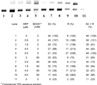
70% BOOH aqueous solution and in its absence!For the standard 1 h incubation at 37 °C, the amount of damage to the SC DNA increased with an increase in the HRP concentration (0.9-9.0 µM) with and without 60 mM BOOH. Typical gels illustrating this point for pUC 19 and pBR 322 are shown in Figures 1 and 2, respectively. This pair of gels not only confirms the greater sensitivity of pUC 19 than pBR 322 to oxidatively induced strand-Figure 1. Band areas (arbitrary units) for pUC 19 incubated under standard conditions.
scission observed previously (2) but also shows that while the HRP/BOOH couple induces more strand-scission in pUC 19 than the same concentration of HRP alone, a similar effect is far from obvious in pBR 322. The increase in strand-scission with HRP concentration and the dif-ference between these two SC DNAs in terms of their sensitivity to oxidatively induced scission and the relative effects of the HRP/BOOH couple vs HRP alone are further illustrated in the Supporting Information in Figures S1-S9 (most of which also illustrate some other point). The extent of damage to SC pUC 19 with 9.0 µM HRP is roughly the same with [BOOH] ) 60 and 6.0 mM, but damage does decrease somewhat when the [BOOH] is further reduced to 0.6 and 0.06 mM (see Figure 3). To quantify (roughly) the amounts of SC, R, and L DNA, the areas of the respective bands in each lane were determined as described under Experimental Section and are given in the figure captions together with the concentrations of any compounds (HRP, BOOH, etc.) added to the incubation medium. These areas are pre-sumed to be approximately proportional to the actual quantity of DNA present in each band. It should be noted
that total DNA and SC DNA levels greater than the levels in the control experiments are probably due both to “edge effects”6and to errors in the injection of 0.6 µL
of the SC DNA preparation into the incubation tube (maximum error ca. (10%). Nevertheless, despite such minor annoyances the overall picture is clear: HRP alone damages SC DNA in a dose-dependent manner.7With
pBR 322, this damage was generally, but not always, slightly enhanced by the addition of commercial BOOH. With pUC 19, this damage was always enhanced and was greater. In those cases where the total DNA is much lower than in the control lanes, it is most probable that there has been extensive degradation of the DNA into small fragments.
The foregoing experiments were repeated several times, always with similar results. Additional experi-ments were also carried out, some of which yielded very surprising results, if the induction of DNA damage by
HRP/BOOH really were due to BOO• radicals. For
example, the addition of 10 mM Trolox [a water-soluble analogue of vitamin E and an outstanding trap for alkylperoxyl radical in aqueous solutions (8)] had no significant protective effect as measured by the total quantity of DNA remaining after an incubation, viz., (SC + R + L), as a percentage of the control (SC DNA incubated without added reagents). Thus, in Figure 3 it can be seen that incubation of pUC 19 with HRP (9.0 µM) + BOOH (60 mM) + Trolox (10 mM) leaves (SC + R + L) DNA ) 58% (lane 12), incubation without the Trolox leaves total DNA ) 60% (lane 2) and 81% (lane 7), and incubation with HRP (9.0 µM) and Trolox (10 mM) leaves total DNA ) 58% (lane 13).8Additional evidence that 10
mM Trolox does not decrease the extent of DNA damage induced by the HRP/BOOH couple (relative to the effect of this couple at the same concentrations on incubations with HRP alone) is presented in Figures S4-S9 in the Supporting Information and below.
Another very surprising result was that catalase (CAT, which would decompose hydrogen peroxide to water and oxygen) increased DNA damage induced by 9.0 µM HRP in a dose-dependent manner. Furthermore, damage to the DNA was greater for incubations with 9.0 µM HRP plus 60 mM BOOH (see Figure S10). Tentatively, we attributed these results to adventitious transition metal ions absorbed on the surface of the CAT. These experi-ments reminded us that in some fairly recent work exploring the formation of peroxynitrate from peroxy-nitrite we had found anomalous results with commercial aqueous BOOH solutions which we attributed to the presence of hydrogen peroxide and which were eliminated 6A relatively unimportant, but annoying problem plagues the edge
lanes of many gels which contain less DNA than other lanes not subjected to extensive DNA damage. This “edge” effect is generally more obvious for the right-hand lane (usually lane 11) but can also manifest itself in there being more DNA in lane 2 that in lane 1, the lane where the DNA areas are arbitrarily set at 100% in the figure caption tables [see, e.g., Figure 2 where (SC+R) DNA ) 23% in lane 11 and 121% DNA in lane 2].
7Results for only two such “blank” experiments are shown in
reference 4. In Figure 1 of this reference, it can be seen that CIP alone increased the R DNA of pBR 322 from 18% to 21%, whereas 60 mM BOOH in the absence of CIP increased the R DNA to 35%. This “direct” effect of BOOH is consistent with our own results. The same quantity of BOOH increased the R DNA to 62% in the presence of the CIP. In Figure 2 of reference 4, it can be seen that lactoperoxidase increased the R DNA from 12% to 18% and with 6.0 mM 2,3-dimethyl-3-hydroperoxy-1-butene to 38%.
8Lane 14 in Figure 3 both is an edge lane and has no appropriate
control, i.e., no [HRP] ) 0, [BOOH] ) 60 mM lane.
Figure 2. Band areas (arbitrary units) for pBR 322 incubated under standard conditions.
Figure 3. Band areas (arbitrary units) for pUC 19 incubated under standard conditions.
by purification of the BOOH (9). In the present work, we had already demonstrated that the incubation of pUC 19 with 12 mM H2O2and HRP caused extensive
degra-dation of the SC DNA in an HRP dose-dependent fashion and, furthermore, with an added 60 mM BOOH all the DNA (i.e., all SC, R, and L DNA) was degraded (see Figure S11 in the Supporting Information). After puri-fication of the BOOH, we found that the extent of damage to both pBR 322 and pUC 19 DNA induced by HRP (1.8 and 9.0 µM) was identical (within our experimental accuracy) to that induced by the same concentration of HRP + 60 mM pure BOOH (compare lanes 2 and 3 with lanes 7 and 8 in Figures 4 and 5) (for duplicate experi-ments, see Figures S7-S9 in the Supporting Informa-tion). Furthermore, even in such ‘clean’ systems 10 mM Trolox exerted little or no protective effect on the SC DNA (see Figures 4, 5, and S7-S9). Damage to pUC 19 by 9 µM HRP and 60 mM pure BOOH was essentially the same for BOOH purified by azeotropic distillation and for that further purified by recrystallization from hexane (see Figure S12).
These results raised the obvious question: Does HRP catalytically decompose, or even react with, BOOH? In other work (3, 10), we have employed HPLC with chemiluminescence detection to measure hydroperoxide concentrations reliably and quantitatively. We therefore employed this same procedure to study the reaction of 9 µM HRP (the highest concentration employed in our DNA experiments) with 60 µM BOOH (the lowest concentra-tion employed) in our standard, metal-ion-free, buffer at 37 °C. In the absence of HRP, there was no measurable decay of the BOOH over 24 h. In the presence of HRP, only 2 µM BOOH decayed in the first hour, i.e., [BOOH]1.0h
) 58 µM. This increased to totals of 6 µM after 2 h and 13 µM after 2.75 h ([BOOH]2.0h) 54 µM, [BOOH]2.75h)
47 µM), but thereafter the reaction essentially ceased with [BOOH]24h) 0.45 µM (see Table S1 in Supporting
Information).
Discussion
In this discussion we will ignore minor differences in DNA damage induced by different reagents in view of the small irreproducibility found for several “repeat” experiments run on different gels and even run in different lanes on the same gel. In this way, we hope to avoid “over interpretation” of our results. Concentrating, therefore, on large differences in DNA damage, it is obvious that all our major results on the HRP/BOOH/ SC DNA systems point to the fact that if any BOO•
radicals (which are neutral peroxyls) actually are pro-duced in the HRP/BOOH reaction (see below) they do not induce any obvious direct single-strand-scission. This conclusion is in full agreement with our earlier work (2) but is in disagreement with the conclusions drawn in the study of various peroxidase/alkyl hydroperoxide/pBR 322 systems (4). The main facts leading to our present conclusion are the following:
(i) HRP induces extensive DNA damage in a dose-dependent manner in the absence of BOOH. For pBR 322, this damage is only slightly enhanced by 60 mM commercial (H2O2-contaminated) BOOH (see Figure 2).
With pUC 19 which, for reasons we have not explored, is more sensitive than pBR 322 to radical-induced strand scission (2), 60 mM commercial BOOH alone induced more extensive DNA damage than with pBR 322, and this damage was greatly enhanced by HRP (see Figure 1). However, neither pBR 322 nor pUC 19 treated with HRP and 60 mM pure BOOH were damaged to a significantly greater extent than when these SC DNAs were treated with the same concentration of HRP only (compare lanes 2 and 3 with lanes 7 and 8 in Figures 4 and 5). Clearly, pure BOOH is simply an innocent
bystander in HRP/DNA reactions.9
(ii) The rate constant for the reaction of Trolox with BOO• radicals will be very much greater (probably by
several orders of magnitude) than the rate constant for the reaction of BOO• with the DNA [which, even in bp
9Even with the maximum concentration of HRP (9 µM) and BOOH
(60 mM) employed in this work, only one gel (pUC 19, Figure S12) gave results which could suggest that pure BOOH may not be completely innocent. In this gel, the odd lanes, 3, 5, 7, and 9, had HRP only, and the means (ranges) of SC and R DNAs (in arbitrary units) were 28 (23-24) and 42 (38-52), respectively, while the even lanes, 4, 6, 8, and 10, with HRP + BOOH have means (ranges) of SC and R DNA of 15 (6-31!) and 58 (50-67), respectively. Since the ranges for SC and also for R DNA in the two systems overlap, we will not “over interpret” these results.
Figure 4. Band areas (arbitrary units) for pBR 322 incubated under standard conditions.
Figure 5. Band areas (arbitrary units) for pUC 19 incubated under standard conditions.
units, is present at very much lower concentrations (viz.,16 µM in bp) than the Trolox (10 mM)]. Since 10 mM Trolox provides no protection to DNA stressed with HRP/BOOH (see Figures 3-5 and Figures S4-S9 in Supporting Information), it is quite clear that BOO•
radicals cannot be the DNA damaging species in these systems.
(iii) Although the HRP/60 mM (pure) BOOH couple generally9 induces no greater DNA damage than that
induced by the same concentration of HRP alone (see Figures 4 and 5 and Figures S7-S9 in the Supporting Information), the HRP/60 mM BOOH/H2O2system causes
considerably more DNA damage than HRP alone at the same concentration. This is generally true for the ad-ventitious H2O2in commercial BOOH (Figures 1-3 and
S1-S3) and for 12 mM H2O2deliberately added (Figure
S11 in the Supporting Information).10This again leads
to the conclusion that the DNA damaging agent in our experiments is not the BOO• radical but is HRP itself
or, more probably, an oxidized form of HRP. Since it has been demonstrated that O2can effect catalytic oxidations
by HRP in the presence of a reducing agent (11), we carried out some experiments with N2-purged solutions
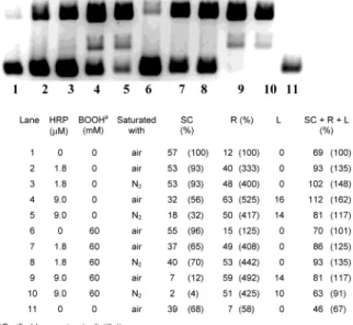
(see Figure 6). Our results argue against an O2-oxidized
form of HRP being the DNA damaging agent. Indeed, if small differences in the degree of DNA damage were to be considered (which we prefer not to do), it would appear that damage may be greater under N2than under air!
Nevertheless, because very little O2would be required
to produce the observed amount of damage (vide infra), we cannot completely rule out a role for O2 in these
reactions.
At this point it is appropriate to consider whether alkylperoxyl radicals are generated in peroxidase/alkyl hydroperoxide systems. A 1988 claim that peroxyl radi-cals (and alkoxyl radiradi-cals) are produced by reaction of HRP (and other heme-proteins) with BOOH was based
on spin-trapping with 5,5-dimethyl-1-pyrroline-N-oxide (DMPO) and assignment of one of the ESR spin-adduct spectra to DMPO-OOB (12). Similarly, one of the ESR spin-adduct spectra in the CIP/BOOH/DMPO system was also assigned to DMPO-OOB (4). However, Dikalov and Mason (13) have demonstrated that all claims for DMPO-OOB (and related) spin-adducts are incorrect, a conclu-sion that has received overwhelming support from the work of Honeywell and Mile (14). Thus, the BOOH-derived spin-adduct assigned by numerous workers to DMPO-OOB is not formed in the absence of O2, instead
the adduct is DMPO-CH3, the CH3radical being produced
via the β-scission of tert-butoxyl (BO•) radicals (13). In
the presence of O2and with a variety of BOOH
“activat-ing” systems (including peroxidases), all the ESR spectra which have been assigned to DMPO-OOB are actually due to DMPO-OCH3 (13).11 Depending on the system
employed, the BO• radicals may have been produced
either by the bimolecular self-reaction of BOO•radicals
and/or by one-electron reduction of BOOH. The only other observation implying peroxyl radical formation in per-oxidase/hydroperoxide systems was the evolution of O2
upon reaction of 3-hydroperoxy-1-butene (3HB, 28 mM) with an extremely high concentration of CIP (740 µM) (4). It is also worth noting that the oxidation of 2′-deoxyguanosine by the CIP/3HB system was reduced by the addition of “radical scavengers” (4). However, one of these “scavengers”, tert-butyl alcohol, is completely un-reactive toward alkylperoxyl radicals, k < 10-2M-1s-1
(though it does scavenge HO•radicals), and all the radical
scavengers employed are certainly less reactive toward peroxyl radicals than Trolox (which had absolutely no DNA-protecting ability in the HRP/BOOH system).
At this point, we decided to check whether 9 µM HRP (the highest concentration used in our DNA experiments) could decompose 60 µM BOOH (the lowest concentration used). The concentrations of BOOH decomposed after 1, 2, 2.75, and 24 h were 2, 6, 13, and 15 µM, respectively (see Table S1). These results prove that HRP can decompose BOOH but that it is not an effective catalyst, there being only a slow and limited reaction. The extent of this reaction is very slight over the 1 h incubation periods employed with the DNA, but it may be that just sufficient oxidized HRP (vide infra) is produced to account for the possible small increase in DNA strand-scission occasionally observed with HRP and pure BOOH relative to HRP alone.9
Our present results on the HRP/BOOH system indi-rectly support our earlier contention (2) that neutral alkylperoxyl radicals are, at best, very inefficient DNA cleaving agents.
Finally, it should be noted that DNA cleavage by a high-valent form of HRP (Compound I or II) is a pos-sibility because the heme edge is exposed in this enzyme (17) and will oxidize other large molecular weight sub-strates, including proteins, lipids, and LDL (18-21). The agent which oxidizes the HRP is unknown, but, provided DNA cleavage by the oxidized HRP is efficient, very little will be required. Indeed, sufficient oxidized HRP may even have been present in our purchased HRP samples (which was one reason for employing HRP purchased from two different sources). That is, for example, 9.0 µM 10H
2O2(12 mM) and BOOH (60 mM) degraded all pUC 19 DNA
even in the absence of HRP (Figure S11). It is likely that adventitious transition metal ions are absorbed on the DNA polyanion and that BOOH (or its products) recycles these ions from their upper to lower valence state where they will react with H2O2to produce the DNA
cleaving HO•radical.
11A claim that DNA damage by BOOH/hemoglobin is due to BOO• -(15), in which the evidence for BOO•formation is also based on spin-trapping with DMPO (16), should be reevaluated.
Figure 6. Band areas (arbitrary units) for pBR 322 incubated both under standard conditions (air saturated) and under similar conditions with nitrogen-purged samples.
HRP produced ca. 71% single-strand cleavage of pBR 322 with no other reagents added (lane 3, Figure 4). In this experiment, the pBR 322 concentration was 3.67 nM. Since the first single-strand break converts the SC DNA to R DNA, then 3.67 × 0.71 ) 2.6 nM of oxidized HRP could “do the job” (assuming that SC DNA cleavage by the oxidized HRP is 100% efficient). This represents only 2.6 × 10⁻⁹/9.0 × 10-6) 2.9 × 10⁻⁴ (i.e., 0.03%) of the
HRP used in the experiment. This simple calculation emphasizes that extremely little oxidized HRP may be required to explain our observation that HRP can, by itself, effect DNA cleavage. If minute traces of oxidized HRP are not present in commercial HRP, only small quantities of adventitious H2O2(or, less probably O2, vide
supra) may be required to produce the observed results.
Acknowledgment. We are extremely grateful to Dr. Thomas Paul for his advice and encouragement through-out the course of this work. We also thank Dr. Cristina Sanchez for help with the first experiment and Sue Lin for purifying the BOOH.
Supporting Information Available: Photographs and band areas for various experiments with pBR 322 and pUC 19 (Figures S1-S12) and effect of 9 µM HRP on 60 µM BOOH (Table S1). This material is available free of charge via the Internet at http://pubs.acs.org.
References
(1) Marnett, L. J. (2000) Oxyradicals and DNA damage.
Carcino-genesis 21, 361-370.
(2) Paul, T., Young, M. J., Hill, I. E., and Ingold, K. U. (2000) Strand cleavage of supercoiled DNA by water-soluble peroxyl radicals. The overlooked importance of peroxyl radical charge. Biochemistry
39, 4129-4135.
(3) Bedard, L., Young, M. J., Hall, D., Paul, T., and Ingold, K. U. (2001) Quantitative studies on the peroxidation of human low-density lipoprotein initiated by superoxide and by charged and neutral alkylperoxyl radicals. J. Am. Chem. Soc. 123, 12439-12448.
(4) Adam, W., Kurz, A., and Saha-Mo¨ller, C. R. (2000) Peroxidase-catalyzed oxidative damage of DNA and 2′-deoxyguanosine by model compounds of lipid hydroperoxides: involvement of peroxyl radicals. Chem. Res. Toxicol. 13, 1199-1207.
(5) Russell, G. A. (1957) Deuterium-isotope effects in the autoxidation of aralkyl hydrocarbons. Mechanism of the interaction of peroxy radicals. J. Am. Chem. Soc. 79, 3871-3877.
(6) Inouye, S. (1984) Site-specific cleavage of double-strand DNA by hydroperoxide of linoleic acid. FEBS Lett. 172, 231-234.
(7) Ha¨ring, M., Ru¨diger, H., Demple, B., Boiteux, S., and Epe, B. (1994) Recognition of oxidized abasic sites by repair endonu-cleases. Nucleic Acids Res. 22, 2010-2015.
(8) Barclay, L. R. C., Locke, S. J., MacNeil, J. M., VanKessel, J., Burton, G. W., and Ingold, K. U. (1984) Autoxidation of micelles and model membranes. Quantitative kinetic measurements can be made using either water-soluble or lipid-soluble initiators with water-soluble or lipid-soluble chain-breaking antioxidants. J. Am.
Chem. Soc. 106, 2479-2481.
(9) Hodges, G. R., and Ingold, K. U. (1999) Cage-escape of geminate radical pairs can produce peroxynitrate from peroxynitrite under a wide variety of experimental conditions. J. Am. Chem. Soc. 121, 10695-10701.
(10) Fowler, G., Daroszewska, M., and Ingold, K. U. (2002) Melatonin does not “directly scavenge hydrogen peroxide”. Demise of another myth. Free Radical Biol. Med. (submitted for publication). (11) Ozaki, S., Watanabe, S., Hayasaka, S., and Konuma, M. (2001)
The activation of molecular oxygen by horseradish peroxidase with sodium sulfite. J. Chem. Soc., Chem. Commun., 1654-1655. (12) Davies, M. J. (1988) Detection of peroxyl and alkoxyl radicals produced by reaction of hydroperoxides with heme-proteins by electron spin resonance spectroscopy. Biochim. Biophys. Acta 964, 28-35.
(13) Dikalov, S. I., and Mason, R. P. (1999) Reassignment of organic peroxyl radical adducts. Free Radical Biol. Med. 27, 864-872. (14) Honeywell, J. D., and Mile, B. (2002) J. Chem. Soc., Perkin Trans.
2, 569-575.
(15) Kanazawa, A., Sawa, T., Akaik, T., and Maeda, H. (2000) Formation of abasic sites in DNA by tert-butyl peroxyl radicals: implication for potent genotoxicity of lipid peroxyl radicals. Cancer
Lett. 156, 51-55.
(16) Akaike, T., Sato, K., Ijiri, S., Miyamoto, Y., Kohno, M., Ando, M., and Maeda, H. (1992) Bactericidal activity of alkyl peroxyl radicals generated by heme-iron-catalyzed decomposition of or-ganic peroxides. Arch. Biochem. Biophys. 294, 55-63.
(17) Ator, M. A., David, S. K., and Ortiz de Montellano, P. R. (1987) Structure and catalytic mechanism of horseradish peroxidase. Regiospecific meso alkylation of the prosthetic heme group by alkyl hydrazines. J. Biol. Chem. 262, 14954-14960.
(18) Aesbach, R., Amado, R., and Nekom, H. (1976) Formation of dityrosine cross-links in proteins by oxidation of tyrosine residues.
Biochim. Biophys. Acta 439, 292-301.
(19) Kim, E. H., and Sevanian, A. (1991) Hematin- and peroxide-catalyzed peroxidation of phospholipid liposomes. Arch. Biochem.
Biophys. 288, 324-330.
(20) Heinecke, J. W., Li, W., Francis, G. A., and Goldstein, J. A. (1993) Tyrosyl radical generated by myeloperoxidase catalyzes the oxidative cross-linking of proteins. J. Clin. Invest. 91, 2866-2872. (21) Wieland E., Parthasarathy, S., and Steinberg, D. (1993) Peroxi-dase-dependent metal-independent oxidation of low-density lipo-protein in vitro: a model for in vivo oxidation? Proc. Natl. Acad.
Sci. U.S.A. 90, 5929-5933.
TX020043O