A cell-based drug delivery platform for
treating central nervous system inflammation
The MIT Faculty has made this article openly available.
Please share
how this access benefits you. Your story matters.
Citation
Levy, Oren et al. "A cell-based drug delivery platform for treating
central nervous system inflammation." Journal of Molecular
Medicine 99, 5 (January 2021): 663–671. © 2021 Springer-Verlag
GmbH Germany, part of Springer Nature
As Published
https://doi.org/10.1007/s00109-020-02003-9
Publisher
Springer Science and Business Media LLC
Version
Author's final manuscript
Citable link
https://hdl.handle.net/1721.1/130518
Terms of Use
Article is made available in accordance with the publisher's
policy and may be subject to US copyright law. Please refer to the
publisher's site for terms of use.
AUTHOR ACCEPTED MANUSCRIPT
© 2020 Springer-Verlag GmbH Germany, part of Springer Nature
A cell-based drug delivery platform for treating central nervous system
inflammation
Cite this article as: Oren Levy, A cell-based drug delivery platform for treating central nervous system
inflammation, Journal of Molecular Medicine, doi: 10.1007/s00109-020-02003-9
This Author Accepted Manuscript is a PDF file of a an unedited peer-reviewed manuscript that
has been accepted for publication but has not been copyedited or corrected. The official version
of record that is published in the journal is kept up to date and so may therefore differ from this
version.
Terms of use and reuse: academic research for non-commercial purposes, see here for full terms.
A cell-based drug delivery platform for treating central nervous system inflammation
Oren Levy1*, Veit Rothhammer2*, Ivan Mascanfroni2*, Zhixiang Tong1, Rui Kuai1,3, Michael De Biasio1,
Qingping Wang4, Tahir Majid5, Christelle Perrault6, Ada Yeste2, Jessica E. Kenison2,Helia Safaee1, Juliet
Musabeyezu1, Martina Heinelt1, Yuka Milton1, Heidi Kuang1, Hao Yue Lan1, William Siders7,
Marie-Christine Multon8, Jonathan Rothblatt9, Salam Massadeh10,11, Manal Alaamery10,11, Ali H. Alhasan11,12,
Francisco J. Quintana2,3,13+, and Jeffrey M. Karp 1,3,13+
1
Department of Anesthesiology, Perioperative and Pain Medicine, Brigham and Women’s Hospital,
Harvard Medical School, Harvard Stem Cell Institute, Boston, MA, USA. 2Center for Neurologic
Diseases, Brigham and Women’s Hospital, Harvard Medical School, Boston, MA, USA. 3Centre of
Excellence for Biomedicine, Brigham and Women’s Hospital, Boston, MA, USA. 4
Sanofi R&D,
Department of Drug metabolism and Pharmacokinetics, Waltham, MA, USA. 5Sanofi R&D, Chemical
Research, Integrated Drug Discovery, Waltham, MA, USA. 6Sanofi R&D, In vitro Pharmacology,
Integrated Drug Discovery, Centre de Recherche Vitry-Alfortville, Vitry-Sur-Seine, France. 7Genzyme
R&D, Neuroimmunology Research, Framingham, MA, USA. 8Sanofi R&D, Translational Sciences,
Centre de Recherche Vitry-Alfortville, Vitry-Sur-Seine, France. 9 Sanofi R&D, Global BioTherapeutics,
Cambridge, MA 02139 USA. 10Developmental Medicine Department, King Abdullah International
Medical Research Center, Ministry of National Guard Health Affairs, Riyadh, Saudi Arabia. 11Centre of
Excellence for Biomedicine, Joint Centers of Excellence Program, King Abdulaziz City for Science and
Technology, Riyadh, Saudi Arabia. 12National Center of Pharmaceutical Technology, Life science and
Environment Research Institute, King Abdulaziz City for Science and Technology, Riyadh, Saudi Arabia.
13
The Broad Institute of Harvard and MIT, Cambridge, MA, USA.
*
Authors contributed equally to this study.
+
Abstract
Mesenchymal stem cells (MSCs) are promising candidates for the development of cell-based drug
delivery systems for autoimmune inflammatory diseases, such as multiple sclerosis (MS). Here, we
investigated the effect of Ro-31-8425, an ATP-competitive kinase inhibitor, on the therapeutic properties
of MSCs. Upon a simple pretreatment procedure, MSCs spontaneously took up and then gradually
released significant amounts of Ro-31-8425. Ro-31-8425 (free or released by MSCs) suppressed the
proliferation of CD4+ T cells in-vitro following polyclonal and antigen-specific stimulation. Systemic
administration of Ro-31-8425-loaded MSCs ameliorated the clinical course of experimental autoimmune
encephalomyelitis (EAE), a murine model of MS, displaying a stronger suppressive effect on EAE than
control MSCs or free 31-8425. 31-8425-MSCs administration resulted in sustained levels of
Ro-31-8425 in the serum of EAE mice, modulating immune cell trafficking and the autoimmune response
during EAE. Collectively, these results identify MSC-based drug delivery as a potential therapeutic
strategy for the treatment of autoimmune diseases.
Key messages
• MSCs can spontaneously take up the ATP-competitive kinase inhibitor Ro-31-8425.
• Ro-31-8425-loaded MSCs gradually release Ro-31-8425 and exhibit sustained suppression of T cells. • Ro-31-8425-loaded MSCs have more sustained serum levels of Ro-31-8425 than free Ro-31-8425. • Ro-31-8425-loaded MSCs are more effective than MSCs and free Ro-31-8425 for EAE therapy.
Introduction
Multiple sclerosis (MS) is an autoimmune disease of the central nervous system (CNS),
characterized by inflammatory lesion formation, demyelination, axonal loss, and progressive
neurodegeneration [1]. In the majority of patients, MS initially shows a relapsing-remitting course
(RRMS), in which acute attacks are followed by a varying degree of recovery [2]. The majority of RRMS
patients eventually develops secondary progressive MS (SPMS), characterized by the irreversible
accumulation of neurological disability [2, 3].
Immunomodulatory strategies for the relapsing remitting phase of MS have been developed, as
exemplified by glatiramer acetate, interferon beta, dimethylfumarate, or natalizumab [4-7]. However,
these strategies often come at the price of severe side effects or partial clinical activity. Therefore, there is
an unmet need for therapeutic approaches for MS, which are not only highly efficacious but also exhibit
minimal side effects. Given these requirements, mesenchymal stem cell (MSC)-based therapies may
represent a novel approach for the treatment of MS and other autoimmune inflammatory diseases. MSCs
are promising candidates for cell therapy due to their safety, scalability, immune-evasive phenotype, and
their potent immunomodulatory activities [8, 9]. MSCs modulate both adaptive and innate immune
responses, suppressing the activation of T cells, B cells, natural killer cells and antigen-presenting cells
[10, 11], making them of particular interest for the treatment of MS [12, 13]. Indeed, MSCs have shown
efficacy in the treatment of the MS animal model, experimental autoimmune encephalomyelitis (EAE),
by modulating the activity of dendritic cells and T cells [12, 14, 15]. Moreover, recent reports suggest that
MSCs can be used to reduce autoimmune inflammation in MS patients [16, 17]. However, their
post-infusion potency is unpredictable [14, 15], raising the need for safe and scalable bioengineering
approaches to further maximize the therapeutic potential of MSCs in the context of MS.
One emerging strategy to develop novel and efficacious cellular therapies for inflammation is to
harness the scalable, clinically safe immunomodulatory activities of MSCs for cell-based drug delivery.
We and others have previously loaded MSCs with anti-cancer drugs and demonstrated their anti-tumor
platform to identify small molecules that boost MSC anti-inflammatory function [20]. Ro-31-8425, a
PKC inhibitor with anti-inflammatory properties [21], also known as bisindoleamine X, upregulates
integrin ⍺L (CD11a), a membrane receptor relevant for cell adhesion, tissue homing, and costimulatory signaling at sites of inflammation [22]. Ro-31-8425-pretreated MSCs displayed improved homing to sites
of inflammation upon systemic administration and a superior anti-inflammatory impact in a murine ear
inflammation model [20].
PKC-governed pathways play a central role in T cell activation and differentiation to regulate the
inflammatory response. Inhibiting different PKC isoforms and dependent pathways has thus been
proposed as a strategy to combat autoimmune disorders [23-27]. Considering the role of PKC in
autoimmune responses and our previous observations of the boosted immunomodulatory effects of
pretreated MSCs in inflammatory settings, we sought to explore the impact of
Ro-31-8425-MSCs in the experimental autoimmune encephalomyelitis (EAE) mouse model of MS. Herein, we report
that with a simple pretreatment protocol, Ro-31-8425 was spontaneously internalized by MSCs and then
released from cells to suppress antigen-specific proliferation of CD4+ T cells in vitro. Furthermore,
systemically infused 8425-pretreated MSCs outperformed the vehicle-MSCs and the free
Ro-31-8425 group in the EAE mouse model, significantly reducing the clinical score. Ro-31-Ro-31-8425-MSCs led to
sustained levels of Ro-31-8425 in the serum, consequently modulating T cell driven autoimmunity.
Collectively, this study demonstrates the potential of MSCs as promising drug delivery vehicles for the
Materials and methods
Mesenchymal stem cell culture and compound pretreatment
Human MSCs were purchased from Lonza and expanded in StemPro® MSC SFM CTS™ with 10% SFM supplement (Gibco) (herein referred to as full StemPro medium). Cells were kept at 37°C with 5% CO2
and media was changed every 3 days. Cells were passaged using 1% trypsin-EDTA solution. MSCs at
passage 3-6 were used for all experiments. For compound pretreatment, pre-confluent MSC were
incubated in StemPro medium for 24 hours with Ro-31-8425 at the indicated concentrations.
Co-culture system of MSCs and splenocytes
A co-culture system of MSCs and splenocyes was used to directly assess the immunosuppressive
properties of Ro-31-8425 pretreated MSCs (cpd-pretreated MSCs). For polyclonal T cell stimulation,
bulk splenocytes from naive wild type C57bl/6 mice were fluorescently labelled with CFSE (5 µM) and
activated with antibodies to CD3 and CD28 (anti-CD3 (clone 17A2) 4 µg/ml plate-bound, anti-CD28
(clone PV-1) 2 µg/ml soluble, both BioXCell). After 72 hours, cells were stained for CD3 and CD4 and
analyzed by FACS for CFSE dilution in the CD3CD4 double positive population. For antigen specific
proliferation, splenocytes were isolated from 2D2 transgenic mice expressing a transgenic T cell receptor
specific for MOG35-55. 100.000 bulk 2D2 splenocytes were activated with MOG35-55 (2 µg/ml) or vehicle
and incubated directly with Ro-31-8425 or Ro-31-8425-pretreated MSCs. During the final 16 h, cells
were pulsed with 1 Ci [3H]thymidine (PerkinElmer), followed by collection on glass fiber filters and
analysis of incorporated [3H]thymidine in a beta-counter (1450 MicroBeta TriLux; PerkinElmer).
Compound storage and release from cpd-pretreated MSCs
15,000 MSCs/well in a 96 well were pretreated with Ro-31-8425 at 3μM for 24h. At 24h, cellular
supernatants were collected and cells were rinsed and then lysed by incubating 1hr at 37°C with lysis
point when the processed was repeated. Each collected sample was vortexed with 3vol of Acetonitril,
centrifuged 15min at 3000g and then analyzed for Ro-31-8425 via MassSpec.
Animal models
All experiments were performed in accordance with guidelines of the Institutional Animal Care and Use
Committee at Harvard Medical School, and mice were kept in a pathogen-free facility at the Harvard
Institutes of Medicine. For EAE model, 8-10 week old female C57BL/6 mice were immunized with a
peptide of amino acids amino acids 35–55 of myelin oligodendrocyte glycoprotein (MOG35–55) in an
emulsion of complete Freund's adjuvant with mycobacterium tuberculosis and pertussis toxin as
previously described [28-30]. C57BL/6 mice developed clinical signs of EAE after 9-13 days, with peak
disease lasting 1-3 days after EAE onset, and mice completely or partially recover within 7-10 days.
Animals were scored daily in a blinded manner using the following scoring system: 1, loss of tone in the
tail; 2, hind limb paresis; 3, hind limb paralysis; 4, tetraplegia; 5, moribund. Animals were sacrificed
during the recovery phase for cellular analyses.
MSC administration
MSCs (native or cpd-pretreated) were stained using violet CellTrace and 0.25x10⁶ cells (in 100 µL PBS)
were administered (via tail vein injection) into each mice. To elucidate the impact of disease stage on
MSC homing to CNS, cells were administered prior to disease onset (days 3 and 8 in the RR EAE b6
model). Cell presence in the CNS was assessed 24h post the second cell infusion and at the end of the
experiment via flow cytometry.
Quantitative measurement of Ro-31-8425 in mouse serum by Liquid Chromatography-Tandem mass spectrometry (LC-MS)
Serum (20 uL) from each group was processed by adding 3 volume of acetonitrile to precipitate protein
system, coupled to an API-6500 mass spectrometry. The analytes were separated using a reverse phase
C18 column (Waters, BEH 1.7 µm, 100Å, 2.1x50 mm) at 50oC and the mobile phases consisted of A:
water containing 0.1% formic acid and B: acetonitrile containing 0.1% formic acid with a flow rate of 0.8
mL/min. The flow was sprayed to API-6500 mass spectrometer at electrospray ionization (ESI) positive
mode in unit resolution and source temperature of 550oC. Multiple reaction monitoring (MRM) was used
to monitor the transition of protonated parent ion 425.1Da to the product ion 305.0 Da for compound
Ro-31-8425, transition of 237.1 Da to 194.4 Da for internal standard carbamazepine. The limit of
quantification (LOQ) of Ro-31-8425 in the mouse serum was 0.1ng/ml. The method was linear in the
range of 0.1–250 ng/ml with a coefficient of correlation greater than 0.994 in the mouse serum.
Detection of MSCs in CNS of EAE mice and distribution in other organs and FACS staining
Assessment of MSC presence in the CNS was performed as previously described [31, 32]. Briefly, mice
were perfused with ice-cold PBS, the brain and spinal cord were removed and incubated with collagenase
type III and DNase in PBS. Tissues were then homogenized and loaded on a Percoll gradient to enrich
CNS infiltrates. Flow cytometry analysis for detection of stained MSCs, as well as DCs, encephalitogenic
and regulatory T cells, was performed. Similarly, the kidneys, lungs, spleen, gut and heart were
homogenized and analyzed for MSC presence. Antibodies for flow cytometry were purchased from
eBioscience or BD Pharmingen and used at a concentration of 1:100 unless recommended otherwise by
the manufacturer. Cells were analyzed on a LSRII or MACSQuant flow cytometer (BD Biosciences and
Miltenyi Biotec, respectively). As outlined in the individual figures, Th1 cells were defined as
CD3+CD4+IFN-𝜸+
IL-17-IL-10-Foxp3-, Th17 cells as CD3+CD4+IFN-𝜸
-IL-17+IL-10-Foxp3-, Treg cells as
CD3+CD4+IFN- 𝜸
Results
Uptake and gradual release of Ro-31-8425 by MSCs
We previously demonstrated that Ro-31-8425 pretreatment improves homing of systemically
administered MSCs to sites of inflammation, resulting in increased suppression of local skin
inflammation [20]. To explore the potential of MSCs for drug delivery, we evaluated the uptake and
release of Ro-31-8425 by MSCs. We treated MSCs with Ro-31-8425 (3 µM) for 24h, and after this
loading period we washed the cells, replaced the media and collected supernatant samples every 24h for
72h. Next, the cells were lysed and Ro-31-8425 levels in cell lysate and supernatant samples were
measured by liquid chromatography-mass spectrometry (LCMS). Following a 24 h loading, MSCs
retained about 25% of Ro-31-8425 available in the pretreatment solution and released it gradually over
time (Fig. 1a). This secretion profile suggests that MSCs may be used for the delivery of
immunomodulatory drugs such as Ro-31-8425, releasing them gradually into the blood stream or target
organs to maximize their therapeutic efficacy.
Ro-31-8425 suppresses CD4+ T cell proliferation
We next evaluated the immunosuppressive effects of free and MSC-delivered Ro-31-8425 in a polyclonal
activation assay and in an antigen-specific system. Therefore, untreated MSCs, Ro-31-8425-loaded MSCs
or free Ro-31-8425, were co-cultured with splenocytes from naive wild type C57bl/6 mice stimulated
with antibodies against CD3/CD28 (Fig. 1b) or instead with splenocytes from 2D2 transgenic mice,
which harbor a transgenic T-cell receptor specific to myelin oligodendroglycoprotein (MOG35-55) and can
be activated by their cognate antigen MOG35-55 presented by antigen presenting cells [33] (Fig. 1c). In
response to CD3/CD28 stimulation, Ro-31-8425 displayed a dose-dependent inhibitory effect on CD4+ T
cell proliferation when incubated directly with the splenocytes or when “stored” inside MSCs that were pretreated with different concentrations of Ro-31-8425. Importantly, Ro-31-8425 loaded MSCs resulted
in a slower, more sustained suppression of T-cell proliferation. Similarly, Ro-31-8425 also inhibited the
sustained immunosuppressive effect of Ro-31-8425 when loaded into MSCs in this co-culture system.
These findings suggest that MSCs store and slowly release Ro-31-8425 to suppress T-cell proliferation in
a dose-dependent manner in response to both polyclonal and antigen-specific activation. Importantly,
Ro-31-8425-loaded MSCs displayed more potent anti-proliferative effects compared to untreated control
MSCs, suggesting that Ro-31-8425-MSCs may also show increased anti-inflammatory activity than
untreated MSCs in vivo.
Improved therapeutic efficacy of MSC-delivered Ro-31-8425
We evaluated the therapeutic impact of Ro-31-8425-loaded MSCs in EAE induced in C57Bl/6 mice by
immunization with MOG35-55 [12]. Vehicle control- or Ro-31-8425-loaded MSCs were administered
intravenously on days 3 and 8 following EAE induction; an additional group received free Ro-31-8425, at
an amount equivalent to that loaded in the MSCs. Clinical signs of EAE were assessed by daily blinded
scoring (Fig. 2a). The administration of Ro-31-8425-loaded MSCs led to a significant amelioration of
EAE, and this therapeutic effect was stronger than the one observed following treatment with free
Ro-31-8425 or vehicle-loaded MSCs only.
Systemic cell-based delivery of RO-31-8425 results in prolonged drug circulation
To elucidate the mechanism responsible for the increased therapeutic effect of RO-31-8425-MSCs, we
analyzed serum samples collected 24 hours after each MSC infusion or free Ro-31-8425 administration
on days 3 and 8 using a validated LC-MS method (Fig. 2b). We found that Ro-31-8425 serum levels in
mice infused with Ro-31-8425-MSCs were higher (>5-fold) than those measured in mice receiving an
equivalent amount of free Ro-31-8425. We also analyzed serum samples at multiple time points post
intravenous administration of a single dose of 8425-MSCs or an equivalent amount of free
Ro-31-8425. We detected a 3-fold larger area under the curve (AUC) in samples from Ro-31-8425-MSCs treated
mice (Fig. 2c), suggesting that MSC-based delivery results in a sustained release of RO-31-8425 into the
RO-31-8425 does not modify MSC bio-distribution
To determine whether Ro-31-8425 affects MSC bio-distribution, we tracked MSCs during EAE in vivo.
Fluorescently labeled vehicle- or Ro-31-8425-loaded MSCs were administered intravenously on days 3
and 8 after induction of EAE. 24h after the last MSC administration, MSCs tissue distribution was
determined by FACS. MSCs were detected in the lung, liver, heart and kidney, and this bio-distribution
was not affected by the loading of Ro-31-8425 into MSCs. Indeed, Ro-31-8425 loading did not promote
MSC homing to the CNS (Fig. 2d). We also examined the drug concentration in the brain, spleen, and
lymph nodes and found the drug concentrations in these organs were not significantly changed by loading
Ro-31-8425 in MSCs (Fig. S1). These results indicate that Ro-31-8425 administration in drug loaded
MSCs does not affect MSC homing in vivo, and does not affect drug concentration in major organs.
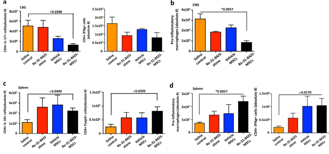
RO-31-8425-MSCs modulate the immune response during EAE
We next analyzed the effect of RO-31-8425-MSCs on the immune response during EAE.
Ro-31-8425-MSC treatment led to a decrease in the number of Th17 (CD3+CD4+IL17+) and Th1 (CD3+CD4+
IFN-𝜸+
IL-10-) cells in the CNS, as determined by flow cytometry (Fig. 3a). Ro-31-8425-loaded MSCs also
decreased the recruitment of pro-inflammatory macrophages (CD11b+CD45+Ly6Chi) to the CNS (Fig.
3b). In addition, Ro-31-8425-MSC administration increased the number of splenic anti-inflammatory
IL-10+ type 1 regulatory cells and Foxp3+ T regulatory cells (Fig. 3c), and it also increased the number of
pro-inflammatory macrophages and Th1 cells in the spleen (Fig. 3d). Taken together, these findings
suggest that Ro-31-8425-MSCs alter immune cell trafficking from the spleen to the CNS, limiting CNS
inflammation. In addition, Ro-31-8425 might also alter the priming and maturation of pro-inflammatory T
cells, as reflected by an increase of anti-inflammatory T-cell phenotypes in the spleen. Overall, our data
highlights the potential of Ro-31-8425 pretreatment in guiding the development of MSC-based therapies
for MS. Instead, MSCs appear to be acting as a living delivery platform, resulting in sustained serum
levels of Ro-31-8425, potentially modulating the immune response by impacting immune cell trafficking
Discussion
MSCs are considered potential candidates for MS therapy [12, 13, 34, 35]. In this study, we
evaluated the use of MSCs for the delivery of the anti-inflammatory compound Ro-31-8425. We have
previously shown that Ro-31-8425 increases the recruitment of MSCs to sites of inflammation, boosting
the immunosuppressive activity of MSCs [20]. In our current study, we show that free and MSC-loaded
Ro-31-8425 suppresses T-cell proliferation. MSCs store Ro-31-8425, releasing it in vivo at a constant rate
to increase its therapeutic activity. Indeed, Ro-31-8425-MSCs displayed improved therapeutic activity in
EAE, outperforming free Ro-31-8425 and vehicle-loaded MSCs. Interestingly, Ro-31-8425 did not affect
MSCs bio-distribution. In addition, it is also possible that Ro-31-8425 changes the secretome of MSCs,
boosting their therapeutic effects. Indeed, in previous studies we found that Ro-31-8425 pretreatment
attenuates the secretion of certain inflammatory cytokines and chemokines (e.g. IL-6 and MCP-1) from
MSCs, while majority of other tested cytokines and chemokines remained unaffected (Levy et al., 2015),
suggesting that Ro-31-8425 has limited effects on the secretome. Thus, future studies are needed to
further polarize the secretome towards superior therapeutic potency [36]. Collectively, these data suggest
that the improved therapeutic activity of 8425-MSCs results from the extended release of
Ro-31-8425 in the circulation, resulting in a stronger modulation of immune cell priming and trafficking during
EAE.
Ro-31-8425 is a cell-permeable kinase inhibitor that is considered a reversible and selective
inhibitor of PKC, a family of kinases that has been shown to participate in the pathogenesis of multiple
inflammatory diseases. For instance, PKC-theta is expressed largely in T cells, governing T-cell
activation and survival [23, 25]. Consequently, PKC-theta signaling in T cells is associated with various
inflammatory diseases and murine models of MS, inflammatory bowel disease, arthritis and asthma [25],
identifying the selective inhibition of PKC-theta as a potential therapeutic approach for T-cell
autoimmunity [25]. Indeed, several PKC-theta inhibitors have shown efficacy in experimental models of
regulates pain sensitivity and locomotor function and its levels are dependent on the degree of motor
function in a murine EAE model [26]. Also, the PKC inhibitor bisindolylmaleimide VII (which belongs to
the same chemical family of bisindoleamides as Ro-31-8425) prevented EAE progression in rats [38]. In
our study, MSC-delivered Ro-31-8425, a compound which was previously reported to inhibit different
PKC isoforms and displayed anti-inflammatory properties in edema and arthritis animal models [21],
modulated immune responses by impacting immune cell trafficking and activation. Indeed, when
delivered by MSCs, serum levels of Ro-31-8425 (Figure 2b and 2c) were above the previously reported
IC50 of Ro-31-8425 on different PKC isoforms (for instance, IC50 = 8 nM for PKCα, 8 nM for PKCβI,
14 nM for PKCβII and 13 nM for PKCγ) [39, 40]. We are currently evaluating the role of different PKC isoforms in our model, but the results presented herein support a potential role of the PKC axis during this
autoimmune response.
Our data also highlights the potential of using MSCs to deliver immunomodulatory drugs as a
strategy to achieve sustained drug levels in the circulation to maximize their therapeutic efficacy. Pessina
et al. previously used MSCs for the delivery of the chemotherapeutic drug Paclitaxel, improving its anti-tumor activity in murine models of leukemia and glioblastoma, demonstrating the broad applicability of
this approach [41] [19]. While systemic administration of drug-loaded erythrocytes was previously
reported to increase the plasma half-life of the loaded drugs [42, 43], our study is the first to demonstrate
sustained circulation levels of a drug loaded directly into MSCs. Collectively, our study, along with other
reports, highlights the potential use of MSCs to advance effective cell-based delivery of certain drugs.
Indeed, this approach, which is cost-effective, simple and scalable, may be attractive for loading MSCs,
or other cell types, with a variety of drugs for treating cancer or autoimmune diseases.
Nevertheless, there are several opportunities to improve the pretreatment approach. Namely in
controlling the drug release kinetics. Furthermore, this simple pre-treatment approach can only be used
with drugs that are uptaken spontaneously following cell pretreatment, potentially enabling the use of
small, hydrophobic drugs, while excluding the use of entire classes of drugs. In addition to
a potential approach to enable more effective and versatile cell-based drug delivery is to load cells with
drug microparticles. Recently, we have reported the encapsulation of a macromolecule anti-prostate
cancer pro-drug (G114) in poly(lactic-co-glycolic acid) (PLGA) microparticles, followed by loading of
drug microparticles into MSCs [18]. G114-MP-MSCs slowly released G114, and were able to kill
prostate cancer cells in vitro as well as suppress prostate cancer tumor growth in a murine in vivo model
[18]. This particle-in-a-cell delivery platform can harness the tropic capabilities of cells to sites of disease,
while enabling encapsulation of a wide array of drugs (including drugs that cannot be uptaken
spontaneously by cells), with tight control over the drug release kinetics from the cells. Furthermore, such
a platform can be effective for controlling multiple facets of cell fate post infusion to advance cell-based
therapies, including modulation of cell innate properties (i.e., boost MSC immunomodulatory potential
[44, 45] and cell imaging (i.e., when loading cells with iron oxide NPs-loaded MPs [46]). Further studies
are needed to evaluate cell fate as well as the PK/PD profiles of MSC-delivered drugs in different disease
models and to harness in vivo and in vitro high throughput screening to identify drugs that activate
specific immunoregulatory pathways and regulatory cell populations better matched for the treatment of
specific disorders [28, 47-52]. These studies will facilitate the development of MSC-based drug delivery
Authors’ contributions
O.L., V.R. and I.M. co-wrote the paper, designed experiments, performed experiments, and analyzed and
interpreted data. Z.T., R.K., M.B., Q.W., T.M., and C.P. designed experiments, performed experiments,
and analyzed and interpreted data. A.Y., J.K., H.S., J.M., M.H., Y.M., H.K. and H.L. performed
experiments and analyzed data. W.S., M.M., J.R.,M.A., S.M. and A.A. designed experiments and
Funding information
This work was supported by a research grant from Sanofi-Aventis U.S. to J.M.K. and FJQ, by a
grant from King Abdulaziz City for Science and Technology to J.M.K. and FJQ, by National Institutes of
Health grants HL095722, and by the Fundação para a Ciência e a Tecnologia through
MIT-Portugal-TB/ECE/0013/2013 (to J.M.K.). V.R. received support from an educational grant from Mallinckrodt
Pharmaceuticals (A219074) and by a fellowship from the German Research Foundation (DFG RO4866
Compliance with ethical standards
All procedures performed in studies involving animals were in accordance with the ethical standards of
the institution at which the studies were conducted and ethical approval was obtained from the
Conflict of interest
Q. W., T. M., C.P., W. S., M.-C. M., and J.R. are employed by Sanofi. JMK has been a paid consultant
and or equity holder for companies including Stempeutics, Sanofi, Celltex, LifeVaultBio, Tissium,
Takeda, Skintifique, Alivio Therapeutics, Altrix Bio, Ligandal, Vyome, Camden Partners, Stemgent,
Gyro Gear, Mirakel, Landsdowne Labs, Biogen, Pancryos, Element Biosciences, Frequency Therapeutics,
Molecular Infusions, Quthero, and Mesoblast. JMK is also an inventor on a patent that was licensed to
Mesoblast. JMK holds equity in Frequency Therapeutics, a company that has licensed IP generated by
JMK that may benefit financially if the IP is further validated. The interests of JMK were reviewed and
are subject to a management plan overseen by his institutions in accordance with its conflict of interest
Consent Statements
Consent to participate: Not Applicable.
Consent for publication: Not Applicable.
References
[1] Nylander A, Hafler DA. Multiple sclerosis. J Clin Invest. 2012;122:1180-8.
[2] Compston A, Coles A. Multiple sclerosis. The Lancet. 2008;372:1502-17.
[3] Rosti-Otajärvi E, Hämäläinen P. Behavioural symptoms and impairments in multiple sclerosis: a
systematic review and meta-analysis. Mult Scler J. 2013;19:31-45.
[4] Ford C, Goodman AD, Johnson K, Kachuck N, Lindsey JW, Lisak R, et al. Continuous long-term
immunomodulatory therapy in relapsing multiple sclerosis: results from the 15-year analysis of the US
prospective open-label study of glatiramer acetate. Multiple sclerosis (Houndmills, Basingstoke,
England). 2010;16:342-50.
[5] Jacobs LD, Cookfair DL, Rudick RA, Herndon RM, Richert JR, Salazar AM, et al. Intramuscular
interferon beta-1a for disease progression in relapsing multiple sclerosis. The Multiple Sclerosis
Collaborative Research Group (MSCRG). Annals of neurology. 1996;39:285-94.
[6] Gold R, Kappos L, Arnold DL, Bar-Or A, Giovannoni G, Selmaj K, et al. Placebo-controlled phase 3
study of oral BG-12 for relapsing multiple sclerosis. The New England journal of medicine.
2012;367:1098-107.
[7] Polman CH, O'Connor PW, Havrdova E, Hutchinson M, Kappos L, Miller DH, et al. A randomized,
placebo-controlled trial of natalizumab for relapsing multiple sclerosis. The New England journal of
medicine. 2006;354:899-910.
[8] Hoogduijn MJ, Popp F, Verbeek R, Masoodi M, Nicolaou A, Baan C, et al. The immunomodulatory
properties of mesenchymal stem cells and their use for immunotherapy. Int Immunopharmacol.
2010;10:1496-500.
[9] Singer NG, Caplan AI. Mesenchymal Stem Cells: Mechanisms of Inflammation. Annu Rev Pathol
Mech. 2011;6:457-78.
[10] Krampera M, Glennie S, Dyson J, Scott D, Laylor R, Simpson E, et al. Bone marrow mesenchymal
stem cells inhibit the response of naive and memory antigen-specific T cells to their cognate peptide.
[11] Tse WT, Pendleton JD, Beyer WM, Egalka MC, Guinan EC. Suppression of allogeneic T-cell
proliferation by human marrow stromal cells: implications in transplantation. Transplantation.
2003;75:389-97.
[12] Uccelli A, Prockop DJ. Why should mesenchymal stem cells (MSCs) cure autoimmune diseases?
Curr Opin Immunol. 2010;22:768-74.
[13] Holloman JP, Ho CC, Hukki A, Huntley JL, Gallicano GI. The development of hematopoietic and
mesenchymal stem cell transplantation as an effective treatment for multiple sclerosis. Am J Stem Cells.
2013;2:95-107.
[14] Zhang J, Li Y, Lu M, Cui Y, Chen J, Noffsinger L, et al. Bone marrow stromal cells reduce axonal
loss in experimental autoimmune encephalomyelitis mice. J Neurosci Res. 2006;84:587-95.
[15] Uccelli A, Morando S, Bonanno S, Bonanni I, Leonardi A, Mancardi G. Mesenchymal Stem Cells
for Multiple Sclerosis: Does Neural Differentiation Really Matter? Stem Cell Res Ther. 2011;6:69-72.
[16] Saeed S, Amir Ali S, Oger J. The use of mesenchymal stem cells in the treatment of multiple
sclerosis: an overview of open labels and ongoing studies. J Neurol Neurophysiol. 2014;5.
[17] Karussis D, Karageorgiou C, Vaknin-Dembinsky A, Gowda-Kurkalli B, Gomori J, Kassis I, et al.
Safety and immunological effects of mesenchymal stem cell transplantation in patients with multiple
sclerosis and amyotrophic lateral sclerosis. Arch Neurol. 2010;67:1187-94.
[18] Levy O, Brennen WN, Han E, Rosen DM, Musabeyezu J, Safaee H, et al. A prodrug-doped cellular
Trojan Horse for the potential treatment of prostate cancer. Biomaterials. 2016;91:140-50.
[19] Pascucci L, Coccè V, Bonomi A, Ami D, Ceccarelli P, Ciusani E, et al. Paclitaxel is incorporated by
mesenchymal stromal cells and released in exosomes that inhibit in vitro tumor growth: A new approach
for drug delivery. J Control Release. 2014;192:262-70.
[20] Levy O, Mortensen LJ, Boquet G, Tong Z, Perrault C, Benhamou B, et al. A small-molecule screen
for enhanced homing of systemically infused cells. Cell Rep. 2015;10:1261-8.
[21] Nixon JS, Bishop J, Bradshaw D, Davis PD, Hill CH, Elliott LH, et al. Novel, potent and selective
[22] Luster A, Alon R, von Andrian U. Immune cell migration in inflammation: present and future
therapeutic targets. Nat Immunol. 2005;6:1182-90.
[23] Boschelli DH. Small molecule inhibitors of PKCTheta as potential antiinflammatory therapeutics.
Curr Top Med Chem. 2009;9:640-54.
[24] Chand S, Mehta N, Bahia MS, Dixit A, Silakari O. Protein kinase C-theta inhibitors: a novel therapy
for inflammatory disorders. Curr Pharm Des. 2012;18:4725-46.
[25] Curnock A, Bolton C, Chiu P, Doyle E, Fraysse D, Hesse M, et al. Selective protein kinase Cθ (PKCθ) inhibitors for the treatment of autoimmune diseases. Biochem Soc Trans. 2014;42:1524-8.
[26] Lieu A, Tenorio G, Kerr BJ. Protein kinase C gamma (PKCγ) as a novel marker to assess the
functional status of the corticospinal tract in experimental autoimmune encephalomyelitis (EAE). J
Neuroimmunol. 2013;256:43-8.
[27] Jimenez J, Boyall D, Brenchley G, Collier P, Davis C, Fraysse D, et al. Design and optimization of
selective protein kinase C θ (PKCθ) inhibitors for the treatment of autoimmune diseases. J Med Chem. 2013;56:1799-810.
[28] Quintana FJ, Basso AS, Iglesias AH, Korn T, Farez MF, Bettelli E, et al. Control of Treg and TH17
cell differentiation by the aryl hydrocarbon receptor. Nature. 2008;453:65-71.
[29] Tsunoda I, Kuang LQ, Theil DJ, Fujinami RS. Antibody association with a novel model for primary
progressive multiple sclerosis: induction of relapsing-remitting and progressive forms of EAE in H2s
mouse strains. Brain Pathol. 2000;10:402-18.
[30] McRae BL, Kennedy MK, Tan LJ, Dal Canto MC, Picha KS, Miller SD. Induction of active and
adoptive relapsing experimental autoimmune encephalomyelitis (EAE) using an encephalitogenic epitope
of proteolipid protein. J Neuroimmunol. 1992;38:229-40.
[31] Starossom SC, Mascanfroni ID, Imitola J, Cao L, Raddassi K, Hernandez SF, et al. Galectin-1
Deactivates Classically Activated Microglia and Protects from Inflammation-Induced Neurodegeneration.
[32] Bai L, Lennon DP, Eaton V, Maier K, Caplan AI, Miller SD, et al. Human bone marrow-derived
mesenchymal stem cells induce Th2-polarized immune response and promote endogenous repair in
animal models of multiple sclerosis. Glia. 2009;57:1192-203.
[33] Bettelli E, Pagany M, Weiner HL, Linington C, Sobel RA, Kuchroo VK. Myelin oligodendrocyte
glycoprotein-specific T cell receptor transgenic mice develop spontaneous autoimmune optic neuritis. J
Exp Med. 2003;197:1073-81.
[34] Cohen JA. Mesenchymal stem cell transplantation in multiple sclerosis. J Neurol Sci. 2013;333:43-9.
[35] Hauser SL, Chan JR, Oksenberg JR. Multiple sclerosis: Prospects and promise. Annals of neurology.
2013;74:317-27.
[36] Ranganath SH, Levy O, Inamdar MS, Karp JM. Harnessing the mesenchymal stem cell secretome
for the treatment of cardiovascular disease. Cell Stem Cell. 2012;10:244-58.
[37] Nagahama K, Ogawa A, Shirane K, Shimomura Y, Sugimoto K, Mizoguchi A. Protein Kinase C θ Plays a Fundamental Role in Different Types of Chronic Colitis. Gastroenterology. 2008;134:459-69.
[38] Zhou T, Song L, Yang P, Wang Z, Lui D, Jope RS. Bisindolylmaleimide VIII facilitates
Fas-mediated apoptosis and inhibits T cell-Fas-mediated autoimmune diseases. Nat Med. 1999;5:42-8.
[39] Merritt JE, Sullivan JA, Tse J, Wilkinson S, Nixon JS. Different sensitivities of neutrophil responses
to a selective protein kinase C inhibitor Ro 31-8425; redundancy in signal transduction. Cell Signal.
1997;9:53-7.
[40] Wilkinson SE, Parker PJ, Nixon JS. Isoenzyme specificity of bisindolylmaleimides, selective
inhibitors of protein kinase C. Biochem J. 1993;294 ( Pt 2):335-7.
[41] Pessina A, Bonomi A, Coccè V, Invernici G, Navone S, Cavicchini L, et al. Mesenchymal stromal
cells primed with paclitaxel provide a new approach for cancer therapy. PLoS One. 2011;6:e28321.
[42] He H, Ye J, Wang Y, Liu Q, Chung HS, Kwon YM, et al. Cell-penetrating peptides meditated
encapsulation of protein therapeutics into intact red blood cells and its application. J Control Release.
[43] Dong X, Niu Y, Ding Y, Wang Y, Zhao J, Leng W, et al. Formulation and Drug Loading Features of
Nano-Erythrocytes. Nanoscale Res Lett. 2017;12:202.
[44] Ankrum JA, Dastidar RG, Ong JF, Levy O, Karp JM. Performance-enhanced mesenchymal stem
cells via intracellular delivery of steroids. Sci Rep. 2014;4.
[45] Ranganath SH, Tong Z, Levy O, Martyn K, Karp JM, Inamdar MS. Controlled inhibition of the
mesenchymal stromal cell pro-inflammatory secretome via microparticle engineering. Stem Cell Reports.
2016;6:926-39.
[46] Xu C, Miranda-Nieves D, Ankrum JA, Matthiesen ME, Phillips JA, Roes I, et al. Tracking
mesenchymal stem cells with iron oxide nanoparticle loaded poly(lactide-co-glycolide) microparticles.
Nano Lett. 2012;12:4131–9.
[47] Quintana F. The aryl hydrocarbon receptor: a molecular pathway for the environmental control of the
immune response. Immunology. 2013;138:183-9.
[48] Rothhammer V, Mascanfroni ID, Bunse L, Takenaka MC, Kenison JE, Mayo L, et al. Type I
interferons and microbial metabolites of tryptophan modulate astrocyte activity and central nervous
system inflammation via the aryl hydrocarbon receptor. Nat Med. 2016;22:586-97.
[49] Quintana FJ, Iglesias AH, Farez MF, Caccamo M, Burns EJ, Kassam N, et al. Adaptive
autoimmunity and Foxp3-based immunoregulation in zebrafish. PLoS One. 2010;5:e9478.
[50] Mimran A, Mor F, Carmi P, Quintana FJ, Rotter V, Cohen IR. DNA vaccination with CD25 protects
rats from adjuvant arthritis and induces an antiergotypic response. J Clin Invest. 2004;113:924-32.
[51] Farez Mauricio F, Mascanfroni Ivan D, Méndez-Huergo Santiago P, Yeste A, Murugaiyan G, Garo
Lucien P, et al. Melatonin contributes to the seasonality of multiple sclerosis relapses. Cell.
2015;162:1338-52.
[52] Mascanfroni ID, Takenaka MC, Yeste A, Patel B, Wu Y, Kenison JE, et al. Metabolic control of type
Figure legends
Fig. 1 MSCs uptake and then release a significant portion of Ro-31-8425, which displays immune-suppressive properties in-vitro. (a) 15,000 MSCs/well were pretreated with Ro-31-8425 (3µM for 24h).
At 24 h (right after completion of pretreatment), 48 h, and 72 h, cells were rinsed and levels of
Ro-31-8425 in the supernatant and cell lysates were then evaluated via LCMS. (*p<0.05, p-values are indicated
in the graph, one-way ANOVA using Tukey’s HSD, error bars represent SD). (b-c) Co-culture systems of
MSCs and splenocytes were established to assess the immunosuppressive properties of Ro-31-8425 w/wo
MSCs. (b) MSCs (vehicle or Ro-31-8425-pretreated at indicated concentrations, 1.5x10⁴ cells/well) were
co-cultured with CFSE labeled CD3/CD28-stimulated splenocytes derived from naïve C57Bl/6 mice
(1x10⁶ cells/well). Other treatment groups included splenocyte incubation for 72h with Ro-31-8425 alone
(without MSCs, at indicated concentrations). Upon 72h of treatment, CD4+ T cell proliferation was
evaluated by measuring CFSE dilution in the CD3CD4 double positive population (n=3, *p<0.05 vs.
CD3/CD28-activated Ro-31-8425-untreated group, p-values are indicated in the graph, one-way ANOVA
using Tukey’s HSD, error bars represent SEM). (c) MSCs (vehicle or Ro-31-8425-pretreated at indicated concentrations, 1x10⁴ cells/well) were co-cultured with MOG-stimulated 2D2 splenocytes (1x10⁵
cells/well). Another group consisted of splenocytes incubated for 72h with Ro-31-8425 alone (without
MSCs, at indicated concentrations). Upon 72h of treatment, proliferation was evaluated using 3
H-thymidine incorporation (n=3, *p<0.05 vs. MOG-activated, Ro-31-8425-untreated group, p-values are
indicated in the graph, one-way ANOVA using Tukey’s HSD, error bars represent SEM).
Fig. 2 Ro-31-8425 pretreatment improves the therapeutic impact of prophylactic MSC administration in EAE, without altering the MSC global biodistribution. C57BL/6 mice were
immunized with MOG35–55/CFA and developed remitting-relapsing EAE after 9-13 days. Ro-31-8425
(18.75µg/kg, 375ng/mouse) or MSCs (vehicle- or Ro-31-8425-pretreated, 0.25x10⁶/mouse) were
administered intravenously twice into each mouse on days 3 and 8 post immunization. (a) Clinical score
independent experiments, n=5 mice per group.* p<0.05, n.s. not significant
,
p-values are indicated in the graph and were determined by two-way analysis of variance (ANOVA). (b) 24 hours after the secondinfusion (Day 9 post MOG immunization), blood samples were obtained, and Ro-31-8425 serum levels
were assessed by LCMS (*p<0.05, p-values are indicated in the graph, one-way ANOVA using Tukey’s
HSD, error bars represent SD). (c) A single dose of 8425 (18.75µg/kg, 375ng/mouse) or
Ro-31-8425-pretreated MSCs (0.25x10⁶/mouse) were administered intravenously into C57BL/6 mice. The serum
concentrations of Ro-31-8425 at indicated time points were quantified by LC-MS. (d) EAE b6 mice
received the above described treatments of Ro-31-8425 alone, vehicle-MSCs or Ro-31-8425-MSCs
treatments (MSCs were fluorescently stained using violet CellTrace prior to each infusion). 24 hours post
the second infusion (Day 9), mice were sacrificed, and MSC presence in the CNS (brain and spinal cord),
kidneys, lungs, spleen, gut and heart were assessed by flow cytometry (n=3 mice per group).
Fig. 3 Ro-31-8425-pretreated MSCs modulate the immune response in EAE
C57Bl/6 mice were immunized with MOG35–55 to develop EAE as described above and Ro-31-8425
(18.75µg/kg, 375ng/mouse) or MSCs (vehicle- or Ro-31-8425-pretreated, 0.25x10⁶/mouse) were
administered (via tail vein injection) twice into each mouse on days 3 and 8 post MOG immunization.
Mice were sacrificed during the recovery phase of the disease (d22) and spleen and CNS were harvested
and homogenized. Flow cytometry analysis for detection of pro-inflammatory macrophages, CD4+ IL-17+
T cells, CD4+ IFN-Ƴ+ T cells, CD4+ IL-10+ T cells and CD4+ Foxp3+ T regulatory cells was performed.
(a) CD4+ IL-17+ T cells, CD4+ IFN-Ƴ+ T cells in the CNS. (b) Pro-inflammatory macrophages in the CNS. (c) CD4+ IL-10+ T cells, CD4+ Foxp3+ T regulatory in the spleen. (d) Pro-inflammatory
macrophages, CD4+ IFN-Ƴ+ T cells in the spleen. Data presented as ±SEM.*p< 0.05, p-values are
indicated in the graph, analyzed by one-way analysis of variance (ANOVA) with Tukey’s multiple
A cell delivery platform for the treatment of central
nervous system inflammation
Fig. 1 MSCs uptake and retain a significant portion of Ro-31-8425, which displays
immunosuppressive properties in-vitro
b
*0.036 *0.024 *0.004 *0.025c
*0.008 *0.034 *0.046a
% of in iti al R o-31 -8425 do se d in the m edi a *0.0004 *0.0055Fig. 2 Ro-31-8425 pretreatment improves the therapeutic impact of prophylactic MSC
administration in a RRMS (b6)-EAE murine model
* * Ab so lu te c el l n um ber (D ay 9 )
c
d
ns *0.0294 *0.0054 *0.016 0.0086 0.0004Fig. 3 Ro-31-8425-pretreated MSCs modulate the EAE immune response
a
b
c
d
0.0388 *0.0057