HAL Id: inserm-00851467
https://www.hal.inserm.fr/inserm-00851467
Submitted on 14 Aug 2013
HAL is a multi-disciplinary open access archive for the deposit and dissemination of sci-entific research documents, whether they are pub-lished or not. The documents may come from teaching and research institutions in France or abroad, or from public or private research centers.
L’archive ouverte pluridisciplinaire HAL, est destinée au dépôt et à la diffusion de documents scientifiques de niveau recherche, publiés ou non, émanant des établissements d’enseignement et de recherche français ou étrangers, des laboratoires publics ou privés.
fibrinolytic microvesicles and fibrinolytic crosstalk
Eduardo Angles-Cano, Laurent Plawinski
To cite this version:
Eduardo Angles-Cano, Laurent Plawinski. Fibrinolysis, new concepts and new mechanisms: fib-rinolytic microvesicles and fibfib-rinolytic crosstalk: Fibrinolysis, new concepts and new mechanisms. Comunicacion Celular y Homeostasis Vascular, Apr 2013, Mazatlan, Mexico. pp.S55-S64. �inserm-00851467�
Programa educativo
Rev Hematol Mex 2013;14 (Supl. 1):S55-S64
Fibrinolysis, new concepts and new mechanisms: fibrinolytic
microvesicles and fibrinolytic crosstalk
Eduardo Ángles-Cano,1 Laurent Plawinski2
1 Inserm UMR_S765. Thrombosis : pathophysiology and new
therapies. Faculty of Pharmaceutical and Biological Sciences, Paris Descartes University
2 UMR CNRS 5248 C.B.M.N.-Université Bordeaux I
Correspondance to: Eduardo Angles-Cano, M.D., Sc. D.
Inserm U765. Faculté de Sciences Pharmaceutiques et Biologiques 4, Avenue de l’Observatoire, Cedex 75270
75006 Paris, France
www.nietoeditores.com.mx
T
he fibrinolytic activity of the intravascularcom-partment is a major mechanism of defense against thrombosis. It allows specific lysis of excess fibrin formed after vascular injury in order to restore vascular integrity and blood flow. Its effectiveness depends on the simultaneous functioning of the fibrin network as (a) a support of the hemostatic clot, (b) a surface for the as-sembly of a ternary complex with plasminogen and tissue activator (tPA), (c) a surface for plasminogen activation and (d) a substrate for plasmin.1
ABSTRACT
Thrombus lysis is the consequence of a restricted number of reac-tions localised to the surface of fibrin. A functional defect or an insuf-ficient fibrinolytic response may lead to thrombosis with severe or fatal clinical consequences, e.g. myocardial infarction and ischemic stroke. Despite this clinical exigency and a real progress in the knowledge of the different components of this system (plasminogen and its activators, inhibitors and receptors), its functional evaluation still remains a challenge in haemostasis. The absolute requirement of a template for molecular assembly of plasminogen and its activa-tors (tissue- and urokinase-type plasminogen activaactiva-tors: tPA and uPA) restricts the formation of plasmin and protects its activity onto the surface of, respectively, fibrin and cells. In contrast, plasmin and tPA released from the clot during its lysis are immediately neutral-ised by their respective inhibitors a2-antiplasmin and plasminogen
activator inhibitor 1, PAI-1). It seems therefore almost impossible to detect fibrinolytic activity in plasma with methods currently in
use. Because of its unavailability, it is also impossible to measure the degree of fibrinolysis directly on the clot. Notwithstanding, it was recently discovered that circulating membrane microvesicles might be indicators of the fibrinolytic response to an inflammatory or prothrombotic process. These cell-derived fibrinolytic microvesicles bear at their membrane the plasminogen activators expressed by the parent cell: tPA from endothelial cells and uPA from leukocytes. These molecules are localised at the membrane surface and have the capacity to activate plasminogen into plasmin in situ. Moreover, it was recently discovered that these microvesicles might participate in a new mechanism of plasmin formation requiring a cross-talk be-tween two different surfaces. In this fibrinolytic cross-talk one of the surfaces bear plasminogen (fibrin, extracellular matrix or platelets) whereas the other surface carry the plasminogen activator, typically leukocyte-derived microvesicles bearing uPA. These new actors and concepts in plasminogen activation represent hitherto unknown pathways in our comprehension of fibrinolysis and potential novel biomarkers in clinical practice.
The tPA is synthesized and released by the endothelium in response to a number of stimuli.2 Released into contact
with the clot, it binds to fibrin but can also generate plasmin
on the endothelial surface.3 Other cellular components
involved in thrombus formation are also involved in its dissolution. For instance, leukocytes that form aggregates with platelets release a second type of plasminogen activa-tor: urokinase (uPA), which can, under certain conditions,
activate plasminogen bound to fibrin.4 However, the
activation of plasminogen by uPA occcurs primarily at
the cell membrane.5 If the membrane of endothelial cells
and leukocytes behave as surfaces for the production of plasmin, the membrane of platelets play an important role as a source of plasminogen within the thrombus.6 Platelets
can also develop a regulatory activity by releasing PAI-1,
the major inhibitor of plasminogen activators.7
Recently, we have shown that beyond the participation of cells, a similar mechanism of activation of plasminogen was present at the membrane of microvesicles from the cell
Moreover, more recent studies have demonstrated the ex-istence of a new mechanism for the formation of plasmin, the fibrinolytic cross-talk, requiring a first surface bearing plasminogen and a second suface bearing the plasminogen activator uPA.9 The formation of plasmin on fibrin or on the
cell membrane is therefore built on the close relationship between the molecular conformation of the plasminogen adsorbed onto a surface and its recognition by activators immobilized on the same surface or on a moving surface.
These two new mechanisms, the fibrinolytic activity of membrane microvesicles and the fibrinolytic cross-talk, represent new pathways for fibrinolysis. They will be analyzed in this review after having defined the role played by conformational changes of plasminogen in the mechanism of plasmin generation.
Plasminogen structure and conformational changes
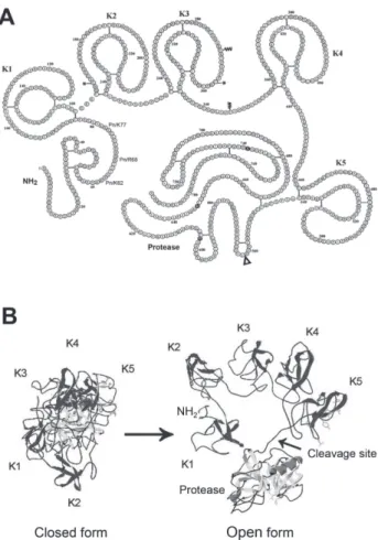
This 92 kDa glycoprotein consists of 791 amino acids (Asn791-Glu1, Glu-plasminogen) that are grouped into five modules called “kringle” and a catalytic region. These modular elements are preceded by an amino-terminal peptide (Glu1-Lys77) (Figure 1A, Table 1). Kringles 1 and 4 contain binding sites for lysine residues (LBS, lysine binding site) of fibrin that allow interaction and effective binding of plasminogen. Kringle 5 contains modified LBS with affinity for the lysine residues of the amino-terminal peptide that promotes the adoption of a closed spiral
con-formation (Figure 1B).10 This plasminogen closed form
predominates in the circulation. The release of the amino-terminal peptide after cleavage by plasmin characterizes a truncated open conformation known as Lys-plasminogen. An open form also occurs when the LBS is occupied by lysine analogues such as epsilon aminocaproic acid
(e-ACA) or tranexamic acid (TXA).11 Indeed, invalidation of
the LBS function by these molecules prevents interaction between the kringle 5 and the amino-terminal peptide. Invalidation of the LBS function by these agents also prevents binding of plasminogen to fibrin and thus its activation by tPA. Surprisingly, it was found that the open form (Figure 1B) induced by low doses of EACA is recog-nized and converted into plasmin by uPA. By homology, it is widely accepted that the direct interaction of native plasminogen (Glu-plasminogen) with the lysine residues of fibrin or membrane glycoproteins leads to a transition from the closed form to the open form. This form is
ef-ficiently activated by tPA linked to fibrin or uPA bound to its receptor. In summary, the binding of plasminogen to fibrin or cell membranes and its transition into the open form are necessary conditions for its transformation into plasmin by plasminogen activators located nearby. The
Figure 1. Structure and conformation of plasminogen.
A. Amino-acids sequence and secondary structure of plasmino-gen. The 791 amino-acids of Glu-plasminogen (Glu1-Asn791) are
assembled in structural modules: an aminoterminal (NH2) peptide followed by 5 kringle modules (K) and a serine-protease region containing the amino acids of the active site (black circles). The site of cleavage by the activators (Arg561-Val562) is indicated by the
open triangle. The cleavage by plasmin at the NH2-terminal peptide (Pn/K77, Pn/R68, Pn/K62) results in the truncated form Lys77-Asp791.
Original figure: http://www.chem.cmu.edu.groups/Llinas/images/ res/res-kringle-PLASMIN.gif
B. The closed form (interaction between the aminoterminal peptide and kringle 5) is the main circulating conformation. The open form is adopted upon binding of plasminogen to C-Lys terminal residues of fibrin or membrane glycoproteins. Original figure (Lähteenmäki K, Edelman S, Korhonen TK. Trends in Microbiology 2005, 13:79) reproduced with permission from Elsevier Limited.
Fibrinolysis, new concepts and new mechanisms
recent description of the crystal structure of plasminogen has confirmed and settled key structure-function
relation-ships among its different constitutive modules.12
Structure of plasminogen activators
Both tPA and uPA have as plasminogen, a mosaic structure
composed of several modules.13 The characteristics of
these molecules are reported in Table 1. The main func-tion of the catalytic region of plasminogen activators is the conversion of plasminogen to plasmin by cleavage of the peptide bond Arg561-Val562. The tPA is secreted by
endothelial cells as a single-chain protein with a very low index of zymogenicity.[2] This indicates that, exception-ally, the single-chain molecule is as active as the two-chain form generated after cleavage of the Arg275-Ile276 of tPA by
plasmin. However, the adsorption of these two forms of tPA onto the surface of fibrin via an interaction between the finger module and the D region of fibrin is a sine qua
non for development of their activity.1,14
The uPA is released by leukocytes as a classical serine protease single-chain zymogen (sc-uPA), which must be transformed into a double-chain form (tc-uPA) in order to display its full protease activity.[5] The cleavage at
the peptide bond Lys158-Ile159 of sc-uPA is mainly made
by plasmin. In contrast, thrombin wipes out the potential activity of scu-PA by cleaving it two residues upstream of the cleavage site of plasmin.[15] At the NH2-terminal posi-tion of the uPA is found the EGF module, which contains a sequence of interaction for its binding to the receptor uPAR. This receptor is itself anchored to the membrane via a glycosylphosphatidylinositol group that has a large transmembrane mobility. Beyond this function, the uPAR in concert with transmembrane glycoproteins activates
Table 1. Main components of the plasminogen activation system.
Plasminogen Plasmin suPAR sc-uPA → tc-uPA tPA
Plasma concentration 0.12- 0.18 mg/mL 0 < 1 ng/mL 3.6 ± 0.9 ng/mL 7.5 ± 2.5 ng/mL
Molar concentration 1.5 to 2 µM 0 < 1.5 pM 50 to 85 pM 70 to 140 pM
Molecular mass 92kDa 84kDa 65kDa 54kDa 70kDa
Cleavage site Arg561-Val562 - - Lys158-Ile159 Arg275-Ile274
Modules P-K1-5-SP K1-5-SP D1-3 EGF-K1-SP F-EGF-K1-2-SP
Polypeptide sequence 1 chain 2 chains 1 chain 1 chain → 2 chains 1 chain / 2 chains
Enzyme activity - ++++ - +/- → ++++ ++++ / ++++
suPAR: uPAR soluble; P: N-terminal peptide; K: kringle domaine; SP: serine protease; D: LU (Ly-6 uPAR) module; EGF: epidermal growth factor-like module; F: finger module
several intracellular signaling pathways involved in cell migration and survival.16
Plasmin is formed on biological surfaces
Upon formation of a fibrin clot, plasminogen and tPA bind to its surface and acquire the molecular conformation nec-essary for the composition of an enzyme/substrate complex leading to in situ production of plasmin.14 Plasmin formed
remains bound to fibrin via its LBS and is thus engaged exclusively in a fibrinolytic function and protected from
inhibition by α2-antiplasmin. Indeed, the formation of a
complex between the catalytic region of plasmin and the reactive site of the α2-antiplasmin requires interaction with
the LBS of kringle 1 of plasmin. Since plasmin remains adsorbed to fibrin it can not be inhibited.17 Thus, to this
surface reaction generating active plasmin can be opposed
the reaction of inhibition by α2-antiplasmin which takes
place in the circulation. Lysis of a clot is therefore the result of a surface reaction with high specificity from the initial formation of plasmin to the acceleration and amplification phases of fibrinolysis.
Molecular interactions on biological surfaces
Plasminogen is immobilized onto lysine residues of fi-brin via the LBS of kringles 1 and 4. The tPA, although having a kringle 2 module with an active LBS, binds to fibrin via its finger module. Inded, the affinity of the finger module for the D region of fibrin (Kd = 1 nM) is 1000 times greater than that of kringle 2 for the lysine residue (Kd = 1 µM). It is precisely this high affinity interaction with fibrin that allows expression of the activity of tPA. In the absence of fibrin tPA has only very limited ability to activate plasminogen. Conformational changes of
plas-minogen and tPA after adsorption onto their sites and their proximity on the surface of fibrin allow the composition of an enzyme/substrate complex resulting in the produc-tion of plasmin and the lysis of the fibrin polymer. In the vascular wall the generation of plasmin at the surface of macromolecules of the extracellular matrix follows the same principle: plasminogen is immobilized via its kringle domains onto fibronectin or laminin where it is
activated by uPA released by inflammatory cells.5
On cell membranes, the molecular assembly occurs on receptor sites for plasminogen (α-enolase,18 tetrameric
complex annexin A2-S100A10,[19] histone H2B,[20] or
the Plg-RKT21) where it is activated by uPA immobilized
on its receptor uPAR. On certain cells such as endothelial cells, smooth muscle cells, or neurons, tPA is attached to transmembrane proteins that converts plasminogen to plasmin.22,23 In contrast to uPA, no specific or unique
receptor for tPA has been described.
Acceleration and amplification of fibrinolysis
Plasmin formed in situ can amplify the activation of plasminogen by generating new binding sites for
plasmi-nogen.24 Indeed, the first molecules of plasmin generated
at the fibrin surface, hydrolyze lysyl bridges and unveils carboxy-terminal (C-Lys) lysine residues that represent
new plasminogen binding sites.25 Plasminogen bound to
C-Lys adopts an open conformation that is recognised and activated by tPA bound nearby or by uPA released from leukocytes. The uPA does not bind to fibrin but
specifi-cally recognizes the plasminogen bound to C-Lys sites.4
The increased binding of plasminogen multiplies the number of molecules of plasmin formed by the activators and enhances the degradation of fibrin and clot lysis. It is the multiplication of the number of binding sites of plas-minogen and the conformational changes thereof which are the major factors for the amplification fibrinolysis. The transformation of sc-uPA into double-chain uPA is a
second acceleration factor in the formation of plasmin.26
Regulation
The plasminogen activation system is finely regulated by (1) serine protease inhibitors (serpins), (2) competitors of plasminogen, or (3) by proteolytic remotion of C-Lys binding sites.
(1) Serpins. Regulation by serpins directly affects plasmin (mainly α2-antiplasmin) or plasminogen
activa-tors (mainly PAI-1). In case of excess tPA or plasmin, plasma inhibitors with less restricted specificity can also act (α2-macroglobulin, C1 esterase inhibitor).27 In the
cen-tral nervous system, other inhibitors of the plasminogen activation system such as neuroserpin and protease nexin-1 (PN-1) have been identifie.28, 29 PN-1 also inhibits plasmin
and thrombin and recent data suggest that PN-1 stored in platelets could play an important role in the vascular
sys-tem.30 PAI-2 is produced by the syncytiotrophoblasts and
monocytes. Its physiopathological role as an inhibitor of uPA and tPA remains an enigma, and seems to especially have intracellular functions.31 It should be noted that in all
cases the inhibition of activators or plasmin occurs mainly in the circulating or liquid phase and that in generally most players in the system of plasminogen activation are partially protected form their inhibitors when bound to their receptors.
(2) Competitors. Lipoprotein(a) or Lp(a) may exercise antifibrinolytic effect and many studies support the clinical
relevance of this mechanism in cardiovascular disease.32
The mechanism antifibrinolytic of this lipoprotein can be explained due to its particular structure, a component similar to the low density lipoprotein (LDL) and a gly-coprotein, apo(a) structurally close to plasminogen but without enzymatic activity: a non-activatable copy of the catalytic region, a copy of the kringle 5 and a variable number of copies of the kringle 4 having a high affinity for fibrin.[33] Thus, competition between plasminogen and apo (a) for fibrin binding limits the amount of bound plasminogen, decreases the formation of plasmin and inhibits fibrinolysis.34
(3) Proteolytic remotion of C-Lys binding sites. The zymogen TAFI (thrombin-activated fibrinolysis inhibitor; procarboxypeptidase U) can be activated by thrombin or plasmin in TAFIa. This exopeptidase cleaves the Lys-C residues of proteins, thereby limiting the binding of plas-minogen to the activation surfaces.35 The in vitro activity
of TAFIa is well established36 but it does not seem to play
a role in physiological fibrinolysis in vivo (mouse mode).37
The important number of studies supporting the clinical interest for this potential regulator of fibrinolysis, has so far provided only associational results.38
Finally, in contrast to the mechanism of regulation of plasminogen receptors by TAFIa, agents that block the LBS of kringle modules can be used as therapeutic in-hibitors of the binding of plasminogen. These are lysine
Fibrinolysis, new concepts and new mechanisms
analogues such as e-ACA and TXA mentioned above. These compounds interact with the LBS of kringle and thus block competitively the binding of plasminogen to fibrin or cells. Their clinical use as an anti-fibrinolytic and anti-haemorrhagic has recently been discussed in several clinico-surgical situations and in a large multi-center study.39,40
Functions of the plasminogen activation system
We distinguish the functions of the plasminogen activation system (fibrinolysis and pericellular proteolysis) depen-ding on the nature of the surface on which the reaction takes place.
- Fibrinolysis: cleavage by plasmin of arginyl and lysyl bridges of fibrin leading to its dissolution and release of degradation products. D-dimer fragments found in the circulation reflect both formation of clot and its dissolution by plasmin.[1] Efficient fibrinolysis al-lows recanalization of the occluded vessel. The use of thrombolytic agents for the treatment of ischemic stroke or coronary events is modeled on this physi-ological model.
- Pericellular proteolysis occurs when the formation of plasmin occurs at the surface of cell membranes or the extracellular matrix.22, 23, 26 At the cellular level,
plasmin activates transmembrane receptors (PAR, protease-activated receptors 1 and 4), induces
intra-cellular signalling41 and a phenotypic response
char-acterized initially by membrane vesiculation.42 The
released microvesicles bear plasminogen activators
synthesized by the parent cell.42 The plasmin formed
in situ induces directly or via the activation of pro-metalloproteases (MMP-3, 9 and 12) the proteolysis of matrix proteins: fibronectin, laminin or
vitronec-tin.43 This proteolysis induces changes in cell
adhe-sion leading to different physiological phenomena
(cellular remodelling, angiogenesis, cell migration).36
The plasmin formed in excess or resulting from lack of regulators (inhibitors) produces a degradation in extenso of the extracellular matrix. This process can result in the loss of cell adhesion and death by ap-optosis as observed in some pathological situations (cell death, weakening/rupture of the atherosclerotic
plaque, aneurysm).44-46 This process of apoptosis
induced by cell detachment may be thwarted by in-hibitors such as PAI-1 and protease nexin-1.44-47 It is
important to differentiate these stages of cell activa-tion and apoptosis to assess the effects of mediators, inhibitors and therapeutic agents.
Fibrinolysis, peculiarities: platelets, microvesicles
The classical pathway of plasminogen activation presented above requires the co-assembly of plasminogen and its activator (uPA or tPA) on the same surface in order to trigger the fibrinolytic or proteolytic process. Moving surfaces such as platelets and microvesicles require special conditions for the production of plasmin.
Mechanism of plasminogen binding to platelets
Like other cell membranes, platelets can adsorb plas-minogen on their surface via C-Lys residues-dependent interactions which number is multiplied by 5 on activated platelets (specific binding, saturable and reversible).[6] This binding is made via the GPIIb / IIIa (αIIb β3) and fibrinogen (fibrin) of platelets activated with thrombin. [48, 49] Plasminogen thus bound adopts an open confor-mation more easily activated by uPA. Platelets may thus contribute to increase the concentration of plasminogen and potentially plasmin within the clot despite their pro-coagulant activity.
Microvesicles and the plasminogen activation system
Microvesicles are membrane vesicles released by activated cells or cells in apoptosis.[50] Of size between 0.1 and 1μm they have exposed phosphatidylserine on their surface, contain no DNA fragments and must not be confused with
apoptotic bodies or exosomes (Figure 2).51 The formation
and release of microvesicles is the result of an extracellular stimulus (physical, chemical or biological) that leads to a massive influx of calcium into the cell. The increase in intracellular calcium alters the activity of phospholipids transporters and stimulates calpains resulting in phosphati-dylserine externalization, changes in the integrity of the cytoskeleton and cell contraction leading to budding of
microvesicles from the cell membrane.52 Many
pathologi-cal conditions such as cardiovascular disease, diabetes, cancer and inflammatory diseases have been associated
with an increase in the number of microvesicles.53,54
These microvesicles carry on their surface and in their cytoplasm proteins of the parent cell. In addition to cell type specific clusters of differentiation (CD), various biomolecules including tissue factor (TF) and
inflamma-tory cytokines can be vectorized by microvesicles.55,56 In
2007, it was shown that microvesicles released from TNF-stimulated HMEC-1 cells (a cell line that synthesizes uPA and its receptor uPAR) with were able to generate plasmin. [8] Indeed, this type of microvesicles bears at their surface uPA / uPAR complexes and uPAR available sites capable of binding exogenous uPA (Figure 2).
The binding of plasminogen to the surface of the microvesicles also involves residues C-Lys. A selective antibody directed against the α-enolase confirmed that this receptor of plasminogen was involved in the binding of plasminogen to the surface of endothelial microvesicles. Recently it has been also shown that the intracellular protein, histone H2B, was involved in the binding of plas-minogen to the surface of microvesicles.[20] The histone H2B is localized to the cell membrane via an interaction with phosphatidylserine exposed in the outer leaflet of the membrane.[57] Thus, procoagulant phospholipid would also increase the number of plasminogen binding sites and promote fibrinolysis.
We confirmed the presence of fibrinolytic microvesicles in the circulation.[58] [è!] These microvesicles have func-tional characteristics similar to those previously described and bear plasminogen activators synthesized by the pa-rental cell (leukocytes: uPA, endothelial cells: tPA). These findings underscore the pathophysiological relevance that such microvesicles could have in vivo.
Figure 2. Plasminogen activation on a microvesicle.
Representation of a fibrinolytic microvesicle bearing on its surface (A) the uPAR receptor anchored to the membrane by the glycos-ylphosphatidylinositol (GPI) group. (B) conformational change of uPAR after binding of sc-uPA. (C) sc-uPA is converted in situ into tc-uPA. (D) plasminogen bound to its receptor adopts the open form. (E) plasmin generated on the membrane by tc-uPA.
A new pathway of plasminogen activation: fibrinolytic cross-talk
Plasminogen adsorbed to fibrin or to the cell membrane adopts the open molecular conformation whose cleavage site is easily accessible to activators located nearby on the same surface. This is the case of the plasminogen-fibrin-tPA ternary complex or of the plasminogen-cell membrane-uPAR/uPA assembly. However, it was found that uPA in solution was an efficient activator of Lys-plasminogen and of Glu-plasminogen complexed to e-ACA. These observations allowed us to hypothesize the existence of an interaction involving two surfaces, one bears plasminogen and the other an activator of plasminogen.[9] It was possible to show that plasminogen carried by platelets can be recog-nized by cells or microvesicles bearing the complex uPA/ uPAR. In a similar fashion the plasminogen bound to fibrin or matrix proteins can be activated to plasmin by uPA/uPAR borne by leukocyte microvesicles. This new mechanism of activation of plasminogen that we have called fibrinolytic cross-talk (Figure 3) is characteristic of uPA and is therefore not sensitive to microvesicles bearing tPA. This specificity could be explained by structural arrangements imposed by the different modules of tPA (finger-EGF-K1-K2-SP) and uPA (EGF-K1-SP) (Table 1). This activation reaction has therefore all the characteristics of a specific and saturable reaction whose efficiency depends on the number of active microvesicles acting on platelets or fibrin. This new acti-vation pathway may have a role physiologically relevant. Indeed, the activation of plasminogen on the surface of platelets by microvesicular uPA generates two times more plasmin that uPA in solution. Recent studies by independent laboratories have confirmed our hypothesis and results.59,61
Two studies have reported the activation of plasminogen
bound to fibrin by uPA borne by leukocytes59,61 while the
third study is focused on the activation of sc-uPA by plasmin formed on the platelet surface.60
Relevance of the fibrinolytic cross-talk mechanism
This novel mechanism of activation of plasminogen to plasmin at the surface of platelets, the extracellular matrix and fibrin by uPA raises the question of its involvement in different pathophysiological situations:
Fibrinolysis
The binding of plasminogen to platelets during clot for-mation and the presence of microvesicles bearing uPA
Fibrinolysis, new concepts and new mechanisms
Figure 3. Fibrinolytic cross-talk (Blood 2010, 115(10) cover illustration).
In inflammatory processes of the vascular wall, fibrinolysis and proteolysis may be induced via a cross-talk between monocytes or cellular microvesicles bearing uPA and platelet-, fibrin-, or extracellular matrix–bound plasminogen. This mechanism of plasmin formation bypasses the requirement for co-assembly of plasminogen and uPA on the same surface. See the article by Dejouvencel et al on page 2048. The fibrinolytic cross-talk refers to the interaction that is established between two biological surfaces, one carrying plasminogen, the other uPA. The surfaces carrying plasminogen (Pg) are represented by the platelet membrane, fibrin or extracellular matrix proteins. The surfaces bearing uPA are represented by microvesicles (MVs) issued from leukocytes (MoMV). The microvesicles are moving surfaces, this inter-surface activation system allows efficient generation of plasmin (Pn) on the inter-surface of fibrin or platelets forming the clot and on proteins of the extracellular matrix. Endothelial MVs (EndMV) bear tPA.
would lead to the formation of plasmin and allow the re-canalisation of an occluded vessel. Indeed, a recent study suggests that the activation of plasminogen bound to fibrin by leukocytes bearing uPA plays a role in endogenous fibrinolysis.61
Cell migration and angiogenesis
Aside from its fibrinolytic function, plasmin formation by the uPA/uPAR system is involved in tissue remodelling via the activation of proMMP and plays a critical role in cell migration and angiogenesis.[16] Indeed, vascular regeneration involves both angiogenesis and vasculogen-esis-dependent endothelial progenitor cells. The ability of endothelial microvesicles to generate plasmin influences
and modulates the repair process of endothelial progenitor cells. A small amount of microvesicles bearing an active plasminogen activation system, promotes cell migration and angiogenesis whereas at high concentrations the ex-cess plasmin leads to matrix degradation, decreased cell adhesiveness and finally apoptosis.8,35
Dissemination of cancer cells
The spread of cancer cells is a consequence of matrix deg-radation and loss of cell adhesion. High amounts of uPA/
uPAR were associated with advanced metastatic cancers.62
It is interesting to note that the described fibrinolytic/ proteolytic cross-talk mechanism is only possible in the presence of an activator of the uPA-type. This
activa-tor is involved in tumour progression and was found on microvesicles emitted by cancerous cells. In addition, mi-crovesicles released by platelets may promote metastasis
and promote angiogenesis.63
Conclusion and potential applications
The structure and function of molecules of the plasmi-nogen activation system including the recent description
of the crystal structure of plasminogen,12 and their role
in the maintenance of haemostasis and thrombosis pre-vention is now well established. However, detection of a dysfunction of this system remains a major challenge for the haematologist and the vascular biologist. The circulating concentration of plasminogen activators is ex-tremely low compared to active concentrations required at the site of injury in the microcirculation. Moreover, the plasminogen activators circulate as an inactive complex with PAI-1 and only the forms located on the cell mem-brane (uPA, tPA) or fibrin (tPA) are active. Furthermore, since all measurements performed in plasma do not take into account the contribution of cellular activators, it is therefore impossible to quantify a lack of tPA or uPA activity that may be the cause of a fibrinolytic default. The recent discovery of cellular fibrinolytic microvesicles and a new mechanism for the formation of plasmin, the
fi-brinolytic cross-talk, opened up new perspectives.8 These
microvesicles would act within the clot, thus explaining the lack of systemic fibrinolysis as demonstrated in vivo
in a mouse model of fibrinolytic platelets.64 We suggest
that the fibrinolytic activity of endothelial and leukocyte microvesicles compensates locally the activity of proco-agulant microvesicles. Is this phenomenon that explains the spontaneous re-canalization observed in 10-20% of patients with acute occlusion of the coronary arteries?65,66
Accordingly, the functional balance between these two types of microvesicles would result in a physiological haemostatic response, while the lack of fibrinolytic mi-crovesicles may promote the formation of a thrombus. The existence of a haemorrhagic syndrome (Quebec platelet disorder) caused by profibrinolytic platelets having an abnormal expression of uPA is consistent with
this hypothesis.67 Using a mouse model of this autosomal
dominant disease it was possible to demonstrate that these animals are resistant to arterial thrombosis and that transfusion of these platelets to control mice prevent the
formation of occlusive arterial thrombi.67
In this context, the presence of plasminogen and its activator uPA or tPA on moving surfaces (respectively platelets and microvesicles) and the identification of the fibrinolytic cross-talk mechanism suggests the possibil-ity of using these materials as vectors of fibrinolysis and pericellular proteolysis.
REFERENCES
1 Mosesson MW. Fibrinogen and fibrin structure and functions.
J Thromb Haemost. 2005; 3: 1894-904.
2 Angles-Cano E, Balaton A, Le Bonniec B, Genot E, Elion J, Sultan Y. Production of monoclonal antibodies to the high fibrin-affinity, tissue-type plasminogen activator of human plasma. Demonstration of its endothelial origin by immunolocalization.
Blood. 1985; 66: 913-20.
3 Suzuki Y, Yasui H, Brzoska T, Mogami H, Urano T. Surface-retained tPA is essential for effective fibrinolysis on vascular endothelial cells. Blood. 2011; 118: 3182-5.
4 Fleury V, Lijnen HR, Angles-Cano E. Mechanism of the enhan-ced intrinsic activity of single-chain urokinase-type plasmino-gen activator during ongoing fibrinolysis. J Biol Chem. 1993; 268: 18554-9.
5 Del Rosso M, Margheri F, Serrati S, Chilla A, Laurenzana A, Fibbi G. The urokinase receptor system, a key regulator at the intersection between inflammation, immunity, and coagulation.
Curr Pharm Des. 2011; 17: 1924-43.
6 Miles LA, Plow EF. Binding and activation of plasminogen on the platelet surface. J Biol Chem. 1985; 260: 4303-11. 7 Podor TJ, Singh D, Chindemi P, Foulon DM, McKelvie R,
Weitz JI, Austin R, Boudreau G, Davies R. Vimentin exposed on activated platelets and platelet microparticles localizes vitronectin and plasminogen activator inhibitor complexes on their surface. J Biol Chem. 2002; 277: 7529-39.
8 Lacroix R, Sabatier F, Mialhe A, Basire A, Pannell R, Borghi H, Robert S, Lamy E, Plawinski L, Camoin-Jau L, Gurewich V, Angles-Cano E, Dignat-George F. Activation of plasminogen into plasmin at the surface of endothelial microparticles: a me-chanism that modulates angiogenic properties of endothelial progenitor cells in vitro. Blood. 2007; 110: 2432-9.
9 Dejouvencel T, Doeuvre L, Lacroix R, Plawinski L, Dignat-George F, Lijnen HR, Angles-Cano E. Fibrinolytic cross-talk: a new mechanism for plasmin formation. Blood. 2010; 115: 2048-56.
10 Cockell CS, Marshall JM, Dawson KM, Cederholm-Williams SA, Ponting CP. Evidence that the conformation of unliganded human plasminogen is maintained via an intramolecular interaction between the lysine-binding site of kringle 5 and the N-terminal peptide. Biochem J. 1998; 333 ( Pt 1): 99-105.
11 Mangel WF, Lin BH, Ramakrishnan V. Characterization of an extremely large, ligand-induced conformational change in plasminogen. Science. 1990; 248: 69-73.
12 Xue Y, Bodin C, Olsson K. Crystal structure of the native plas-minogen reveals an activation-resistant compact conformation.
Fibrinolysis, new concepts and new mechanisms
13 Schaller J, Gerber SS. The plasmin-antiplasmin system: structural and functional aspects. Cell Mol Life Sci. 2011; 68: 785-801.
14 Longstaff C, Thelwell C, Williams SC, Silva MM, Szabo L, Kolev K. The interplay between tissue plasminogen activator domains and fibrin structures in the regulation of fibrinolysis: kinetic and microscopic studies. Blood. 2011; 117: 661-8. 15 Braat EA, Levi M, Bos R, Haverkate F, Lassen MR, de Maat
MP, Rijken DC. Inactivation of single-chain urokinase-type plasminogen activator by thrombin in human subjects. J Lab
Clin Med. 1999; 134: 161-7.
16 Smith HW, Marshall CJ. Regulation of cell signalling by uPAR.
Nat Rev Mol Cell Biol. 2010; 11: 23-36.
17 Rouy D, Angles-Cano E. The mechanism of activation of plasminogen at the fibrin surface by tissue-type plasminogen activator in a plasma milieu in vitro. Role of alpha 2-antiplasmin.
Biochem J. 1990; 271: 51-7.
18 Miles LA, Dahlberg CM, Plescia J, Felez J, Kato K, Plow EF. Role of cell-surface lysines in plasminogen binding to cells: identification of alpha-enolase as a candidate plasminogen receptor. Biochemistry. 1991; 30: 1682-91.
19 Madureira PA, Surette AP, Phipps KD, Taboski MA, Miller VA, Waisman DM. The role of the annexin A2 heterotetramer (AIIt) in vascular fibrinolysis. Blood. 2011.
20 Das R, Burke T, Plow EF. Histone H2B as a functionally im-portant plasminogen receptor on macrophages. Blood. 2007; 110: 3763-72.
21 Andronicos NM, Chen EI, Baik N, Bai H, Parmer CM, Kios-ses WB, Kamps MP, Yates JR, 3rd, Parmer RJ, Miles LA. Proteomics-based discovery of a novel, structurally unique, and developmentally regulated plasminogen receptor, Plg-RKT, a major regulator of cell surface plasminogen activation.
Blood. 2010; 115: 1319-30.
22 Ho-Tin-Noe B, Enslen H, Doeuvre L, Corsi JM, Lijnen HR, Angles-Cano E. Role of plasminogen activation in neuronal organization and survival. Mol Cell Neurosci. 2009; 42: 288-95. 23 Flood EC, Hajjar KA. The annexin A2 system and vascular
homeostasis. Vascul Pharmacol. 2011; 54: 59-67.
24 Weisel JW, Nagaswami C, Korsholm B, Petersen LC, Suenson E. Interactions of plasminogen with polymerizing fibrin and its derivatives, monitored with a photoaffinity cross-linker and electron microscopy. J Mol Biol. 1994; 235: 1117-35. 25 Fleury V, Angles-Cano E. Characterization of the binding of
plasminogen to fibrin surfaces: the role of carboxy-terminal lysines. Biochemistry. 1991; 30: 7630-8.
26 Ellis V, Behrendt N, Dano K. Plasminogen activation by recep-tor-bound urokinase. A kinetic study with both cell-associated and isolated receptor. J Biol Chem. 1991; 266: 12752-8. 27 Bennett B, Croll A, Ferguson K, Booth NA. Complexing of
tis-sue plasminogen activator with PAI-1, alpha 2-macroglobulin, and C1-inhibitor: studies in patients with defibrination and a fibrinolytic state after electroshock or complicated labor. Blood. 1990; 75: 671-6.
28 Bouton MC, Boulaftali Y, Richard B, Arocas V, Michel JB, Jandrot-Perrus M. Emerging role of serpinE2/protease nexin-1 in hemostasis and vascular biology. Blood. 2012; 119: 2452-7. 29 Miranda E, Lomas DA. Neuroserpin: a serpin to think about.
Cell Mol Life Sci. 2006; 63: 709-22.
30 Boulaftali Y, Ho-Tin-Noe B, Pena A, Loyau S, Venisse L, Francois D, Richard B, Arocas V, Collet JP, Jandrot-Perrus M,
Bouton MC. Platelet protease nexin-1, a serpin that strongly influences fibrinolysis and thrombolysis. Circulation. 2011; 123: 1326-34.
31 Lee JA, Cochran BJ, Lobov S, Ranson M. Forty years later and the role of plasminogen activator inhibitor type 2/SERPINB2 is still an enigma. Semin Thromb Hemost. 2011; 37: 395-407. 32 Nordestgaard BG, Chapman MJ, Ray K, Boren J, Andreotti F,
Watts GF, Ginsberg H, Amarenco P, Catapano A, Descamps OS, Fisher E, Kovanen PT, Kuivenhoven JA, Lesnik P, Masana L, Reiner Z, Taskinen MR, Tokgozoglu L, Tybjaerg-Hansen A. Lipoprotein(a) as a cardiovascular risk factor: current status.
Eur Heart J. 2010; 31: 2844-53.
33 Angles-Cano E, Rojas G. Apolipoprotein(a): structure-function relationship at the lysine-binding site and plasminogen activa-tor cleavage site. Biol Chem. 2002; 383: 93-9.
34 Hervio L, Durlach V, Girard-Globa A, Angles-Cano E. Multiple binding with identical linkage: a mechanism that explains the effect of lipoprotein(a) on fibrinolysis. Biochemistry. 1995; 34: 13353-8.
35 Mosnier LO, Bouma BN. Regulation of fibrinolysis by thrombin activatable fibrinolysis inhibitor, an unstable carboxypeptidase B that unites the pathways of coagulation and fibrinolysis.
Arterioscler Thromb Vasc Biol. 2006; 26: 2445-53.
36 Guimaraes AH, Laurens N, Weijers EM, Koolwijk P, van Hinsbergh VW, Rijken DC. TAFI and pancreatic carboxypep-tidase B modulate in vitro capillary tube formation by human microvascular endothelial cells. Arterioscler Thromb Vasc Biol. 2007; 27: 2157-62.
37 Morser J, Gabazza EC, Myles T, Leung LL. What has been learnt from the thrombin-activatable fibrinolysis inhibitor-deficient mouse? J Thromb Haemost. 2010; 8: 868-76. 38 Heylen E, Willemse J, Hendriks D. An update on the role of
carboxypeptidase U (TAFIa) in fibrinolysis. Front Biosci. 2011; 17: 2427-50.
39 Martin K, Knorr J, Breuer T, Gertler R, Macguill M, Lange R, Tassani P, Wiesner G. Seizures after open heart surgery: comparison of epsilon-aminocaproic acid and tranexamic acid.
J Cardiothorac Vasc Anesth. 2011; 25: 20-5.
40 Shakur H, Roberts I, Bautista R, Caballero J, Coats T, Dewan Y, El-Sayed H, Gogichaishvili T, Gupta S, Herrera J, Hunt B, Iribhogbe P, Izurieta M, Khamis H, Komolafe E, Marrero MA, Mejia-Mantilla J, Miranda J, Morales C, Olaomi O, Olldashi F, Perel P, Peto R, Ramana PV, Ravi RR, Yutthakasemsunt S. Effects of tranexamic acid on death, vascular occlusive events, and blood transfusion in trauma patients with significant hae-morrhage (CRASH-2): a randomised, placebo-controlled trial.
Lancet. 2010; 376: 23-32.
41 Pendurthi UR, Ngyuen M, Andrade-Gordon P, Petersen LC, Rao LV. Plasmin induces Cyr61 gene expression in fibroblasts via protease-activated receptor-1 and p44/42 mitogen-activa-ted protein kinase-dependent signaling pathway. Arterioscler
Thromb Vasc Biol. 2002; 22: 1421-6.
42 Doeuvre L, Plawinski L, Goux D, Vivien D, Angles-Cano E. Plasmin on adherent cells: from microvesiculation to apoptosis.
Biochem J. 2010; 432: 365-73.
43 Meilhac O, Ho-Tin-Noe B, Houard X, Philippe M, Michel JB, Angles-Cano E. Pericellular plasmin induces smooth muscle cell anoikis. FASEB J. 2003; 17: 1301-3.
44 Rossignol P, Angles-Cano E, Lijnen HR. Plasminogen activator inhibitor-1 impairs plasminogen activation-mediated vascular
smooth muscle cell apoptosis. Thromb Haemost. 2006; 96: 665-70.
45 Horowitz JC, Rogers DS, Simon RH, Sisson TH, Thannickal VJ. Plasminogen activation induced pericellular fibronectin proteolysis promotes fibroblast apoptosis. Am J Respir Cell
Mol Biol. 2008; 38: 78-87.
46 Kochtebane N, Choqueux C, Passefort S, Nataf P, Messika-Zeitoun D, Bartagi A, Michel JB, Angles-Cano E, Jacob MP. Plasmin induces apoptosis of aortic valvular myofibroblasts.
J Pathol. 2010; 221: 37-48.
47 Rossignol P, Ho-Tin-Noe B, Vranckx R, Bouton MC, Meilhac O, Lijnen HR, Guillin MC, Michel JB, Angles-Cano E. Protease nexin-1 inhibits plasminogen activation-induced apoptosis of adherent cells. J Biol Chem. 2004; 279: 10346-56.
48 Miles LA, Ginsberg MH, White JG, Plow EF. Plasminogen in-teracts with human platelets through two distinct mechanisms.
J Clin Invest. 1986; 77: 2001-9.
49 Adelman B, Rizk A, Hanners E. Plasminogen interactions with platelets in plasma. Blood. 1988; 72: 1530-5.
50 Doeuvre L, Plawinski L, Toti F, Angles-Cano E. Cell-derived microparticles: a new challenge in neuroscience. J Neurochem. 2009; 110: 457-68.
51 Gyorgy B, Szabo TG, Pasztoi M, Pal Z, Misjak P, Aradi B, Laszlo V, Pallinger E, Pap E, Kittel A, Nagy G, Falus A, Buzas EI. Membrane vesicles, current state-of-the-art: emerging role of extracellular vesicles. Cell Mol Life Sci. 2011; 68: 2667-88. 52 Morel O, Jesel L, Freyssinet JM, Toti F. Cellular mechanisms
underlying the formation of circulating microparticles.
Arterios-cler Thromb Vasc Biol. 2011; 31: 15-26.
53 Amabile N, Rautou PE, Tedgui A, Boulanger CM. Micropar-ticles: key protagonists in cardiovascular disorders. Semin
Thromb Hemost. 2010; 36: 907-16.
54 Tushuizen ME, Diamant M, Sturk A, Nieuwland R. Cell-derived microparticles in the pathogenesis of cardiovascular disease: friend or foe? Arterioscler Thromb Vasc Biol. 2011; 31: 4-9. 55 Tual-Chalot S, Leonetti D, Andriantsitohaina R, Martinez MC.
Microvesicles: intercellular vectors of biological messages.
Mol Interv. 2011; 11: 88-94.
56 Zwicker JI, Trenor CC, 3rd, Furie BC, Furie B. Tissue factor-bearing microparticles and thrombus formation. Arterioscler
Thromb Vasc Biol. 2011; 31: 728-33.
57 Das R, Plow EF. Phosphatidylserine as an anchor for plas-minogen and its plasplas-minogen receptor, histone H2B, to the macrophage surface. J Thromb Haemost. 2011; 9: 339-49. 58 Lacroix R, Plawinski L, Robert S, Doeuvre L, Sabatier F,
Mar-tinez de Lizarrondo S, Mezzapesa A, Anfosso F, Leroyer AS, Poullin P, Jourde N, Njock MS, Boulanger CM, Angles-Cano E, Dignat-George F. Leukocyte- and endothelial-derived micro-particles: a circulating source for fibrinolysis. Haematologica. 2012; 97: 1864-72.
59 Lishko VK, Yermolenko IS, Ugarova TP. Plasminogen on the surfaces of fibrin clots prevents adhesion of leukocytes and platelets. J Thromb Haemost. 2010; 8: 799-807.
60 Baeten KM, Richard MC, Kanse SM, Mutch NJ, Degen JL, Booth NA. Activation of single-chain urokinase-type plasmi-nogen activator by platelet-associated plasmiplasmi-nogen: a me-chanism for stimulation of fibrinolysis by platelets. J Thromb
Haemost. 2010; 8: 1313-22.
61 Bai X, Weitz JI, Gross PL. Leukocyte urokinase plasminogen activator receptor and PSGL1 play a role in endogenous arterial fibrinolysis. Thromb Haemost. 2009; 102: 1212-8. 62 Dass K, Ahmad A, Azmi AS, Sarkar SH, Sarkar FH. Evolving
role of uPA/uPAR system in human cancers. Cancer Treat
Rev. 2008; 34: 122-36.
63 Janowska-Wieczorek A, Wysoczynski M, Kijowski J, Marquez-Curtis L, Machalinski B, Ratajczak J, Ratajczak MZ. Microvesicles derived from activated platelets induce metastasis and angiogenesis in lung cancer. Int J Cancer. 2005; 113: 752-60.
64 Kufrin D, Eslin DE, Bdeir K, Murciano JC, Kuo A, Kowalska MA, Degen JL, Sachais BS, Cines DB, Poncz M. Antithrombotic thrombocytes: ectopic expression of urokinase-type plasmi-nogen activator in platelets. Blood. 2003; 102: 926-33. 65 Huisse MG, Lanoy E, Tcheche D, Feldman LJ, Bezeaud A,
Angles-Cano E, Mary-Krause M, de Prost D, Guillin MC, Steg PG. Prothrombotic markers and early spontaneous recanali-zation in ST-segment elevation myocardial infarction. Thromb
Haemost. 2007; 98: 420-6.
66 Swan HJ. Acute myocardial infarction: a failure of timely, spon-taneous thrombolysis. J Am Coll Cardiol. 1989; 13: 1435-7. 67 Hayward CP, Rivard GE. Quebec platelet disorder. Expert Rev