Die „Beckenkammspan-Interpositionsarthrodese“ des oberen
Sprunggelenks
Interposition Arthrodesis of the Ankle
Patrick Vienne1Zusammenfassung Operationsziel
Stabile und korrekt positionierte Arthrodese des oberen Sprunggelenks (OSG) zur Wiederherstellung einer schmerzfreien Belastbarkeit der Gliedmaße.
Indikationen
Invalidisierende, schmerzhafte OSG-Arthrose mit umfang-reichem Knochendefekt bei Status nach Trauma, Infekt oder bei schweren Deformitäten wie z.B. kongenitalen Fehlbildungen oder diabetischen Osteoarthropathien.
Kontraindikationen Akuter Gelenkinfekt.
Schwere arterielle Verschlusskrankheit der betroffenen unteren Extremität.
Operationstechnik
Lateraler Zugang zur distalen Fibula. Fibulaosteotomie 7 cm proximal der Fibulaspitze. Zurückklappen der distalen Fibula nach dorsal und Darstellen des OSG. Vollständige Entknorpe-lung der Gelenkflächen und Débridement des Knochende-fekts. Bestimmung der Defektgröße. Entnahme eines ent-sprechenden trikortikalen Beckenkammspans. Entnahme von autogener Spongiosa am Beckenkamm oder an der pro-ximalen lateralen Tibia. Einbringen der trikortikalen Späne und Ausfüllen des Defekts mit abstützender Wirkung. Fixa-tion mit 6,5-mm- und 3,5-mm-Titan-AO-Zugschrauben. Je nach Ausdehnung des Defekts zusätzliche Stabilisierung des Spans mit einer Titanplatte. Laterale Stabilisierung durch di-rekte Verschraubung der distalen Fibula an Talus und Tibia. Schichtweiser Wundverschluss. Gespaltener Unterschenkel-liegegipsverband in Neutralstellung des OSG.
Weiterbehandlung
Erster Wechsel des Gipsverbands nach 48 h. Mobilisation im abnehmbaren Unterschenkelliegegipsverband mit Vollentlastung für 6 Wochen. Teilbelastung mit 15 kg für weitere 6 Wochen. Erste klinische und radiologische Ver-laufskontrolle 6 Wochen postoperativ.
Ergebnisse
Zwischen Januar 2002 und Januar 2004 wurden fünf Pati-enten (vier Frauen, ein Mann, Durchschnittsalter 57 Jahre
Abstract Objective
Bony fusion of the ankle in a functionally favorable posi-tion for restituposi-tion of a painless weight bearing while avoiding a leg length discrepancy.
Indications
Disabling, painful osteoarthritis of the ankle with exten-sive bone defect secondary to trauma, infection, or serious deformities such as congenital malformations or diabetic osteoarthropathies.
Contraindications Acute joint infection.
Severe arterial occlusive disease of the involved limb. Surgical Technique
Lateral approach to the distal fibula. Fibular osteotomy 7 cm proximal to the tip of the lateral malleolus and pos-terior flipping of the distal fibula. Exposure of the ankle. Removal of all articular cartilage and debridement of the bone defect. Determination of the size of the defect and harvesting of a corresponding tricortical bone graft from the iliac crest. Also harvesting of autogenous cancellous bone either from the iliac crest or from the lateral part of the proximal tibia. Insertion of the tricortical bone graft and filling of the remaining defect with cancellous bone. Fixation with three 6.5-mm titanium lag screws. Depend-ing on the extent of the defect additional stabilization of the bone graft with a titanium plate. Fixation of the later-al fibula on tlater-alus and tibia with two 3.5-mm titanium screws for additional support. Wound closure in layers. Split below-knee cast with the ankle in neutral position.
Results
Between January 2002 and January 2004 this technique was used in five patients with extensive bone defects (four women, one man, average age 57 years [42–77 years]). No intra- or early postoperative complications.
Oper Orthop Traumatol 2005;17:502–17
DOI 10.1007/s00064-005-1141-y
Vorbemerkungen
Die Arthrodese des oberen Sprunggelenks (OSG) ist ein anerkanntes Behandlungsverfahren bei sympto-matischen Arthrosen primärer, posttraumatischer oder postinfektiöser Art sowie bei schweren Deformi-täten des Rückfußes oder als Rettungsmaßnahme bei gelockerten OSG-Totalendoprothesen [9, 16]. Ver-schiedene Techniken wurden bereits beschrieben [1, 2, 7, 8, 10, 12, 13, 15, 19, 20]. Biomechanische Untersu-chungen haben eine größere Stabilität der Schrauben-arthrodese gezeigt [6, 14, 18]. Umfangreiche Knochen-defekte stellen bei jeder Technik eine besondere Her-ausforderung für Operateur und Patienten dar, seien es freie vaskularisierte Verpflanzungen von Fibulat-ransplantaten mit oder ohne Weichteilrekonstruktio-nen [4, 17] oder sog. Überbrückungsplastiken mit Be-ckenkammspänen.
Wir bevorzugen dabei eine Technik mit einem tri-kortikalen Beckenkammspan, eine sog. Beckenkamm-span-Interpositionsarthrodese. Der Span dient sowohl der Defektfüllung als auch der strukturellen Abstüt-zung. Durch eine schonende Entnahmetechnik am Beckenkamm können die Komplikationen an der Ent-nahmestelle minimiert werden. Die hohe Primärstabi-lität der Schraubenosteosynthese begünstigt die Inte-gration des Knochenspans und den Durchbau in kor-rekter Stellung.
Introductory Remarks
Ankle arthrodesis is a universally accepted treatment for symptomatic primary, posttraumatic, or postinfec-tious osteoarthritis as well as for severe hindfoot de-formities or for salvage procedures of a loosened total ankle arthroplasty [9, 16]. Various techniques have been described [1, 2, 7, 8, 10, 12, 13, 15, 19, 20]. Biome-chanical investigations have shown a greater stability of internal fixation with screws than with external fixa-tion [6, 14, 18]. Extensive bone defects pose a challenge to surgeons and patients irrespective of the technique employed. Free pedicled fibular transplantations with or without soft-tissue reconstruction [4, 17] or so-called bridging plasties with iliac crest grafts have been rec-ommended.
We prefer an interposition arthrodesis using a tri-cortical bone graft harvested from the iliac crest. The graft serves not only for filling of the defect, it acts also as a structural support between tibia and talus. Com-plications at the harvesting site can be minimized by a gentle harvesting technique. The initial high stability of the internal screw fixation favors the integration of the bone graft and consolidation in a perfect position.
[42–77 Jahre]) mit schweren Knochendefekten im OSG be-handelt. Keine intra- oder frühen postoperativen Kompli-kationen. Der AOFAS-Score (American Orthopedic Foot and Ankle Society) konnte von 23 Punkten präoperativ auf 76 Punkte (Maximum 86 Punkte) postoperativ verbessert werden (Durchschnittsverlauf 25 Monate). Zwei Patienten entwickelten eine Pseudarthrose und mussten sich einer erneuten Operation mit einem Sprunggelenkarthrode-sennagel unterziehen. Eine Valgusfehlstellung nach der Arthrodese wurde bei einer Patientin mit einer supramal-leolären Korrekturosteotomie behandelt.
Schlüsselwörter
Arthrodese · Oberes Sprunggelenk · Becken-kammspan · Knochendefekt
The AOFAS (American Orthopedic Foot and Ankle Society) Score was improved from 23 points preoperatively to 76 points postoperatively (average follow-up time of 25 months). Two patients developed a nonunion and under-went a revision with an ankle arthrodesis nail. A valgus malposition after arthrodesis in one patient was corrected with a supramalleolar osteotomy.
Key Words
Arthrodesis · Ankle joint · Iliac crest graft · Bone defect
Vorteile
• Minimaler Längenverlust der Gliedmaße.
• Erhöhung der Primärstabilität der Osteosynthese durch Abstützung mit strukturellem trikortikalen autogenen Knochenspan.
• Wiederherstellung der Konturen des OSG.
Nachteile
• Mögliche Beschwerden an der Entnahmestelle am Beckenkamm.
• Defekte > 5 cm nicht ohne Längenverlust korrigier-bar.
Indikationen
• Schmerzhafte und invalidisierende Arthrose des OSG mit schwerem Knochendefekt bei posttrau-matischen oder postinfektiösen Zuständen.
• Schwere, schmerzhafte Deformitäten des OSG bei kongenitalen Fehlbildungen und diabetischen oder neuropathischen Osteoarthropathien.
• Gelockerte OSG-Endoprothesen mit gleichzeiti-gen, umfangreichen Knochendefekten.
Kontraindikationen
• Akute Gelenkinfektion.
Relativ
• Periphere arterielle Verschlusskrankheit.
• Knochendefekte > 6 cm Länge.
• Schlechte Weichteilsituationen und dann nur in Kombination mit freier muskulokutaner Deckung.
Patientenaufklärung
• Allgemeine Operationsrisiken.
• Intraoperative Komplikationen wie Gefäß-, Ner-ven- oder Sehnenverletzungen. Besonders gefähr-det sind Arteria tibialis anterior, Nervi peronei pro-fundus und superficialis, Nervus suralis sowie die Sehnen der Musculi peronei longus und brevis.
Advantages
• Minimal leg length discrepancy.
• Increase of primary stability of the internal fixation with a tricortical autogenous bone graft.
• Restoration of ankle contours.
Disadvantages
• Possible discomfort at the harvesting site.
• Defects > 5 cm cannot be corrected without result-ing in a leg length discrepancy.
Indications
• Painful and disabling osteoarthritis of the ankle ac-companied by major bone defects secondary to trauma or infection.
• Severe painful ankle deformities secondary to con-genital malformations or to diabetic or neuropathic osteoarthropathies.
• Loosened total ankle arthroplasty components ac-companied by extensive bone defects.
Contraindications
• Acute joint infections.
Relative
• Peripheral arterial occlusive disease.
• Bone defects measuring > 6 cm in length.
• Poor soft-tissue coverage which requires a simulta-neous free musculocutasimulta-neous transplantation.
Patient Information
• Usual surgical risks.
• Risk of intraoperative vessel, nerve, or tendon inju-ries. Particularly exposed are anterior tibial artery, deep and superficial peroneal nerve, sural nerve, long and short peroneal tendons.
• Risk of postoperative wound healing disturbances, infections or reflex sympathetic dystrophy.
Operationsprinzip und -ziel
Arthrodese des OSG in funktionell günstiger Posi-tion unter Verwendung eines strukturellen trikortika-len autogenen Beckenkammspans. Osteosynthese mit Zugschrauben oder zusätzlicher Titanplatte zur Wiedererlangung einer schmerzfreien Belastbarkeit der Gliedmaße.
Surgical Principles and Objective
Ankle fusion in a functionally favorable position us-ing autogenous tricortical iliac crest grafts for struc-tural support and filling of the remaining defect with cancellous bone. Internal fixation with lag screws and, if necessary, additional titanium plate. The goal is a return to a pain-free weight bearing.
• Postoperative Komplikationen wie Wundheilungs-störungen, Infektion oder Dystrophie.
• Knochenheilungsstörung, besonders häufig bei Diabetikern, Rauchern und Patienten mit beste-henden Durchblutungsstörungen.
• Mögliche Komplikationen im Entnahmegebiet des Beckenkammspans: Verletzung des Nervus cuta-neus femoris lateralis, Nachblutung, Infektion, sog. Insuffizienzfraktur.
• Restbeschwerden.
• Korrekturverlust durch verzögerte oder ausblei-bende Heilung: Sekundäre Korrekturosteotomien können erforderlich werden.
• Pseudarthrose und Möglichkeit der Revisionsope-ration.
• Implantatbruch oder Implantatlockerung mit Not-wendigkeit der Implantatentfernung.
• Mögliche Unterschenkelamputation bei sehr schwe-ren, nicht beherrschbaren Komplikationen, wie z.B. Infektionen.
• Bei Erfolg der Arthrodese: Verbesserung der Geh-funktion und Schmerzfreiheit.
• Ausführliche Informationen über die Position des Fußes und die Folgen für den Schuhkomfort.
• Ruhigstellung im Liegegipsverband ohne Belastung für mindestens 6 Wochen notwendig, dann 6 Wo-chen Teilbelastung im Gehgipsverband bis zum vollständigen Knochendurchbau.
• Volle Belastung frühestens nach 3 Monaten mög-lich.
• Notwendigkeit einer optimalen Schuhversorgung zur Verbesserung der Gehfähigkeit.
Operationsvorbereitungen
• Konventionelle Röntgenaufnahmen des OSG ante-roposterior (a.p.) und lateral im Stehen.
• Bei komplexen Deformitäten dreidimensionale Computertomographie des OSG.
• Photographische Dokumentation.
• Bei Diabetikern und Patienten mit peripherer arte-rieller Verschlusskrankheit ausführliche angiologi-sche Abklärungen, einschließlich Angiographie und evtl. präoperativer PTA (perkutane translumi-nale Angioplastie) zur Verbesserung der Gefäßsi-tuation.
• Enthaarung, Desinfektion und steriles Einpacken des Unterschenkels am Abend vor dem Eingriff.
• Antibiotische Prophylaxe mit einmaliger Gabe ei-nes Cephalosporins der zweiten Generation 30 min präoperativ.
• Delayed consolidation, particularly in diabetic pa-tients, smokers and patients with impaired periph-eral circulation.
• Possible complications at the harvesting site at the iliac crest: injury to the lateral femorocutaneous nerve, hemorrhage, infection, and fractured iliac crest.
• Persisting symptoms.
• Loss of correction due to delayed union or non-union: revision surgery may become necessary.
• Nonunion possibly requiring revision.
• Implant breakage or loosening necessitating im-plant removal.
• Risk of below-knee amputation in extremely seri-ous, uncontrollable complications such as infec-tions.
• Successful arthrodesis will result in improved gait and freedom from pain.
• Detailed explanation of the postoperative foot posi-tion and its influence on shoe selecposi-tion.
• Immobilization in a below-knee non-walking cast for at least 6 weeks followed by 6 weeks partial weight bearing in a walking cast until complete bony fusion.
• Full weight bearing at the earliest after 3 months.
• Need for specially adapted shoes to improve gait.
Preoperative Work Up
• Anteroposterior (AP) and lateral weight bearing radiographs.
• For complex deformities: three-dimensional com-puted tomography.
• Photographic documentation.
• For diabetic patients and those with peripheral arte-rial occlusive disease: detailed vascular investiga-tions including angiography and possibly percuta-neous transluminal angioplasty to improve circula-tion.
• Epilation, disinfection, and sterile wrapping of low-er leg at the evening before surglow-ery.
• A single shot of second-generation cephalosporin 30 min before surgery.
Surgical Instruments and Implants
• Basic orthopedic set.
• Oscillating saw.
• Bone spreader, straight and slightly curved chisels (Lambotte chisels), curettes.
• Set for bone graft harvesting from iliac crest.
• Drill, 4.5-mm and 3.2-mm drill bits for 6.5-mm lag screws, 3.5-mm and 2.5-mm drill bits for 3.5-mm
Instrumentarium und Implantate
• Orthopädisches Grundsieb.
• Oszillierende Säge.
• Knochenspreizer, gerade und leicht gebogene Mei-ßel (Lambotte-MeiMei-ßel), scharfe Löffel.
• Set für Knochenspanentnahme am Beckenkamm.
• Universalbohrer (4,5-mm- und 3,2-mm-Bohrer für 6,5-mm-Zugschrauben, 3,5-mm- und 2,5-mm-Boh-rer für 3,5-mm-Schrauben).
• 6,5-mm- und 3,5-mm-AO-Titanschrauben, Drittel-rohr-AO-Titanplatten.
• Steril abgedeckter Bildwandler.
Anästhesie und Lagerung
• Allgemeinnarkose oder Regionalanästhesie (Spi-nalanästhesie).
• Poplitealkatheter zur postoperativen Analgesie [5]. Der Poplitealblock wird mit 50 ml Ropivacain 0,5% durchgeführt. Der Block wird für 48 h durch patientenkontrollierte Anästhesie mit Ropivacain 0,2% oder 0,3% 5 ml/h erhalten.
• Rückenlage, sterile Abdeckung des ipsi- oder kon-tralateralen Beckenkamms.
• Sterile Abdeckung bis auf Höhe des Kniegelenks: Klinische Beurteilung der Beinachse einschließlich der Rotation des Unterschenkels (Abbildung 1).
• Pneumatische Oberschenkelblutsperre.
screws, 2.0-mm drill bit for freshening of resection surfaces.
• 6.5-mm and 3.5-mm AO titanium screws, third tu-bular titanium AO plates.
• Sterile-draped image intensifier.
Anesthesia and Positioning
• General or regional (spinal) anesthesia.
• Popliteal catheter for postoperative analgesia [5]. The popliteal block is performed with ropivacaine 0.5%, 50 ml through a perineural catheter. The block is maintained for 48 h and patient-controlled anesthesia with ropivacaine 0.2% or 0.3% 5 ml/h.
• Supine, sterile draping of the ipsi- or contralateral iliac crest.
• Sterile draping up to the level of the knee: clinical assessment of the leg axis including rotation of the lower leg (Figure 1).
• Tourniquet at thigh.
Abbildung 1
Rückenlage, sterile Abdeckung des ipsi- oder kontralate-ralen Beckenkamms, sterile Abdeckung bis auf Höhe des Kniegelenks.
Figure 1
Supine, sterile draping of the ipsi- or contralateral iliac crest, sterile draping up to the level of the knee.
Operationstechnik
Abbildungen 2 bis 13
Surgical Technique
Figures 2 to 13
N. suralis Mm. peronei long. et brev.
Sinus tarsi
N. cutaneus dors. intermed.
Fibula Syndesmose Syndesmosis
Abbildung 2
Ventrolateraler Hautschnitt entlang der distalen Fibula, distal leicht nach ventral bis auf Höhe des Sinus tarsi laufend.
Figure 2
Anterolateral skin incision overlying the distal fibula, distally turning slightly anterior up to the level of the sinus tarsi.
Abb.3 Luer Rongeur Fibula Ventrale Syndesmose Anterior syndesmosis Abbildung 3
Längsspalten des subkutanen Gewebes und Darstellen der ventralen Syndesmose. Die ventrale Syndesmose wird durch-trennt und mit dem Luer reseziert.
Figure 3
Longitudinal division of the subcutaneous tissues and expo-sure of the anterior syndesmosis. The anterior part of the syn-desmosis is divided and resected with a rongeur.
Syndesmosenreste Remnants of syndesmosis
Abbildung 4
Darstellen der Fibula bis ca. 10 cm proximal der Außenknö-chelspitze. Schräge Fibulaosteotomie mit der oszillierenden Säge 7 cm proximal der Fibulaspitze. Resektion eines 1 cm breiten Fibulafragments.
Figure 4
Exposure of the fibula up to 10 cm proximal to the tip of the lateral malleolus. Oblique fibular osteotomy with an oscillat-ing saw 7 cm proximal to the tip of the lateral malleolus. Re-section of a 1 cm broad segment of the fibula.
Defekt in der Tibia Defect in tibia Talus Facies articularis mall. lat. Retraktion der distalen Fibula nach dorsal Posterior flipping of distal fibula Abbildung 5
Retraktion der distalen Fibula nach dorsal. Darstellen des OSG.
Figure 5
Posterior flipping of the distal fibular fragment and exposure of the ankle.
Talus
Arthrodesenspreizer Arthrodesis spreader Meißel
Chisel
Defekt in der Tibia Defect in tibia
Meißel Chisel
Defekt in der Tibia Defect in tibia Arthrodesenspreizer Arthrodesis spreader Meißel Chisel Meißel Chisel a b c Abbildungen 6a bis 6c
Vollständiges Entknorpeln der Gelenkflächen mit dem gera-den und leicht gebogenen Meißel von lateral nach medial (a). Bei diesem Schritt sollten die Konkavität der distalen Tibiaflä-che und die Konvexität der TalusoberfläTibiaflä-che möglichst erhal-ten bleiben. Vollständiges Débridement des Knochendefekts bis in gesunde Spongiosabezirke (b). Durch maximale Innen-drehung des Fußes Darstellen und Entknorpeln der medialen Gelenkfläche zwischen medialem Malleolus und medialer Ta-lusfläche (c). Abmessen der Größe des Knochendefekts.
Figures 6a to 6c
Complete removal of cartilage of all articular surfaces with straight and slightly curved chisels proceeding from lateral to medial (a). In doing so the concavity of the remaining distal tibial joint surface and the convexity of the talar dome should be preserved as much as possible. Complete debride-ment of the bone defect down to healthy cancellous bone (b). With the foot in maximal internal rotation exposure of the joint surfaces between medial malleolus and medial talar surface and removal of articular cartilage (c). Assessment of the size of the bone defect.
Entnahme des trikortikalen Beckenkammspans Harvesting of tricortical iliac crest graft Abbildung 7
Gerader Hautschnitt von ca. 6 cm Länge 1 cm unterhalb der Crista iliaca anterior. Längsspalten der Subkutis und subperi-ostale Darstellung der Crista. Entnahme eines trikortikalen Beckenkammspans, der in Länge und Höhe dem OSG-Defekt entspricht. Entnahme von autogener Spongiosa mit dem scharfen Löffel.
Einbringen von hämostatischem Material (z.B. Spongostan®, Johnson & Johnson) in den Beckenkammdefekt. Legen eines Schmerzkatheters. Fortlaufender Verschluss des Periosts mit Vicryl-Faden 1.0. Schichtweiser Wundverschluss, Einlegen ei-ner subkutanen Redon-Drainage.
Figure 7
Straight, 6 cm long skin incision, 1 cm below the anterior part of the iliac crest. Longitudinal division of the subcutaneous tissues and subperiosteal exposure of the crest. Harvesting of a tricortical iliac crest graft corresponding in length and height to the defect in the distal tibia. Also harvesting of can-cellous bone with a curette.
Packing of the defect with hemostatic material such as Spon-gostan® (Johnson & Johnson). Placement of a catheter for analgesia. Running suture of the periosteum with Vicryl 1.0. Wound closure in layers, subcutaneous suction drain.
Präparieren des Spans Preparing of bone graft
Abbildung 8
Vorbereitung des trikortikalen Beckenkammspans: Entfer-nung der Weichteile und Schnitt des Spans auf die gewünsch-te Länge.
Figure 8
Preparation of the tricortical bone graft: freeing from soft tis-sues and tailoring of the graft to the desired length.
Angefrischte Talus-Gelenkfläche Freshened articular surface of talus
Abbildung 9
„Anfrischung“ der Resektionsflächen im OSG mit dem 2,0-mm-Bohrer.
Figure 9
Freshening of the resection surfaces of the ankle with a 2.0-mm drill bit.
Abbildungen 10a bis 10c
Nach Einstellen des Gelenks in der gewünschten Stellung (90° Dorsal-/Plantarflexion, 10° Außenrotation, Fuß planti-grad) Einbringen des Spans in den Defekt (a).
In dieser Stellung Einbringen einer 6,5-mm-AO-Titanzug-schraube (Bohren: 4,5 mm und 3,2 mm!) vom medialen Malleo lus in Richtung Talus in einem Winkel von 60° (b). Eine zweite 6,5-mm-AO-Titanzugschraube wird von der ven-tralen Tibia in Richtung des dorsalen Taluskörpers einge-bracht (c). Die Stellung der Arthrodese ist jetzt gegeben.
Figures 10a to 10c
After positioning of the ankle in the determined neutral null position (90° dorsiplantar flexion, 10° external rotation, foot plantigrade), placing of the bone graft into the defect (a). With the foot in the determined position insertion of a 6.5-mm AO titanium lag screw (drilling: 4.5 mm and 3.2 mm) from the medial malleolus in direction of the talus at an angle of 60° (b). A second 6.5-mm AO titanium lag screw is inserted into the an-terior tibia in direction of the posan-terior part of the talar body (c). The definite position of the arthrodesis has now been obtained. Fibula
Trikortikaler Beckenkammspan im Tibiadefekt
Tricortical bone graft in tibial defect
Angefrischte Talus-Gelenkfläche Freshened articular surface of talus
Trikortikaler Beckenkammspan im Tibiadefekt
Tricortical bone graft in tibial defect Ventrale AO-Titanzugschraube Anterior AO titanium lag screw Mediale Zugschraube
Medial lag screw
a
b
Fünf-Loch-Drittelrohrplatte Five-hole third tubular plate
Abbildung 11
Stabilisierung des Spans mit einer ventral gelegenen Drit-telrohrplatte; sie überbrückt den Span und wird in Talus und Tibia verschraubt. Eine dritte 6,5-mm-AO-Titanzug-schraube wird von der dorsalen Tibia in Richtung Talus-kopf eingebracht.
Figure 11
Stabilization of the bone graft with a third tubular plate placed anteriorly. The plate bridges the bone graft and is attached with screws to talus and tibia. A third 6.5-mm AO titanium lag screw is introduced from the posterolat-eral aspect of the tibia into the talar head.
Titan-zugschrauben Dorsale Titanzugschraube
Posterior titanium lag screw
Titanium lag screws
Abbildungen 12a und 12b
Vorbereitung der distalen Fibula mit horizontalem Absä-gen der Knorpeloberfläche (a). Fixation der distalen Fibula lateral am Talus und an der Tibia mit zwei 3,5-mm-AO-Ti-tanzugschrauben (b).
Figures 12a and 12b
Preparation of the distal fibular fragment: horizontal re-section of the articular cartilage with a saw (a), fixation of the fragment to talus and tibia with two 3.5-mm AO tita-nium lag screws (b).
a b
Trikortikaler Beckenkammspan im Tibiadefekt
Tricortical bone graft in tibial defect
Abb.13
Fünf-Loch-Drittelrohrplatte Five-hole third tubular plate
Abbildung 13
Einlegen einer subkutanen Redon-Drainage und schicht-weiser Wundverschluss mit subkutanen Einzelnähten (Monocryl 4.0) und fortlaufender Hautnaht (Ethicrin 4.0).
Figure 13
Subcutaneous suction drain. Wound closure in layers with subcutaneous interrupted 4.0 sutures and running 4.0 skin suture.
Postoperative Behandlung
• Steriler Verband. Gespaltener Unterschenkelliege-gipsverband in Neutralstellung des OSG.
• Antibiotische Kurzprophylaxe mit Cefuroxim (Zi-nacef®) 3 × 1,5 g intravenös für 24 h.
• Rückenlage, operiertes Bein auf Sandkissen.
• Erster Verbands- und Gipsverbandwechsel 48 h postoperativ, Applikation eines gespaltenen Gips-verbands aus Kunststoff (Scotch-Cast®, 3M, 3M
Center, St. Paul, MN, USA).
• Entfernung der Redon-Drainage und des Popliteal-katheters 48 h postoperativ.
• Mobilisation am Gehwagen mit voller Entlastung des operierten Beins ab dem 1. postoperativen Tag.
• Mobilisation an zwei Unterarmstöcken ab dem 2. postoperativen Tag im Unterschenkelliegegips-verband mit voller Entlastung für 6 Wochen.
• Thrombose-/Embolieprophylaxe mit niedermole-kularem Heparin (z.B. Fraxiparin®-Fertigspritzen
einmal täglich) gemäß Körpergewicht für die Zeit der Ruhigstellung im Gipsverband.
• Steriler Verbandswechsel alle 48 h bis zur Faden-entfernung 14 Tage postoperativ.
• Erste klinische und radiologische Verlaufskontrolle 6 Wochen postoperativ.
• Bei beginnender Konsolidierung Teilbelastung mit 15 kg im Unterschenkelgehgipsverband für weitere 6 Wochen.
• Physiotherapie zur allgemeinen Kräftigung, Geh-schule und Lymphdrainage ab der 7. postoperativen Woche.
• Zweite klinische und radiologische Verlaufskon-trolle 12 Wochen postoperativ.
• Beim regelrechten Durchbau der Arthrodese vol-le Belastung im stabivol-len hohen Schuh (z.B. Künz-li Schuh®, Künzli AG, Hauserstraße 47, 5310
Windisch, Schweiz) erlaubt. Beenden der Anti-koagulation.
• Bei vorwiegend sitzender Tätigkeit Wiederaufnah-me der Arbeit mit 50%igem Pensum ab der 13. Wo-che für 4 WoWo-chen und 100%igem Pensum ab der 17. Woche erlaubt.
• Bei vorwiegend stehender Tätigkeit Beginn mit 50%igem Pensum ab der 17. Woche bis Ende des 6. Monats. Steigerung auf 100% ab dem 7. postope-rativen Monat.
• Optimale Schuhversorgung mit orthopädischem Serienschuh mit Pufferabsatz, rückversetzter Ab-rollhilfe und Fußbettung nach Maß.
• Nächste klinische und radiologische Verlaufskon-trolle 6 Monate und 1 Jahr postoperativ.
Postoperative Management
• Sterile dressing. Split below-knee cast with the an-kle in neutral position.
• Zinacef® (cefuroxime sodium) 3 × 1.5 g during first
24 h.
• Supine, operated leg on sand bag.
• First dressing and cast change after 48 h, split fiber-glass (Scotch cast®, 3M, 3M Center, St. Paul, MN,
USA).
• Removal of suction drain and of popliteal catheter on day 2.
• Mobilization with a walker without any weight bear-ing on the operated limb on day 1.
• Use of forearm crutches from day 2 on without any weight bearing on the below-knee walking cast for 6 weeks.
• Prevention of thrombophlebitis with low molecular heparin (such as fragmin in prefilled syringes ac-cording to body weight, once daily) during time of cast immobilization.
• Sterile dressing change every 2 days up to the mo-ment of suture removal at day 14.
• First clinical and radiologic control at 6 weeks.
• Once consolidation has begun, partial weight bear-ing (15 kg) in below-knee walkbear-ing cast for an addi-tional 6 weeks.
• Strengthening and gait exercises as well as lymph drainage starting at week 7.
• Second clinical and radiologic control at 12th week.
• Once consolidation is complete, full weight bearing in a solid boot (such as Künzli Shoe®, Künzli AG,
Hauserstraße 47, 5310 Windisch, Switzerland). Dis-continuation of anticoagulation.
• For sedentary workers 50% resumption of work at week 13 for the duration of 4 weeks and full-time work at week 17.
• For work requiring standing resumption of 50% ac-tivities at week 17 until the end of 6th month, increase to 100% starting the 7th postoperative month.
• Provision of proper orthopedic shoes with elastic heel, rocker-bottom sole and made to measure insoles.
• Clinical and radiologic control after 6 and 12 months.
Errors, Hazards, Complications
• Incomplete removal of articular cartilage, inadequate freshening of contact surfaces, improper screw posi-tioning, and insufficient compression: absent consoli-dation, nonunion, malalignment: revision.
• Faulty position of tricortical bone graft: subsidence into resection surfaces, inadequate support, healing
Fehler, Gefahren, Komplikationen
• Unvollständige Entknorpelung der Gelenkflächen und unvollständige Anfrischung der Gelenk- und Knochenflächen, nicht optimale Schraubenlage und ungenügende Kompression: Fehlender Durchbau, Pseudarthrose, Fehlstellung: Revisionsoperation.
• Fehlerhafte Platzierung des trikortikalen Spans: Einsinken der Resektionsflächen, ungenügende Abstützung, Heilung in Fehlstellung: Revisionsope-ration evtl. erforderlich.
• Intraoperative Verletzung wichtiger anatomischer Strukturen wie Sehnen, Nerven und Gefäße: Naht, ggf. unter mikrochirurgischen Bedingungen. Beson-ders gefährdet sind die Peronealsehnen direkt dorsal des distalen Fibulafragments und der Nervus pero-neus superficialis bei seinem Austritt aus der Pero-nealloge am ventrolateralen Unterschenkel. Die an-deren wichtigen anatomischen Strukturen wie Arte-ria tibialis anterior und Nervus peroneus profundus sind besser geschützt und daher bei dem beschriebe-nen Zugang nicht unmittelbar gefährdet.
• Frühe postoperative Infektion: Sofortige Revision mit Débridement, Drainage und Gewebebiopsien zur bakteriologischen Untersuchung. Gezielte intravenö-se antibiotische Therapie gemäß Antibiogramm.
• Fehlender radiologischer Durchbau und anhalten-de belastungsabhängige Schmerzen nach 6 Mona-ten: Computertomographie. Revisionsoperation erwägen.
• Arthrodese in Fehlstellung: Sekundäre Korrektur-osteotomie.
• Störende Schrauben bei gesichertem Durchbau der Arthrodese: Entfernung der Schrauben, frühestens nach 6 Monaten.
Ergebnisse
Zwischen Januar 2002 und Januar 2004 wurden fünf Pa-tienten (vier Frauen, ein Mann, Durchschnittsalter 58 Jahre [42–77 Jahre]) mit schweren Knochendefekten im OSG mit einer Beckenkammspan-Interpositionsarthro-dese behandelt. Ursache des Knochendefekts war in ei-nem Fall (Patientin D.D.) ein Zustand nach schwerer, offen reponierter und verschraubter Pilon-tibiale-Frak-tur mit konsekutivem devaskularisierten Knochenfrag-ment im Bereich der distalen Tibia. Der Patient S.S. litt an den Folgen einer offen reponierten und verschraub-ten multifragmentären intraartikulären Pilon-tibiale-Fraktur mit konsekutiver Infektion, ausgeprägtem Dé-bridement und Implantation eines Zementspacers. Die Patientin R.E. wies einen Zustand nach offener Reposi-tion und Schrauben-/Plattenosteosynthese einer
bimalle-with malalignment: revision may become neces-sary.
• Intraoperative injury to important anatomic struc-tures such as tendons, nerves, and vessels: suture, if necessary using microsurgical techniques. Particu-larly prone to injury are the peroneal tendons that lie directly posterior to the distal fibular fragment and the superficial peroneal nerve at its exit point from the peroneal compartment at the anterolateral aspect of the lower limb. Other important structures such as the anterior tibial artery and deep peroneal nerve are better protected and not exposed to injury by the described approach.
• Early postoperative infection: immediate revision with debridement, drainage, and tissue biopsies for culture and sensitivity test. Specific intravenous an-tibiotic therapy based on result of sensitivity test.
• Radiologically proven absent consolidation and persistent pain on weight bearing after 6 months: computed tomography, consider revision.
• Malunion of arthrodesis: corrective osteotomy.
• Screws giving rise to symptoms after complete consolidation: screw removal at the earliest after 6 months.
Results
Between January 2002 and January 2004 we operated five patients (four women and one man, average age 57 years [42–77 years]) who presented with a severe bone defect of the ankle and were treated with an in-terposition arthrodesis. A tibial plafond fracture open-ly reduced and fixed by screws leading subsequentopen-ly to a devitalized bone fragment at the level of the distal tibia was the cause of the defect in one patient (D.D.). Patient S.S. suffered from a comminuted intraarticular tibial plafond fracture, that was treated by open reduc-tion and internal screw fixareduc-tion. An infecreduc-tion ensued necessitating an extensive debridement and implanta-tion of a cement spacer. The third patient (R.E.) pre-sented with a fracture dislocation of the ankle treated with open reduction and internal fixation with subse-quent infection and malalignment in valgus and exter-nal rotation. The fourth patient (F.U.) had a congeni-tal nonunion of the tibia treated by repeated attempts at stabilization resulting in a marked valgus malalign-ment of the hindfoot and nonunion of the distal fibula. The last patient (S.A.) consulted for a rupture of the deltoid ligament and valgus instability of the hindfoot after total ankle arthroplasty.
All patients had an average of four previous inter-ventions (three to seven; Table 1). In none of the
ar-olären Luxationsfraktur mit nachfolgender Infektion und Fehlheilung in Valgus- und Außenrotationsfehlstel-lung auf. Die Patientin F.U. zeigte nach mehrmaligen Stabilisierungsoperationen bei kongenitaler Tibiapseud-arthrose eine massive Valgusfehlstellung des Rückfußes und Pseudarthrose der distalen Fibula. Die Patientin S.A. wies einen Zustand nach Implantation einer OSG-Total-prothese mit konsekutiver Ruptur des Ligamentum del-toideum und instabiler Valgusfehlstellung des Rückfußes auf.
Alle Patienten waren im Durchschnitt viermal (drei- bis siebenmal) voroperiert worden (Tabelle 1). Bei keiner Arthrodese traten intra- oder frühe post-operative Komplikationen auf.
Alle Patienten wurden klinisch und radiologisch nach durchschnittlich 25 Monaten (12–36 Monate) nachkontrolliert. Bei drei Patienten war die Arthrode-se durchgebaut. Eine Patientin wies eine Valgusfehl-stellung auf, und zwei Patienten litten an einer Pseud-arthrose (Tabelle 2).
Abbildung 14 zeigt den radiologischen Verlauf bei der 71-jährigen Patientin R.E. (Abbildung 14a: Zu-stand vor der Arthrodese; Abbildung 14b: 16 Monate postoperativ). Die Patientin weist einen vollständigen
throdeses intra- or early postoperative complications were encountered.
The patients were examined clinically and radio-logically after an average of 25 months (12–36 months). A bony consolidation occurred in three patients, one patient showed a valgus malalignment, and two pa-tients experienced a nonunion (Table 2).
Figure 14 documents the radiologic evolution in the 71-yeold patient R.E. (Figure 14a: state before ar-throdesis; Figure 14b: findings 16 months postopera-tively). The complete bony consolidation in perfect position is evident, and the patient is free of pain ever since.
Figure 15 shows the radiologic course of patient S.S. After treatment of a tibial plafond fracture (Figures 15a and 15b) he developed an infection that was treated with a temporarily implanted cement spacer (Figure 15c). An arthrodesis with interposition of a 5 cm long iliac crest graft was performed after the infection had healed (Figure 15d). Subsequently, a nonunion devel-oped (Figure 15e). On account of the poor talar bone stock we resorted to an ankle arthrodesis nail. 6 months after this intervention a beginning consolidation was noted allowing full weight bearing (Figure 15f).
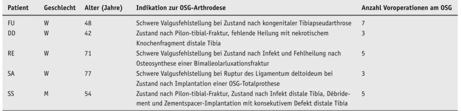
Tabelle 1
Patientendaten. M: männlich; OSG: oberes Sprunggelenk; W: weiblich.
Patient Geschlecht Alter (Jahre) Indikation zur OSG-Arthrodese Anzahl Voroperationen am OSG
FU W 48 Schwere Valgusfehlstellung bei Zustand nach kongenitaler Tibiapseudarthrose 7 DD W 42 Zustand nach Pilon-tibial-Fraktur, fehlende Heilung mit nekrotischem 3
Knochenfragment distale Tibia
RE W 71 Schwere Valgusfehlstellung bei Zustand nach Infekt und Fehlheilung nach 5 Osteosynthese einer Bimalleolarluxationsfraktur
SA W 77 Schwere Valgusfehlstellung bei Ruptur des Ligamentum deltoideum bei 3 Zustand nach Implantation einer OSG-Totalprothese
SS M 54 Zustand nach Pilon-tibial-Fraktur, Zustand nach Infekt distale Tibia, Débride- 5 ment und Zementspacer-Implantation mit konsekutivem Defekt distale Tibia
Table 1
Patient collective. F: female; M: male.
Patient Sex Age (years) Indication for ankle arthrodesis Number of previous
operations
FU F 48 Pronounced valgus malalignment, state after congenital nonunion of tibia 7 DD F 42 State after tibial plafond fracture, absent consolidation, necrotic bone 3
fragment distal tibia
RE F 71 Pronounced valgus malalignment, state after infection and malunion after 5 internal fixation of fracture-dislocation
SA F 77 Pronounced valgus malalignment, rupture of the deltoid ligament, state 3 after total ankle arthroplasty
SS M 54 State after tibial plafond fracture, infection of distal tibia treated with 5 debridement and implantation of cement spacer resulting in defect of distal tibia
Durchbau der Arthrodese in korrekter Stellung auf und ist völlig beschwerdefrei.
In Abbildung 15 ist die Rekonstruktion bei dem Pati-enten S.S. dargestellt. Nach Versorgung einer Pilon-tibia-le-Fraktur (Abbildungen 15a und 15b) kam es zu einer Infektion, welche vorübergehend mit einem
Zement-Patient D.D., a heavy smoker and alcoholic with pa-ralysis of the ipsilateral upper limb, also suffered from a nonunion necessitating a revision consisting of im-plantation of cancellous bone harvested from the proximal ipsilateral tibia. In spite of the intervention the nonunion persisted leading to the insertion of an
Tabelle 2
Ergebnisse. AOFAS: American Orthopedic Foot and Ankle Society; M: männlich; W: weiblich.
Patient Geschlecht Alter Länge Becken- AOFAS-Score AOFAS-Score postop. Komplikationen Revisionsoperationen
(Jahre) kammspan (cm) präop. (max. 86 Punkte)
FU W 48 2,5 31 82 Keine Keine
DD W 42 3,5 27 42 Pseudarthrose Revisionsarthrodese mit Nagel
RE W 71 4,0 4 82 Keine Keine
SA W 77 5,0 34 64 Valgusfehlstellung Korrekturosteotomie
SS M 54 5,0 27 52 Pseudarthrose Revisionsarthrodese mit Nagel
Table 2
Results. AOFAS: American Orthopedic Foot and Ankle Society; F: female; M: male.
Patient Sex Age Length of AOFAS AOFAS Score postop. Complications Revision surgery
(years) bone graft (cm) Score preop. (max. 86 points)
FU F 48 2.5 31 82 None None
DD F 42 3.5 27 42 Nonunion Revision arthrodesis with nail
RE F 71 4.0 4 82 None None
SA F 77 5.0 34 64 Valgus malalignment Corrective osteotomy
SS M 54 5.0 27 52 Nonunion Revision arthrodesis with nail
Abbildungen 14a und 14b
Patientin R.E., 71 Jahre alt.
a) Präoperative Röntgenbilder (OSG a.p. und seitlich). Zustand nach Versorgung einer bimalleolären Fraktur und anschlie-ßender Infektion. Vollständige Destruktion des OSG und gro-teske Valgusdeformität mit lateralem Knochendefekt. b) Postoperative Röntgenbilder (OSG a.p. und seitlich) 16 Mo-nate nach Beckenkammspan-Interpositionsarthrodese. Physio-logische Stellung des Rückfußes, Wiederherstellung der Län-ge und vollständiLän-ge Integration des Spans.
Figures 14a and 14b
Patient R.E., 71 years old.
a) Preoperative AP and lateral radiographs. State after bimal-leolar fracture-dislocation, infected subsequently. Note the complete destruction of the ankle joint, the impressive valgus deformity as well as the lateral bone defect.
c) AP and lateral radiographs taken 16 months postoperative-ly. Optimal hindfoot position, restoration of length, and com-plete integration of bone graft.
spacer behandelt wurde (Abbildung 15c). Nach Abhei-lung der Infektion wurde die Arthrodese mit Interposi-tion eines 5 cm langen Beckenkammspans durchgeführt (Abbildung 15d). Im Verlauf entwickelte sich eine Pseudarthrose (Abbildung 15e). Aufgrund der schlech-ten Knochenqualität im Bereich des Talus entschieden wir uns bei diesem Patienten für einen Verfahrenswech-sel auf einen Sprunggelenkarthrodesennagel. 6 Monate nach diesem Eingriff zeigt der Patient eine beginnende Konsolidierung im Bereich der Arthrodese und belastet seinen Fuß voll (Abbildung 15f).
Die 42-jährige Patientin D.D., eine starke Rauche-rin und AlkoholikeRauche-rin, mit einer Lähmung des ipsila-teralen Arms zeigte ebenfalls eine Pseudarthrose und musste erneut operiert werden. Es wurde eine Revisi-onsarthrodese mit Spongiosaplastik aus der proxima-len ipsilateraproxima-len Tibia durchgeführt. Trotz dieses erneuten Eingriffs kam es nicht zum Durchbau der
Ar-ankle arthrodesis nail. Early signs of consolidation were observed after 6 months at the time when the pa-tient was able to bear full weight.
The patients R.E., S.A., and F.U. having the longest follow-up (32 months [28–38 months]) were submitted to the hindfoot score of the American Orthopedic Foot and Ankle Society (AOFAS) [11]. The score im-proved from 23 points (4–34 points) preoperatively to 76 points (64–82 points) postoperatively (maximum 86 points). A symptomatic valgus malalignment of the hindfoot in patient S.A. necessitated a supramalleolar varus osteotomy. None of the three patients showed evidence of degenerative arthritis. The follow-up of our series is rather short, and longer clinical and radio-logic follow-up examinations are needed to find out whether this tendency will persist.
Only a few publications in regard to the technique of iliac crest bone graft interposition into the ankle are
d e f
Abbildungen 15a bis 15f
Patient S.S., 54 Jahre alt. a) Schwere Pilon-tibiale-Fraktur.
b) Platten- und Schraubenosteosynthese.
c) Infektsanierung nach Entfernung des Osteosynthesemate-rials und Implantation eines Zementspacers. Stabilisierung mit Fixateur externe.
d) Beckenkammspan-Interpositionsarthrodese. Ein 5 cm lan-ger Span wurde lateral eingebracht und mit einer AO-Drittel-rohrplatte stabilisiert.
e) Entwicklung einer Pseudarthrose mit Kollaps des Spans und Bruch des Osteosynthesematerials.
f) Situation 6 Monate nach definitiver Versorgung mit Sprunggelenkarthrodesennagel.
Figures 15a to 15f
Patient S.S., 54 years old. a) Severe tibial plafond fracture.
b) Internal fixation with plate and screws.
c) Removal of implants, treatment of infection, and insertion of cement spacer. Stabilization with external fixator. d) Interposition arthrodesis with iliac bone graft. The 5 cm long graft had been inserted from lateral and stabilized with an AO third tubular plate.
e) Development of a nonunion, subsidence of the bone graft, and breakage of the implants.
f) Status 6 months after revision and internal fixation with an ankle arthrodesis nail.
throdese, und es wurde auf einen Sprunggelenkarthro-desennagel gewechselt. 6 Monate nach diesem Eingriff zeigt die Patientin eine beginnende Konsolidierung und belastet ihren Fuß voll.
Bei den drei Patientinnen R.E., S.A. und F.U. mit der längsten Verlaufsbeobachtung (durchschnittlich 32 Mo-nate [28–38 MoMo-nate]) konnte der Rückfußscore der American Orthopedic Foot and Ankle Society (AO-FAS-Score [11]) von 23 Punkten (4–34 Punkte) prä-operativ auf 76 Punkte (64–82 Punkte; Maximum 86 Punkte) postoperativ verbessert werden. Bei der 77-jäh-rigen Patientin S.A. war die Arthrodese in einer stören-den Valgusstellung konsolidiert und musste durch eine zusätzliche supramalleoläre Varisationsosteotomie kor-rigiert werden. Keine der drei Patientinnen entwickelte weitere degenerative Veränderungen der Nachbargelen-ke. Der Nachuntersuchungszeitraum der gesamten Serie ist sehr kurz, und weitere klinische und radiologische Kontrollen werden nötig sein, um diese Tendenz zu be-urteilen.
In der Literatur findet man wenige Publikationen, die spezifisch über die Technik der Beckenkammspan-Inter-positionsarthrodese des OSG referieren. Bei den kon-ventionellen Arthrodesetechniken ohne Interposition eines Beckenkammspans werden Pseudarthrosenraten bei OSG-Arthrodese bis zu 35% und Infekthäufigkeiten zwischen 3% und 25% berichtet [3]. In einer Serie von elf Patienten mit Beckenkammspan-Interpositionsarthro-dese nach gelockerten OSG-Totalprothesen berichten Groth & Fitch über eine erfolgreiche Fusion in „fast allen Fällen“ ohne weitere Angaben zu Komplikationen oder Ergebnissen [9].
Die beschriebene Technik ist anspruchsvoll. Sie kann aber bei ausreichender Knochenqualität und op-timaler Mitarbeit des Patienten zu einer stabilen Ar-throdese ohne wesentlichen Längenverlust führen. Größere Fallserien sind sicher nötig, um die Erfolgs- und Misserfolgsfaktoren noch besser zu definieren.
Literatur – References
1. Adams JC. Arthrodesis of the ankle joint. J Bone Joint Surg Br 1948;30:506–11.
2. Albert E. Zur Resektion des Kniegelenkes. Wien Med Press 1879; 20:705.
3. Bauer G, Kinzl L. Arthrodesen des oberen Sprunggelenkes. Orthopäde 1996;25:158–65.
4. Bishop AT, Wood MD, Sheetz KK. Arthrodesis of the ankle with free vascularized autogenous graft. Reconstruction of segmental loss of bone secondary to osteomyelitis, tumor, or trauma. J Bone Joint Surg Am 1995;77:1867–75.
5. Borgeat A, Blumenthal S, Karovic D, et al. Clinical evaluation of a mod-ified anatomical approach to performing the popliteal block. Reg Anesth Pain Med 2004;29:290–6.
to be found. Conventional methods without bone graft interposition are said to result in an incidence of non-union of up to 35% and of infection varying between 3% and 25% [3]. Groth & Fitch [9] reported on a series of eleven patients treated with bone graft interposition for loosened ankle prosthesis; the fusion was success-ful in “nearly all patients”, however, this report did not mention any complication.
The surgical technique is demanding. An arthrode-sis without loss of limb length can only be expected in the presence of adequate bone quality and optimal pa-tient compliance. A greater number of papa-tients is de-finitively needed to document the incidence of results and failures.
6. Breitfuss H, Muhr G, Mönnig B. Fixateur oder Schraube bei Arthrode-sen am OSG. Unfallchirurg 1989;92:245–53.
7. Charnley J. Compression arthrodesis of the ankle and shoulder. J Bone Joint Surg Br 1951;33:180–91.
8. Dennis DA, Clayton M, Wong DA, et al. Internal fixation compression arthrodesis of the ankle. Clin Orthop 1990;253:212–20.
9. Groth HE, Fitch HF. Salvage procedures for complications of total an-kle arthroplasty. Clin Orthop 1987;224:244–50.
10. Holt ES, Hansen ST, Mayo KA, et al. Ankle arthrodesis using internal screw fixation. Clin Orthop 1991;268:1–28.
11. Kitaoka HB, Alexander IJ, Adelaar RS, et al. Clinical rating system for the an-kle-hindfoot, midfoot, hallux and lesser toes. Foot Ankle 1994;7:349–53. 12. Marcus R, Balourdas GM, Heiple KG. Ankle arthrodesis by chevron
fu-sion with internal fixation and bone grafting. J Bone Joint Surg Am 1988;65:833–8.
13. Morgan CD. Arthroskopische Arthrodese des OSG. Orthopäde 1991; 20:99–103.
14. Pommer A, David A, Hahn MP, et al. Biomechanische Untersuchung zur initialen Stabilität verschiedener Arthrodesentechniken des OSG. Unfallchirurg 1995;957:1–5.
15. Scranton PE. An overview of ankle arthrodesis. Clin Orthop 1991; 268:96–101.
16. Stauffer RN. Salvage of painful total ankle arthroplasty. Clin Orthop 1982;170:184–8.
17. Thodarson DB, Markolf K, Cracchialo A. Arthrodesis of the ankle with cancellous-bone screws and fibular strut graft. J Bone Joint Surg Am 1990;72:1359–63.
18. Thodarson DB, Markolf K, Cracchialo A. Stability of an ankle arthrode-sis fixed by cancellous-bone screws compared with that fixed by an external fixator. J Bone Joint Surg Am 1992;74:1050–5.
19. Thodarson DB, Markolf K, Cracchiolo A. External fixation in arthrode-sis of the ankle. J Bone Joint Surg Am 1994;76:1541–4.
20. Willms R, Gotzen L. Monolaterale externe Kompressionsarthrodese des OSG. Unfallchirurg 1990;93:115–9.
Korrespondenzanschrift – Address for Correspondence
Dr. Patrick Vienne Leiter Fuß-/Sprunggelenkschirurgie Uniklinik Balgrist Forchstraße 340 CH-8008 Zürich Telefon (+41/1) 386-1277, Fax -1279 E-Mail: [email protected]