Biomaterials from a Modular Peptide Scaffold by
Wade Wang B.S. Biochemistry
The University of Texas at Austin, 2013
SUBMITTED TO THE DEPARTMENT OF CHEMISTRY IN PARTIAL FULFILLMENT OF THE REQUIREMENTS FOR THE DEGREE OF
DOCTOR OF PHILOSOPHY IN CHEMISTRY AT THE
MASSACHUSETTS INSTITUTE OF TECHNOLOGY June 2019
0 2019 Massachusetts Institute of Technology. All rights reserved.
Signature of Author:
Signature redacted
Department of Chemistry May 1, 2019
Certified by:
Signature redacted
Paula T. Hammond David H. Koch Professor in Engineering Thesis Supervisor Accepted by: MASSACHUSETTS INST OF TECHNOLOGY
JUL 0
2
2019
LIBRARIES
Signature redacted
ITUTE Robert W. Field
Haslam and Dewey Professor of Chemistry Chair, Departmental Committee on Graduate Students
This doctoral thesis has been examined by a committee of the Department of Chemistry as follows:
Signature redacted
Professor Jeremiah A. Johnson...
Th s Committee Chair
Professor Paula T. Hammond...
Associate Professor of Chemistry
Signature redacted
Thesis Supervisor David H. Koch Professor in Engineering
Signature redacted
Professor Timothy M . Swager... N..
'4hesis CommitteeA4 ember
Biomaterials from a Modular Peptide Scaffold By
Wade Wang
Submitted to the Department of Chemistry
on April 2 3rd, 2019 in Partial Fulfillment of the
Requirements for the Degree of Doctor of Philosophy
Abstract
Modular polymer scaffolds are an attractive solution to address the needs of biomaterial design and engineering. Simple modification of the scaffold enables both screening an array of materials as well as optimization of a single material. This thesis describes the development and applications of two different polypeptide scaffolds that may be functionalized with click chemistry on both the side chain and end group. We demonstrate the utility of these scaffolds with the synthesis of biomaterials and biopolymers for drug delivery and tissue engineering applications.
One area of focus is the synthesis of a bioinspired polypeptide-hyaluronic acid conjugate. Proteoglycans are an interesting class of biomacromolecules whose applications have been limited by reproducible isolation from natural sources. Synthetic proteoglycans provide an alternative as a reproducible and scalable solution, though many synthetic systems lack biological activity. We have developed a method to synthesize polypeptide-hyaluronic acid conjugates of various architectures that more closely mimic the composition of proteoglycans found in nature. These conjugates exhibit biological activity distinct from native hyaluronic acid of various sizes. The conjugates were also successfully employed in a three dimensional
vasculogenesis application. The synthesis of bulk hydrogels based off end to end linking of a polypeptide scaffold was also investigated. This endeavor required optimization of the
polymerization conditions to achieve the desired end functionality. Ultimately, these polymers may be end-linked to form a soft hydrogel. Finally, the effects of secondary structure on polymer-drug conjugate efficacy are interrogated by grafting the anticancer drug doxorubicin and poly(ethylene glycol) to polypeptide scaffolds that exhibit different degrees of a-helicity. The drug release, toxicity, and conjugate association with cells was evaluated by in vitro assays.
Thesis Supervisor: Paula T. Hammond David H. Koch Professor in Engineering
Acknowledgements
There have been many people that have helped me through the arduous journey of graduate school. First and foremost, I would like to thank my advisor Paula Hammond for her patience in guiding a chemist through an engineering lab heavily focused on biology applications. I've learned much in this interdisciplinary setting that has made me a well-rounded researcher. I would like to thank Jeremiah Johnson, my thesis chair, for being knowledgeable, relatable, and approachable and Tim Swager for his help in getting me oriented as a first year graduate student. I would not be in graduate school if it wasn't for my undergraduate advisor, Grant Willson. My tenure in his lab laid the foundation of research experience for me to build upon.
It has been great to be in the Hammond group inside and outside the lab. As the resident chemist, it was a pleasure to collaborate with most of the lab in some form. Jiahe has been a great colleague and friend. I will always remember our cycling trips together. Brett, Santi, Erik, and others never failed to keep things entertaining with their shenanigans. Most of my time was spent at the ISN, and I need to thank Bryan and Colin for their company. Donna and Marlisha do an amazing job maintaining the lab and office space. I've had the pleasure of mentoring two very talented MIT undergraduate students, Aofei Liu and Brian Zhong. Both were extraordinary in their aptitude and willingness to learn, and I know they will achieve greatness in their graduate studies at Stanford. Justin Wolfe was my first friend at MIT. We even met during a visit weekend before MIT, became first year roommates, and have seen each other through many adventures. I'm lucky to have such an amazing friend group in my chemistry cohort: Kenny, Angela, Ethan, Alex, Dani, Kathleen, Jess, Sterling, Tim, Salima. I could fill volumes with the good times we've had in each other's company, especially during the early years. I joined the MIT cycling club team halfway through graduate school and my only regret is not starting sooner. There is no better group of people to ride, train, race, and party with. Instead of listing names, I will direct you to cycling.mit.edu. I even managed to convince the team to let me be treasurer. I look forward to reuniting with my friends wherever my career takes me.
Two people played a big part of my personal growth in graduate school, Cathy and Amanda. Thank you both for the lessons and adventures. Finally, I'd like to thank my family for their patience while I pursued a Ph.D. If you've read this far, there's a good chance you're looking for your name, and I apologize if you couldn't find it. Get in touch with me and let me make it up to you.
Preface
This thesis is comprised of material adapted from the publications below. Peer reviewed:
Wang, W. and Hammond, P. T. Hydrolysis Resistant Functional Polypeptide Scaffold for Biomaterials. Polym. Chem. 2018, 9 (3), 346-351.
In preparation:
Wang, W.; Brown A. T.; Griffith L. G.; Hammond, P.T. Synthesis and Biological Activity of Polypeptide-Hyaluronan Conjugates. In preparation.
Zhong, B.; Wang, W.; Hammond, P. T. Secondary Structure Influences Efficacy of Polymer-drug Conjugates Derived from "Clickable" Polypeptide Scaffolds. Submitted.
Relative Contributions
Chapters 1 and 3 are the sole work of the author. Chapter 2 is the sole work of the author with the exception of the 3D vasculogenesis application. Chapter 4 is a collaboration between the author and an undergraduate at MIT.
Materials and funding for this research were provided by NSF DMR (Award# 1307064). The authors wish to express their appreciation to the Institute for Soldier Nanotechnologies at MIT, supported by the Army Research Office and Army Research Laboratories, whose facilities and/or equipment were used to conduct the research reported in this paper. The Biophysical Instrumentation Facility for the Study of Complex Macromolecular Systems (NSF-0070319) is gratefully acknowledged. This work was supported in part by the Koch Institute Support (core) Grant P30-CA 14051 from the National Cancer Institute.
Table of contents
A b stract ... 5 Acknowledgem ents... 7 P refac e... 8 Relative Contributions ... 9 Table of contents... 10Chapter 1: Developing a peptide-based, hydrolysis-resistant polymer scaffold... 11
1. 1 Introduction ... 12
1.2 Results and Discussion... 14
1.3 Conclusion... 19
1.4 Experim ental ... 20
3.5 References ... 51
Chapter 2: Biological activity of synthetic polypeptide-hyaluronan conjugates... 53
2.1 Introduction ... 54
2.2 Results and discussion... 55
2.4 Experim ental ... 66
3.5 References ... 87
Chapter 3: Exploration of end linked polypeptide scaffold networks ... 89
3.1 Introduction ... 90
3.2 Results/Discussion ... 92
3.3 Conclusion... 103
3.4 Experim ental... 104
3.5 References ... 129
Chapter 4: Secondary Structure Influences Efficacy of Polymer-drug Conjugates Derived from "Clickable" Polypeptide Scaffolds ... 130
4.1 Introduction ... 131
4.2 Results and Discussion... 133
4.3 Conclusions ... 141
4.4 Experim ental ... 142
Chapter 1: Developing a peptide-based, hydrolysis-resistant
polymer scaffold
1.1 Introduction
A biomaterial is defined by the International Union of Pure and Applied Chemistry (IUPAC) as
"material exploited in contact with living tissues, organisms, or microorganisms".1 These materials are commonly designed or engineered for healthcare, which is by far their most popular application. While biomaterials may be comprised of metals or composites, polymeric materials have proven to be among the most versatile with regard to chemical and physical properties.2
Polymeric biomaterials may be divided into two categories: natural and synthetic. Natural polymers are isolated from biological sources. Some well-known natural polymeric biomaterials include cellulose cotton (cellulose), wool/silk (keratin), and gelatin (collagen). Such polymers have been used for millennia in biomaterials applications ranging from wound healing to drug delivery to tissue culture because of their abundance and inherent biocompatibility or biological activity.3 However, these polymers are commonly ill defined, difficult to isolate, difficult to modify, and susceptible to batch to batch variation.4 On the other hand, synthetic biomaterials
allow for precise control and modification of chemical structure and functionality through advances in living polymerization and click chemistry. While many simple polymers, such as poly(ethylene glycol), have been used to much success in biomaterial applications, it is difficult to endow biological activity to these polymers through chemical modification. Other synthetic polymers that are created by controlled polymerization but more easily modified are excellent candidates to replace simple synthetic polymers when complex functionality or biological activity
are required.
Polypeptides produced by N-carboxyanhydride ring opening polymerization are an appealing material for the replacement of natural polymers in biological applications. The polymer backbone is inherently biocompatible and polymers with varying side chains can be synthesized to suit specific applications.5 In particular, poly(y-propargyl-L-glutamate) (PPLG) is an a-helical polypeptide that may be modified through copper-catalyzed azide-alkyne cycloaddition (CuAAC). The controlled polymerization and modification methods enable precise control of structure, making it a promising material for the replacement of natural polymers in biological applications.6
13 However, the limited lifetime of ester bonds on the side chain precludes PPLG from being used
in long term applications or under harsh conditions where polymer degradation is undesirable such
functionalization for implant coatings, and scaffolds for tissue engineering. In addition, the increased lability of the ester bond under basic conditions or high temperatures limits the employment of otherwise useful post polymerization reactions or processing conditions.
One approach to synthesis of hydrolysis resistant functional polypeptides involves coupling of amines to poly(L-glutamate). This may be accomplished by displacement of a glutamic acid ester with an amine through nucleophilic acyl substitution. Despite being the most direct route to poly(L-glutamate) derivatives, this approach requires the use of excess amine and organic solvents. Commercially available poly(y-benzyl-L-glutamate) (PBLG) reacts with amines, though achieving high conversion requires the use of catalysts or aggressive reaction conditions that may result in side reactions.'4 Poly(y-trichloroethyl-L-glutamate) features a better leaving group as the
ester, resulting in greater reactivity, but requires additional monomer synthesis and polymerization of the amino acid monomer. In addition, complete conversion without excess amine is still not guaranteed.' 5" 6 Alternatively, amines may be coupled to poly(L-glutamate) by reagents such as EDC or DMT-MM. 7",1 8 While this method has generated interesting materials, grafting density is limited. Literature examples of these couplings routinely report incomplete grafting, ranging from
50% to 90%, despite stoichiometric or excess amounts of amine. Synthesis of complex biomaterials often involves conjugation of high molecular weight components in low concentrations and aqueous medium, which would further reduce the grafting efficiency.
Thus, replacing amide bond coupling with a more robust chemistry, such CuAAC, would be beneficial in the synthesis of these materials. CuAAC, commonly referred to as click chemistry, has recently generated great attention due to its ease of execution, effectiveness, and tolerance of diverse functional groups. Previous examples of glutamic acid derivatives capable of post polymerization modification with CuACC have not demonstrated hydrolytic stability9 or quantitative modification of every repeat unit with azides or alkynes." To improve upon other approaches in synthesizing functional polypeptides, we have designed a hydrolysis resistant variant of PPLG through post polymerization modification of PBLG. Deprotection followed by amide bond coupling yields a polymer that retains a-helical structure, modular nature, and low dispersity of ester PPLG, but greatly improves hydrolytic stability by replacing the ester bond with an amide. Furthermore, we have found the synthetic route to be highly robust and scaleable. The
direct transformation of the poly(L-glutamate) backbone allows access to a broad molecular weight range.
1.2 Results and Discussion
A polypeptide with a functional group connected by an amide linkage on the side chain may be synthesized though either direct polymerization of a monomer containing an amide group or post polymerization modification of a polymer to install the functional group through amide bond coupling. A few examples of ring opening polymerization of NCA monomers containing an amide group on the side chain5 encouraged attempts to synthesize the polymer through polymerization
of a monomer bearing an amide linked alkyne group on the side chain. Propargyl groups were selectively installed on the c-amine of lysine and y-carboxylate of L-glutamate by blocking the a-amine and a-carboxylic acid with protecting groups or chelation with copper.'9 However, attempted synthesis of the NCA monomer by the Fuchs-Farthing method with triphosgene0 did not yield the expected product. Alternative methods of NCA synthesis by protection of the a-carboxylic acid with tert-butyldimethylsilyl chloride followed by oxalyl chloride and DMF were
also unsuccessful (Figure 1.7).
Ultimately, a post polymerization modification strategy succeeded in the synthesis of amide linked PPLQ. Synthesis of PPLQ begins with the synthesis and subsequent polymerization of y-Benzyl-L-glutamate N-carboxyanhydride to form poly(y-benzyl-y-Benzyl-L-glutamate). The ester is saponified under basic conditions to produce the free carboxylic acid which is then coupled with propargylamine (Figure 1.1).
0 R iN NH2 0 O H (COC12)3 m O o R NH2 N2THF HN-& DMF 00 NH2 00 0 EDC, HOBt LH R NH2 HNR<N NH2 LiOH H n 2N - H ' THF, H20 DMF HO 0 NO
Figure 1.1 Synthesis of amide linked PPLQ by post polymerization modification.
The end of the PPLQ polymer chain may also be tuned by choice of amine initiator to give greater control over the final structure. The synthesis of end-functional polymers is prerequisite to forming end linked networks or attachment of targeting ligands. A norbornene group on the end of the polymer enables end functionalization after grafting by reactions with either a thiol or tetrazine. The norbornene group was installed on the end of PPLQ by initiating the polymerization of PBLG with a norbornene amine, and the subsequent post polymerization modifications are designed to preserve the norbornene group. Various conditions were screened for the deprotection of PBLG including hydrobromic acid in acetic acid, anisole in methanesulfonic acid2 2, and trimethylsilyl
iodide.2324 Only basic deprotection in lithium hydroxide preserved the norbornene end group while maintaining the a-helical structure of the polypeptide (Figure 1.8). Likewise, a few conditions were screened for the coupling of propargylamine to poly(L-glutamate). Uronium/aminium type coupling reagents as well as EDC alone or EDC with NHS were not successful. However, EDC with HOBt yielded the desired polymer (Figure 1.10).
The deprotection of benzyl esters on a polypeptide under basic conditions is not standard and does result in some racemization of the polypeptide when compared to known acidic deprotection methods that do not induce racemization. However, poly(L-glutamate) obtained by this deprotection method is still predominantly a-helical. Furthermore, the effect of racemization on secondary structure is diminished in both PPLQ and PPLQ-g-E02 as determined by circular dichroism and FTIR (Figure 1.12-1.14). Because the secondary structure of PPLQ and PPLQ-E02
is similar despite deprotection method, basic deprotection as described is suitable for when end group functionality is a priority. Otherwise, PPLQ may still be synthesized by acidic deprotection of PBLG with no subsequent changes to the procedure.
The synthesis of amide linked PPLQ has some notable advantages compared to the synthesis of ester linked PPLG. First, y-benzyl glutamic acid is commercially available in both the L and D isomer. The convenient source of starting material, purification of the NCA by recrystallization, and quantitative post polymerization modification enables easy scale up of the synthesis to ten gram scale and larger. Moreover, the polymerization of the NCA monomer, y-benzyl-L-glutamate NCA, is one of the most studied and characterized of all NCA polymerizations.25 If desired, the
NCA monomer as well as PBLG or poly(L-glutamate) are widely commercially available, enabling the synthesis of amide linked PPLQ in labs that may not be well equipped for organic chemistry.
Amide linked PPLQ retains all the positive characteristics of ester linked PPLG while bolstering the hydrolytic stability. NMR shows quantitative conversion of glutamic acid repeat units to y-propargyl-L-glutamate (Figure 1.2) and GPC shows that the low dispersity characteristic of NCA polymerization is retained (Figure 1.3). In this regard, amide linked PPLQ synthesized by post polymerization modification is comparable to the hypothetical properties of amide linked PPLQ synthesized by polymerization of a NCA monomer bearing an alkyne. Furthermore, all alkyne groups on the polymer are accessible to modification by CuAAC with various azides. This suggests that the polymer forms a stable a-helix in solution, effectively displaying the alkynes on the side chains to enhance their reactivity'. The a-helical structure is further confirmed by CD spectroscopy (Figure 1.4). However, upon grafting, the secondary structure shifts to a random coil with some elements of a PPII helix (Figure 1.13). This structure may be favored by the possibility of side chain to backbone hydrogen bonding enabled by the amide bonds on the side chain.26
11 11 1 1 ii 2 NH2 8 3 5_ 4 6 7
J
5 ,6 4 2 3 8.5 8.0 7.5 7.0 6.5 6.0 5.5 5.0 4.5 4.0 3.5 3.0 2.5 2.0 6 (ppm)Figure 1.2. 1H NMR of amide linked PPLG in DMSO-d6. Additional peaks at 8 2.50 and 3.33 correspond to DMSO and water, respectively. Residual signals from HOBt, EDC, and DMF are from 8 7.0 - 8.0 and 8 2.5 - 3.0.
--- PBLG - PPLQ /~\I % %% 16 Elution time 18
Figure 1.3. GPC trace of initial PBLG and derived PPLQ. For PPLQ, the calculated M. is 11.6 kDa and PDI 1.11. For PBLG, the calculated M. is 5.1 kDa and PDI 1.17. Mn and PDI were calculated based off PMMA standards.
Jf
8 7 d i 14 P4 k 11
100-N
E 50--50 190 200 210 220 230 240 250 X (nm)Figure 1.4. Circular dichroism spectrum of PPLQ in hexafluoroisopropanol (0.17 mg/mL).
To test the accessibility of the norbornene end group, PPLQ was first grafted with an azide modified diethylene glycol to render the polymer water soluble. The grafted product, PPLQ-g-E02, was reacted with
P-mercaptoethanol
as a model thiol using LAP as a photoinitiator and illumination by a UV lamp. NMR confirmed the consumption of norbornene groups (Figure 1.5).1 NH2 HN 0
LHO/
_ _ _ _L 1 7.8 7.0 6.2 5.4 4.6 a (ppm)Figure 1.5. 'H NMR analysis in D20 detailing consumption of the norbornene end group of PPLQ in a model reaction with
P-mercaptoethanol.
The bottom spectrum is before illumination; the top spectrum is following illumination. The insert depicts the highlighted vinyl protons of norbornene on the end of PPLQ-g-E02 prior to and after the photoreaction. The norbornene is and mix of endo and exo isomers. Thus, stereochemical complexity results in multiple peaksThe hydrolytic stability of amide PPLQ was evaluated over time by NMR spectroscopy. Various aqueous environments of pH 4.5, 8, 12, and 14 at room temperature were tested as well as a physiologically relevant environment of pH 7.4 at 370C. Ester PPLG was quickly hydrolyzed in pH 12 and nearly instantly hydrolyzed at pH 14. The hydrolysis of ester PPLG at either slightly basic pH and physiological conditions was slower, but apparent. Amide PPLQ showed no detectable hydrolysis other than at pH 14. Even at extremely basic pH, the hydrolysis of amide PPLQ is slow. As hypothesized, converting the side chain linkage from ester to amide greatly improved hydrolytic stability (Figure 1.6).
30 A + Ester pH 8 (RT) de-U- Ester pH 12 (RT) 20- Ester pH 7.4 (37*C) 0.+ Amide pH 14 (RT) M -+ Amide pH 8,12 (RT) 10 or pH 7.4 (37*C) 0 0 500 1 000 1500 Time (hours)
Figure 1.6. Hydrolysis over time for various functional groups of both ester and amide PPLG at different pH. Hydrolysis was undetectable for amide PPLQ at pH < 14 and both ester PPLG and amide PPLQ at acidic pH.
1.3 Conclusion
By converting the side chain linkage from an ester to an amide, the stability of PPLG in neutral and basic aqueous environments, especially at elevated temperatures, has been greatly improved. The improved stability makes amide PPLQ suitable for use in long term cell culture or other biological settings in addition to harsher pH environments. Although the propargyl group is installed after polymerization, the polymer shows complete modification comparable to polymerization of a propargyl containing monomer. In addition, other unique characteristics of ester PPLG such as the potential for quantitative grafting by CuAAC are retained. Amide PPLQ
exhibits many desirable properties for biomaterial applications such as facile functionalization and controlled synthesis. There is potential for modification to endow a diverse set of properties and mimic the structure and function of natural polymers. This new polymer, PPLQ, expands the already versatile nature of PPLG.
1.4 Experimental
Triphosgene, 3-(3 -Dimethylaminopropyl)-1 -ethyl-carbodiimide hydrochloride (EDC), 1-Hydroxybenzotriazole hydrate (HOBt), and 7-Benzyl-L-glutamate were obtained from Chem-Impex. 5-Norbomene-2-methylamine was obtained from TCI America. N,N-Dimethylformamide (DMF) was dried and stored over 3A molecular sieves under an argon atmosphere prior to use. Lithium phenyl-2,4,6- trimethylbenzoylphosphinate (LAP)27 and 2-(2-azidoethoxy)ethanol 28 were synthesized according to literature procedure. Regenerated cellulose 1 kDa molecular weight cutoff dialysis membrane was obtained from Spectrum Labs. All other reagents and materials were purchased from Sigma-Aldrich and used as received.
'H and 13C NMR spectra were obtained in CDCl3, dimethyl sulfoxide-d6 or deuterium oxide (Cambridge Isotope Laboratories) using a Bruker Avance 400 MHz NMR spectrometer at 25 'C. Circular dichroism (CD) spectroscopy was done on an Aviv model 202 CD spectrometer at 25 0.1 OC, sampling every 1 nm with a 3 s averaging time over a range of 190-260 nm (bandwidth 1.0 nm). Measurements were taken in a quartz cell of 1 mm path length at a concentration between 0.1 - 0.5 mg/mL. Gel permeation chromatography (GPC) measurements in DMF with 10 mM
LiBr were carried out using a Waters 1525 binary pump system equipped with two Polypore columns operated at 75 0C, series 2414 refractive index detector, and 717plus autosampler. Waters
Breeze Chromatography Software Version 3.30 was used for data collection as well as data processing. The instrument was calibrated against narrow molecular weight poly(methyl methacrylate) standards. FT-IR was performed on a Thermo Nicolet Nexus 870 equipped with a KBr beamsplitter, DTGS-KBr detector, and DuraSamplIR 1I ATR accessory (SENSIR).
0 0
S 0 10
y-Benzyl-L-glutamate N-carboxyanhydride
To a dispersion of y-benzyl-L-glutamate (25.0 g, 112 mmol) in THF (200 mL) in a 1 L two neck round bottom flask equipped with a magnetic stir bar and rubber septum was added triphosgene (30.0 g, 100 mmol) in one portion. The mixture was stirred and sparged with a steady stream of argon under reflux for 2 h then cooled to RT. Hexanes (700 mL) were added and the mixture was allowed to sit for 7 h. The resulting precipitate was collected by vacuum filtration and washed 4 times with hexanes under a blanket of argon. The resulting solid was added to a flame dried I L flask, dissolved in anhydrous THF (300 mL), and recrystallized via solvent diffusion under a layer of hexanes (700 mL) in an argon atmosphere at RT. The solids were collected by vacuum filtration under a blanket of argon, washed 4 times with hexanes, and dried in vacuo to give white crystals (25.7 g, 103 mmol, 92%). 'H NMR (400 MHz, CDC 3) 6: 7.46 - 7.31 (m, 5H), 6.49 (s, I H), 5.37 - 5.01 (m, 2H), 4.40 (s, I H), 2.63 (t, J= 6.3 Hz, 2H), 2.42 - 2.23 (m, 1 H), 2.23 - 2.01 (m, I H). "C NMR (100 MHz, CDCl3) 8: 172.57, 169.46, 151.76, 135.28, 128.86, 128.77, 128.55, 67.30, 57.16, 30.11, 27.08. 0 R +N NH2 H 0 0 poly(y-benzyl-L-glutamate) (PBLG)
A flame dried 100 mL Schlenk flask equipped with a magnetic stir bar and rubber septum was charged with NCA (3.28 g, 13.2 mmol) and DMF (30 mL). 5-Norbornene-2-methylamine (16.9 p L, 0.132 mmol) was added via micropipette with stirring and the reaction was sparged with a steady stream of argon at RT for 48 h. The reaction was added to water and the resulting precipitate was collected by centrifugation, washed 3 times with water, and dried in vacuo to give a white powder (2.44 g, 11.9 mmol repeat units, 90%). 'H NMR (400 MHz, DMSO) 6: 8.37 (s, I H), 7.48
0
R +N NH2
H
N O
poly(y-propargyl-L-glutamine) (PPLQ)
A round bottom flask equipped with a stir bar was charged with PBLG (6.00 g, 27.4 mmol repeat
units) and THF (72 mL). The solution was cooled to 0 'C and a chilled solution of lithium hydroxide (1.70 g, 41.1 mmol) in water (24 mL) was added. The reaction was vigorously stirred at 4 'C for 12 h, diluted with water, and extracted 3 times with diethyl ether (3 x 100 mL). The organic layer was discarded and the pH of the aqueous layer was adjusted to 1-2 with 6M HCl. Residual ether was evaporated by sparging with compressed air and the solution was frozen and lyophilized to give a white solid. The solid was added to a round bottom flask equipped with a stir bar and DMF (25 mL), HOBt hydrate (ca. 20% water) (6.95 g, 41.1 mmol), and propargylamine (2.46 mL, 38.4 mmol) were added. EDC (6.83 g, 35.6 mmol) was added portionwise with stirring. After stirring for 24 h methanol (250 mL) was added. The precipitate was collected by centrifugation, washed with water, and dried in vacuo to give a white powder (2.63 g, 15.8 mmol repeat units, 58%). 'H NMR (400 MHz, DMSO) 6: 8.22 (s, IH), 8.09 (s, IH), 6.16 - 5.93 (m,
0.02H), 4.21 (s, I H), 3.85 (s, 2H), 3.07 (s, I H), 2.15 (s, 2H), 1.82 (d, J= 48.3 Hz, 2H). 0 R +N NH2 H NN 0 N 0 HO Poly(y-((1-(2-(hydroxymethoxy)ethyl)-1H-1,2,3-triazol-4-yl)methyl)-L-glutamine) (PPLQ-g-E02)
PPLQ (0.40 g, 2.4 mmol repeat units), DMF (10 mL), 2-(2-azidoethoxy)ethanol (0.34 mL, 2.9
mmol), and PMDETA (50 jtL, 0.24 mmol) were added to a centrifuge tube. The tube was capped
The tube was quickly capped, vortexed for 3 minutes, and allowed to sit for 12 h. Diethyl ether (40 mL) was added to the reaction and the resulting emulsion was separated by centrifugation. The diethyl ether was discarded and the remaining liquid was resuspended in DI water (5 mL). Dowex M4195 resin was added until the solution no longer appeared blue. The resin was removed by gravity filtration, the solution was dialyzed against water (MWCO 1000, RC), frozen, and lyophilized to give a white powder (0.65 g, 2.2 mmol repeat units, 91 %). I H NMR (400 MHz, D20) 6: 8.02 (s, 1 H), 6.19 - 5.81 (m, I H), 4.56 (s, 2H), 4.41 (s, I H), 4.26 (s, I H), 3.91 (s, 2H), 3.61 (s, 2H), 3.53 (s, 2H), 2.36 (s, 2H), 2.16 - 1.80 (m, 2H).
Photochemical reaction
PPLQ-g-E02 (35 mg),
p-mercaptoethanol
(0.28 p L), and LAP (0.44 mg) were added to 0.70 mL of deuterium oxide in a quartz cuvette. The solution was illuminated for 10 minutes by a 100 W long wave mercury UV lamp placed 7 cm above the sample.Hydrolysis study
Hydrolysis of amide linked PPLQ and ester linked PPLG was determined by NMR. Polymer samples were rendered water soluble by grafting with an oligomeric ethylene glycol, dissolved in deuterium oxide (20 mg/mL), and the pH was adjusted by the addition of monosodium dihydrogen phosphate, sodium bicarbonate, or potassium carbonate to a final concentration of 10 mM. Sodium duteroxide was added to a final concentration of 150 mM. These additives gave solutions of pH 4.5, 8.0, 12, and 14, respectively, as determined by pH paper with pH increments of 0.5. A 23:77 ratio of monosodium dihydrogen phosphate to disodium monohydrogen phosphate was added to a final concentration of 250 mM for a solution of pH 7.4 at 37 'C. For PPLG and PPLQ, the peak corresponding to the aromatic proton on the triazole ring was used to determine extent of hydrolysis. Prior to hydrolysis, the peak is located at 68.08 ppm. After hydrolysis, the peak is located at 68.04 ppm. For determining PPLQ hydrolysis at pH 14, the triazole peak diminished over time, possibly due to proton exchange with the deuterated solvent. Instead, the peaks at 63.90 ppm and 63.85 ppm corresponding to protons on the oligomeric ethylene glycol were used to determine hydrolysis (see Fig. S4).
1) Lysine, CuSO4, 0 NaHCO3 0 H2O/Acetone 0 _ /N 2 - H NH 0 2) 8-hydroxyquinoline, 0 CHC13IH2O 0 NH2 H 0 OC(OCC3)2 0 ,, EtOAc H 0 0 N HCI (anhy) Dioxane HO NH2N 0 0 0 OC(OCC3)2 EtOAc H HN 0 0 H O _ I OH O NH O 0H OTBS TBDMS-CI, DIPEA O NH EtOAc H0 (CC0)2, DMF (cat) HN DMx -DCM 0
Figure 1.7. Strategies for synthesis of NCA monomer with an amide bond on the side chain. Formation of the amino acid was not an issue, but subsequent reactions proved unsuccessful. Synthesis of NCA by phosgene or triphosgene (Fuchs-Farthing method) begins by a dispersion of amino acid in organic solvent. Normally, the dispersion becomes a homogenous solution as the insoluble amino acid is converted to soluble NCA over the course of the reaction. For all conditions tested, the amino acid remained insoluble, indicating no conversion of amino acid to NCA. Various solvents (ethyl acetate, dichloroethane, dioxane, dichloromethane, tetrahydrofuran, acetonitrile) and additives (3 eq. pinene to amino acid, added prior to triphosgene) were screened by the same general procedure for NCA synthesis. A different amino acid structure resembling y-propargyl-L-glutamine was also tested. None of the conditions described changed the outcome of the reaction. Changing the method of NCA formation by protection of the a-carboxylic acid by TBDMS followed by treatment with oxalyl chloride/DMF was also tested. TLC of the reaction did not show formation of product but rather reversion of the starting material to the unprotected carboxylic acid.
Hydrolysis resistant alkyne containing polypeptides based off an ether linkage Br
HO OH KOH - HO
H0
TsCI. ET3N _ TsO 0 Boc-Ser. NaH
DCM DMF 0 O> O OH HN 0 O O OH 0 O / NH2 HBTU, DIPEA DMF OY N luo
< Br
HO OH KOH HO o
f O TsCI, ET3N TO 0 Boc-Ser NaH
2 H20 2 CM 2 OMF
0 OH TBDMS-CI. DIPEA
EtOAc 0 ) -0~
; ONHK (COC)2 DMF (cat) + O
0 _0 CM 3 H
Figure 1.8. Synthesis of modified serines bearing an alkyne on the side chain attached by a hydrolysis resistant functional group. A modified amino acid based off an ether linkage was also investigated for the purpose of creating a water stable, a-helical polypeptide. This amino acid was synthesized by reacting an alkyne modified oligoethylene glycol and a N-Boc protected serine. Two variants of different spacer lengths were tested. While the N-carboxyanhydride was synthesized, the polymerization was not attempted. This route was deemed unsuitable due to the difficulty in preparation of large quantities of amino acid precursor in addition to purification of the carboxyanhydride monomer; high purity monomer is critical for success of N-carboxyanhydride polymerizations.
HO - O"-'
2-(prop-2-yn-1-yloxy)ethan-i-ol
To a chilled, stirring solution of potassium hydroxide (21.2 g) in water (33 mL) over an ice water bath was added ethylene glycol (52.7 mL, 0.943 mol) followed by dropwise propargyl bromide (80 wt% in toluene, 21.0 mL, 0.235 mol) over 30 minutes. The ice water bath was removed and the reaction was stirred at room temperature for 2 days, quenched with I M HCI (150 mL), diluted with water (50 mL), and extracted three times with dichloromethane. The combined organic extracts were dried over sodium sulfate, and the solvent was removed in vacuo. Distillation under reduced pressure afforded a mix of the mono product with a small amount of disubstituted byproduct as a clear oil (7.65 g) which was used in subsequent steps without purification. ' H NMR (400 MHz, Chloroform-d) 6 4.22 (d, J= 2.3 Hz, 3H), 3.81 - 3.74 (m, 2H), 3.73 (s, I H), 3.70 -3.63 (in, 2H), 2.46 (t, J= 2.4 Hz, I H), 2.44 (t, J= 2.4 Hz, 0.3H), 2.10 (s, I H).
TsO " 0
To a chilled, stirring solution of 2-(prop-2-yn-1-yloxy)ethan-1-ol (7.5 g, 75 mmol) and triethylamine (31.5 mL, 226 mmol) in dichloromethane (250 mL) over an ice water bath was added portionwise tosyl chloride (21.4 g, 112 mmol) over 15 minutes. The ice water bath was removed and the reaction was stirred at room temperature for 26 hours, quenched with 200 mL I M HCI, and extracted five times with dichloromethane. The combined organic extracts were died over magnesium sulfate and the solvent was removed in vacuo. Purification by flash chromatography with silica gel eluted by a 10% to 40% ethyl acetate:hexanes gradient afforded a yellow oil (10.3 g, 40.6 mmol, 54%).'H NMR (400 MHz, Chloroform-d) 6 7.80 (d, J = 8.3 Hz, 2H), 7.35 (d, J =
7.9 Hz, 2H), 4.22 - 4.16 (m, 2H), 4.11 (d, J= 2.4 Hz, 3H), 3.76 - 3.66 (m, 2H), 2.45 (s, 4H). Rf 0.36 (40% EtOAc:Hex, visualized by UV)
0 OH
HN 0
N-(tert-butoxycarbonyl)-O-(2-(prop-2-yn-1-yloxy)ethyl)-L-serine
A flame dried schlenk flask equipped with a magnetic stir bar was charged with N-Boc serine (0.672 g, 3.28 mmol) and N,N-dimethylformamide (15 mL). The solution was cooled to -20'C with a 10% ethanol:ethylene glycol bath over dry ice. Sodium hydride (60% dispersion in mineral oil, 0.302g, 7.53 mmol) was added portionwise with stirring under positive pressure of argon over 15 minutes. After addition, the cooling bath was removed and the reaction was stirred for 40 minutes. 2-(prop-2-yn- I -yloxy)ethyl 4-methylbenzenesulfonate (1.0 g, 3.9 mmol) was added dropwise over 2 minutes and the reaction was stirred for 24 hours. The reaction was then diluted with cold water and extracted with ethyl acetate. The organic phase was discarded, and the pH was adjusted to 1 by addition of cold 3M HCl. The aqueous solution was extracted three times with ethyl acetate. The combined organic extracts were rinsed with brine, dried over magnesium sulfate, and the solvent was removed in vacuo. Purification by flash chromatography with silica gel eluted by a 5% to 8% methanol:dichloromethane gradient afforded a yellow oil (482 mg, 1.68 mmol, 51%).'H NMR (400 MHz, Chloroform-d) 6 5.54 (s, I H), 4.38 (s, I H), 4.22 (s, 2H), 3.95 (s, 1 H), 3.71 (s, 5H), 2.46 (d, J = 2.5 Hz, 1H), 1.45 (s, 9H), 1.25 (s, IH). Rf 0.36 (6% MeOH:DCM,
HO'__ O,- - _ o
2-(2-(2-(prop-2-yn-1-yloxy)ethoxy)ethoxy)ethan-1-ol
To a chilled, stirring solution of triethylene glycol (33.5 mL, 250 mmol) and potassium hydroxide (5.61 g, 100 mmol) in water (10 mL) over an ice water bath was added dropwise propargyl bromide (80 wt% in toluene, 5.6 mL, 50 mmol) over 20 minutes. Upon completion, the cooling bath was removed and the reaction was stirred at room temperature for 3 days. The reaction was quenched with 50 mL of I M HCl, diluted with 50 mL water, and extracted three times with dichloromethane. The combined organic extracts were dried over sodium sulfate and the solvent was removed in
vacuo to give a yellow-orange oil (6.3 g, 33 mmol, 67%).'H NMR (400 MHz, Chloroform-d) 6
4.21 (d, J= 2.4 Hz, 2H), 3.76 - 3.65 (m, IOH), 3.62 (q, J= 4.6 Hz, 2H), 2.53 (s, I H), 2.44 (t, J
2.4 Hz, 1 H).
TsO 0
2-(2-(2-(prop-2-yn-1-yloxy)ethoxy)ethoxy)ethyl 4-methylbenzenesulfonate
To a chilled, stirring solution of 2-(2-(2-(prop-2-yn-1-yloxy)ethoxy)ethoxy)ethan-1-o (5.98 g,
31.8 mmol) and triethylamine (13.5 mL, 95.6 mmol) in dichloromethane (100 mL) over an ice water bath was added portionwise tosyl chloride (9.12 g, 47.8 mmol) over 7 minutes. The ice water bath was removed and the reaction was stirred at room temperature for 28 hours, quenched with 100 mL I M HCl, and extracted three times with dichloromethane. The combined organic extracts were died over magnesium sulfate and the solvent was removed in vacuo. Purification by flash chromatography with silica gel eluted by a 40% to 60% ethyl acetate:hexanes gradient afforded a
yellow oil (6.95 g, 20.3 mmol, 64%).'H NMR (400 MHz, Chloroform-d) 6 7.83 - 7.75 (m, 2H), 7.35 (d, J= 7.9 Hz, 2H), 4.23 - 4.09 (m, 4H), 3.74 - 3.60 (m, 6H), 3.59 (s, 3H), 3.52 (s, 1 H), 2.45
(s, 3H), 2.44 - 2.41 (m, I H). Rf 0.16 (40% EtOAc:Hex, visualized by UV) 0.67 (4% MeOH:DCM, visualized by UV)
OH O
O 9 ONH
(S)-2-((tert-butoxycarbonyl)amino)-4,7,10,13-tetraoxahexadec-15-ynoic acid
A flame dried schlenk flask equipped with a magnetic stir bar was charged with N-Boc serine (0.672 g, 3.28 mmol) and N,N-dimethylformamide (15 mL). The solution was cooled to -20'C with a 10% ethanol:ethylene glycol bath over dry ice. Sodium hydride (60% dispersion in mineral oil, 0.302g, 7.53 mmol) was added portionwise with stirring under positive pressure of argon over 15 minutes. After addition, the cooling bath was removed and the reaction was stirred for 40 minutes. 2-(2-(2-(prop-2-yn- 1 -yloxy)ethoxy)ethoxy)ethyl 4-methylbenzenesulfonate (1.34 g, 3.9 mmol) was added dropwise over 2 minutes and the reaction was stirred for 21 hours. The reaction was then diluted with cold water and extracted with diethyl ether followed by extraction with ethyl acetate. The organic phase was discarded, and the pH was adjusted to I by addition of cold 3M HCl. The aqueous solution was extracted three times with ethyl acetate. The combined organic extracts were rinsed with brine, dried over magnesium sulfate, and the solvent was removed in
vacuo. Purification by flash chromatography with silica gel eluted by a 5% to 10%
methanol:dichloromethane gradient afforded a yellow oil (0.68 g, 1.8 mmol, 55%).'H NMR (400 MHz, Chloroform-d) 6 5.62 (s, I H), 4.38 (s, IH), 4.24 (d, J= 1.5 Hz, 2H), 3.93 (dd, J= 9.6, 3.6 Hz, 1 H), 3.82 - 3.56 (m, 14H), 2.45 (t, J= 2.4 Hz, I H), 1.45 (s, 9H). Rf 0.25 (10% MeOH:DCM, visualized by anisaldehyde stain).
0 0 IN
H
(S)-4-(2,5,8,11-tetraoxatetradec-13-yn-1-yl)oxazolidine-2,5-dione
To a chilled, stirring solution of (S)-2-((tert-butoxycarbonyl)amino)-4,7,10,13-tetraoxahexadec-15-ynoic acid (102 mg, 0.273 mmol) in ethyl acetate (2 mL) was added TBDMS-Cl (47 mg, 0.32 mmol) in one portion. The solution was stirred for two minutes and triethylamine (48 pgL, 0.27 mmol) was added dropwise over 30 seconds. The reaction was stirred at 00C for 1.5 hours. The crude reaction mix was filtered through a 0.45 pm PTFE syringe filter after which the solvent was removed in vacuo. Dichloromethane (2 mL) was added to dissolve the intermediate and the solution was cooled by an ice water bath. Oxalyl chloride (30 p L, 0.34 mmol) followed by DMF (10 tL) was added while stirring resulting in evolution of gas. The cooling bath was removed and the reaction was stirred at room temperature for 1 hour, the solvent was removed in vacuo, and
tetrahydrofuran was added, resulting a crystalline precipitate. The solution was filtered and the solids were washed with tetrahydrofuran to give a crystalline solid (16 mg, 0.053 mmol, 19%).' H NMR (400 MHz, Chloroform-d) 6 7.51 (s, I H), 4.41 (t, J= 3.4 Hz, I H), 4.17 (d, J= 2.4 Hz, 2H), 4.08 - 3.89 (m, 2H), 3.89 - 3.45 (m, 12H), 2.45 (t, J= 2.4 Hz, I H).
0 0
0
4-Pentynoic acid N-hydroxysuccinimide ester
To a stirring solution of 4-pentynoic acid (4.49 g, 45.7 mmol) and N-hydroxysuccinimide (5.54 g, 48.2 mmol) in DCM (185 mL) was added EDC HCl (9.24 g, 48.2 mmol) in one portion. The reaction was stirred overnight. Afterwards the solution was washed thrice with water (150 mL), rinsed with brine, and dried over MgSO4. The solvent was removed in vacuo to yield a
yellow-white solid (5.51 g, 28.2 mmol, 610%). 'H NMR (400 MHz, CDCl3) 6 2.94 - 2.86 (m, 2H), 2.84 (s, 4H), 2.67 - 2.57 (in, 2H), 2.06 (t, J= 2.7 Hz, 1 H). 13C NMR (101 MHz, Chloroform-d) 6 169.03, 167.14, 80.98, 70.16, 30.43, 25.70, 14.22. H0 N OH O NH2 c-6-Pentynoic-L-lysine19
To a stirring solution of L-lysine (0.47 g, 2.56 mmol) and sodium bicarbonate (0.90 g, 10.24 mmol) in water (7 mL) was added a solution of Cu(II) sulfate (0.32 g, 1.28 mmol) in water (3.5 mL) dropwise over 1 minute. The solution was stirred for 10 minutes after which a solution of 4-Pentynoic acid N-hydroxysuccinimide ester (0.50 g, 2.56 mmol) in acetone (3.5 mL) was added
dropwise over 2 minutes. The reaction was stirred for 17 hours after which the precipitate was
removed by filtration and washed with water followed by acetone. The isolated solid (0.66 g) was dissolved in a 1:1 mixture of chloroform and water (30 mL). 8-Hydroxyquinoline (0.55 g, 3.84 mmol) was added in one portion and the solution was stirred for 10 minutes. The precipitate was removed by filtration and the aqueous solution was washed four times with chloroform (30 mL). Residual chloroform was removed by sparging with N2 and the aqueous solution was lyophilized
to give a white powder (0.46 g, 2.01 mmol, 79%).'H NMR (400 MHz, D20) 6 3.73 (t, J= 6.1 Hz, I H), 3.23 (t, J= 6.8 Hz, 2H), 2.56 - 2.39 (m, 4H), 1 .98 - 1.79 (m, 2H), 1.65 - 1.50 (m, 2H), 1.50 - 1.30 (m, 2H). H 00 O N 0 y-Propargyl-Boc-L-Gln-OtBu
To a stirring solution of Boc-L-Glu-OtBu (3.00 g, 9.89 mmol) and DMF (90 mL) was added HBTU (4.50 g, 11.7 mmol) in one portion. Upon complete dissolution of the solids, propargylamine (0.665 mL, 10.4 mmol) followed by DIPEA (4.31 mL, 24.7 mmol) were added. The reaction was stirred for 3 hours after which EtOAc (360 mL) was added and the solution was washed with water (3 x 100 mL), rinsed with brine, and dried over MgSO4. The solvent was removed in vacuo and the residue was purified by flash chromatography (10% followed by 30% EtOAc:DCM) yielding a white solid (2.47 g, 7.26 mmol, 73%).'H NMR (400 MHz, CDC13) 8 6.53 (s, 1 H), 5.25 (d, J=
7.5 Hz, I H), 4.06 (dd, J = 5.2, 2.6 Hz, 2H), 2.35 - 2.25 (m, 2H), 2.23 (t, J= 2.6 Hz, I H), 2.22
-2.11 (m, 1H), 1.93 - 1.80 (m, 1H), 1.46 (s, 9H), 1.45 (s, 9H).13C NMR (101 MHz, Chloroforn-d)
6 171.95, 171.47, 82.52, 80.19, 79.71, 71.56, 60.52, 53.51, 32.61, 29.68, 29.37, 28.44, 28.11. Rf 0.67 (20% EtOAc:DCM, visualized with CAM).
HON
HO NH2
0
y-Propargyl-L-glutamine
To a stirring solution of 4M HCl in dioxane (3.8 mL, 15.2 mmol) was added 7-propargyl-Boc-L-Gln-OtBu (0.496 g, 1.38 mmol) in one portion, and the reaction was stirred for 3 hours. EtOAc was added and the precipitate was collected by centrifugation and dried in vacuo yielding an off white powder (291 mg, 1.32 mmol, 96%). 'H NMR (400 MHz, D20) 6 4.05 (t, J= 6.4 Hz, I H),
3.97 (d, J= 2.2 Hz, 2H), 2.61 (s, I H), 2.59 - 2.35 (m, 2H), 2.35 - 2.13 (m, 2H).'"C NMR (101 MHz, Deuterium Oxide) 6 174.07, 79.57, 71.73, 52.56, 31.00, 28.78, 27.02, 25.57. H 0OH 00 OH O yNH 01 Nu-Boc-Nr-5-hexynoic-L-lysine
To a stirring solution of NHS (2.22 g, 19.0 mmol) and 5-hexynoic acid (2.03 g, 18.1 mmol) in DCM (76 mL) was added solid EDC (3.74 g, 19.5 mmol) over 30 seconds. The reaction was stirred overnight, after which consumption of the starting material was confirmed by TLC. The solution was washed three times with water, rinsed with brine, dried over MgSO4, and the solvent was removed in vacuo. The resulting oil was used without further purification. 'H NMR (400 MHz, Chloroform-d) 6 2.84 (s, 4H), 2.78 (t, J= 7.4 Hz, 2H), 2.35 (td, J= 6.9, 2.7 Hz, 2H), 2.02 (t, J= 2.6 Hz, I H), 1.96 (q, J= 6.9 Hz, 2H)."C NMR (101 MHz, Chloroform-d) 6 169.07, 168.17, 82.41, 69.83, 29.67, 25.59, 23.34, 17.60. Rf 0.3 (30% EtOAc:Hexane, visualized with KMnO4).
To a chilled, stirred solution of Boc-Lys-OH (4.46 g, 18.1 mmol) in saturated sodium bicarbonate (45 mL) was added the 5-hexynoic acid NHS ester in a solution of THF (45 mL) dropwise over 5 minutes. The reaction was stirred at room temperature for 24 hours. The THF was removed in
vacuo and the resulting aqueous solution was cooled to 00C by an external ice bath. The pH was adjusted to 2 by dropwise addition of 1N HCI with stirring. Afterwards, the aqueous solution was extracted by EtOAc (75 mL) and the organic extract was rinsed with brine and dried over MgSO4. Removal of solvent in vacuo yielded a white solid (5.12 g, 15.0 mmol, 83% over 2 steps).'H NMR (400 MHz, Chloroform-d) 6 10.07 (s, 1 H), 5.88 (s, 1 H), 5.27 (d, J= 7.7 Hz, I H), 4.29 (d, J= 6.8 Hz, I H), 3.26 (qd, J= 6.9, 2.7 Hz, 2H), 2.34 (t, J= 7.4 Hz, 2H), 2.25 (td, J = 6.8, 2.6 Hz, 2H), 1.99 (t, J= 2.6 Hz, I H), 1.86 (p, J= 7.0 Hz, 3H), 1.80 - 1.65 (m, I H), 1.65 - 1.49 (m, 2H), 1.45 (s, 9H), 1.44 - 1.36 (n, 2H).'3C NMR (101 MHz, Chloroform-cd) 6 175.52, 173.45, 172.22, 83.57,
80.28, 69.49, 53.26, 39.38, 35.18, 32.14, 29.02, 28.48, 24.31, 22.50, 17.93.
To a chilled, stirring solution of Na-Boc-N,-5-hexynoic-L-lysine (228 mg, 0.674 mmol) in EtOAc (4 mL) was added TBDMS-Cl (114 mg, 0.756 mmol) in one portion. The solution was stirred for two minutes and DIPEA (120 p L, 0.67 mmol) was added dropwise over 30 seconds. The reaction was stirred at 00C for 1 hour, after which the starting material was completely consumed (TLC). Rf 0.69 (10% MeOH:DCM, visualized with KMnO4). The salt was removed by filtration with a 0.45 pm PTFE syringe filter after which the solvent was removed in vacuo. DCM (2 mL) was added to dissolve the intermediate and the solution was cooled by an ice water bath. Oxalyl chloride (72 pL, 0.84 mmol) followed by DMF (10 ptL) was added while stirring resulting in evolution of gas. TLC (10% MeOH:DCM, visualized with KMnO4) showed presence of the starting amino acid.
The procedure was also performed with isolation of the intermediate TBDMS protected amino acid by flash chromatography (5% MeOH:DCM) prior to treatment with oxalyl chloride. The outcome of the reaction was the same.
Screening of deprotection conditions
CHCI, N H2 TA, O0 AcON H Ms O le NH2 -- - -- --- H 0 UWA 0 0H Nq HO 0 THF:Water LIOH THF: Waer
Figure 1.9. Screening of conditions for benzyl ester deprotection and preservation of norbornene end groups. All tested methods were adequate for the conversion of benzyl esters to the free carboxylic acid. However, only deprotection with LiOH demonstrated preservation of the norbornene end group as determined by NMR.
TMSI deprotection2 3,2 4
To a stirring solution of PBLG (0.50 g, 2.28 mmol repeat units) in chloroform (5 mL) was added TMS-I (0.39 mL, 2.73 mmol) dropwise over 1 minute. The reaction was stirred at 500C for 2 hours
after which diethyl ether was added and the precipitate was collected by centrifugation, washed with water, and dried in vacuo to yield an off white powder (256 mg, 1.98 mmol repeat units, 87%).
HBr deprotection
To a stirring solution of PBLG (0.52 g, 2.37 mmol repeat units) in trifluoroacetic acid (3 mL) was added 33 wt% HBr in AcOH (1.1 mL). The reaction was stirred for 1 hour after which diethyl ether was added and the precipitate was collected by filtration, washed with diethyl ether, and dried
in vacuo to yield an off white powder (0.27 g, 2.06 mmol repeat units, 87%).
MsOH, Anisole deprotection22
To a chilled stirring solution of PBLG (1.0 g, 4.56 mmol repeat units) in trifluoroacetic acid (10 mL) was added methanesulfonic acid (9.5 mL) dropwise over 1 minute followed by anisole (1.6 mL) dropwise over 1 minute. The reaction was stirred at 00C for 20 minutes after which the cooling bath was removed and the reaction was stirred for an additional 20 minutes. Diethyl ether was added and the precipitate was collected by centrifugation, washed with diethyl ether, and dissolved in sat. NaHCO3. The solution was exhaustively dialyzed against deionized water across a 2 kDa regenerated cellulose membrane. Afterwards the pH of the solution was adjusted to 2 by dropwise addition of 1 M HCl and the precipitate was collected by centrifugation and dried in vacuo to yield a white powder (0.41 g, 3.19 mmol repeat units, 70%).
NaOH deprotection
To a chilled stirring solution of PBLG (1.0 g, 4.56 mmol repeat units) in THF (8 mL) was added a chilled solution of NaOH (0.37 mg, 9.1 mmol) in water (I mL) dropwise over 2 minutes. The reaction was stirred for 20 hours at room temperature after which the solution was cooled to 00C
and the pH adjusted to 2 by dropwise addition of I M HCL. The precipitate was collected by centrifugation and dried in vacuo yielding a white powder (88 mg, 0.68 mmol repeat units, 15%)
HATUIPEA o - DMF NH2 HBTU, 1PEA_ NH2 HO 0 DMF N 0 NH2 EDC, HOBt DMF ED NHS DMF
Figure 1.10. Screening of amide bond coupling conditions. Reactions were determined to be unsuccessful by spectroscopy or inability to isolate product by precipitation. In general, no reaction between the amine terminus of the polypeptide with carboxylic acid was observed. This reaction is likely suppressed by the use of excess propargylamine and the consumption of the amine terminus by side reactions during NCA polymerization.
Representative procedure for uronium/aminium coupling of propargylamine
To a stirring solution of poly(glutamate) (395 mg, 3.06 mmol repeat units) in DMF (10 mL) was added HBTU (1.53 g, 3.98 mmol) in one portion. Upon complete dissolution of the solid, DIPEA (1.6 mL, 9.2 mmol) was added and the solution stirred for 15 minutes. Afterwards, propargylamine (274 pL, 4.28 mmol) was added and the solution stirred 12 hours. The product was precipitated by addition of isopropanol, collected by centrifugation, and dried in vacuo to give a white powder (0.338 g) (see Fig. S3b).
Coupling by EDC
Coupling of poly(L-glutamate) and propargylamine by only EDC with no additives followed the same procedure as outlined in the main manuscript with exclusion of HOBt (see Fig. S3c). Coupling of poly(L-glutamate) and propargylamine by EDC and NHS followed the same procedure as outlined in the main manuscript with substitution of HOBt with NHS. No product was isolated from precipitation and the reaction was determined to be incomplete.
2 | -f 8 7 r-77*~-.~ 6 I I I
J
9.5 9.0 8.5 8.0 7.5 7. 0 6.5 6.0 5.5 5.0 4.5 4.0 3.5 3.0 2.5 2.0 1.5 1.0 0.5 (PPni)Figure 1.11. 1H NMR spectrum of the product by HATU coupling in DMSO-d6 exhibited extra peaks and incomplete integrations. Known solvents and impurities are located at 8 7.95, 2.89, 2.73
IT U~ I
J
TTd/
."-
/
J T T~T~L 9.0 8.5 8.0 7.5 7.0 6.5 6.0 5.5 5.0 4.5 4.0 3.5 3.0 2.5 2.0 1.5 1.0 a (ppm)Figure 1.12. 'H NMR spectrum of the product by EDC coupling with no additives in DMSO-d6 exhibited extra peaks and incomplete integrations. Known solvents and impurities are located at 8
0 N; /H2 R-'N NH2 H X=NHorO 0 X H N N ~o
I1
8.0 7.5 7.0 6.5 6.0 5.5 5.0 8 (ppm) 4.5 4.0 3.5 3.0 2.5 2.0Figure 1.13. Protons monitored by 'H NMR to evaluate hydrolysis. The black trace represents PPLG grafted with an oligomeric ethylene glycol. The red trace represents the polymer sample after complete hydrolysis. The protons are highlighted on the structure and spectrum by corresponding shapes.
b)
--- Poly(L-glutamate) (HBr) - Poly(L-glutamate) (LiOH) 0 210 220 230 240 250 ), (nm) E E V. 60- 40- 2 0- -20-- PPLQ (HBr) -PPL(OH P 15 E E .0 M xFigure 1.14. (a) Circular dichroism of poly(L-glutamate) obtained through deprotection by either HBr or LiOH as denoted in legend. Spectrum was taken at 0.5 mg/mL in 100 mM sodium acetate
pH 4.5. (b) Circular dichroism of PPLQ obtained through deprotection by either HBr or LiOH as
denoted in legend. Spectrum was taken at 0.5 mg/mL in hexafluoroisopropanol.
LIOH deprotection
- PPLQ-g-EO2 (water) -PPLQ-g-E02 (HFIP)HBr deprotection
E5 E '0 V C.) 0) x 210 220 230 240 250 X (nm) 1 0- -10- -20--30- ---PPLQ-g-E02 (water) -PPLQ-g-EO2 (HFIP) 190 200 210 220 230 240 250 1 (nm)Figure 1.15. Circular dichroism of PPLQ-g-E02 obtained through deprotection by LiOH or HBr. Spectra were taken at 0.5 mg/mL in either water or hexafluoroisopropanol as denoted in legend.
a)
40 201 0 -20- -40--601 20 190 200 210 220 230 240 250 X (nm) 1 0- -10- -20-E E V. 190 200Amide I Amide II U Ester -o0 Amide III a E 0
z
1800 1700 1600 1500 1400 1300 1200 Wavenumber (cm 1) PPLQ (HBr) -PPLQ (LiOH) -PPLGFigure 1.16. Overlaid FTIR spectra of ester PPLG as well as amide PPLQ synthesized through PBLG deprotection by either HBr or LiOH. Though the spectrum of PPLQ and PPLG differ, likely
due to the differences of the side chain ester or amide, PPLQ synthesized through acidic and basic deprotection are similar. Spectra are normalized to peak amide I absorbance.
0 0 \/ I I \l~
I
r
L~ifU-16 15 14 13 12 11 10 9 8 7 6 5 4 3 2 1 0 -1 -2 -3 6 (ppm)Figure 1.17. 'H NMR spectrum of y-Benzyl-L-glutamate N-carboxyanhydride in CDC13
I IN-~ 0 0 0 II 210 190 170 150 130 110 90 6 (ppm) 70 50 30 10 -10
Figure 1.18. 13C NMR spectrum of y-Benzyl-L-glutamate N-carboxyanhydride in CDC13
-1
I
1- 1
0
0
a
16 15 14 13 12 11 10 9 8 7 6 5 4 3 2 1 0 -1 -2 -3
6 (ppm)
Figure 1.19. 1H NMR spectrum of poly(y-benzyl-L-glutamate) in (CD3)2SO. Integration around 8
2.20 is artificially high due to peak from DMSO. Peak at 8 3.34 is from water. Peaks at 8 6.12 and 6.02 are from vinyl protons on a norbornene based initiator.
HOJH
16 15 14 13 12 11 10 9 8 7 6 5 4 3 2 1 0 -1 -2 -3
6 (ppm)
Figure 1.20. 'H NMR spectrum of poly(y-((1-(2-(hydroxymethoxy)ethyl)-1H-1,2,3-triazol-4-yl)methyl)-L-glutamine) in D20.
IM
I
(Mix of monoldisubstituted)
vi
[I
16 15 14 13 12 11 10 9 8 7 6 5 4 3 2 1 0 -1 -2 -3
6 (ppm)
Figure 1.21. 'H NMR spectrum of 2-(prop-2-yn-1-yloxy)ethan-1-ol in CDC13
/
4 ;-z N 15 14 13 12 11 10 9 8 7 6 5 6 (ppm) 4 3 2 1 0 -1 -2 -3Figure 1.22. 'H NMR spectrum of 2-(prop-2-yn-1-yloxy)ethyl 4-methylbenzenesulfonate in CDC13. Peaks at 8 4.11, 2.04, and 1.26 are from ethyl acetate.
16
II
I
I
0 OH-cp /iff 16 15 14 13 12 11 10 J f 9 8 7 6 5 4 3 2 1 0 -1 -2 -3 6 (ppm)
Figure 1.23. 1H NMR spectrum of N-(tert-butoxycarbonyl)-O-(2-(prop-2-yn-1-yloxy)ethyl)-L-serine in CDC13.
II
Ii
A 16 15 14 13 12 11 10 9 8 7 6 5 4 3 2 1 0 -1 -2 -3 6 (ppm)Figure 1.24. 1H NMR spectrum of 2-(2-(2-(prop-2-yn-1-yloxy)ethoxy)ethoxy)ethan-1-ol in CDC13.
I
1,
.
V
I I
/1
I
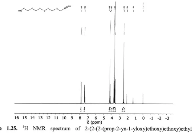
I I I 16 15 14 13 12 11 10 9 8 7 6 5 4 3 2 1 0 -1 -2 -3 6 (ppm)Figure 1.25. 'H NMR spectrum of 2-(2-(2-(prop-2-yn-1-yloxy)ethoxy)ethoxy)ethyl 4-methylbenzenesulfonate in CDC13. Peaks at 8 4.11, 2.04, and 1.26 are from ethyl acetate.
I x \\ \ i 1 J, f
~niLi
*T~ ~'4~V ~ Y ~ B 16 15 14 13 12 11 10 9 8 7 6 5 6 (ppm) 4 3 2 1 0 -1 -2 -3 -A o~s CHI
if 11 OH CHSFigure 1.26. 'H NMR spectrum of
(S)-2-((tert-butoxycarbonyl)amino)-4,7,10,13-tetraoxahexadec-1 5-ynoic acid in CDC13. Peak at 8 5.30 is from dichloromethane. Peak at 8 3.49
is from methanol.
I lI I
16 15 14 13 12 11 10 9 8 7 6 5 4 3 2 1 0 -1 -2 -3
6 (ppm)
Figure 1.27. 1H NMR spectrum of (S)-4-(2,5,8, 11 -tetraoxatetradec-13-yn-1 -yl)oxazolidine-2,5-dione in CDC13.