HAL Id: tel-01214578
https://tel.archives-ouvertes.fr/tel-01214578
Submitted on 12 Oct 2015HAL is a multi-disciplinary open access archive for the deposit and dissemination of sci-entific research documents, whether they are pub-lished or not. The documents may come from
L’archive ouverte pluridisciplinaire HAL, est destinée au dépôt et à la diffusion de documents scientifiques de niveau recherche, publiés ou non, émanant des établissements d’enseignement et de
Hydration water dynamics of the tau protein in its
native and amyloid states
Yann Fichou
To cite this version:
Yann Fichou. Hydration water dynamics of the tau protein in its native and amyloid states. Modeling and Simulation. Université Grenoble Alpes, 2015. English. �NNT : 2015GREAY021�. �tel-01214578�
THÈSE
Pour obtenir le grade de
DOCTEUR DE L’UNIVERSITÉ DE GRENOBLE
Spécialité : Physique pour les sciences du vivant Arrêté ministériel : 7 Aout 2006
Présentée par
Yann FICHOU
Thèse dirigée parMartin WEIK
préparée au sein de l’Institut de Biologie Structurale et del’école doctorale de physique
Dynamique de l’eau d’hydratation
de la protéine tau dans ses formes
native et amyloïde
Thèse soutenue publiquement le11 mars 2015, devant le jury composé de :
Pr Antonio Cupane
Université de Palerme, Palerme, Italie, Rapporteur
Pr Damien Laage
Ecole normale supérieure, Paris, France, Rapporteur
Dr Martin Blackledge
Institut de biologie structurale, Grenoble, France, Examinateur
Pr Marc Jamin
Institut de biologie structurale, Grenoble, France, Examinateur
Dr Véronique Receveur-Bréchot
Centre de recherche en cancérologie de Marseille, Marseille, France, Examinatrice
Pr Douglas Tobias
Université de Californie Irvine, Californie, Etats-unis, Examinateur
Dr Giuseppe Zaccai
THÈSE
Pour obtenir le grade de
DOCTEUR DE L’UNIVERSITÉ DE GRENOBLE
Spécialité : Physique pour les sciences du vivant Arrêté ministériel : 7 Aout 2006
Présentée par
Yann FICHOU
Thèse dirigée parMartin WEIK
préparée au sein de l’Institut de Biologie Structurale et del’école doctorale de physique
Hydration water dynamics of the
tau protein in its native and
amy-loid states
Thèse soutenue publiquement le11 mars 2015, devant le jury composé de :
Pr Antonio Cupane
Université de Palerme, Palerme, Italie, Rapporteur
Pr Damien Laage
Ecole normale supérieure, Paris, France, Rapporteur
Dr Martin Blackledge
Institut de biologie structurale, Grenoble, France, Examinateur
Pr Marc Jamin
Institut de biologie structurale, Grenoble, France, Examinateur
Dr Véronique Receveur-Bréchot
Centre de recherche en cancérologie de Marseille, Marseille, France, Examinatrice
Pr Douglas Tobias
Université de Californie Irvine, Californie, Etats-unis, Examinateur
Dr Giuseppe Zaccai
Acknowledgements - Remerciements
First of all, I would like to thank Damien Laage and Antonio Cupane for reviewing this manuscript. I am also grateful to Véronique Receveur-Bréchot, Marc Jamin, Martin Black-ledge, Douglas Tobias, Martin Weik and Giuseppe Zaccai to have been part of my defense committee.
My most sincere acknowledgment goes to my advisor, Martin Weik, who made this whole experience possible. He trained me to carry out proper research and initiated many projects on which I was honored to work. I want to thank Martin for having given me the freedom of research in the different projects, although he was always available for advices and discussions. It was scientifically and personally very enriching to work with such a great person. Martin, es hat mich sehr gefreut mit dir zu arbeiten.
I’m truly grateful to Douglas Tobias, who welcomed me in his group at UCI for about 7 months. He allowed me to learn a lot about MD simulations and to apply it to my research projects. Our discussions have always been very pleasant and fruitful.
Je ne vois pas comment remercier François-Xavier Gallat si ce n’est en utilisant la langue de Molière, si chère à son coeur. Merci FX de m’avoir appris ce qu’il fallait savoir de cette fameuse protéine tau, ainsi que de m’avoir initié à la diffusion de neutron. Ce fut toujours un réel plaisir d’échanger avec toi.
Je voudrais remercier Giorgio Schirò, pour nos discussions scientifiques sur la diffusion de neutron. Avec un sens de l’humour à la hauteur de sa connaissance des différentes techniques de diffusion, il m’a beaucoup appris pendant ma dernière année de thèse. Je voudrais remercier tous les membres, passés et présents, de l’équipe DYNAMOP. Tout d’abord, Mathilde lethier, qui m’a enseigné les bases de la biochimie en faisant preuve de beaucoup de pédagogie. Mes interactions avec Antoine, Jacques, Virgile, Chady, Maryam, Virginia, Joyce, Damien et Céline furent toujours plaisantes. Merci également à Aude Vernet qui a beaucoup accompli lors de son stage dans notre équipe. Gianluca, ce fut un plaisir d’être à tes cotés du début à la fin de ma thèse. Enfin, je tiens à remercier Linda Ponet pour l’immense travail administratif qu’elle a accompli pour moi.
ACKNOWLEDGEMENTS - REMERCIEMENTS
I would like to especially thank Matthias Heyden who actually taught me everything about MD simulations. He was always available and resourceful regarding all my requests. His curiosity and open mindedness is just as great as his knowledge and cleverness. I thank for their kindness all the members of the Tobias group, James, Abe, Krista, Scott, Eric, Alfredo, and in particular Vera.
I would like to thank Martina Havenith for making the collaboration on the THz spec-troscopy project possible. Thanks to Gudrun and Valeria for being part of this project as well.
Pendant ce projet de thèse, j’ai côtoyé de nombreuses personnes qui m’on beaucoup apporté scientifiquement. Merci à Franck Gabel, Pau Bernado, Alessandro Paciaroni, Catherine Picart, et Daphna Frenel. Merci également à Martine Moulin et à toute l’équipe du Dlab pour m’avoir accueilli pendant ces quelques semaines. Des remerciements particuliers iront à Giuseppe Zaccai qui a beaucoup apporté aux différents projets.
Many thanks go to Rachel Martin, Domarin Khago and Carolyn Kingsley, for our collab-oration on the crystallin project.
Comment oublier la “Team Porquerolles”, avec qui, en octobre 2011, nous avions tous em-barqué dans le même bateau, Louise, Widade, Yann, Gianluca, Mathieu, Hicham, Didier. Merci pour ces discussions toujours très intellectuelles du mercredi midi.
Evidemment, beaucoup d’amis, parfois de longues dates, ont été à mes cotés pendant ces trois années. A ce titre, que tous mes amis ayant un jour habité avec moi Mellac et sa banlieue (Quimperlé, Clohars,...), ou encore cette belle ville de Rennes, se sentent assurés de tous mes remerciements. Merci à Kaelig qui, à sa façon, m’a encouragé à suivre cette voie. Je ne manquerai pas de remercier tous mes amis Grenoblois, qu’ils soient swingueurs ou scientifiques. Merci à Joséphine pour nos discussions prandiales bien sympathiques. Ce fut également un plaisir d’être en colloc avec mon cher homonyme pendant près de 2 ans.
Comme la pression de fin de rédaction à souvent tendance à se propager dans l’entourage du malheureux thésard, je remercie Marie-Noëlle, Coraline, François Xavier et Florian pour m’avoir aidé à finaliser ce manuscrit.
Cette section ne serait pas complète si je n’y exprimais pas ma sincère gratitude à Coraline, pour avoir été à mes cotés tout au long de cette thèse, et en particulier dans les moments difficiles.
Enfin, je tiens à remercier du plus profond de mon coeur ceux sans qui rien de tout ça n’aurait pu exister, ma Famille. Merci à mes parents, Marie-Jeanne et André, et à mon frère Elouan, pour leur soutient sans faille, leur intérêt pour un métier pourtant compliqué à expliquer, et surtout merci pour leur affection qui a été pour moi une ressource inestimable et irremplaçable. Un grand merci à mes parents pour avoir su faire naître chez moi la curiosité à l’égard de tout ce qui nous entour. Enfin, ma dernière pensée ira à mes grand parents Louis et Marie qui, de part leur seule présence, inspirent tant de qualités humaines.
Preface
Long before being a scientific quest, water had been a central substance throughout the history of human civilizations, as described in the book H2O: a biography of water, by
Philip Ball. In ancient Egypt, water was this flowing substance that one day could bring life and prosperity, and the other day could spread death and misery over the world. Water was once the ultimate element before dissociation, which could turn into essentially everything present on earth. For about two millenniums, water had remained one of the four fundamental elements from which derives everything in the universe. Then, No wonder that water has long been the object of many fantasies and mysteries. Lavoisier pioneered the rigorous work that provides the knowledge of water as a mere chemical compound, in the 18th century. Yet, even modern science has trouble to remain objective whenever water is involved, as evidenced by several passionate debates that have animated the scientific community in the 20th century. Nevertheless, it seems that water has not yet revealed all its secrets, as reviewed in chapter 1, and hopefully this work will contribute to a better understanding of the substance in a biological context.
The work on the dynamics of the tau protein and its hydration water follows up on the re-search carried out by Dr. Franc¸ois-Xavier Galla and Dr. Kathleen Wood, on the dynamical properties of different classes of proteins, using incoherent neutron scattering. The work presented in this manuscript was mostly achieved in the DYNAMOP group at the Institut de Biologie Structurale (IBS), under the supervision of Dr. Martin Weik. In addition, I had the honor to receive a Fulbright fellowship that allowed me spending almost seven months in the group of Prof. Douglas Tobias at the University of California Irvine (UCI). The MD simulation studies presented here were carried out at UCI under the supervision of Douglas Tobias. Thanks to the collaboration between these two groups, I was able to address the proposed issues by using a combination of computational and experimental techniques, which turned out to be a powerful approach. The work on THz spectroscopy was achieved in collaboration with the group of Prof. Martina Havenith at the University of Bochum, in Germany.
The results of the thesis work are presented as a gathering of manuscripts, which are submitted or in a final stage before submission. The first chapter sets the background
PREFACE
and motivations of the work, before introducing the different methods used in the second chapter. The five following sections present the different results in the form of journal articles, with the exception of chapter 6. Each chapter ends with a brief summary, which can all be read independently in order to obtain rapidly a complete overview of the thesis in a few minutes. A detailed summary of the manuscript is provided in French in appendix C.
Contents
1 Introduction 1
1.1 Water, the matrix of life . . . 2
1.1.1 Molecular crowding . . . 2
1.1.2 Water and biological function . . . 3
1.1.3 Hydration water . . . 4
1.1.4 Water and entropy . . . 4
1.2 Protein dynamics . . . 5
1.2.1 Energy landscape . . . 5
1.2.2 Dynamics-function relationship in proteins . . . 7
1.2.3 The dynamical transition . . . 7
1.3 Intrinsically disordered protein (IDPs) . . . 9
1.3.1 Generalities . . . 10
1.3.2 Biological functions and advantages of IDPs . . . 12
1.3.3 IDPs and diseases . . . 14
1.3.4 Protein aggregation and IDPs . . . 14
1.4 The tau protein . . . 17
1.4.1 Generalities . . . 17
1.4.2 Dynamical coupling with hydration water . . . 19
1.4.3 Tau amyloid fibers and diseases . . . 20
1.5 Summary of the introduction and issues addressed in this thesis . . . 23
2 Biophysical methods 25 2.1 Neutron scattering and molecular dynamics . . . 26
2.1.1 Introduction . . . 26
2.1.2 Neutron sources and instrumentation . . . 26
2.1.3 Scattering theory . . . 28
2.1.4 Incoherent and coherent scattering . . . 30
2.1.5 Deuteration is a powerful tool . . . 32
2.1.6 Elastic, quasi-elastic and inelastic scattering . . . 32
CONTENTS
2.2 Small angle scattering . . . 35
2.3 THz spectroscopy . . . 37
2.4 Molecular dynamics (MD) simulations . . . 40
2.4.1 Introduction and equation of motion . . . 40
2.4.2 Force fields . . . 40
2.4.3 Water models . . . 41
2.4.4 MD simulation and neutron scattering . . . 43
2.5 Time scale diagram : methods and biological processes . . . 44
3 Translational diffusion of hydration water correlates with functional mo-tions in folded and intrinsically disordered proteins 45 3.1 Manuscript . . . 45
3.2 Supplementary information . . . 69
3.3 Summary and lead-in for the next chapter . . . 82
4 Comparing collective water dynamics around intrinsically disordered and globular proteins 83 4.1 Manuscript . . . 83
4.2 Summary and lead-in for the next chapter . . . 102
5 Hydration water mobility is enhanced around tau amyloid fibers 103 5.1 Manuscript . . . 103
5.2 Supplementary information . . . 123
5.3 Summary and lead-in for the next chapter . . . 136
6 Dynamical diversity of protein aggregates 137 6.1 Introduction . . . 137
6.2 Materials and methods . . . 138
6.2.1 Proteins aggregation and samples preparation . . . 138
6.2.2 Elastic incoherent neutron scattering (EINS) experiments . . . 140
6.2.3 Data treatment . . . 140
6.3 Results . . . 141
6.3.1 EINS experiments on MBP . . . 141
6.3.2 EINS experiments on lysozyme . . . 143
6.3.3 EINS experiments on the hexapeptides VQIVYK and VQIINK . . . 145
6.4 Discussion and conclusion . . . 147
7 Molecular dynamics simulations of a powder model of the intrinsically disordered human tau protein 150 7.1 Manuscript . . . 150
7.2 Summary . . . 175 8 General discussion, perspectives and concluding remarks 176 A Paper : A Polymer Surfactant Corona Dynamically Replaces Water in
CONTENTS
and Activity 181
B SAXS experiments on γ-S crystallin 186
C Résumé de la thèse en français 190
C.1 Introduction . . . 191
C.2 Méthodes biophysiques . . . 192
C.3 Résumé des travaux effectués . . . 193
C.3.1 Nature des mouvements de l’eau d’hydratation impliqués dans la transition dynamique de la protéine . . . 193
C.3.2 Comparaison de la dynamique collective de l’eau d’hydratation des protéines Tau et MBP . . . 194
C.3.3 Mobilité de l’eau d’hydratation de la protéine tau dans sa forme native et fibrillaire . . . 194
C.3.4 Une diversité dynamique au sein des agrégats de protéines . . . 195
C.3.5 Modèle d’une poudre hydratée de la protéine tau . . . 195
C.4 Conclusion . . . 196
Bibliography 198
Résumé 211
Chapter
1
Introduction
This chapter sets the scientific background of the issues investigated in this manuscript. First, we argue that water is an essential substance in biology, so important that it has been named the matrix of life (Szent-Gyorgyi, 1979). We provide several examples demon-strating that water molecules should be considered as biomolecules on their own, which are required to interact with all biological entities, including proteins and nucleic acids. Second, the focus will be put on proteins and their dynamics. After recalling that the dynamical features of a protein are as important as its structural properties, we introduce the notion of energy landscape as a means of understanding protein stability and dynamics. Then, we focus on a recently-discovered class of proteins that is composed of the intrinsi-cally disordered proteins (IDPs). Although research on IDPs has skyrocketed only about fifteen years ago, it is now established that they are ubiquitous in living systems and they fulfilled a large variety of roles. IDPs have drawn a particular attention, because their aggregation is involved in many diseases including neurodegenerative disorders. Finally we focus on a particular IDP called tau that has been the main object of this thesis. Present in neuronal cells where it regulates microtubule stability, the tau protein is of particular interest because its fibrillation is one of the hallmarks of the Alzheimer disease.
1.1. WATER, THE MATRIX OF LIFE
1.1
Water, the matrix of life
1.1.1 Molecular crowdingWe have all been taught that 70% of our body mass is made of water. However it is somewhat hard to transpose this value at a molecular level and get an intuition of what the interior of a living cell is like. It is crowded, very crowded. The total concentration of macromolecules inside cells can be up to 400 mg/ml, meaning that they occupy up to 40% of the total volume (Ellis and Minton, 2003). The average distance between macromolecules is roughly 20 angstrom (Å; 1 Å = 10−10
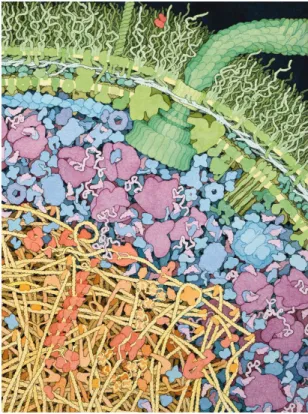
meter), which corresponds to about six layers of water. Those numbers are well illustrated by David Goodsell (Goodsell, 2009) who made an elegant and realistic painting of the inside of the bacteria Escherichia coli (see figure 1.1).
Figure 1.1: Schematic representation of the molecular crowd inside the bacteria E. coli extracted from (Goodsell, 2009).
While moving in a random-walk manner, supplied by thermal energy, macromolecules bump into each other, sometimes ricocheting, and sometimes interacting to accomplish their vital function. One might naively think of water as a biologically inert medium that only fills the gap between biomolecules. In reality, this view of water being a mere bystander in the play of life has been severely reviewed. Water molecules are actually
1.1. WATER, THE MATRIX OF LIFE
active components, ubiquitous in life, that have to be considered as biomolecules on their own. Macromolecular interactions are mediated by their surrounding water molecules, the so-called hydration shell, thereby enabling the complex machinery of life to function. Thus, in order to deepen our understanding of biological processes, scientists have studied how water influences biomolecules such as proteins and reciprocally how proteins influence water, so I have done in the present work.
1.1.2 Water and biological function
Water fulfills a variety of tasks in different biological processes, as thoroughly reviewed by Philip Ball (Ball, 2008). I report in this section a few examples that are directly or indirectly relevant to the work presented in the manuscript.
One essential role of water in biology is its participation in protein folding, mainly through the hydrophobic effect (Baldwin, 2014). Although the precise mechanisms remain incom-pletely understood, the basic principle is that proteins collapse upon their hydrophobic residues to hide them from the surrounding water, therefore maximizing water entropy. The folding process is probably twofold, the protein first collapse to a near-native confor-mation that retains water in its interior, followed by a slower expulsion of its residual water (Cheung et al., 2002; Garcia and Onuchic, 2003). Water molecules also act as temporary linkers between hydrophilic residues during the folding process, holding thereby the folded chain together until it is ready for final compaction (Papoian et al., 2004). Indeed, because one water molecule can form multiple hydrogen bonds, it can mediate protein-protein and protein-ligand interactions. Philip Ball (Ball, 2008) wrote “In other words, it is not simply the case that water molecules can bridge two proteins: such contacts can be imbued with significant information content that allows the interactions to be discriminating. Thus, protein surfaces in a sense extend the range of their influence via their hydration shells". And this does not only apply to proteins but also to nucleic acids (Robinson and Sligar, 1993). When proteins achieve their biological function, dynamical and structural changes in their hydration water are quite often observed (Barillari et al., 2007; Autenrieth et al., 2004; Royer et al., 1996; Kornblatt and Hoa, 1990; Colombo et al., 1992; Grossman et al., 2011). The importance of these changes for macromolecular functions is, however, hard to evaluate and often remains speculative. We add one piece to that statement in chapter 5 by reporting the enhancement of hydration water dynamics after the aggregation of a protein. Recently, it was proposed that a gradient in hydration water dynamics at the protein surface might drive a ligand to its right location, acting as a “hydration funnel” (Conti Nibali and Havenith, 2014).
Proteins involved in electron and proton transfer play a central role in many biological processes, including photosynthesis and protein fluorescence. Not only water molecules can participate in proton and electron transfer, but they can form organized water wires of several molecules. Electron transfer can be promoted by water chains of up to 10 Å in length (Francisco et al., 2004) while a water wire of 23 Å in Rhodobacter sphaeroides has been suggested to act as a proton pathway (Ermler et al., 1994). Finally, hydration water has an essential role in the properties of nucleic acids. Without water and its high
1.1. WATER, THE MATRIX OF LIFE
dielectric permittivity to shield charges, the DNA would not maintain the nice double helix that we know. Moreover some researchers (see for instance (Fuxreiter et al., 2005)) have proposed that a given DNA sequence has a ”hydration fingerprint” that determines its binding properties.
1.1.3 Hydration water
After reporting the role of water in some biological processes, let us now focus on how water is influenced by the presence of biomolecules. At a molecular level, water and biomolecules interact with one another, mainly by forming hydrogen bonds, which disturb their struc-ture and dynamics. The term hydration water refers to water that feastruc-tures dynamical or structural properties different from those of bulk water because of its proximity to a biomolecule. It is generally considered that water molecules are slowed down and adopt a structure distinct from bulk water within at most the two first water layers around pro-teins. The first hydration shell was shown by simulation, x-ray and neutron scattering to be 10-15% more dense than bulk water (Svergun et al., 1998; Seki et al., 2002; Merzel and Smith, 2002). Some water molecules, sometimes refered as ”immobile” or crystallographic water, are slowed down by a minimum of 3 orders of magnitude (residence time superior to 1 ns), while others are almost not affected. However, there is no consensus on the average water slowdown at the protein surface. The retardation ranges in the literature from a mere twofold to several orders of magnitude (see (Halle, 2004) for a critical review of these values).
In addition to the individual-molecule dynamics mentioned above, the collective dynamics of water is influenced in the vicinity of proteins. In particular, recent experimental and theoretical research has shown that protein surface motions are correlated with water motions up to 10-20 Å away from the protein (Heyden and Tobias, 2013; Meister et al., 2013). These correlations are THz vibrations that propagate as sound wave through the water hydrogen bond network. In other words, the vibrational properties of water hydrogen bond network are modified in the vicinity of biomolecules.
1.1.4 Water and entropy
Because of its interaction with proteins, mainly through the formation of hydrogen bonds, hydration water has a lower entropy than bulk water. By modulating the water-protein hydrogen bond network, the protein can change the entropy of its hydration water. In the case where the hydration water entropy increases, this thermodynamics fuel can be used by the protein. This is known as entropy compensation, which can be either an entropy-entropy or an entropy-entropy-enthalpy compensation, depending on whether it compensates for an entropic or enthalpic cost. Thus, the notion of water being a reservoir of entropy for biomolecules has emerged. Using the terminology of Erwin Schrodinger in his book “what is life?”, one can say that macromolecules feed upon the entropy of their hydration water. Several experimental and computational studies have quantified this effect in different
1.2. PROTEIN DYNAMICS
systems for protein aggregation (Thirumalai et al., 2012), protein folding (Harano and Kinoshita, 2004) and ligand binding (Breiten et al., 2013).
1.2
Protein dynamics
The discovery of protein tertiary structure (by Dorothy Wrinch in the late 1930’s) had led the scientific community to think, for some time at least, that the function of a protein is strictly defined by its 3D structure. This picture was backed up by the lock-and-key theory first postulated in 1894 by Emil Fischer. It claims that the active side of a molecule, let’s says an enzyme, has a unique geometric shape that is complementary to the geometric shape of the substrate molecule, similar to the fit of puzzle pieces. This structure-function relationship went along with the tremendous development of protein crystallography, made possible by new technologies developed in the second half of the twentieth century. This structure-function paradigm has now evolved to include a third component, the dynam-ics. As well as particular structural features, a protein requires a proper dynamics, or conformational motions, to be functional.
1.2.1 Energy landscape
The free energy landscape of a protein is a mapping of all possible conformations as a function of their free energy. In other words, the knowledge of this landscape gives the energetically most favorable conformations. The energy landscape theory is often used to describe protein folding and stability, as thoroughly reviewed by Plotkin and Onuchic (Plotkin and Onuchic, 2002a,b), as well as protein misfolding and aggregation (see figure 1.2 and 1.7). In their normal condition, globular proteins possess a global minima cor-responding to their well-folded states (figure 1.2), while intrinsically disordered proteins (IDPs) lacks of such minima in their monomeric state (see the introduction to IDPs in section 1.3). Hans Frauenfelder and coworkers introduced the concept of conformational substates when studying the rebinding kinetics of carbon monoxide to myoglobin after photodissociation (Austin et al., 1975). This concept states that even globular proteins have no unique conformation, but rather multiple conformations that are energetically very close. In terms of energy landscape, this means that within a global energy minimum corresponding to a well-folded state, one finds many accessible substates (see figure 1.2), as if the ground state of a protein was highly degenerated. Transitions from one minimum to another correspond to dynamical changes in the structure of the protein that take it from one substate to another. The timescale of the conformational switching can range from the picosecond (ps), for energetically-close substates, to the second, for conformational changes involving ligand binding for instance. The relation between fast and slow con-formational changes is nicely discussed by Henzler-Wildman and Kern (Henzler-Wildman and Kern, 2007). Although the concept of conformational substates was introduced in the mid-seventies, it is worth noticing that already in 1959 Linderstrom-Lang and Schellman stated
1.2. PROTEIN DYNAMICS
“...a protein cannot be said to have "a" secondary structure but exists mainly as a group of structures not too different from one another in free energy, but frequently differing considerably in energy and entropy. In fact the molecule must be conceived as trying out every possible structure..." (Linderstrom-Lang and Schellman, 1959).
Figure 1.2: Energy landscape of globular proteins. The protein energy landscape is repre-sented by the free energy of the protein as a function of some reaction coordinate. Tran-siently unfolded proteins are in a higher state of energy. When exploring their energy landscape proteins transit spontaneously through different folding states, before eventu-ally ending up in their native state, which is the lowest energy state. This phenomenon is known as the folding funnel. Within the minimum of the native state, a multitude of sub-states, known as conformational subsub-states, are constantly explored. In conditions where inter-protein interactions are possible, a new domain of the landscape opens up with new minima, including oligomers, amorphous and amyloid aggregates. This domain is further discussed in section 1.3.4. Figure adapted from (Jahn and Radford, 2005).
The energy landscape of a protein very much depends on its environment, especially on its hydration state. Many experiments, particularly using neutron scattering, have shown that protein dynamics is significantly reduced in the absence of water (Rupley and Careri, 1991). Therefore, the picture of hydration water acting on the protein as a plasticizer has emerged (Doster, 2008). Fenimore and colleagues (Fenimore et al., 2004) have used the following formulation : “The large number of conformational substates is essential; proteins cannot function without this reservoir of entropy, which resides mainly in the
1.2. PROTEIN DYNAMICS
hydration shell". It is worth noticing that this view has been recently challenged by a solvent-free polymer-protein hybrid that was found to preserve the function and dynamics of the protein in the absence of water (see (Perriman et al., 2010) and (Gallat et al., 2012a), which is included in appendix A).
1.2.2 Dynamics-function relationship in proteins
The loss of biological function when hindering protein motions, either by lowering the tem-perature or by removing hydration water (Rasmussen et al., 1992; Zaccai, 1987; Ferrand et al., 1993; Doster et al., 1993), provided strong evidence for a dynamics-function rela-tionship. Since then, many examples of dynamical processes involved in protein functions have been identified, as reviewed for instance by (Henzler-Wildman and Kern, 2007). Slow dynamics, involving displacements of entire domains, are typically required for macro-molecular activity such as in enzyme catalysis or signal transduction. Even conforma-tional changes that occur very rarely (i.e. poorly-populated conformaconforma-tional states) have been shown to be essential for protein function, as shown for instance in lysozyme (Mulder et al., 2001) or the protein gpW (Sanchez-Medina et al., 2014). A very good illustration of functional large-amplitude motions is the class of IDPs, which constantly undergo large conformational changes and very often collapse upon binding to their biological partners (see section 1.3). Dynamics on shorter timescales, involving energetically close substates, have also been shown to take part in protein function. For instance the binding of car-bon monoxyde to myoglobin requires flexibility of protein side chains (Schotte et al., 2003). More precisely, the concept of proteinquake proposed in (Ansari et al., 1985) illustrates that local allosteric changes are collectively triggered to allow the release of a ligand. Another example of functional dynamics is at play in photoactivable proteins that undergo global conformational rearrangements after their chromophore have absorbed photons (Lukyanov et al., 2005). A recent study on a photoactivable flavoprotein showed that the interruption of the proteinquake by mutageneis short-circuits the protein function (Brust et al., 2013). The notion of a proteinquake is a nice concept that illustrates how protein dynamics on a very broad range of timescales (from fs to second) can be correlated to enable biological function.
1.2.3 The dynamical transition
In the framework of the dynamics-function relationship, protein dynamics have been as-sessed over a wide temperature range, from a few kelvins, up to 400 K. A typical profile of temperature-dependent protein dynamic on the ns time scale is shown in figure 1.3. A first onset of anharmonic dynamics is observed around 100-150 K followed by a drastic increase around 220-240 K, the so-called dynamical transition (Doster et al., 1989). The first increase has been assigned to the rotation of methyl groups entering the instrumental time window (Roh et al., 2005; Wood et al., 2010; Krishnan et al., 2008; Schiro et al., 2010b,a), while the dynamical transition corresponds to the onset of large amplitude mo-tions. This transition has been studied in particular by incoherent neutron scattering for
1.2. PROTEIN DYNAMICS
a large variety of biomolecules, including globular proteins (Wood et al., 2008), membrane proteins (Wood et al., 2007), intrinsically disordered proteins (Gallat et al., 2012b), en-tire cell proteomes (Tehei et al., 2004), amino acids (Schiro et al., 2011), nucleic acids (Caliskan et al., 2006) and sugars (Jasnin et al., 2010). It has been proposed that the large amplitude motions responsible for the dynamical transition are necessary for the protein biological activity (Rasmussen et al., 1992; Ferrand et al., 1993; Ostermann et al., 2000; Pieper et al., 2007), although some counter-examples have been reported (Daniel et al., 1998). In addition, the dynamical transition is suppressed in the absence of water (Roh et al., 2006), consistent with the picture outlined above of water promoting protein dynam-ics and allowing macromolecular biological activity. The role of water in this transition is discussed in detail in chapter 3. The origin of the dynamical transition remains controver-sial as evoked in chapters 3 and 7. Briefly, there are two main explanations of the observed dynamical onset at around 220-240 K (Becker et al., 2004). The first hypothesis is that the protein responds to a change of dynamics occurring in the solvent at the temperature of the dynamical transition. In particular, the confined hydration water would undergo a fragile-to-strong crossover originating from the transition between a high density liquid and a low density liquid phase (Chen et al., 2006; Lagi et al., 2008). This hydration water transition would correspond to the crossing of the so-called Widom line (Xu et al., 2005) at atmospheric pressure. On the other hand, the dynamical transition has been interpreted as an effect purely originating from the instrumental resolution (Khodadadi et al., 2008; Doster et al., 2010). The transition seen at 220-240 K would correspond to the temper-ature at which thermally activated protein motions enter the space-time window of the spectrometer.
1.3. INTRINSICALLY DISORDERED PROTEIN (IDPS) 2.5 2.0 1.5 1.0 0.5 0.0
Mean Square Displacement <u
2 > (Å 2 ) 300 250 200 150 100 50 Temperature (K) Dry protein Hydrated protein
Figure 1.3: Mean square displacements (MSDs) of a dry and a hydrated protein measured by neutron scattering (Gallat et al., 2012b). The MSDs presented here are representative of the average protein dynamics on the ns timescale. The onset of dynamics at around 140 K originates from the rotation of methyl groups entering the instrumental time window and does not depend on the hydration level. The so-called dynamical transition is visible at around 240 K in hydrated proteins and is suppressed in the absence of water.
1.3
Intrinsically disordered protein (IDPs)
For most of the twentieth century, biologists had considered that every proteins had to have a well-defined structure. The primary origin of this paradigm was the ’lock and key’ hypothesis, formulated in 1894 by Emil Fischer to explain the astonishing specificity of the enzymatic hydrolysis of glucoside multimers by different types of similar enzymes. Although the notion of disordered proteins emerged in the 1990’s, it is not before the beginning of this millennium that they became popular among the scientific community and thus intensively studied. A breakthrough in this field was to be expected when looking at the situation in the early 2000’s:
• The biology community realized that disordered proteins were very abundant in the proteome (in particular due to the work of Dunker and coworkers (Dunker et al., 2000)),
1.3. INTRINSICALLY DISORDERED PROTEIN (IDPS)
• many diseases were found to involve natively disordered proteins, • yet, this new class of protein was almost uncharacterized.
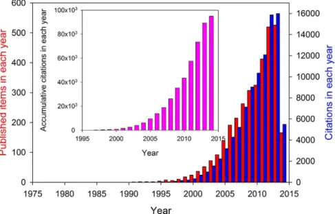
Therefore, it is no surprise that research efforts on IDPs have grown exponentially in the past decade, as shown in figure 1.4.
Figure 1.4: Growing interest of researchers in intrinsically disordered proteins. Number of publications (red bars) and corresponding citations (blue bars) related to IDPs by year, from 1991 to the beginning of 2014. The inset represents accumulative citations in each year. Figure extracted from (Uversky, 2014)
1.3.1 Generalities
Definition There is no exact definition of intrinsically disordered proteins (IDPs), be-cause no strict delimitation between disordered and ordered classes proteins exists. Pro-teins rather fall onto a structural continuum, from tightly folded single domains, to multi-domain proteins that might have flexible or disordered regions, to compact but disordered entities and, finally, to highly extended unstructured states (Dyson and Wright, 2005). A general definition of IDPs would be any protein that exists in different structures, either at the secondary or tertiary level. This lack of precise definition is reflected in the variety of names that can be found in the literature. A list of these terms would include floppy, pliable, rheomorphic, flexible, mobile, partially folded, natively denatured, natively un-folded, natively disordered, intrinsically unstructured, intrinsically denatured, intrinsically unfolded, intrinsically disordered, vulnerable, chameleon, malleable, 4D, protein clouds, dancing proteins, proteins waiting for partners, 32 proteins (Uversky, 2014). Several
1.3. INTRINSICALLY DISORDERED PROTEIN (IDPS)
acids to define the class of intrinsically disordered proteins. The notion of unfoldome and unfoldomics has been recently introduced to refer to the realm of IDPs and its field of study (Uversky et al., 2009). Rather than with a unique conformation, as one can be used to see in the Protein DataBank (PDB), the “structure” of an IDP is described by an ensemble of conformations. The conformations composing this ensemble are selected to be the smallest group of conformations that is able to describe some measured observable, such as small-angle scattering (SAS) spectra (Bernado et al., 2007). Thus, there is not a unique ensemble describing an IDP. The combination of nuclear magnetic resonance (NMR) spectroscopy and SAS has proven to be a valuable tool to provide structural characterizations of IDPs (Sibille and Bernadó, 2012).
IDP classification Despide the blurred line between ordered and disordered proteins, a classification was proposed by Dunker, Dyson, and coworkers (Dunker et al., 2001; Dyson and Wright, 2005), as shown in figure 1.5 extracted from (Uversky and Dunker, 2010). Briefly, four different states of disorder are proposed:
• The entirely disordered proteins that possess neither tertiary nor secondary (or al-most not) structures and are essentially composed of extended random coils. They are almost like polymers.
• The pre-molten globules, which have some residual elements of secondary structure and are more compact that pure random coil proteins.
• The molten globules, which are disordered but compact. The proteins in this inter-mediate state have no (or only a trace of) rigid tertiary structure. However, they are characterized not only by a well-developed secondary structure, but also by the presence of some topology, i.e., relatively fixed mutual positioning of the secondary structure elements.
• The ordered proteins, where no or only a few residues are disordered.
Collapsed globules is another term sometimes found in the literature and that can be identified to molten or pre-molten globules.
Natural occurrence The occurrence of intrinsically disordered protein in the living kingdom is strikingly high. A bioinformatics analysis from Dunker and coworkers (Dunker et al., 2000) estimated that:
• 9-37% of proteins from archea • 6-33% of proteins from bacteria • 35-51% of proteins from eukaryotes
contain disordered segments longer than 40 amino acids (see table 1.6). Moreover 3-18% of proteins in eukaryotic organisms were predicted to be entirely disordered.
1.3. INTRINSICALLY DISORDERED PROTEIN (IDPS)
Figure 1.5: Illustratition of a classification of order among proteins. Top line: Order (O), collapsed (molten globule-like, MG) disorder; extended (pre-molten globule-like, PMG) disorder; and (coil-like, coil) disorder. The figures represent model structures of a 100 residue-long polypeptide chain. Middle line: Relative hydrodynamic volumes occupied by a 100 residue-long polypeptide chain in these four conformations. The figure is extracted from (Uversky and Dunker, 2010).
1.3.2 Biological functions and advantages of IDPs
IDPs are ubiquitous among the proteome and, therefore, it is interesting to investigate what are the advantages of being unfolded in the biomolecular world. This topic was nicely reviewed by H. Jane Dyson in (Dyson, 2011) and is briefly evoked here :
• Promiscuous activity : One feature that comes to mind is the possibility that a dis-ordered domain could bind in different conformations to different partners. One example is the carboxyl-terminal activation domain (CAD) of the hypoxia-inducible factor α (Hif-1α) domain. In complex with the CREB-binding protein, the CAD of Hif-1α takes up a largely helical configuration, but when bound to the enzyme that catalyzes the hydroxylation of Hif-1α, this same sequence is present as an extended structure.
• Enhanced specificity : IDPs that fold upon binding can have very complex interaction with their partner, thus making the interaction highly specific (Sugase et al., 2007). Several examples have been observed for the interaction between transcription factors and DNA where some disordered part of the transcription factors participate in high-affinity binding.
• Complex connection : Many central functions of the cell, such as gene transcription, require the assembly of multi-domain complexes. Flexible linkers are essential to bind all the domains together despite their different architectures.
• Post-translational modifications : After translation, the sites of phosphorylation, methylation, hydroxylation, etc., remain accessible, thus allowing for facilitated post-translational modifications.
1.3. INTRINSICALLY DISORDERED PROTEIN (IDPS)
Figure 1.6: Prediction of disorder from 34 different genomes. L represents the number of consecutive disordered amino acids. The right column represents the predicted percentage of entirely disordered proteins according to a cumulative distribution function analysis. The figure was extracted from (Dunker et al., 2000).
• Regulation by proteolysis : The abundance of IDPs in the cell, which may precip-itate into pathological states if perturbed, has been shown to be tightly regulated by several cellular processes, including mRNA transcript clearance, translational rate and proteolytic clearance. IDPs can undergo ubiquitin-independent degradation that allow for rapid clearance of potentially harmful proteins, without the necessity for specific intact ubiquitination sites, which could potentially be inactivated by muta-tion.
• Higher capture radius : The concept of “fly-casting” suggests that disorder in a polypep-tide chain enhances the capture radius of the chain, with consequences on the kinetics of complex formation.
• Bulk physicochemical effects : Since the solvent-accessible surface of a disordered ensemble is greater than that of a globular folded protein of the same size, it might be expected to have a proportionally greater effect on the bulk physical chemistry of
1.3. INTRINSICALLY DISORDERED PROTEIN (IDPS)
the solution. This effect is partially discussed in this manuscript through the coupling of the tau protein with its hydration water.
The functions carried out by IDPs are manifold and take part in the regulation of key cellular processes, such as transcription, translation, signal transduction and the cell cy-cle. Using the Swiss-Prot database, a thorough statistical analysis of order- and disorder-associated functions showed that the latter cover essentially all functional categories (Xie et al., 2007).
Nevertheless, it has been proposed that disordered proteins functions fall into five main categories (Tompa, 2002). The first one is that of entropic chains, with functions that directly stem from the flexibility, pliability and plasticity of the protein. They rely entirely on an extended random coil conformation that has to remain in constant motion during functioning. The second class, the effectors, modify the activity of a single partner protein or assembled proteins. The third class is that of scavengers, which store and/or neutralize small ligands. The fourth class are the assemblers, which assemble, stabilize and regulate large multi-protein complexes, such as the ribosome, cytoskeleton, transcription preini-tiation complex. The fifth class, the display sites, mediate regulatory post-translational modifications, such as phosphorylation or limited proteolysis. Such modifications often require intrinsic disorder, which enables high specificity/low affinity interactions with the active site of the modified enzyme. This classification is summarized and illustrated with examples in the table 1.1 extracted from (Tompa, 2002).
1.3.3 IDPs and diseases
It has been shown that IDPs are assiociated with a large range of diseases, including inher-ited and acquired maladies. The connection between disorder and diseases such as cancer, cardiovascular diseases, amyloidoses, neuro-degenerative diseases and diabetes has been es-tablished (Uversky et al., 2009). A detailed review of the different diseases related to IDPs, as well as a discussion on what makes IDPs so common in human diseases was written by Uversky and coworkers (Uversky et al., 2008). The authors concluded that these diseases, which have been loosely grouped as “conformational diseases”, are characterized not only by protein misfolding but also by failures of post-translational modification and inability for proteins to interact correctly with their physiological partners. The largest group of conformational diseases, including numerous neurodegenerative disorders, originates from the conversion of specific proteins from their soluble functional states into stable, highly ordered, filamentous protein aggregates, known as amyloid fibrils, and from the deposi-tion of these aggregated materials into a variety of organs and tissues (Chiti and Dobson, 2006).
1.3.4 Protein aggregation and IDPs
IDPs’ predisposition in the aggregation pathway Although there is no consensus on a unique protein aggregation pathway, a general process could be the following (Ross
1.3. INTRINSICALLY DISORDERED PROTEIN (IDPS)
IDP Target/partner Function/action
Entropic chains
MAP2 Not applicable Entropic bristle (spacing in microtubule
architecture)
Titin Not applicable Entropic spring (passive contractile force in muscle)
Effectors
Calpastatin Ca2+-activated protease
(calpain)
Inhibitor of calpain
Stathmin Tubulin dimers Microtubule disassembly
Scavengers
Caseins Calcium phosphate Nanocluster formation, inhibition of pre-cipitation in milk
Desiccation stress pro-tein 16
Water Retention of water to prevent desiccation of plants
Assemblers
MAP2 Tubulin dimers Microtubule polymerization, bundling
Tau Tubulin Microtubule polymerization, bundling
Caldesmon Ca2+/calmodulin, F-actin,
myosin, tropomyosin
Actin polymerization, bundling Display sites
MAP2 Protein kinases Regulation by phosphorylation
Tau Protein kinases Regulation by phosphorylation
Table 1.1: Functional classification of IDPs.
and Poirier, 2004). First the protein reaches a pathological partially unfolded (or partially folded) state. This step can be triggered by several factors including covalent modifications (such as phosphorylation, partial cleavage, etc...), and environmental changes (such as pH, temperature, concentration of osmolyte, etc...). This structurally altered state, prone to self-assemble, forms intermediate oligomers, before eventually ending up in stable aggre-gates. The reason why IDPs eagerly aggregate was proposed to be found in the first step of this pathway (Uversky and Fink, 2004). Because of their unfolded nature and their high conformational flexibility, IDPs can readily switch to the partially unfolded states required to trigger the aggregation. This is in contrast to stable gobular proteins that need to un-dergo more significant conformational changes to reach the same partially unfolded state. The predisposition of IDPs for aggregation is illustrated in the protein energy landscape scheme shown in figure 1.7.
Amyloid and amorphous aggregates Protein aggregates fall into two structural cat-egories : amorphous aggregates and structured, β-rich amyloid aggregates. The latter have attracted a particular attention because of their implication in many diseases. They are structurally characterized by intermolecular β-sheets, so-called cross-β, and their presence
1.3. INTRINSICALLY DISORDERED PROTEIN (IDPS)
Figure 1.7: The protein energy landscape is represented by the free energy of the protein as a function of some reaction coordinate. When folding upon binding their partners, IDPs (center of the scheme) fall into lower states of energy. The propensity of IDPs to interact with various partners (labels 1, 2 and 3) determines their biological functions in recognition of various binding partners (ligands, nucleic acids and other proteins), in regulation of almost all cellular processes, and in signal transduction. In contrast to the folded globular proteins that have to unfold in order to aggregate (see figure 1.2), IDPs seem to be always ready for such intermolecular interactions, as illustrated by the small energy barrier separating them from stable aggregates. Amyloid aggregates, as the most stable form of aggregates, possess the lowest free energy. The figure is adapted from (Turoverov et al., 2010).
is often demonstrated by powder diffraction, featuring an inter β-sheet distance of 4.7 Å (Astbury et al., 1935; Sunde et al., 1997). A schematic view of an amyloid structure is shown in figure 1.8. Amyloid aggregates are thought to be the most stable form that a protein can adopt (Baldwin et al., 2011), as it is illustrated in its energy landscape in figure 1.7. In this view, the native state of a protein (unfolded or globular) would be just a metastable state. It is worthy to note that although amyloids are found in many dis-eases, the so-called functional amyloids are amyloid aggregates that play a biological role (Turoverov et al., 2010). An example is the storage of protein hormones in some secretary cells under the form of high density protein clusters, so-called granules. These granules, which were recently found to be amyloid aggregates (Maji et al., 2009), allow storing a large quantity of hormone proteins until a signal triggers their release, at which point the cell can secrete hormones at a fast rate. The main advantage of this process is that the rate of hormones secretion is no longer limited by the rate of peptide synthesis.
1.4. THE TAU PROTEIN
Figure 1.8: (A) Amyloid aggregates are fibrillar-like and are visible in negatively stained transmission electron micrographs. (B) Schematic diagram of the amyloid structure : the intermolecular β sheets along the fiber axis, so-called cross-β sheets, are spaced by 4.7 Å. The so-called steric zippers stabilized two parallel β sheets, separated by 6 to 11 Å perpendicularly to the fiber axis. (C) The diffraction pattern reflecting the characteristic distances of the cross-β sheets (black dashes) and the steric zippers (white dashes). Figure modified from (Greenwald and Riek, 2010).
1.4
The tau protein
1.4.1 GeneralitiesThe tau protein, essentially present in neurons, is a protein that modulates the stability of axonal microtubules. Thereby, It belongs to the class of microtubule associated proteins (MAPs). Tau is absent in dendrites and is active primarily in the distal portions of axons where it provides microtubule stabilization by binding to them. The microtubule-binding domain is composed of three or four repeat domains, as well as the proline-rich flanking domains on each side of the repeat domains (Amos, 2004). The N-terminal fragment does not bind to microtubules, but projects away from their surface and hence is termed “projection domain”. Tau is thought to have other marginal binding partners, whose role is not clearly understood (Mandelkow and Mandelkow, 2012). Although the tau protein is essentially disordered, a few transient secondary structure elements have been identified (see figure 1.9).
1.4. THE TAU PROTEIN
(a) (b)
Figure 1.9: (a) Schematic view of tau bound to a microtubule, adapted from (Amos, 2004). (b) Top : structural elements in htau40. Most of the protein is unfolded (black lines), with a few short and transient elements of secondary structures (α-helix in red, β-strand in yel-low, poly-proline helix in green). The red frame indicates the region of the two hexapeptide motifs (275V QIIN K280 and 306V QIV Y K311) essential for tau fibrillation. Middle :
do-main subdivision. R1-R4 represent the repeat dodo-mains, which essentially coincide with the microtubule interaction domain. Bottom: approximate location of interaction sites with other proteins. Figure adapted from (Mandelkow and Mandelkow, 2012).
There are mainly six isoforms of tau expressed in the adult human brain, all of which are derived from a single gene by alternative splicing. They differ from each other in the number of repeat domain (either three or four) and in the presence or absence of either one or two 29 amino-acid-long inserts at the N-terminal portion of the protein. The longest isoform, which is studied in this work, is refered to as htau40 and possesses 441 amino acids. It is composed of four repeat domains and has an average radius of gyration of 62 Å in solution, being thereby considerably more extended than a globular protein of the same molecular weight (Gallat et al., 2012b). Although the six isoforms appear to be broadly functionally similar, each is likely to have precise, and to some extent distinctive, roles. The tau protein is overall positively charged and possesses a high heterogeneity of charge distribution along its surface that is important for its interaction with other proteins as well as for aggregation. The repeat domains bind to specific pockets in β-tubulin at the inner surface of the microtubules, whereas the positively charged proline-rich regions are tightly bound to the negatively charged microtubule-surface, and the negatively charged projection domain branches away from the microtubule surface (see figure 1.9a). When tau is bound to a microtubule it stabilizes the structure and promotes its growth. It is interesting to note that tau is in a constant dynamic binding equilibrium, on and off the microtubules, constantly provoking polymerization and depolymerization. Each tau protein remains on average 4 seconds on a macrotubule surface. The main mechanism that modulates the affinity of tau for microtubule is its phosphorilation state, which is regulated by kinase and phosphatase enzymes. Interestingly, as an IDP, tau has been identified to bind marginally to many other partners such as actin filaments or nucleic acids. However the biological importance of the alternative binding to other partners is hardly known.
1.4. THE TAU PROTEIN
1.4.2 Dynamical coupling with hydration water
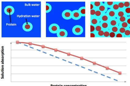
As an IDP, the tau protein lacks a tertiary structure and thus possesses a solvent-accessible surface greater than a globular protein of a similar molecular weight. It implies a larger protein-water hydrogen bond network and, therefore, one might expect the coupling be-tween the tau protein and its hydration water to differ from that of globular proteins. Previous research carried out in our laboratory has addressed the question of the dynam-ical coupling between the tau protein and its hydration water (Gallat et al., 2012b), and compared it to that of a globular protein of the same molecular weight, the maltose binding protein (MBP). The MBP is located in the periplasm and binds maltose, being involved in the transport process of maltodextrin. When comparing the dynamics of proteins and hydration water, as a function of temperature, they found that both follow each other up to 250 K for tau, while they separate at 220 K for MBP. François-Xavier Gallat and colleagues concluded that an enhanced coupling exists between the tau protein and its hydration water as compared to the globular protein. They suggested an extension of this conclusion to the entire class of IDPs and proposed a gradient of protein-hydration water coupling across different protein classes. From looser to tighter coupling, the membrane proteins would come first, followed by the globular proteins, and finally the IDPs with the tightest coupling (see figure 1.10).
A) B) C)
Figure 1.10: Protein-water dynamical coupling for different protein classes. The lower panels show mean square displacements (representative of the dynamics of the considered atoms) of hydration water and proteins. The upper panel represents a schematic view of three protein classes : A) the tau protein as an IDP, B) the maltose binding protein (MBP) as a globular protein and C) the bacteriorhodopsin as a membrane protein. A decreased dynamical coupling between hydration water and proteins is observed from the class A) to C). The figure is extracted from the thesis of François-Xavier Gallat (Gallat, 2011).
1.4. THE TAU PROTEIN
1.4.3 Tau amyloid fibers and diseases
Tau has been much more studied than some other MAPs because of its implication in many diseases. Indeed the neurofibrillary tangles (NTFs) found in several neurodegenerative diseases are composed of the tau protein in the form of straight or paired helical filaments (SFs or PHFs, respectively). Diseases involving tau filaments have been grouped under the term of tauopathies and include Alzheimer disease (AD) and the frontotemporal dementias. A list of these diseases is shown in table 1.2.
Alzheimer disease
Amyotrophic lateral sclerosis/parkinsonism-dementia complex Argyrophilic grain disease
Autosomal-dominant Parkinson’s disease Autosomal-dominant parkinsonism
Autosomal-recessive juvenile parkinsonism Corticobasal degeneration
Dementia pugilistica
Diffuse neurofibrillary tangles with calcification Down syndrome
Familial British dementia
Frontotemporal dementia and parkinsonism linked to chromosome 17 Gerstmann-Straussler-Scheinker disease
Guadeloupean parkinsonism Hallervorden-Spatz disease Myotonic dystrophy
Niemann-Pick disease, type C
Non-Guamanian motor neuron disease with neurofibrillary tangles Pick’s disease
Postencephalitic parkinsonism
Prion protein cerebral amyloid angiopathy Progressive subcortical gliosis
Progressive supranuclear palsy Subacute sclerosing panencephalitis Tangle only dementia
Table 1.2: Diseases in which tau deposits have been described (tauopathies), extracted from (Goedert et al., 2006).
PHFs were first discribed as twisted fibrillar structures, composed of two β-rich amyloid subfibers. Although it has now been shown that the term “tau fibers” is probably more appropriate than “tau PHFs” (see (Mandelkow and Mandelkow, 2012) for more details), we use indifferently both terms throughout this manuscript as it is common practice in the literature. Although the precise structure of tau PHFs remains unknown, it can be divided into two different regions, a rigid β-rich core (denoted as the fiber core), which is
1.4. THE TAU PROTEIN
essentially composed of the four repeat domains, and the remainder, so-called fuzzy coat, which is highly flexible and remains disordered. An essential factor for tau fibrillation is its propensity to form β-structures, encoded in the short hexapeptides 275V QIIN K280
and306V QIV Y K311, present in the repeat domains (see figure 1.9b). Disruption of these
motifs abrogates tau’s tendency to aggregate. Another important feature that promotes tau amyloid formation is the charge compensation of the basic middle part of tau by polyanions. In vitro, this can be achieved by the presence of sulfated glycosaminoglycans (for instance, heparin was used in this work to trigger fiber formation), which helps to overcome the nucleation barrier.
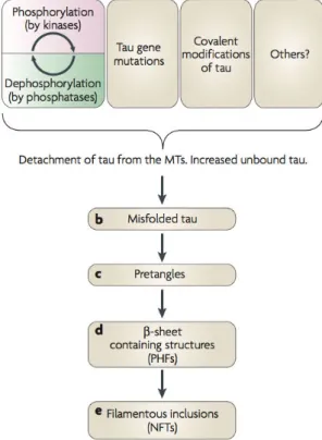
Very little is known about what causes the tau protein to aggregate. It is likely that a higher cytosolic concentration of unbound tau increases the chances of pathogenic conformational changes that, in turn, lead to the aggregation and fibrillation of tau. However, the potential origins of such an increase are multiple (figure 1.11 illustrate a proposed pathway of tau aggregation). Throughout the tauopathies, the tau proteins included in NFTs are always hyperphosphorylated, which is likely to be related to the detachment of tau from the microtubules. Whether this feature is the direct cause of tau fibrillation or a consequence of an early disruption remains to be elucidated. One step of the fibrillation process that has recently attracted considerable attention is the formation of transient soluble oligomers, also called pretangles. They have been proposed to be the toxic species in the aggregation process, while the mature PHFs would merely be a means of storing the excess of proteins. For further reading about the tau protein and the associated tauopathies, I recommend a review from Ballatore and coworkers published in Nature Reviews Neuroscience in 2007 (Ballatore et al., 2007) and a review written by Eva-Maria and Eckhard Mandelkow in 2012 (Mandelkow and Mandelkow, 2012).
1.4. THE TAU PROTEIN
Figure 1.11: Schematic view of a possible aggregation pathway, extracted from (Ballatore et al., 2007). Several factors can cause the detachment of tau from the microtubules, including genetic mutation and the disturbance of the phosphorylation/dephosphorylation equilibrium. These modifications lead to a misfolding of the protein, which will aggregate in pre-tangles (soluble oligomers) before forming the PHFs incorporated in the NFTs.
1.5. SUMMARY OF THE INTRODUCTION AND ISSUES ADDRESSED IN THIS THESIS
1.5
Summary of the introduction and issues addressed in this
thesis
Water is an essential component in any biological system known today and, for that reason, has been named as the “matrix of life”. At the molecular level, water molecules play a key role in a large variety of situations, such as ligand binding, DNA and protein stability, elec-tron and proton transfer, etc. Water and macromolecules, including proteins, are in a con-stant give-and take relationship. On the one hand, water in the vicinity of macromolecules, so-called hydration water, is perturbed and possesses properties distinct from those of bulk water. On the other hand, the presence of water is essential for proteins to acquire their proper structure and dynamics, which they require to achieve their function. Among the proteome, intrinsically disordered proteins (IDPs) composed a recently-discovered class of proteins that lack a well-defined three dimensional structure. Their “spaghetti-like” struc-tures make their dynamics and their interaction with water quite different from globular proteins. IDPs have attracted particular attention, because their aggregation is involved in a large variety of diseases, including neurodegenerative disorders. The intrinsically dis-ordered protein tau, which is the main object of this thesis, regulates the microtubule activity in neuronal cells. Tau has been particularly studied because its fibrillation into amyloid aggregates is one of the hallmarks of Alzheimer disease.
In this thesis, we intend to reveal whether or not the dynamical properties of protein hydra-tion water are biologically relevant and could be used to further understand and modulate biological processes. In order to do so, we have used incoherent neutron scattering, THz spectroscopy, small angle X-ray scattering, and all-atom molecular dynamics simulations to investigate water dynamics around the intrinsically disordered tau protein, compared to the structured maltose binding protein (MBP), as well as around functional and aggregated proteins.
After introducing in chapter 2 the biophysical methods used throughout this work, the manuscript presents an attempt to answer the following questions :
Chapter 3 What are the dynamical features of hydration water that allow a protein to be biologically active? The question was addressed by combining quasi-elastic neutron scattering and molecular dynamics (MD) simulations on the IDP tau as well as on the globular MBP.
Chapter 4 Does the collective dynamics of hydration water differ significantly for different types of proteins? This point was addressed by applying THz spectroscopy on tau and MBP.
Chapter 5 What changes occur in the hydration water dynamics when a protein forms amyloid fibers? This issue was investigated by carrying out neutron scattering and MD simulations on the tau protein in its native and amyloid states.
Chapter 6 Is there a dynamical signature of protein aggregates. If yes, is this signature related to the protein-water interactions? We address this question using elastic
1.5. SUMMARY OF THE INTRODUCTION AND ISSUES ADDRESSED IN THIS THESIS
neutron scattering on different types of aggregates, including amorphous and amyloid aggregates.
Chapter 7 Can we use MD simulations to model a protein powder of an IDP, and what can one learn from it? This issue was investigated by comparing MD simulations of a IDP tau powder model and an MBP powder model.
Chapter
2
Biophysical methods
This chapter presents the different biophysical methods used in this work. We examine the main features of the different techniques, as well as their main applications. A partic-ular attention is paid to neutron scattering (NS) and all-atom molecpartic-ular dynamics (MD) simulation, on which relies a large part of this PhD work. These two techniques were used in a complementary manner to evaluate dynamical properties of proteins and water on the picosecond-to-nanosecond timescale. THz spectroscopy is also described as a means of probing collective water dynamics. The descriptions remain general and the theoretical background of specific data analysis procedure will be detailed in the relevant chapters.
2.1. NEUTRON SCATTERING AND MOLECULAR DYNAMICS
2.1
Neutron scattering and molecular dynamics
2.1.1 IntroductionScattering is a general physical process where waves or particles are deviated after crossing an obstacle. In scattering experiments, the sample constitutes the obstacle and the prop-erties of the scattered beam, such as its angle and energy, reflects the propprop-erties of this sample. In order to obtain information at the atomic level, the wavelength of the incoming particle must be on the order of inter-atomic distances, i.e. a few Angstrom (Å; 1 Å = 10−10
m). This criterium can be fulfilled by several particles such as photons, electrons and neutrons.
Neutron beams possess several advantages over other particle beamss. As neutrons are neutral particles, they interact with atom nuclei. It results in a weak interaction with matter and, therefore, a deep penetration into it. Moreover, it implies that the strength of interaction with an atom, characterized by its cross section σ, is not linear with its atomic number. Especially σ will be sensitive to different isotopes, allowing for isotopic labeling, as detailed in section 2.1.5. Another interesting property lies in the low energy of few-Å-wavelength neutrons, which therefore preserves the samples. That is why neutron scattering is of such a high interest for biological samples, which are particularly sensitive to radiation damages. For instance, a neutron of 1 Å wavelength has an energy of 82 meV, while an X-ray photon of the same wavelength has an energy of 12.4 keV, i.e. six orders of magnitude higher. Furthermore, the energy of a neutron associated with a wavelength of atomic distances (Å) is on the order of thermal energy (meV), thus allowing to measure molecular dynamics. In particular, one can study the energy exchange between neutrons and the sample in quasi-elastic and inelastic scattering experiments. Finally, neutrons possess a non-zero spin (either 1/2 or -1/2), which allows to exploit incoherent and coherent scattering, as detailed in section 2.1.4.
The main disadvantage of neutrons over X-ray has to do with their production. Neutron sources are considerably less intense than X-ray sources, requiring therefore a much longer sample exposure time and larger sample amounts.
2.1.2 Neutron sources and instrumentation Neutron sources
Two types of technologies are used today to produce high-flux neutron sources : nuclear reactors and spallation sources. The first one is based on the chain fission of 235U and
produces a continuous flux of neutrons. The second uses an accelerated and pulsed proton beam that hits a heavy metal target. The high-energy protons strike the target nuclei and break them apart, resulting in the emission of high-energy neutrons. Neutrons produced by either spallation or reactor sources are very fast, having energies in the MeV regime. To be useful in condensed matter research, neutrons must have their energy reduced by