Directed evolution of split APEX peroxidase By
Yisu Han B.S. Chemistry (2013) University of California, Berkeley Submitted to the Department of Chemistry In Partial Fulfillment of the Requirements for the
Degree of Doctor of Philosophy at the
Massachusetts Institute of Technology Sepiember 20 18
C 2018 Massachusetts Institute of Technology All rights reserved
Signature of the author:
Certified by: Accepted by
iignature redacted
I
VSignature redacted
Department of Chemistry August 21, 2018 Alice Y. Ting Professor of Genetics, Biology, and Chemistry (by courtesy), Stanford UniversitySignature redacted
Thesis SupervisorMASSACHUSETTS redacteTd MASSACHLISETS INSTITUTE OF TECHNOWGY
NOV 14
2018
LIBRARIES
ARCHIVES
Robert W. Field Haslam and Dewey Professor of Chemistry Chair,Department Committee on Graduate Students
I
This doctoral thesis has been exainid b" e &xtr-ittee of the Department of Chemistry as foll011ows:
Signature redacted
S.1
'Aradley L. Pettelute Associ e Professor of ChemistrySignature redacted
Alice Y. Ting Professor of Genetics, Biology, and Chemistry (by courtesy), Stanford University Thesis Supervisor
Signature redacted
V'Elizabeth M. Nolan Associate Professor of Chemistry
Directed evolution of split APEX peroxidase By
Yisu Han
Submitted to the Department of Chemistry on August 21, 2018 in partial fulfillment of the Requirements for the Degree of the Doctor of Philosophy
ABSTRACT
APEX is an engineered peroxidase that catalyzes the oxidation of a wide range of substrates, facilitating its use in a variety of applications, from subcellular staining for electron microscopy to proximity biotinylation for spatially restricted proteomics and transcriptomics. While this strategy has provided access to many cellular regions and organelles, there are still many compartments and structures that cannot be accessed; this strategy is limited by the specificity of genetic targeting; there are cellular regions that cannot be exclusively targeted by a single genetic tag (Chapter 1).
To further advance the capabilities of APEX and address the need for an interaction-dependent proximity labeling tool, this thesis describes the development of a split APEX2 system. Short enzymatic reconstitution times are also desired, to further ensures both organelles' morphological integrity. Thus, it is critical that split APEX2 reconstitute peroxidase activity both rapidly and robustly. We first performed two subsequent rounds of structure-guided screening to determine the most optimal cut site (Chapter 2). We then used directed evolution on the top candidate pair to engineer a split APEX tool (sAPEX). Selections were performed via FACS on yeast-displayed fragment libraries, and 20 rounds of evolution produced a 200-amino acid N-terminal fragment (with 9 mutations relative to APEX2) called "AP" and a 50-amino acid C-terminal fragment called "EX". AP and EX fragments were each inactive on their own, but reconstituted to give peroxidase activity when driven together by a molecular interaction (Chapter 3). Our resulting split APEX2 fragment pair has significantly diverged from its parental sequence and shows interaction-dependent reconstitution in multiple contexts in living mammalian cells
(Chapter 4).
Our split APEX tool adds to the proximity labeling toolkit (Chapter 5 and 6), and in the future, should extend the utility of APEX-based approaches to new areas of biology at higher spatiotemporal resolution.
Thesis Supervisor: Alice Y. Ting
Acknowledgements
First, I would like to thank Alice, my thesis advisor, for giving me the opportunity to join her lab. I was able to explore my passions in science and methodology development, and coming from a synthetic chemistry background, I am grateful that she had taken a chance on me to allow me to learn a whole new field. The past five years in her lab, I have learned the most about being a rigorous scientist. Her mentorship provided insight into how to pragmatically design the best experiments, with the most rigorous questions -asking the right questions, to generate the most impactful and significant data. Furthermore, I would also like to thank Matt Shoulders, Bradley Pentelute, and Elizabeth Nolan for being on my thesis committee. Their mentorship and guidance through annual meetings and interactions within classes have been memorable and helpful.
Furthermore, I would like to thank all the mentors I've had in my life who have given me a chance to explore science. From my high school chemistry teacher, Mrs. Celia Reidler, to the lab at Johns Hopkins Applied Physics Laboratory (George, Lance, Andrew, Tracy, Edd, Carl, Sarah) that allowed to do real bench work and solidify my love for science. Thank you for trusting me, believing in me, and encouraging me. I remember the care packages you've sent over the years. I also have to thank my undergrad mentors Carolyn Bertozzi and Chelsea Gordon, for emboldening me to apply to my dream grad schools. Thank you for giving me a chance to learn and expand my passions.
I have also been blessed to meet some of the most amazing people during my time in the Ting lab. I would like to thank the following people for their friendship, conversation and companionship, hot chocolate dates, cookie runs, and a lot of commiseration over food over the years: Cathy Amaya, Tess Branon, Kelvin Cho, Kurt Cox, Robert Coukos, Elbeg Erdenee, Shuo Han, Victoria Hung, Chai Kaewsapsak, Tina Kim, Stephanie Lam, Jeff Martell, Monica Neugebauer, Oom Pattarabanjird, Mateo Sanchez, Wenjing Wang, and Boxuan Zhao. I would especially like to extend my gratitude towards Jeff Martell and Wenjing Wang for being the most valuable wealth of resource; you guys have been amazing mentors and teachers in not just how to do science and troubleshoot, but how to be a scientist and a good person.
Graduate school was not always easy, and I would also like to thank my furbabies LC and Ollie for providing me a constant source of comfort and fluff. I would also like to extend the deepest gratitude towards my boyfriend, Stephen Jedidiah Van Wyck. I met you just as I was reaching the last few months of my graduate career. I appreciate your curiosity and passion for science and nature, your calm demeanor, and your kindness. Thank you for bringing me flowers and feeding me dumplings, boba, hot chocolate, and In-n-Out. For pushing me, (kindly), to finish my thesis with accountability texts. Your rational voice-of-reason resonates love whenever I felt overwhelmed and you provided waves of calm. Thanks for being the most supportive and best surprise to ever come into my graduate life. You bring more light into my life every day. I love you.
Finally, I am most thankful to my parents and extended family. Without their sacrifices, their motivation, their guidance, and love, I would be nowhere. My parents provide me the unwavering support to pursue my dreams. They are there to lift me up when I trip on rocky roads, but also my biggest cheerleaders when I do succeed. I am so thankful for my role models to also be my best support crew. Thank you. I love you. This thesis is dedicated to my parents, for none of this would be possible without them.
Table of Contents
T itle P ag e ... 1
Signature P age... . . 2
A b stract ... . 3
A cknow ledgem ents... 4
T able of contents ... 5
L ist of figures... . ..7
L ist o f tab les...9
L ist of abbreviations... 10
Chapter 1. Introduction to APEX... 12
Background and development...13
A pplications ... 18
Splitting APEX to make a protein complementation assay (PCA)...20
D iscussions... 21
R eferences ... 22
Chapter 2. Rationally-selected screening of APEX2 cut sites...27
Introduction ... 28
Amplex UltraRed Screen of rationally-selected APEX2 cut sites for enzymatic reconstitution in HEK 293T cells...29
Live biotin-phenol labeling assay of potential split APEX pairs...32
D iscussion ... 34
Experimental methods...36
R eferences ... 4 1 Chapter 3. Directed Evolution of split APEX... 42
Introduction ... 43
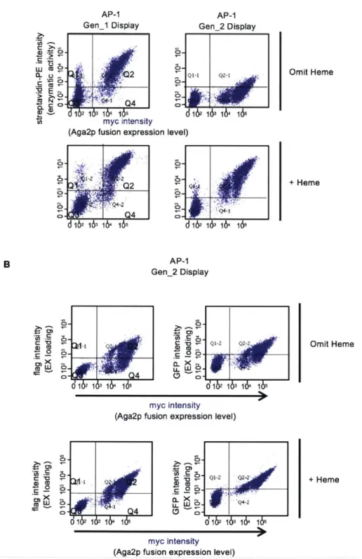
Establishing a platform...44
Yeast display evolution of AP (large fragment) ... 49
FACS-based negative selections for reduced fragment affinity...54
Modulating heme concentrations ... 56
Experimental methods...63
R eferences ... 69
Chapter 4. Testing different clones of split APEX in Mammalian Cells...70
Introduction ... 7 1 Determining the best clone from each generation of directed evolution...71
Comparing finalized split APEX (AP +EX) against earlier generations ... 90
Comparing a published split APEX21, our original split APEX2, and evolved split A P E X 2 ... . 95
D iscu ssion ... 9 8 Experimental methods... 99
R eferences ... 104
Chapter 5. Applying sAPEX to the mito-ER junction for EM imaging and proteomic m ap p in g ... 10 5 Introduction to the mito-ER junction...106
Applying sYFP and sGFP at the mito ER junction...108
Optimizing sAPEX targeting and expression levels for mito-ER applications...113
Preliminary testing of sAPEX at the mito-ER junction for proteomics in C O S 7 cells...118
Using sAPEX for DAB labeling across the MITO-ER...123
D iscu ssion ... 12 5 Experimental Methods...126
R eferences...13 1 Chapter 6. Reconstitution of sAPEX on a target RNA motif...134
Introduction ... 135
Applying sAPEX and optimizing expression levels of AP and EX fragment ... 137
D iscussion ... 148
Experimental methods...149
List of Figures
Figure 1-1: The mechanism of Class I peroxidases, model structures obtained from the Gumiero,
et al. paper2 . . . . 14
Figure 1-2: The binding site of horseradish peroxidase (HRP) with ferulic acid (FA) in the active site ... . . 15
Figure 1-3: APEX2 is an engineered product of structure-guided mutagenesis and yeast display-directed evolution... 16
Figure 1-4: APEX2 can be utilized as both a reporter for EM and proteomics...18
Figure 1-5. Overview of protein complementation assays (PCAs) ... 20
Figure 2-1. Selection of potential split APEX cut sites...28
Figure 2-2. split APEX cut site screening platform...29
Figure 2-3. Screening of potential split APEX cut sites...30
Figure 2-4. Focused Amplex UltraRed screening of potential sAPEX cut sites with single fragm ent controls... 31
Figure 2-5. Testing split APEX pairs using biotin-phenol as substrate...33
Figure 2-6. Biotin-phenol assay of the 3 most promising split APEX2 cut site pairs at the mito-E R junction ... . . 35
Figure 3-1. Displaying split APEX2 candidates as circular permutations on the yeast surface... .44
Figure 3-2. Displaying split APEX2 pairs as fusions to different yeast surface proteins... 47
Figure 3-3. Comparing split display (SD200) and circularly permutated (CP89) platforms to full length APEX2 fused to Aga2p...48
Figure 3-4. Generation 1 yeast display-based directed evolution...50
Figure 3-5. Generation 2 yeast display-based directed evolution...52
Figure 3-6. Generation 3 yeast display-based directed evolution...53
Figure 3-7. Generation 4 yeast display-based directed evolution...55
Figure 3-8. Using heme biosynthesis inhibitor, succinyl acetone, to reduce CP89 peroxidase activ ity ... . . 57
Figure 3-9. Testing exogenous heme supplementation prior to EX-GFP incubation...59
Figure 3-10. Yeast-display directed evolution and results...62
Figure 4-1. Amplex UltraRed Assay of AP-0 mutants derived from first generation of yeast display evolution... 73
Figure 4-2. Biotin-phenol labeling on live HEK 293T cells comparing the top mutants of Generation 1 evolution against APEX2, and starting template AP-0...75
Figure 4-3. Amplex UltraRed and Biotin-phenol labeling of AP-1 mutants derived from second generation of yeast display evolution...78
Figure 4-4. DAB labeling of AP-1 mutants derived from second generation of yeast display evolution ... . 79
Figure 4-5. Biotin-phenol labeling comparing AP-0, AP-1 AP-2 in HEK 293T cells stably expressing EX-HA-FRB ERM...81
Figure 4-6. Biotin-phenol labeling of AP-2 mutants derived from third generation of yeast display evolution in stable EX-FRB-ERM cells. ... 82
Figure 4-7. Diaminobenzidine (DAB) labeling of AP-2 mutants derived from third generation of yeast display evolution in stable EX-FRB-ERM cells...84
Figure 4-8. Biotin-phenol and Amplex UltraRed labeling of AP-3 mutants derived from fourth
generation of yeast display evolution in monoclonal stable EX-FRB-ERM COS7 cells...86
Figure 4-9. Biotin-phenol labeling of finalized split APEX in monoclonal stable EX-FRB-ERM COS7 cells infected with FKBP-AP...86
Figure 4-10. Western blot analysis of biotin-phenol labeling of finalized split APEX in stable FR B -EX H E K 293T ... 89
Figure 4-11. Comparing the different generations of evolved split APEX clones in mammalian cells... . . .. 9 1 Figure 4-12. Comparison of sAPEX variants in the mammalian cytosol, with biotin-phenol labeling as readout of peroxidase activity...93
Figure 4-13. Examining accumulated AP mutations in full length APEX2...94
Figure 4-14. Comparing a published split APEX210, our original split APEX2, and evolved split A P E X 2 ... . .. 96
Figure 5-1: ER-mitochondria encounter structure (ERMES) consists of four proteins...107
Figure 5-2. Testing split GFP at the mito-ER junction...109
Figure 5-3. Testing split YFP at the mito-ER junction...111
Figure 5-4. Testing mito and ER targeting sequences fused to the FKBP/FRB scaffold...114
Figure 5-5. Testing new mito and ER targeting sequences fused to the FKBP/FRB scaffold for sY FP reconstitution ... 116
Figure 5-6. Testing sAPEX at mito-ER contact sites...118
Figure 5-7. Examining sAPEX mito-ER targeting and morphology...119
Figure 5-8. Western blot analysis of live biotin-phenol labeling of sAPEX targeted to the mito-E R ... 12 2 Figure 5-9. DAB labeling of sAPEX targeted to the mito-ER...124
Figure 6-1. Testing MS2 stem-loop coating protein fused to full length APEX2 to target it to an R N A binding site m otif...136
Figure 6-2. Testing split APEX targeted to an RNA binding site motif. ... 138
Figure 6-3. Noncoding RNA construct design...141
Figure 6-4. Streptavidin-enrichment of biotin-phenol labeled HEK 293T cells expressing split A P E X ... 14 3 Figure 6-5. Testing Split APEX2 P2A constructs...145
Figure 6-6. Testing HEK 293T cells stably expressing PCP-EX against double stable cells expressing PCP-EX and MCP-AP...147
List of tables
Table 2: Plasmids used in Chapter 2...38
Table 3: Plasmids used in Chapter 3...68
Table 4: Plasmids used in Chapter 4...103
Table 5: Plasmids used in Chapter 5...131
List of abbreviations
8-oxo-dGTP ... 8-oxo-2'-deoxyguanosine-5'-triphosphate A F ... alexaflu or Agalp...yeast surface glycol protein A-agglutinin, one of the two-subunits Aga2p... yeast surface glycol protein A-agglutinin, one of the two-subunits AP... evolved, finalized N-terminal large fragment of sAPEX AP-0...starting template, unevolved N-terminal large fragment of split APEX AP-I...,,...evolved first generation N-terminal split APEX fragment AP-2... evolved second generation N-terminal split APEX fragment AP-3... evolved third generation N-terminal split APEX fragment APEX...first generation enhanced APX
APEX2...second generation APEX APX ... wild-type ascorbate peroxidase
A TP...adenosine triphosphate
A U ... arbitrary unit
B P ... b iotin-phenol B irA ... E . coli biotin ligase B SA ... bovine serum album in
CAS9...CRISPR associated protein 9 C FP ... cyan fluorescent protein CMV...Cytomegalovirus promoter
COS7...african green monkey kidney cells
C P ... circularly perm utated CRISPR...clustered regularly-interspaced short palindromic repeats D A B ... 3,3'-diam inobenzidine
DMEM...Dulbecco's modified Eagle's medium
D N A ... deoxyribonucleic acid DPBS ... Dulbecco's phosphate-buffered saline dPTP... 2'-deoxy-P-nucleoside-5'-triphosphate D T T ... dithiothreitol ER ... endoplasm ic reticulum ERM... endoplasmic reticulum membrane EX ... C-terminal small fragment of sAPEX
E M ... electron m icroscopy FACS... fluorescence activated cell sorting FB S ... fetal bovine serum FKBP...FK506 binding protein
Flag...synthetic epitope tag FP ... fluorescent protein FRB...FKBP-12, rapamincin binding domain
GFP... green fluorescent protein
HA...epitope tag derived form human influenza hemagglutinin surface glycoprotein H 20 2... . . . ..hydrogen peroxide
His6... histidine hexamer protein tag H R P ... horseradish peroxidase IMS...mitochondrial intermembrane space KDEL...endoplasmic reticulum retention sequence MAVS ... mitochondrial antiviral signaling protein MCP...MS2 stem loop coating protein MEM...minimum essential medium Mito-ER...mitochondrial-endoplasmic reticulum M S ... m ass spectrom etry M W ... m olecular w eight Myc...epitope tag derived from c-myc NEB... New England BioLabs N E S ... nuclear export signal N LS...nuclear localization signal Ni-NTA... nickel nitrilotriacetic acid OMM... outer mitochondrial membrane PCA...protein complementation assay PCP ... PP7 stem loop coating protein PCR...polymerase chain reaction PMSF... phenylmethylsulfonylfluoride PPI...protein-protein interaction R B P ... R N A binding protein RIPA...radioimmunoprecipitation assay R N A ... ribonucleic acid SA-HRP...strepavidin-conjugated horse radish peroxidase sAPEX...finalized evolved split APEX PCA system consisting of AP and EX S D ... sp lit disp lay SGCAA... synthetic galactose plus casein amino acid SDCAA ... synthetic dextrose plus casein amino acid SD S... sodium dodecyl sulfate SDS-PAGE ... sodium dodecyl sulfate polyacrylamide gel electrophoresis T E V ... tobacco etch virus V5...epitope tag derived from paramyxovirus of simian virus 5
W C L ... w hole cell lysate YFP...yellow fluorescent protein
Background and development
In order to understand a complex system, it is critical to first identify its components. Typically to understand the components, the region of interest is isolated and examined in detail, and although standard subcellular fractionation increases the spatial information of a mass spectrometry proteomic experiment, the inherent purification process is subject to four major drawbacks. First, the lysis procedure may decrease spatial information depending on the harshness of the protocol. Second, the purification process can introduce false positives from contaminants and false negatives from the loss of material. Third, the purification procedure may be lengthy and laborious, in which the physiology of the region of interest may change. Fourth, there are cellular regions of interest that simply cannot be accessed by traditional centrifugation methods. Proximity-dependent labeling in living cells offer a novel way to map proteomes by bypassing complex organelle purification. Furthermore, all information is recorded in the context of a living cell. To address this need, the Alice Ting laboratory engineered and developed APEX2, which is derived from soybean ascorbate peroxidase (APX)2, a heme-dependent class I peroxidase. Wild type APX
is a dimer that scavenges hydrogen peroxide in the cytosol through the oxidation of ascorbate to monodehydroascorbate, which reduces hydrogen peroxide to water and prevents oxidative damage due to hydrogen peroxide and resultant hydroxyl radicals37. In addition to recognition of ascorbate
as a reducing agent, ascorbate peroxidase has some promiscuity for the oxidation of aromatic substrates4,6- 9.
Class I peroxidases generally lack disulfide bonds, and Ca2' dependence, which allows for peroxidase activity within the cytosol. The heme co-factor is not covalently bound but is necessary for catalytic activity. These heme-dependent peroxidases are a class of enzymes that catalyze the H202-dependent oxidation of a wide variety of substrates using a 2-equivalent oxidized intermediate known as Compound I see equations below:
1. Peroxidase + H202-+ Compound I + H20
2. Compound I + Substratereuced -> Compound II + Substrateoxidized
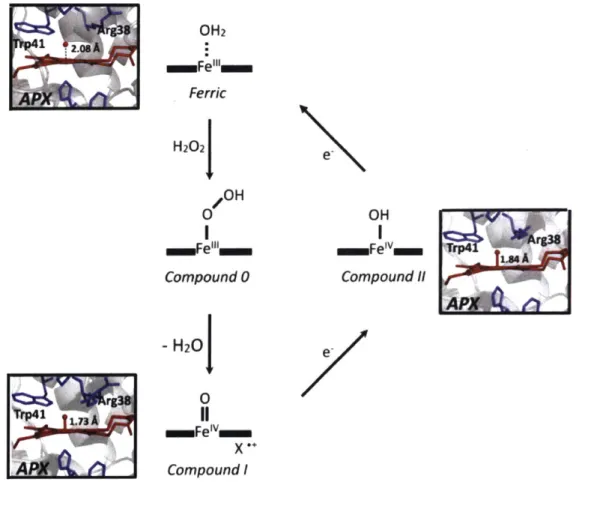
The proposed mechanism4 lead to future studies on in which x-ray crystal structures provided insight towards the mechanism for this class of peroxidases as well as possible intermediate structures. Figure 1-1 depicts the how peroxidases utilizes the heme cofactor to oxidize substrates.
OH2 aaFe"'ll m Ferric H202J OH ammFei" Compound 0 - H20 0 m=FeIv Compound I OH Compound 11
Figure 1-1: The mechanism of Class I peroxidases, model structures obtained from the Gumiero, et al. paper3. APX originates in a resting ferric state (top), but upon replacement of the presence of H202, the occupancy changes the ferric to ferric hydroperoxyl, also known as
Compound 0 (center). The activated compound I causes a loss of 1 water, and 1 electron (bottom). Once reducing substrate binds to the active site again, it donates one proton and one electron to Compound 1. Compound I transforms into Compound II (right), which lacks the radical cation in the porphyrin ring. After a second equivalence of a reducing agent binds to the active site, the cycle repeats and the donation of a proton and electron yield another oxidized substrate product
and finally returns the enzyme back into the ferric resting state. rg38 S .os A APX] 1.73 A *1 e. e.
The scavenging of APX binds hydrogen peroxide and replacing the water, which otherwise occupies the binding site. The starting compound 0 structure still contains Fe in the +3 oxidation state. However once reaching the activated compound I form, in which the Fe is in a +4 oxidation state, the sequential binding and donation of proton and electron by a reducing agent, such as biotin-phenol for instance, can return the enzyme back to its original ferric group state (Figure 1-I).
Phe68 ;8 His42 2 Phe41he41
Arg38
Phe142 Phe179
Hisl70
Figure 1-2: The binding site of horseradish peroxidase (HRP) with ferulic acid (FA) in the active site. The figure is adapted from Veitch'O and shows the carton representation taken from the x-ray crystal structure of the ternary complex: the substrate (ferulic acid), the cyanided-ligated
HRP (variant C), and the heme co-factor necessary for catalysis (this is the molecule in red). The catalytic resides, Arg38, His42, and His170 are also shown, but most importantly the four aromatic phenylalanines are shown from the binding site. This hydrophobic patch of aromatic rings could contribute the high peroxidase activity of HRP towards a large range of aromatic substrates.
Using structure-guided mutagenesis, members of the Ting lab engineered APEX from APX through 3 mutations - Kl4D, El 12K, W41F. The mutations K14D and El 12K are charge reversal mutations that was designed to monomerize APEX (27 kDA), rather than its wild-type form of constitutive dimer. The W41F mutation was modeled after the active site of horseradish peroxidase (HRP) to improve the activity towards aromatic substrates. HRP is a prototypical class III plant peroxidase with a wide substrate tolerance, oxidizing almost any phenol or analine to the corresponding radicals and releasing them from the active site. The substrate binding pocket of HRP contains a hydrophobic patch with 4 phenylalanines (Figure 1-2), thus leading to the W41F
mutation in APX. By replacing the indole with an aromatic phenyl-group increases the pi-pi stacking within the substrate pocket',. Through the utilization of yeast display directed evolution on first generation APEX, the Ting lab engineered second generation APEX2, which introduces the fourth mutation A134P, which further improves enzymatic activity for aromatic substrates
(Figure 1-3 for summary of engineering).
K14D, E112K, W, Rational desigr
Soybean Ascorbate Peroxidase Pdb: 1VOH
A134P Engineered Ascorbate
Directed evolution' Peroxidase (APEX2)
Figure 1-3: APEX2 is an engineered product of structure-guided mutagenesis and yeast display-directed evolution. The starting template of soybean APX underwent one round of rational design to monomerize and enhance peroxidase activity towards aromatic substrates. Generation 1 APEX underwent a second round of engineering where the single mutation Al 34P provided enhanced peroxidase activity for aromatic substrates as well. This finalized engineering product is APEX2
Proximity labeling methodology requires the use of a genetically targeted enzyme that can promiscuously covalently tag proximal endogenous proteins with a chemical handle, such as biotin, for purification and further analysis without performing any organelle purification. When catalyzed by the addition of H202, APEX2 uses its enhanced peroxidase activity to perform one
electron oxidation on the aromatic reducing substrate, biotin-phenol, and generates a short-lived biotin-phenoxyl radical that can diffuse out of the active site to reacts with proximal surface
exposed electron side chains such as tyrosine residues to covalently attached a biotin. The generated biotin-phenoxyl radical product is very reactive has a half-life < 1 ins, resulting in a small labeling radius of APEX2 (< 20 nm) and only proteins proximal to APEX2 will be sufficiently tagged". Furthermore, proximity labeling experiments by APEX2 are performed with high temporal resolution. APEX2, with a kcat = 299 s1, can generate sufficient quantities of biotin-phenoxyl radicals to tag proximal proteins within 1 minute. This feature of APEX2 enables dynamic analysis of protein interaction networks, such as the mapping of GPCR signaling pathways in real time'213 . Furthermore, the biotin-phenoxyl radical not only reacts with proteins,
but also nucleic acids, enabling mapping of the spatial distribution of endogenous RNAs throughout the cell" .This feature of APEX2, in addition to its versatile ability to catalyze the H202-dependent one-electron oxidation of a wide variety of small molecule substrates, has led to its widespread use for a variety of applications, including proteomic mapping of organelles' 1,15-18,
proximity labeling of protein interactomes12,13
,19, spatial mapping of cellular RNA", electron microscopy2,2025, H
202 sensing26, and protein topology determination2" ,25*
Two other types of enzymes have been commonly utilized for proximity labeling of proteins, promiscuous biotin ligase and HRP. Roux and coworkers used a promiscuous mutant of Escherichia coli biotin ligase - pBirA. (BirA + RI 18G = pBirA) in a method called BiolD to biotinylate proteins proximal to the pBirA fusion to obtain proteomes of the mammalian nuclear lamina, trypanosome biolobe, nuclear pore, and centriole27-31. The proposed mechanism is that
pBirA accepts biotin and ATP to generate a biotin adenylate ester, which diffuses from the active site to biotinylate lysine residues of proximal proteins. However, this enzyme has slow kinetics: the rate-limiting step, the dissociation of the biotin adenylate ester from the active site, has a kof= 0.119 s-. pBirA requires 18-24 hours of labeling to generate product levels comparable to one minute of APEX2 labeling25,2 7. This extended biotin incubation time could lead to toxicity.
Furthermore, the biotin-adenylate ester has a half-life of minutes, yielding a larger labeling radius and further loss of spatiotemporal resolution when compared to phenoxyl radicals generated by APEX2". Additionally, due to the need for such length labeling times, the BioLD methodology is
limited when investigating dynamic biological processes.
Unlike pBirA, HRP has much faster kinetics for aromatic substrates. The substrate binding pocket of HRP contains a hydrophobic patch with 4 phenylalanines, thus inspiring the W41F mutation in APX. This hydrophobic region in the substrate binding pocket, combined with its
highly solvent exposure nature may explain HRPs robust activity in oxidizing a broad array of aromatic substrates32,. However, HRP has four disulfide bonds and requires Ca2
+ 11,34,35. As a
result, HRP is inactive in the reducing and Ca2+ scarce environment of the cytosol. Contrastingly,
APEX2 is active within the cytosol since it does not require disulfide bonds or Ca2+ 2,1
Applications
In general, the use of APEX2 begins with fusing it to a protein or peptide in order to target it to a subcellular region or macromolecular complex of interest. For instance, we have targeted APEX2 to the outer mitochondrial membrane (OMM) and the endoplasmic reticulum membrane (ER membrane, or ERM) of mammalian cells by fusing the APEX2 gene to the C-terminal 31 amino acids of the native OMM mitochondrial antiviral signaling protein (MAVS), or to the N-terminal 27 amino acids of the native ERM protein P450 oxidase 2C1, respectively6,25. These
constructs were used for both EM2 5 and proteomic analysis' 6 of the OMM and ERM environment.
In the presence of hydrogen peroxide and DAB, APEX2 catalyzes the polymerization of DAB in
Generation of EM Contrast Biotinylation of Proximal Proteins
[ H]
O, Labeling radius
Osmiophillic polymer H 3 Biotin-phenol
OsO4j H201H2O
Endogenous proteins
-EM Contrast H2N DABNH
Engineered Ascorbate biotin
Peroxidase (APEX2) =
Figure 1-4: APEX2 can be utilized as both a reporter for EM and proteomics. In the presence of diaminobenzidene (DAB) and H202, APEX2 can oxidize DAB, initiating oxidative polymerization to form a locally deposited precipitate that provides EM contrast with subsequent treatment of osmium. For proteomic applications, in the presence of biotin-phenol and H202,
APEX2 generates a biotin phenoxyl radical that proximally labels proteins. The covalently attached biotin allows for streptavidin-enrichment of these proximal proteins for further analysis.
fixed cells2. The localized DAB polymer gives EM contrast upon further treatment with electron-rich osmium4. Alternatively, in the presence of hydrogen peroxide and biotin-phenol, APEX2 in APEX2 in live cells generates a short-lived, membrane-impermeable phenoxyl radical that biotinylates surface-exposed tyrosine residues in a proximity dependent manner' 25 (Figure 1-4). Biotinylated proteins can then be enriched and identified utilizing tandem MS'5
.
While this strategy has provided access to many cellular regions and organelles, there are numerous compartments and structures that cannot be cleanly accessed by such an approach. This technique is limited by the specificity of genetic targeting; there are cellular regions that cannot be exclusively targeted by a single genetic tag. For example, there is great interest in the biology of organelle-organelle contact sites, such as the junctions between mitochondria and ER, which participate in calcium signaling36,3 7, lipid synthesis
38-41, and mitochondrial fission4 243. These
junctions have been visualized by microscopy9, and the complex that tethers the two organelles
has been identified in yeast as the ER-mitochondria encounter structure (ERMES)44.
Yet all APEX2 fusion constructs we have evaluated or considered, such as to the proteins dynamin-l-like protein, Drp 142, mitofusin2, Mfn245
-4 7, synaptojanin 2 binding protein, SYNJ2BPI 16, and PDZ domain containing protein 8, PDZD848 would also target the peroxidase
to protein pools outside of mito-ER contacts, such as into the cytosol49, along the cytoskeleton50,
and over the entire OMM6. As a result, one cannot use conventional APEX2 fusion approaches to specifically target the mito-ER junctions.
Another application for which the APEX2 genetic fusion strategy may be unsuitable is for profiling the interactome of specific cellular RNAs. While there are several robust methods to identify RNA interaction partners for specific proteins of interest5 3, the converse
problem-identifying proteins that interact with a particular RNA species-is much more challenging. One could envision fusing the APEX2 protein to a high-affinity RNA-binding protein (RBP; for example, the bacteriophage MS2 coat protein5 4) allowing the peroxidase to be ectopically targeted to transcripts that are tagged with that RBP's cognate RNA motif. However, a major technical challenge would be the large pool of excess, catalytically active APEX2-RBP fusion protein that is not docked to the tagged RNA, resulting in background labeling that masks the specific signal.
A general solution to these problems and related ones could be a split form of APEX2, in which two inactive fragments of APEX2 reconstitute to give an active peroxidase. One could use an intersectional approach to restrict APEX2 activity specifically to sites of interest, such as mito-ER contacts only, or specific RNA binding sites only - thus eliminating the background labeling from protein overexpression and off-targeting.
Splitting APEX to make a protein complementation assay (PCA)
Split protein sensors, also referred to as protein fragmentation complementation assays (PCA), are employed to improve specificity for a variety of applications from protein-protein interaction (PPI) detection to genome editing55 56. This strategy relies on the conditional reconstitution of enzymatic activity using a protein that has been strategically split into two fragments55. This principle was first observed in 1958 when Richards noted that the two fragments of Ribonuclease, generated by the first subtilisin proteolysis step, results in almost native level enzymatic activity57. In a PCA, a reporter protein is split into two inactive fragments, and each
fragment is genetically fused to a separate protein of interest (Figure 1-5). Interaction of the two proteins of interest drives the reconstitution of the reporter fragments, restoring protein activity.
Reassembled Active Protein
Active Protein Inactive Split Protein Fragments
interacting Domains
Figure 1-5. Overview of protein complementation assays (PCAs) A reporter protein is divided into two inactive fragments, and these fragments are each fused to one protein, A or B, in a pair of proteins whose interaction one wishes to detect. In the absence of a PPI, the reporter fragments remain unfolded, and no reporter protein signal is observed. When a PPI occurs between A and B,
complex with the same activity as the parent reporter protein. Figure is adapted from Ghosh, et al. in Split-Protein systems: Beyond Binary Protein-Protein Interactions 55
Split ubiquitin was the first intentionally designed split protein system; fragments only reconstituted when fused to interacting protein pairs due to the increase in local concentration 8.
The reassembled ubiquitin is then cleaved after the C-terminus, which provides a read out for the original non-covalent protein-protein interaction26. Since then, split proteins and split enzymes
have been developed for many workhorses of cell biology, including green fluorescent protein5 960,
HRP6 1, dihydrofolate reductase62, ubiquitin5 8, luciferase63
-65, beta-galactosidase66-69, TEV protease70, and Cas95671-73. These diverse split proteins have enzymatic activities that include
site-specific proteolysis, cleavage of reconstituted ubiquitin, genome editing, and provide fluorescence, positron emission tomography, and host survival readouts2 3. Many applications of these split protein systems are driven together by in vivo macromolecular interactions to screen for interactions2 3. Other applications extend the utility of split proteins via temporally-controlled
reconstitution through fusion with FK506 binding protein (FKBP) and FKBP-12 rapamycin binding domain (FRB), which undergoes chemically inducible dimerization upon addition of rapamycin2 3. Our lab has initiated efforts to split both APEX2 and HRP. A former graduate student,
Jeff Martell, has successfully published on his engineered split HRP. Jeff also started to split first generation APEX by screening and assaying several rationally selected cut sites. Jeff and I have collaborated to rationally select and test APEX2 cut sites (Chapter 2).
Discussion
Splitting APEX2 presents new challenges, however. First, APEX2 requires a heme cofactor for its activity, and most cut sites would split apart the heme-binding pocket. Second, in order for split APEX2 to be useful for a broad range of applications, the inactive fragments should have relatively low affinity for one another, such that reconstitution only occurs when the fragments are driven together by a molecular interaction. Not many known split proteins have low-affinity fragments, and it is challenging to engineer such a property in conjunction with high activity upon reconstitution.
The ideal successful split APEX2 system requires two low affinity fragments that reconstitute enzymatic activity rapidly only in a proximity dependent manner. For instance, for an
application at the organelle-organelle contact site, mito-ER, each fragment alone must not only remain monomeric and inactive, but also avoid perturbing the localization and expression of native mitochondrial and ER proteins. They also must be well-folded and non-aggregating for proper trafficking and localization. Their mutual low affinity is necessary to prevent artificially induced mito-ER junctions.
Short enzymatic reconstitution times are also desired, to further ensures both organelles' morphological integrity. Thus, it is critical that split APEX2 reconstitute peroxidase activity both rapidly and robustly. The development of split APEX2 requires systematic screening to determine an appropriate fragmentation site, as well as yeast-display directed evolution to improve expression, enzymatic activity, and dynamic range.
To address the need for an interaction-dependent proximity labeling tool, this thesis describes the development of a split APEX2 system. We used a combination of rational design (Chapter 2) and a novel yeast display-based directed evolution approach that incorporates positive and negative selection steps (Chapter 3) to evolve sAPEX in order to obtain targeting of regions that are not membrane bound and/or cannot be targeted by a single gene fusion. Our engineering efforts focused on attempting to minimize background association of the peptide fragments while maintaining high peroxidase activity upon reconstitution. Our resulting split APEX2 fragment pair has significantly diverged from its parental sequence and shows interaction-dependent reconstitution in multiple contexts in living mammalian cells (Chapter 4). Comparison against competitor split APEX73 demonstrated the superiority of the reconstitution of out split APEX tool after directed evolution (Chapter 4). Our split APEX tool adds to the proximity labeling toolkit (Chapter 5 and 6), and in the future, should extend the utility of APEX-based approaches to new areas of biology at higher spatiotemporal resolution.
References
1. Martell, J. D. et al. Engineered ascorbate peroxidase as a genetically encoded reporter for electron microscopy. Nat. Biotechnol. 30, 1143-1148 (2012).
2. Gumiero, A., Metcalfe, C. L., Pearson, A. R., Raven, E. L. & Moody, P. C. E. Nature of the ferryl heme in compounds I and 1I. J. Biol. Chem. (2011). doi:10.1074/jbc.M 110.183483
3. Jones, D. K., Dalton, D. A., Rosell, F. I. & Raven, E. L. Class I heme peroxidases: Characterization of soybean ascorbate peroxidase. Arch. Biochem. Biophys. (1998). doi: 10. 1006/abbi. 1998.0941
4. Sharp, K. H., Mewies, M., Moody, P. C. E. & Raven, E. L. Crystal structure of the ascorbate peroxidase-ascorbate complex. Nat. Struct. Biol. 10, 303-307 (2003).
5. Lad, L., Mewies, M. & Raven, E. L. Substrate binding and catalytic mechanism in ascorbate peroxidase: Evidence for two ascorbate binding sites. Biochemistry (2002). doi:10. 1021/bi0261591
6. Gumiero, A., Murphy, E. J., Metcalfe, C. L., Moody, P. C. E. & Raven, E. L. An analysis of substrate binding interactions in the heme peroxidase enzymes: A structural perspective. Archives of Biochemistry and Biophysics (2010). doi: 10.1016/j .abb.2010.02.015
7. Efimov, I. et al. The redox properties of ascorbate peroxidase. Biochemistry (2007). doi: 10.1021 /bi7006492
8. Macdonald, I. K., Badyal, S. K., Ghamsari, L., Moody, P. C. E. & Raven, E. L. Interaction of ascorbate peroxidase with substrates: A mechanistic and structural analysis. Biochemistry (2006). doi:10.1021/bi0606849
9. Veitch, N. C. Horseradish peroxidase: A modern view of a classic enzyme. Phytochemistry (2004). doi: 10.101 6/j.phytochem.2003.10.022
10. Rhee, H.-W. et al. Proteomic Mapping of Mitochondria in Living Cells via Spatially Restricted Enzymatic Tagging. Science (80-.). 339, 1328-1331 (2013).
11. Paek, J. et al. Multidimensional Tracking of GPCR Signaling via Peroxidase-Catalyzed Proximity Labeling. Cell 169, 338-349.el 1 (2017).
12. Lobingier, B. T. et al. An Approach to Spatiotemporally Resolve Protein Interaction Networks in Living Cells. Cell 169, 350-360.el2 (2017).
13. Kaewsapsak, P., Shechner, D. M., Mallard, W., Rinn, J. L. & Ting, A. Y. Live-cell mapping of organelle-associated RNAs via proximity biotinylation combined with protein-RNA crosslinking. doi.org 153098 (2017). doi:l0..1101/153098
14. Hung, V. et al. Proteomic Mapping of the Human Mitochondrial Intermembrane Space in Live Cells via Ratiometric APEX Tagging. Mol. Cell 55, 332-341 (2014).
15. Hung, V. et al. Proteomic mapping of cytosol-facing outer mitochondrial and ER membranes in living human cells by proximity biotinylation. Elife 6, (2017).
16. Loh, K. H. et al. Proteomic Analysis of Unbounded Cellular Compartments: Synaptic Clefts. Cell 166, 1295-1307.e21 (2016).
17. Chen, C.-L. et al. Proteomic mapping in live Drosophila tissues using an engineered ascorbate peroxidase. Proc. Natl. Acad Sci. U S. A. 112, 1-6 (2015).
18. Han, S. et al. Proximity Biotinylation as a Method for Mapping Proteins Associated with mtDNA in Living Cells. Cell Chem. Biol. 24, 404-414 (2017).
19. Shvets, E., Bitsikas, V., Howard, G., Hansen, C. G. & Nichols, B. J. Dynamic caveolae exclude bulk membrane proteins and are required for sorting of excess glycosphingolipids. Nat. Commun. 6, (2015).
20. Ludwig, A., Nichols, B. J. & Sandin, S. Architecture of the caveolar coat complex. J. Cell Sci. jcs.191262 (2016). doi:10.1242/jcs.191262
21. Liu, L. K., Choudhary, V., Toulmay, A. & Prinz, W. A. An inducible ER-Golgi tether facilitates ceramide transport to alleviate lipotoxicity. J. Cell Biol. 216, 131-147 (2017). 22. Hyenne, V. et al. RAL-1 controls multivesicular body biogenesis and exosome secretion. J
23. Joesch, M. et al. Reconstruction of genetically identified neurons imaged by serial-section electron microscopy. Elife 5, (2016).
24. Lam, S. S. et al. Directed evolution of APEX2 for electron microscopy and proximity labeling. Nat. Methods 12, 51-54 (2014).
25. Dwyer, D. J. et al. Antibiotics induce redox-related physiological alterations as part of their lethality. Proc. Natl. Acad. Sci. 111, E2100-E2109 (2014).
26. Roux, K. J., Kim, D. I., Raida, M. & Burke, B. A promiscuous biotin ligase fusion protein identifies proximal and interacting proteins in mammalian cells. J. Cell Biol. (2012). doi:10.1083/jcb.201112098
27. Belousov, V. V. et al. Genetically encoded fluorescent indicator for intracellular hydrogen peroxide. Nat. Methods (2006). doi: 10.103 8/nmeth866
28. Li, X. W. et al. New insights into the DT40 B cell receptor cluster using a proteomic proximity labeling assay. J. Biol. Chem. (2014). doi:10.1074/jbc.M1 13.529578
29. Lee, N., Moss, W. N., Yario, T. A. & Steitz, J. A. EBV noncoding RNA binds nascent RNA to drive host PAX5 to viral DNA. Cell 160, 607-618 (2015).
30. Adams, J. C. Biotin amplification of biotin and horseradish peroxidase signals in histochemical stains. J. Histochem. Cytochem. (1992). doi:10. 1177/40.10.1527370
31. Gajhede, M., Schuller, D. J., Henriksen, A., Smith, A. T. & Poulos, T. L. Crystal structure of horseradish peroxidase C at 2.15
A
resolution. Nat. Struct. Biol. (1997). doi: 10. 1038/nsb1297-103232. Veitch, N. C., Gao, Y., Smith, A. T. & White, C. G. Identification of a critical phenylalanine residue in horseradish peroxidase, Phe179, by site-directed mutagenesis andlH-NMR: Implications for complex formation with aromatic donor molecules. Biochemistry (1997). doi: 10.102 1/bi971 8402
33. Jiang, S. et al. A proteomics approach to the cell-surface interactome using the enzyme-mediated activation of radical sources reaction. Proteomics (2012). doi:10.1002/pmic.201100551
34. Hopkins, C., Gibson, A., Stinchcombe, J. & Futter, C. Chimeric molecules employing horseradish peroxidase as reporter enzyme for protein localization in the electron microscope. Methods Enzymol. (2000). doi:10.1016/S0076-6879(00)27265-0
35. Szabadkai, G. et al. Chaperone-mediated coupling of endoplasmic reticulum and mitochondrial Ca2+ channels. J. Cell Biol. 175, 901-911 (2006).
36. Rizzuto, R. et al. Close contacts with the endoplasmic reticulum as determinants of mitochondrial Ca2+ responses. Science (80-.). 280, 1763-1766 (1998).
37. Kornmann, B., Osman, C. & Walter, P. The conserved GTPase Gemi regulates endoplasmic reticulum-mitochondria connections. Proc. Natl. Acad. Sci. 108, 14151-14156
(2011).
38. Kornmann, B. et al. An ER-mitochondria tethering complex revealed by a synthetic biology screen. Science (80-.). 325, 477-481 (2009).
39. Lewin, T. M., Van Horn, C. G., Krisans, S. K. & Coleman, R. A. Rat liver acyl-CoA synthetase 4 is a peripheral-membrane protein located in two distinct subcellular organelles, peroxisomes, and mitochondrial-associated membrane. Arch. Biochem. Biophys. 404,
263-270 (2002).
40. Rusifiol, A. E., Cui, Z., Chen, M. H. & Vance, J. E. A unique mitochondria-associated membrane fraction from rat liver has a high capacity for lipid synthesis and contains
pre-Golgi secretory proteins including nascent lipoproteins. J. Biol. Chem. 269, 27494-27502 (1994).
41. Friedman, J. R. et al. ER Tubules Mark Sites of Mitochondrial Division. Science (80-.). 334, 358-362 (2011).
42. Murley, A. et al. ER-associated mitochondrial division links the distribution of mitochondria and mitochondrial DNA in yeast. Elife 2013, (2013).
43. Murley, A. et al. ER-associated mitochondrial division links the distribution of mitochondria and mitochondrial DNA in yeast. Elife 2013, (2013).
44. De Brito, 0. M. & Scorrano, L. Mitofusin 2 tethers endoplasmic reticulum to mitochondria. Nature 456, 605-610 (2008).
45. Filadi, R. et al. Mitofusin 2 ablation increases endoplasmic reticulum-mitochondria coupling. Proc. Natl. Acad Sci. 112, E2174-E2181 (2015).
46. Cosson, P., Marchetti, A., Ravazzola, M. & Orci, L. Mitofusin-2 Independent Juxtaposition of Endoplasmic Reticulum and Mitochondria: An Ultrastructural Study. PLoS One 7, (2012).
47. Hirabayashi, Y. et al. ER-mitochondria tethering by PDZD8 regulates Ca 2 dynamics in mammalian neurons. Science (80-.). 358, 623-630 (2017).
48. Smirnova, E., Griparic, L., Shurland, D.-L. & Bliek, A. M. van der. Dynamin-related Protein Drpl Is Required for Mitochondrial Division in Mammalian Cells. Mol. Biol. Cell 12, 2245-2256 (2001).
49. Henning, M. S. et al. PDZD8 is a novel moesin-interacting cytoskeletal regulatory protein that suppresses infection by herpes simplex virus type 1. Virology 415, 114-121 (2011).
50. Hendrickson, D., Kelley, D. R., Tenen, D., Bernstein, B. & Rinn, J. L. Widespread RNA binding by chromatin-associated proteins. Genome Biol. 17, (2016).
51. Sibley, C. R. Individual nucleotide resolution UV cross-linking and immunoprecipitation (iCLIP) to determine protein-RNA interactions. in Methods in Molecular Biology 1649, 427-454 (2018).
52. Garzia, A., Morozov, P., Sajek, M., Meyer, C. & Tuschl, T. PAR-CLIP for discovering target sites of RNA-binding proteins. in Methods in Molecular Biology 1720, 55-75 (2018). 53. Peabody, D. S. The RNA binding site of bacteriophage MS2 coat protein. EMBO J. 12,
595-600 (1993).
54. Shekhawat, S. S. & Ghosh, I. Split-protein systems: Beyond binary protein-protein interactions. Current Opinion in Chemical Biology (2011). doi:10.1016/j.cbpa.2011.10.014
55. Zetsche, B., Volz, S. E. & Zhang, F. A split-Cas9 architecture for inducible genome editing and transcription modulation. Nature Biotechnology 33, 139-142 (2015).
56. Richards, F. M. ON THE ENZYMIC ACTIVITY OF SUBTILISIN-MODIFIED RIBONUCLEASE. Proc. Natl. Acad Sci. (1958). doi:10.1073/pnas.44.2.162
57. Johnsson, N. & Varshavsky, A. Split ubiquitin as a sensor of protein interactions in vivo. Proc. Natl. Acad Sci. U S. A. 91, 10340-4 (1994).
58. Cabantous, S., Terwilliger, T. C. & Waldo, G. S. Protein tagging and detection with engineered self-assembling fragments of green fluorescent protein. Nat. Biotechnol. 23, 102-107 (2005).
59. Ghosh, I., Hamilton, A. D. & Regan, L. Antiparallel leucine zipper-directed protein reassembly: Application to the green fluorescent protein [12]. Journal of the American
60. Martell, J. D. et al. A split horseradish peroxidase for the detection of intercellular protein-protein interactions and sensitive visualization of synapses. Nat. Biotechnol. 34, 774-780 (2016).
61. Pelletier, J. N., Campbell-Valois, F.-X. & Michnick, S. W. Oligomerization domain-directed reassembly of active dihydrofolate reductase from rationally designed fragments. Proc. Natl. Acad Sci. 95, 12141-12146 (1998).
62. Paulmurugant, R. & Gambhir, S. S. Monitoring protein-protein interactions using split synthetic renilla luciferase protein-fragment-assisted complementation. Anal. Chem. 75,
1584-1589 (2003).
63. Luker, K. E. et al. Kinetics of regulated protein-protein interactions revealed with firefly luciferase complementation imaging in cells and living animals. Proc. Natl. Acad Sci. 101,
12288-12293 (2004).
64. Ozawa, T., Kaihara, a, Sato, M., Tachihara, K. & Umezawa, Y. Split luciferase as an optical probe for detecting protein-protein interactions in mammalian cells based on protein splicing. Anal. Chem. 73, 2516-2521 (2001).
65. Olson, K. R. & Eglen, R. M.
#
Galactosidase Complementation: A Cell-Based Luminescent Assay Platform for Drug Discovery. Assay Drug Dev. Technol. 5, 137-144 (2007).66. Broome, A. M., Bhavsar, N., Ramamurthy, G., Newton, G. & Basilion, J. P. Expanding the utility of -galactosidase complementation: Piece by piece. Mol. Pharm. 7, 60-74 (2010). 67. Ullmann, A., Jacob, F. & Monod, J. Characterization by in vitro complementation of a
peptide corresponding to an operator-proximal segment of the P-galactosidase structural gene of Escherichia coli. J Mol. Biol. 24, 339-343 (1967).
68. Rossi, F., Charlton, C. a & Blau, H. M. Monitoring protein-protein interactions in intact eukaryotic cells by beta-galactosidase complementation. Proc. Natl. Acad Sci. U S. A. 94,
8405-10 (1997).
69. Wehr, M. C. et al. Monitoring regulated protein-protein interactions using split TEV. Nat. Methods 3, 985-993 (2006).
70. Wright, A. V. et al. Rational design of a split-Cas9 enzyme complex. Proc. Natl. Acad Sci. 112, 2984-2989 (2015).
71. Truong, D. J. J. et al. Development of an intein-mediated split-Cas9 system for gene therapy. Nucleic Acids Res. 43, 6450-6458 (2015).
72. Nihongaki, Y., Kawano, F., Nakajima, T. & Sato, M. Photoactivatable CRISPR-Cas9 for optogenetic genome editing. Nat. Biotechnol. 33, 755-760 (2015).
73. Xue, M. et al. Optimizing the fragment complementation of APEX2 for detection of specific protein-protein interactions in live cells. Sci. Rep. 7, (2017).
Introduction
We first sought to identify potentially fruitful cut sites in the APEX2 enzyme; previously, Jeff Martell had performed a cut site screen with first-generation APEX. Together with his guidance, we rationally selected possible fragmentations of APEX2 for screening. There were several considerations in picking which cut sites to screen. First, cut sites were not made within regions of known secondary structure as the disruption could lead to aggregation and/or degradation. Second, with the exception of cut sites on the first N-terminal loop, all fragments lack a fully formed heme-binding pocket. Third, the new N and C-termini generated from cut sites have side chains that do not contain hydrophobic residues to avoid collapse and burial of the termini and significant alteration of secondary structure. In the first round, we scanned all potential loop regions; the second round focused on the successful loops and more exhaustively screening cut sites along those loops.
A APEX2 sequence (red lines show cut sites tested)
aal aa125 aa126 aa250 B 7 (T) / 8 (V) 126 (P) /127 (P) 29 (E) / 30 (K) 128 (E) / 129 (G) 50 (G) / 51 (T) 130 (R) /131 (L) 52 (K) / 53 (T) 135 (T) /136 (K) 62 (H) / 63 (P) 146 (G) / 147 (K) 68 (H) / 69 (S) 170 (K) /171 (E) 89 (P) / 90 (1) 173 (S) /174 (G) 108 (T) / 109 (G) 181 (S) /182 (N) 111 (P)/112 (K) 197 (G) /198 (E) 120 (E) /121 (D) 200 (E) / 201 (G) 121 (D) /122 (K) 214 (D) / 215 (P) 123 (P) / 124 (E) 226 (A) /227 (D
Figure 2-1. Selection of potential split. APEX cut sites. (A) The first screen tested 24 different cut sites. Their locations in the APEX2 protein sequence are indicated by the red vertical lines. Squiggles denote alpha helices. Grey arrows denote beta sheets. Areas shaded green are part of the heme-binding pocket. (B) The 24 different cut sites; newly generated N- and C- terminal amino acid identity abbreviated in parenthesis.
Amplex UltraRed Screen of rationally-selected APEX2 cut sites for enzymatic reconstitution in HEK 293T cells
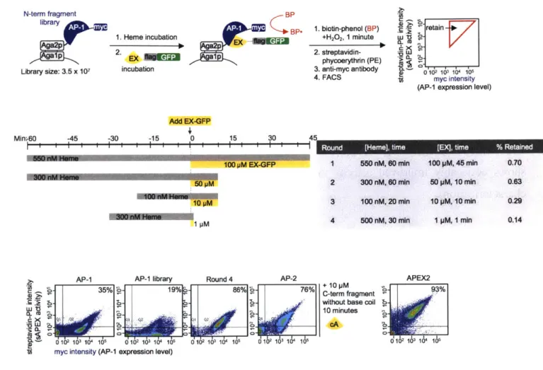
Using these guidelines, we selected 24 preliminary cut sites within solvent-exposed loops and turns between secondary structural elements (alpha helices and beta sheets) based on the crystal structure of wild-type ascorbate peroxidase5 (Figure 2-1). We sought to find the best cut sites in the APEX2 enzyme using a chemically-inducible protein association system as a test platform. We cloned each of the 24 fragment pairs as fusions to FKBP and FRB, whose interaction can be induced with the small molecule rapamycin (Figure 2-2a, 2-2b). We introduced the fragment pairs into HEK 293T cells and evaluated peroxidase activity in the presence or absence of rapamycin. Catalytic activity was measured using an established assay based on the membrane-permeable fluorogenic probe Amplex UltraRed which is colorless, but upon peroxidase-catalyzed oxidization, generates the red-fluorophore Resorufin74' 75 (Figure 2-2c). Ideal split APEX2 candidates are those that display strong Resorufin signal only with the addition of rapamycin, indicating low affinity fragments and robust reconstitution.
A
Full-length APEX2 flag FRB APE
N-term APEX2 fragment flag-FRB-AP fg FR A C-term APEX2 fragment V5-FKBP-EX Rapamycin protein-protein interaction B I+ Inactive split APEX fragments active reconstituted sAPEX HEK 293T cells
1. Transfect with DNA
2. +/- 400 nM Rapamycin (24hrs)
HO O OH
) F
AO0
H2O2
Amplex UltraRed labeling Image cells by
confocal
HO N 0microscopy
R nF Resorufin= "
-Figure 2-2. split APEX cut site screening platform. (A) N- and C-terminal sAPEX fragments selected for testing were fused to FRB and FKBP, respectively. Full length APEX2 was used cloned and used as both a benchmark and a positive control. (B) Schematic overview of split
MIE
C
APEX (sAPEX). Two inactive fragments (grey) can reconstitute to give active peroxidase (green) when driven together by a protein-protein interaction (PPI) such as that of FKBP and FRB under the presence of rapamycin, yellow diamond. (C) Cartoon of the Amplex UltraRed assay -Pairs of constructs were introduced into HEK 293T cells by transient transfection, along with a CFP-NLS (nuclear localization signal) co-transfection marker. Cells were either treated with rapamycin for 20-24 h (left) or left untreated (right) in (Figure 2-3). Subsequently Amplex UltraRed, a fluorogenic small-molecule peroxidase substrate, and H202 were added for 25 minutes, after which
cells were fixed and imaged. Resorufin is the fluorescent product of Amplex UltraRed oxidation and indicates peroxidase activity.
Of the 24 fragment pairs tested, seven produced significant Resorufin product, indicative of reconstituted activity (Figure 2-3a). With the exception cut sites 7/8 and 29/30, all of the fragments we screened are expected to lack a completely formed heme-binding pocket. Cut sites within the heme binding cavity (130/131, 134/135, 146/147, 170/171, 173/174, and 181/182) exhibited the most dramatic reduction in catalytic function; these fragment pairs completely failed to reconstitute activity (Figure 2-la; 2-3a).
+ Rapamycin omit Rapamycin + Rapamycin omit Rapamycln B + Rapamycin omit Rapamycin Resorufin CFP Resorufin CFP Resorufin CFP Resorufin CFP Resorufin CFP Resorufin CFP
Full-length -3Full-length APEX2 123 APEX2U
...
CFP-NLSO EM 12-0N only 1267 7l 7 f O' N 128 fllNo 8f lll 29 ff ME 130OffME 900 0f 50 Of fO N 134 f l 1E 'OM E Nf
a 52 f l O . 146 flflfa) 480 0 fO N 62EE 170 U*UU 49 U U 68 f f f f 173 l M E r)51 M EfE f 89 f f f f 181 f -Efl 52 f l l l 108 fllME 197No 53 No 111 200 fl No 198fllff
120 O ll f 214 f l l l 199ffl
lf
121 226E . - 200 oM AFigure 2-3. Screening of potential split APEX cut sites. (A) Initial Amplex UltraRed screen of cut sites, fragmentation occurs after the listed amino acid. For instance, cut site 7 APEX2 between aa 7 and 8. Scale bars, 20 tm. performed. (B) Second cut site screen, focused on residues
surrounding T7, G50, and E200. Same assay as in (A) with a total of two replicates.
To more finely map the optimal cut sites, we performed a second round of screens, evaluating cut sites 1-3 residues away from the most promising sites identified in our initial screen (Figures 2-3B). This yielded three optimal starting positions-51/52, 89/90, and 200/201- all of which fell in solvent-exposed loops distal (>15 angstroms away) from the APEX2 active site and heme-binding pocket. To test if any of the fragments contained peroxidase activity on its own, fragment pairs that generated the most robust resorufin signals were tested with single fragment controls. Omission of either fragment showed that the fragments alone lack any detectable peroxidase activity at all cut sites except for the cut site 7 (Figure 2-4).
only only
flag-FRB-AP V5-FKBP-EX
A Cut site + Rapamycin omit Rapamycin + Rapamycin + Rapamycin after Re-nr CFP Resorufin CFP Resorufin Resorufin amino acid: 29M EU 502ME 520 E H 89 -flflflflf 123 EU..