Rechtsmedizin 2007 · 17:40–43 DOI 10.1007/s00194-006-0425-8 Online publiziert: 12. Januar 2007 © Springer Medizin Verlag 2007
Virtopsy Team
1, 2· L. Oesterhelweg
11
Zentrum für Forensische Bildgebung/Virtopsy, Institut
für Rechtsmedizin, Universität Bern, Bern
2
Institut für Diagnostische, Interventionelle und
Pädiatrische Radiologie, Inselspital, Bern
Atmosphere of departure
in forensic medicine?
Virtopsy Basic Course
Leitthema
Regarding autopsies, imaging procedures
like x-ray and surface scanning are
meth-ods in their childhood in forensic
sci-ence but are becoming more and more
a valuable tool in institutes of legal
med-icine in Europe, America and Asia. Byron
G. Brogdon gave a vivid statement in his
book Forensic Radiology: „The sad truth
is that a century af ter the first x-ray was
introduced as evidence in a law court,
there is no general appreciation of the
ex-tent of the radiology poex-tential in the
fo-rensic sciences“[1]. This statement
pub-lished in 1998 was one of the triggers for
research projects like the Virtopsy project
in Bern. Since 1998 many scientific
pub-lications on the use of computed
tomog-raphy, magnetic resonance imaging,
sur-face scanning and 3D reconstruction have
been published by various working groups
[1, 2, 3, 4, 5, 6, 7, 8]. These few groups
es-tablished the mentioned procedures as
rou tine tools in their daily forensic work.
The question of education in this special
interdisciplinary field of forensic sciences
was becoming more apparent which led
to the first Virtopsy basic course in Bern,
Switzerland.
Virtopsy basic course
In September 2006 participants from
Aus-tria, Germany, Italy, Japan, Switzerland,
Turkey and the USA met in Bern for the
first Vir topsy basic course (
.Fig. 1). This
cour se was designed as a hands-on
work-shop, using surface scanning, CT-scanning
and CT-workstation reconstruction and
was held by forensic pathologists,
radiolo-gists and measurement engineers. In
addi-tion to the practical work, the theoretical
background was presented in lectures.
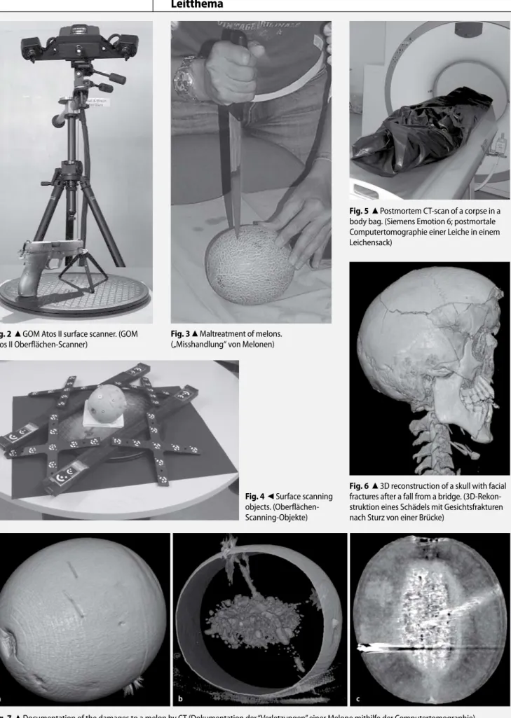
Surface scanning. In a demonstration by
the measurement engineers the
partici-pants were introduced to the technique of
photogrammetry and 3D surface scanning
of different tools – possible instruments in
forensic cases. The components of the
procedures – the digital measurement
camera (Nikon D2X, Nikon AG,
Switzer-land) and the surface scanner (GOM Atos
II, Gesellschaft für optische Messtechnik
mbH, Deutschland;
.Fig. 2) were
ex-plained. In the practical part, melons
(Cu-cumis melo) were „maltreated“ by the
participants with different instruments –
knife, ham mer, airgun, etc. – leaving
im-printed traces on the surface of the melon
(
.Fig. 3). After these procedures the
melons were prepared with reference
markers for the surface scans and a
com-plete surface documentation was
per-formed by the participants (
.Fig. 4).
CT-scanning. After an overview on the
de-velopment and the technique of computed
tomography, a demonstration of multislice
computed tomography (Siemens Somatom
Emotion 6, Siemens Medical Solutions,
Germany) by the Institute of Forensic
Med-icine, Bern was given on a scan of a corpse
that was transferred to the Institute of
Fo-rensic Medicine for radiological
documen-tation and autopsy (
.Fig. 5). The
partici-pants were able to perform adjustments of
the scan. In a second practical part the
par-ticipants used the abovementioned melon
model to scan by themselves.
Fig. 1
7
Participants of the first Virtopsy basic course and the Virtopsy team. (Teilnehmer und Team des ersten Virtopsy-Grundkurses)
Zusammenfassung · Abstract
Rechtsmedizin 2007 · 17:40–43 DOI 10.1007/s00194-006-0425-8 © Springer Medizin Verlag 2007
Virtopsy Team · L. Oesterhelweg
Aufbruchstimmung in der Rechtsmedizin? Virtopsy Basic Course
Zusammenfassung
Kernaufgaben der Rechtsmedizin sind die Er-hebung und die Dokumentation forensisch relevanter Befunde an Lebenden und To-ten für die Ermittlungsorgane und die Jus-tiz. Während in den Arbeitsbereichen der fo-rensischen Molekularbiologie und der foren-sischen Toxikologie bereits „High-tech-Ver-fahren“ den täglichen Arbeitsablauf dominie-ren, bedient sich die forensische Pathologie nach wie vor der Jahrhunderte alten Sekti-onstechniken und des Befundprotokolls. Mo-derne Dokumentationsverfahren wie Ober-flächen-Scanning und radiologische Schnitt-bildverfahren wie Computertomographie (CT) und „magnetic resonance imaging” (MRI) stehen mehr und mehr im Fokus der
foren-sischen Wissenschaften und finden zuneh-mend Eingang in die Routinemethoden ein-zelner Institute. Als Zeichen des steigenden Interesses an diesen Verfahren fand im Sep-tember 2006 der erste „Virtopsy basic course“ im Zentrum für forensische Bildgebung/Vir-topsy im Institut für Rechtsmedizin der Uni-versität Bern statt. Teilnehmer aus Deutsch-land, Italien, Japan, Österreich, der Schweiz, der Türkei und den USA nahmen an diesem ersten Workshop in forensischer Radiologie und Oberflächendokumentation teil.
Schlüsselwörter
Forensische Radiologie · Oberflächen-dokumentation · Virtopsy
Atmosphere of departure in forensic medicine?
Virtopsy Basic Course
Abstract
Forensic medicine aims for the documenta-tion of medical and other forensic findings in living and deceased persons for the police and the judiciary system. While in forensic ge-netics and forensic toxicology, high technol-ogy procedures are part of the daily work, fo-rensic pathology is still using the old estab-lished techniques from former centuries. New methods like 3D-surface scanning and mod-ern radiology procedures like computed to-mography (CT) or magnetic resonance imag-ing (MRI) are becomimag-ing more and more part of scientific research in forensic sciences and are today part of the routine workflow in a
some institutes of legal medicine. As a sign of this increasing interest the first Virtopsy ba-sic course was held in September 2006 at the Center for Forensic Imaging/Virtopsy in the Institute of Forensic Medicine in Bern, Swit-zerland. Participants from Austria, Germany, Italy, Japan, Switzerland, Turkey and the USA took part in this first hands-on course in fo-rensic radiology and surface scanning.
Keywords
Forensic radiology · Surface scanning · Virtopsy
2D and 3D data reconstruction. One of
the most important parts is the
documen-tation of cases and the reconstruction of
data in 2 and 3 dimensions. Therefore the
workstation (Siemens Leonardo, Siemens
Medical Solutions, Germany) and
applica-tions for adjustment of the 2-dimensional
images and the 3-dimensional virtual
mod-els were presented using the data of the
scanned corpse (
.Fig. 6). In the practical
part the participants had to reconstruct the
injuries on the melon produced by the
mal-treatment and got to know the
reconstruc-tions of gunshot channels and stab wounds
(
.Fig. 7). Also a basic overview of the
da-ta storage possibilities and the software
so-lutions was given.
Conclusions
The resonance of the first Virtopsy
ba-sic course was highly positive and the
de-mand for a Virtopsy advanced course was
voiced. This seemed to be a basis for an
ex-ten sion of imaging techniques to forensic
sciences and will hopefully be a platform
for scientific research projects and
knowl-edge exchange.
Corresponding author
Dr. L. Oesterhelweg
Zentrum für Forensische Bildgebung/Virtopsy, Institut für Rechtsmedizin, Universität Bern Bühlstrasse 20, CH-3012 Bern
lars.oesterhelweg@irm.unibe.ch
Conflict of interest. There is no conflict of interest.
The corresponding author affirms that he/she has no connection with any company whose product is mentioned in this article or any company that markets a competing product. The presentation of the findings is free of bias and product-neutral.
Acknowledgement. The Virtopsy team gratefully
acknowledge the support of the first Virtopsy basic course by Siemens Medical Solutions, Germany; GOM International AG, Switzerland; Sectra, Linköping, Sweden and the Virtopsy Foundation.
41
Rechtsmedizin 1 · 2007|
Fig. 2
8
GOM Atos II surface scanner. (GOM Atos II Oberflächen-Scanner)Fig. 3
8
Maltreatment of melons.(„Misshandlung“ von Melonen)
Fig. 4
9
Surface scanning objects. (Oberflächen-Scanning-Objekte)Fig. 5
8
Postmortem CT-scan of a corpse in abody bag. (Siemens Emotion 6; post mortale Computertomographie einer Leiche in einem Leichensack)
Fig. 6
8
3D reconstruction of a skull with facial fractures after a fall from a bridge. (3D-Rekon-struktion eines Schädels mit Gesichtsfrakturen nach Sturz von einer Brücke)Fig. 7
8
Documentation of the damages to a melon by CT (Dokumentation der “Verletzungen” einer Melone mithilfe der Computertomographie)42 |
Rechtsmedizin 1 · 2007References
1. Brogdon BG (1998) Forensic radiology. CRC, Boca Raton 2. Dirnhofer R, Jackowski C, Vock P et al. (2006)
Virt-opsy: minimally invasive. Imaging-guided virtual autopsy. Radiographics 26: 1305–1333 3. Myers JC, Okoye MI, Kiple D et al. (1999)
Three-dimensional (3-D) imaging in post-mortem exami-nations: elucidation and identification of cranial and facial fractures in victims of homicide utilizing 3-D computerized imaging reconstruction tech-niques. Int J Legal Med 113: 33–37
4. Paperno S, Riepert T, Krug B et al. (2005) Prospekti-ve Untersuchung zur Wertigkeit der postmortalen Computertomographie im Vergleich zur Autopsie. Rofo 177: 130–136
5. Poulsen K, Simonsen J (2006) Computed tomogra-phy as routine in connection with medico-legal autopsies. Forensic Sci Int (in press)
6. Stein KM, Bahner ML, Merkel J et al. (2000) Detec-tion of gunshot residues in routine CTs. Int J Legal Med 114: 15−18
7. Thali MJ, Braun M, Dirnhofer R (2003) Optical 3D surface digitizing in forensic medicine: 3D docu-mentation of skin and bone injuries. Forensic Sci Int 137: 203–208
8. Thali MJ, Yen K, Schweitzer W et al. (2003) Virtopsy, a new imaging horizon in forensic pathology: virtu-al autopsy by postmortem multislice computed to-mography (MSCT) and magnetic resonance imaging (MRI) – a feasibility study. J Forensic Sci 48: 1–18
43
Rechtsmedizin 1 · 2007|
Thiemann, H.-H., I. Nitz, A. Schmeling(Hrsg.)
Röntgenatlas der normalen
Hand im Kindesalter
Stuttgart: Georg Thieme Verlag 2006, 149 S., 148 Abb., 74 Tab.,
(ISBN 3-13-766603-1), 89.00 EUR Der „Thiemann-Nitz“ gehört seit Jahren zu den wichtigsten – leider nicht überall be-kannten – Standardwerken über die normale Ossifikation der menschlichen Hand. Anfäng-lich war die Erkennung von Wachstumsstö-rungen das Hauptanwendungsgebiet, später die Errechnung der vermutlichen Endgröße von Kindern einschließlich eventueller therapeutischer Maßnahmen. Schließlich kam in letzter Zeit die Bedeutung für die forensische Altersdiagnostik hinzu. Dem wur-de die 3. Auflage durch Hinzuziehung von forensischen Experten der Altersschätzung gerecht.
Der Textteil gliedert sich in eine kurze „Einfüh-rung“, den Abschnitt „Methodik“ zur Durch-führung und Auswertung der Röntgenauf-nahmen, das Kapitel „Allgemeine Befunde“ mit Erläuterung der Knochenentwicklung, das Kapitel „Berechnung der zu erwartenden Endgröße“ nach den Methoden von Bayley und Pinneau sowie von Roche, Wainer und Thissen, jeweils mit den entsprechenden Tabellen. Den Textteil beschließt das kurze Kapitel „Forensische Altersdiagnostik im Strafverfahren“.
Den Hauptteil machen die Bildtafeln aus. Sie sind sehr übersichtlich angeordnet, getrennt nach Geschlecht und Altersgruppen (für das Neugeborenenalter in Vierteljahresschritten, für das Kindesalter in Halbjahresschritten, und für Jugendliche in Jahresabständen). Während der Buchtitel nur das Kindesalter berücksichtigt, umfasst das Bildmaterial auch das Jugendalter (bis zu 18 Jahren). Zu jedem Fall gibt es eine Röntgenaufnahme, ein er läuterndes Schema sowie eine Tabelle mit den Abmessungen der Handknochen der entsprechenden Altersgruppe unter Angabe von Mittelwerten und Standardabweichun-gen. Diese Einfügung der Streubreiten stellt eine wichtige Neuerung dar. Die Qualität der Abbildungen ist tadellos, gleiches gilt für die gesamte Aufmachung des Buches.
Die Aktualisierung und Erweiterung des Röntgenatlas ist ausdrücklich zu begrüßen und wird seiner weiteren Verbreitung dienen. Das Buch wird speziell für Genetiker, Ortho-päden, Pädiater, Radiologen und Rechtsme-diziner eine wichtige Hilfe sein.
G. Geserick (Berlin)