Rheumatol Int (2009) 29:807–810 DOI 10.1007/s00296-008-0762-x
123
O R I G I N A L A R T I C L ELymphocyte counts in patients with ANCA-associated vasculitis
Andreas Holbro · Philipp Schuetz · Christoph Berger ·Christoph Hess · Thomas Daikeler
Received: 5 August 2008 / Accepted: 21 October 2008 / Published online: 8 November 2008 © Springer-Verlag 2008
Abstract How lymphocyte counts relate to treatment-response in patients with ANCA-associated vasculitis (AAV) is controversial, and data on short-term variability of lymphocyte counts are lacking. Retrospective single cen-ter evaluation of disease activity and lymphocyte counts in patients with AAV, and of lymphocyte counts in kidney transplant-recipients, were done; both at the University Hospital Basel, Switzerland. Twenty-three patients with AAV were included. Remission was achieved in all patients. Ten patients experienced a relapse after a median of 66 weeks (range 15–189 weeks). Median lymphocyte counts at diagnosis were signiWcantly higher than at remis-sion (1.38 £ 109/L vs. 0.99 £ 109/L; P = 0.007). By con-trast, median lymphocyte counts at remission and relapse did not diVer signiWcantly. However, intra-individual vari-ability of lymphocyte counts early after diagnosis was high [median lymphocyte variability-range during the Wrst 3 weeks of treatment 1.57 (range 0.27–3.95), n = 17]. This variability was not speciWc to patients with AAV, but was
also observed in patients after kidney transplantation [vari-ability of 1.76 (range 0.74–3.95, n = 31)]. The signiWcantly higher median lymphocyte counts at diagnosis of AAV make lymphocyte counts a valuable surrogate for the treat-ment-eYciency in clinical studies. By contrast, on a patient-level, variability of lymphocyte counts impedes meaningful interpretation of individual measurements. Keywords Vasculitis · ANCA · Lymphocytes · AAV · Prognostic · Variability
Introduction
Therapeutic options for severe AAV are limited, and ther-apy—mostly cyclophosphamide (CYC)-based—remains a balancing act between disease- and treatment-related mor-bidity and mortality [1, 2]. Factors predicting treatment-response would hence be important.
Lymphocytes, both of the T- and B-cell lineage, are thought to be involved in the pathogenesis of AAV [3–6]. How circulating lymphocyte counts relate to disease activ-ity, however, remains controversial. One study reported that treatment-induced lymphopenia (·0.5 £ 109/L) is associated with a high likelihood to achieve sustained remission [3, 7]. By contrast, another group suggested that, analogous to patients with systemic lupus erythematodes, lymphopenia in AAV reXects disease activity, with lym-phocyte counts actually increasing in patients achieving remission [8, 9]. While the reason for these contradictory Wndings remain unclear, neither study has taken into account short-term variability of lymphocyte counts and how this might impact study results.
Here, we investigated how in AAV-patients (1) median lymphocyte counts relate to disease activity on a population C. Hess and T. Daikeler are equal contributors.
A. Holbro
Division of Hematology, University Hospital Basel, 4031 Basel, Switzerland
P. Schuetz
Division of Endocrinology, Institute of Clinical Epidemiology, University Hospital Basel, 4031 Basel, Switzerland
C. Berger · C. Hess
Department of Internal Medicine,
University Hospital Basel, 4031 Basel, Switzerland
T. Daikeler (&)
Department of Rheumatology,
University Hospital Basel, 4031 Basel, Switzerland e-mail: [email protected]
808 Rheumatol Int (2009) 29:807–810
123
level, and (2) assessed, in these same individuals, short-term lymphocyte count variability.
Patients and methods Study populations
After obtaining approval by the institutional review board, disease activity and lymphocyte counts in patients with AAV (diagnosed between 1/1992 and 12/2007), and lym-phocyte counts in kidney transplant-recipients (transplanted within the living-donor program between 1/2004 and 12/ 2006) were retrospectively assessed in patients treated at the University Hospital Basel, Switzerland. Medical charts from all patients with a deWnite diagnosis of AAV were reviewed to obtain demographic, clinical and laboratory data. Diagnosis and follow-up of AAV
AAV was diagnosed according to American College of Rheumatology (ACR) criteria [10], the Birmingham Vas-culits Activity Score (BVAS) [11] was calculated initially and for every subsequent visit.
Laboratory measurements
Lymphocyte counts were determined via routine Xow-cytometry (ADVIA® Hematology system, Siemens), ANCA titers were measured using indirect immunoXuores-cence (INOVA®, San Diego), and ANCA were speciWed [myeloperoxydase (MPO) vs. proteinase3 (PR3)], and quantiWed using enzyme linked immunosorbent systems (DLD Diagnostika®, Hamburg).
Outcome parameters
Remission of AAV was deWned as complete absence of clinical symptoms and a daily prednisone-dose ·7.5 mg, according to published criteria [12]. Relapse was deWned as de novo disease activity according to the BVAS [11]. Statistical analysis
Discrete variables are expressed as counts (percentage), continuous variables as medians and ranges. Frequency comparison was done by Chi-square test. Two-group com-parison of normally distributed data was performed by paired or unpaired Student’s t test. For not normally distributed
Table 1 Patients with AAV:
disease manifestation, treatment and response to treatment
Sex Diagnosis Age at diagnosis (years) Manifestation Initial BVAS Initial therapy TTR weeks TSR weeks m WG 43 K, P, I, M, A, S, E 35 CYC 32 16+ m WG 63 K, A, E 14 CYC 16 2+ m WG 68 K, M 13 CYC 31 343+ m WG 66 K, P, A, S, E 26 CYC 28 438+ m WG 44 K, P, M, A, S, N 23 CYC 25 32 m WG 29 K, P, M 10 CYC 34 223+ m WG 55 K, P, M, A, S, N 13 CYC 58 110+ f WG 70 P, A, S 9 PRED 19 1+ f WG 65 K, M, A, N 22 AZA 17 189 f WG 47 K, P, A, S 14 CYC 24 254+ f WG 77 K, P, M, N 18 CYC 83 15 f WG 33 M 6 MTX 10 93 f WG 22 P, M, A, S 12 CYC 87 38 f WG 71 K, I, M 28 CYC 13 122+ f WG 51 K, M, A 21 CYC 42 167 f WG 27 P, M, S 13 MTX 17 5+ f WG 60 M, A, S, N 15 MTX 38 18 f WG 60 M, A, E 9 MTX 19 168 m CSS 33 P, M, N 17 CYC 26 35 f CSS 67 P, M, A 6 CYC 25 179 f CSS 51 P, S, N 10 CYC 29 294+ m AAV nc 58 K 12 CYC 40 15+ f AAV nc 44 K, P, A 12 AZA 13 66+ Abbreviations: WG Wegener granulomatosis, CSS Churg– Strauss syndrome, AAV nc ANCA-associated vasculitis not otherwise classiWed. Manifesta-tion: organ involvement at pre-sentation, K kidney, P lung, I Intestinal, M sinus/mucous,
A arthritis, S skin, E eye, N neurologic, BVAS
Birming-ham Vasculitis Activity Score,
CYC cyclophosphamide, MTX
methotrexat, PRED prednisone alone, AZA azathioprin, TTR time-to-remission in weeks,
TSR time since remission, +
Rheumatol Int (2009) 29:807–810 809
123
data, the Mann–Whitney U test for unpaired data and theWilcoxon’s matched pair signed rank test for paired data were used. All testing was two-tailed, and P values <0.05 were considered to indicate statistical signiWcance. For all statistical analysis STATA 9.2 (Stata Corp®, College Sta-tion, TX) was used.
Results
Patient’s characteristics
Of the 33 AAV patients identiWed, 23 (70%) were included in the analysis, 10 patients (30%) had to be excluded from the analysis because of missing data, being lost during fol-low-up, or due to concurrent diagnoses other than AAV necessitating immunosuppressive therapy. Wegener’s gran-ulomatosis was diagnosed in 18/23 patients (78%), Churg– Strauss Syndrome in 3/23 patients (13%), two patients had AAV not otherwise classiWed. A total of 14/23 patients were female (61%), the median age at diagnosis was 55 years (range, 22–77 years). Relevant baseline characteristics of
the AAV study population are summarized in Table1. Median BVAS at diagnosis was 13 (range 6–35). Sixteen out of twenty-three patients had CYC as part of their induc-tion therapy. Median CYC dose-to-remission was 13.5 g (range, 1.5–76.5 g). All 23 patients (100%) achieved remission [median time-to-remission, 26 weeks (range 10–87 weeks)], 10/23 patients (43%) experienced a relapse [median time-to-relapse: 66 weeks (range 15–189)]. The control-cohort of kidney transplant-recipients (n = 31), transplanted con-secutively between 2004 and 2006) received immunosup-pressive regimens containing glucocorticoids, mycophenolate mofetil, and tacrolimus. Ten/31 (33%) transplant-recipients were female; the median age at transplantation was 50 years (range, 19–68 years).
Lymphocyte counts
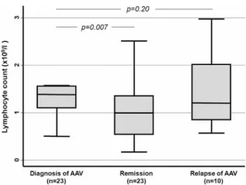
Lymphocyte counts were analyzed (1) cross-sectional (i.e., on a population level) at diagnosis and in remission, and (2) longitudinally (i.e., intra-individually) during the Wrst 3 weeks after diagnosis. On a population level, median lymphocyte counts at diagnosis [Wrst measurement 1.38 £ 109L (range: 0.5–3.11)] were signiWcantly higher than median lymphocyte counts in remission [Wrst measure-ment in remission, 0.99 £ 109L (range, 0.17–3.1),
P = 0.007] (Fig.1). Relapsing and non-relapsing patients had similar lymphocyte counts both at diagnosis and in remission [median lymphocyte counts at diagnosis, relaps-ing patients: 1.54 (range 0.77–3.11), non-relapsrelaps-ing patients: 1.34 (range 0.5–2.56), P = 0.15, median lymphocyte counts in remission, relapsing patients: 0.94 (range 0.17–3.1), non-relapsing patients: 0.99 (range 0.41–2.82), P = 0.88]. Time-to-remission was independent of initial lymphocyte counts (P = 0.8, Cox regression).
However, serial lymphocyte counts early after diagnosis [deWned here as ¸4 (range 4–17) measurements during the Wrst 3 weeks after diagnosis; available in n = 17 AAV-patients] revealed a high intra-individual variability of lymphocyte counts, even if measured in very short (daily) intervals [median intra-individual variability-range; 1.57
Fig. 1 Lymphocyte counts at diagnosis, in remission and at relapse of
AAV. Boxes denote median and interquartile ranges; whiskers denote ranges
Fig. 2 Variability of serial
lym-phocyte counts during the Wrst 3 weeks of therapy in patients with AAV (left panel) and in pa-tients after kidney transplanta-tion (right panel). Boxes denote median and interquartile ranges;
810 Rheumatol Int (2009) 29:807–810
123
(range 0.27–3.599)]. The lymphocyte count variability in patients with AAV was compared to the variability in a cohort of kidney transplant-recipients during the Wrst 3 weeks after transplantation (n = 31, 4–21 measurements). The median lymphocyte count at transplantation was 0.95 £ 109/L (range, 0.12–2.8), the median intra-individual variability-range 1.76 (range 0.74–3.95). Variability of lymphocyte counts in AAV-patients and transplant-recipi-ents was similar, excluding an AAV-speciWc phenomenon (P = 0.48) (Fig.2).
Discussion
The key observations of this study are that (1) in patients with AAV median lymphocyte counts on a cohort level are higher at presentation than in remission, and (2) in a given AAV-patient intra-individual short-term variability of lym-phocytes is making their value as an indicator of treatment-eYciency questionable. The major limitations of our study are its retrospective design, and the small number of patients that were studied. Despite these limitations, the range of the observed short-term lymphocyte count vari-ability—which is essentially unaVected by the study design—may have implications possibly extending beyond the Weld of clinical AAV-research.
In the context of AAV, our data—on a cohort level—are in-line with the plausible notion stemming from a relatively large study stating that lymphocyte counts inversely associ-ate with sustained remission [7]. However, by combining cohort-derived data and analyses in individual patients, our results indicate that the value of individual lymphocyte counts in reXecting treatment-eYciency is limited.
While for now remaining a provocative hypothesis, short-term (day-by-day!) variability of lymphocyte counts may also inXuence clinical decision making when judging immuno-competence, e.g., in patients infected with HIV or in transplant-recipients.
In summary, our study establishes a surprising short-term variability of lymphocyte counts both in patients with AAV, and in a heterogeneous group of kidney transplant-recipients. Larger, ideally prospective studies integrating short-term variability of peripheral lymphocyte counts and disease activity are needed to more deWnitely establish the value of lymphocyte counts as biomarkers for the treat-ment-eYciency in patients suVering from AAV.
Acknowledgments We thank Alan Tyndall, Jürg A SchiVerli and
Jürg Steiger for ongoing support.
References
1. HoVman GS, Kerr GS, Leavitt RY, Hallahan CW, Lebovics RS, Travis WD et al (1992) Wegener granulomatosis: an analysis of 158 patients. Ann Intern Med 116(6):488–498
2. Talar-Williams C, Hijazi YM, Walther MM, Linehan WM, Halla-han CW, Lubensky I et al (1996) Cyclophosphamide-induced cys-titis and bladder cancer in patients with Wegener granulomatosis. Ann Intern Med 124(5):477–484
3. Kallenberg CG (2007) Antineutrophil cytoplasmic autoantibody-associated small-vessel vasculitis. Curr Opin Rheumatol 19(1):17–24. doi:10.1097/BOR.0b013e3280119842
4. Brouwer E, Stegeman CA, Huitema MG, Limburg PC, Kallenberg CG (1994) T cell reactivity to proteinase 3 and myeloperoxidase in patients with Wegener’s granulomatosis (WG). Clin Exp Immu-nol 98(3):448–453
5. Bosch X, Guilabert A, Font J (2006) Antineutrophil cytoplasmic antibodies. Lancet 368(9533):404–418. doi: 10.1016/S0140-6736(06)69114-9
6. Flossmann O, Jones RB, Jayne DR, Luqmani RA (2006) Should rituximab be used to treat antineutrophil cytoplasmic antibody associated vasculitis? Ann Rheum Dis 65(7):841–844. doi:10.1136/ard.2005.048900
7. Villa-Forte A, Clark TM, Gomes M, Carey J, Mascha E, Karafa MT et al (2007) Substitution of methotrexate for cyclophospha-mide in Wegener granulomatosis: a 12-year single-practice expe-rience. Medicine (Baltimore) 86(5):269–277
8. Izzedine H, Cacoub P, Launay-Vacher V, Bagnis C, Deray G (2002) Lymphopenia in Wegener’s granulomatosis. A new clinical activity index? Nephron 92(2):466–471. doi:10.1159/ 000063303
9. Vila LM, Alarcon GS, McGwin G Jr, Bastian HM, Fessler BJ, Reveille JD (2006) Systemic lupus erythematosus in a multiethnic US cohort, XXXVII: association of lymphopenia with clinical manifestations, serologic abnormalities, disease activity, and dam-age accrual. Arthritis Rheum 55(5):799–806. doi:10.1002/art. 22224
10. Hunder GG, Arend WP, Bloch DA, Calabrese LH, Fauci AS, Fries JF et al (1990) The American College of Rheumatology 1990 cri-teria for the classiWcation of vasculitis. Introduction. Arthritis Rheum 33(8):1065–1067
11. Luqmani RA, Bacon PA, Moots RJ, Janssen BA, Pall A, Emery P et al (1994) Birmingham Vasculitis Activity Score (BVAS) in sys-temic necrotizing vasculitis. QJM 87(11):671–678
12. Hellmich B, Flossmann O, Gross WL, Bacon P, Cohen-Tervaert JW, Guillevin L et al (2007) EULAR recommendations for con-ducting clinical studies and/or clinical trials in systemic vasculitis: focus on anti-neutrophil cytoplasm antibody-associated vasculitis. Ann Rheum Dis 66(5):605–617. doi:10.1136/ard.2006.062711