HAL Id: hal-01522822
https://hal.sorbonne-universite.fr/hal-01522822
Submitted on 15 May 2017
HAL is a multi-disciplinary open access
archive for the deposit and dissemination of
sci-entific research documents, whether they are
pub-lished or not. The documents may come from
teaching and research institutions in France or
abroad, or from public or private research centers.
L’archive ouverte pluridisciplinaire HAL, est
destinée au dépôt et à la diffusion de documents
scientifiques de niveau recherche, publiés ou non,
émanant des établissements d’enseignement et de
recherche français ou étrangers, des laboratoires
publics ou privés.
Distributed under a Creative Commons Attribution| 4.0 International License
Patients with ANCA-associated vasculitis admitted to
the intensive care unit with acute vasculitis
manifestations: a retrospective and comparative
multicentric study
Julien Demiselle, Johann Auchabie, Francois Beloncle, Philippe Gatault,
Steven Grangé, Damien Du Cheyron, Jean Dellamonica, Sonia Boyer, Dimitri
Titeca Beauport, Lise Piquilloud, et al.
To cite this version:
Julien Demiselle, Johann Auchabie, Francois Beloncle, Philippe Gatault, Steven Grangé, et al..
Patients with ANCA-associated vasculitis admitted to the intensive care unit with acute
vasculi-tis manifestations: a retrospective and comparative multicentric study. Annals of Intensive Care,
SpringerOpen, 2017, 7, pp.39. �10.1186/s13613-017-0262-9�. �hal-01522822�
RESEARCH
Patients with ANCA-associated vasculitis
admitted to the intensive care unit with acute
vasculitis manifestations: a retrospective
and comparative multicentric study
Julien Demiselle
1,2, Johann Auchabie
1, François Beloncle
1, Philippe Gatault
3, Steven Grangé
4,
Damien Du Cheyron
5, Jean Dellamonica
6, Sonia Boyer
6, Dimitri Titeca Beauport
7, Lise Piquilloud
1,8,
Julien Letheulle
9, Christophe Guitton
10,11, Nicolas Chudeau
12, Guillaume Geri
13, François Fourrier
14,
René Robert
15, Emmanuel Guérot
16, Julie Boisramé‑Helms
17,18, Pierre Galichon
19, Pierre‑François Dequin
20,
Alexandre Lautrette
21, Pierre‑Edouard Bollaert
22, Ferhat Meziani
17,18, Loïc Guillevin
23, Nicolas Lerolle
1*and Jean‑François Augusto
2Abstract
Purpose: Data for ANCA‑associated vasculitis (AAV) patients requiring intensive care are scarce.
Methods: We included 97 consecutive patients with acute AAV manifestations (new onset or relapsing disease),
admitted to 18 intensive care units (ICUs) over a 10‑year period (2002–2012). A group of 95 consecutive AAV patients with new onset or relapsing disease, admitted to two nephrology departments with acute vasculitis manifestations, constituted the control group.
Results: In the ICU group, patients predominantly showed granulomatosis with polyangiitis and proteinase‑3
ANCAs. Compared with the non‑ICU group, the ICU group showed comparable Birmingham vasculitis activity score and a higher frequency of heart, central nervous system and lungs involvements. Respiratory assistance, renal replace‑ ment therapy and vasopressors were required in 68.0, 56.7 and 26.8% of ICU patients, respectively. All but one patient (99%) received glucocorticoids, 85.6% received cyclophosphamide, and 49.5% had plasma exchanges as remission induction regimens. Fifteen (15.5%) patients died during the ICU stay. The following were significantly associated with ICU mortality in the univariate analysis: the need for respiratory assistance, the use of vasopressors, the occurrence of at least one infection event in ICU, cyclophosphamide treatment, sequential organ failure assessment at admission and simplified acute physiology score II. After adjustment on sequential organ failure assessment or infection, cyclo‑ phosphamide was no longer a risk factor for mortality. Despite a higher initial mortality rate of ICU patients within the first hospital stay (p < 0.0001), the long‑term mortality of hospital survivors did not differ between ICU and non‑ICU groups (18.6 and 20.4%, respectively, p = 0.36). Moreover, we observed no renal survival difference between groups after a 1‑year follow‑up (82.1 and 80.5%, p = 0.94).
Conclusion: This study supports the idea that experiencing an ICU challenge does not impact the long‑term prog‑
nosis of AAV patients.
Keywords: Anti‑neutrophil cytoplasmic antibody, ANCA‑associated vasculitis, Intensive care unit, Mortality
© The Author(s) 2017. This article is distributed under the terms of the Creative Commons Attribution 4.0 International License (http://creativecommons.org/licenses/by/4.0/), which permits unrestricted use, distribution, and reproduction in any medium, provided you give appropriate credit to the original author(s) and the source, provide a link to the Creative Commons license, and indicate if changes were made.
Open Access
*Correspondence: nicolas.lerolle@univ‑angers.fr
1 Département de Réanimation Médicale et de Médecine Hyperbare, Centre Hospitalier Universitaire, 4 rue Larrey, 49933 Angers Cedex 9, Fra nce
Page 2 of 9 Demiselle et al. Ann. Intensive Care (2017) 7:39
Background
Anti-neutrophil cytoplasmic antibodies (ANCAs)-asso-ciated vasculitis (AAV) are life-threatening multisys-tem autoimmune diseases characterized by necrotizing inflammation of small- to medium-sized vessels [1, 2]. There are three differentiated entities based on clinical and pathological criteria: microscopic polyangiitis (MPA), granulomatosis with polyangiitis (GPA) and eosinophilic granulomatosis with polyangiitis (EGPA) [3]. Their clini-cal spectrum partially overlaps. Indeed, rapidly progres-sive glomerulonephritis is the typical renal presentation of MPA and GPA, but is rarely present in EGPA [1]. Dif-fuse alveolar hemorrhage (DAH) is the most critical lung injury observed with all entities, but more frequently with MPA and GPA [4, 5]. Other respiratory presentations include pulmonary infiltrates and nodules, the latter being observed predominantly in GPA [6]. Even though ANCA negativity does not exclude AAV diagnosis, diffuse forms of AAV are usually associated with serum positivity for ANCAs [1, 7]. Given their high level of specificity, ANCA detection is critical for AAV diagnosis, and ANCA posi-tivity with a compatible clinical diagnosis usually allows the initiation of immunosuppressive treatments [8, 9].
The prompt initiation of immunosuppressive drugs to induce remission is critical for AAV patient prognosis. In generalized and severe forms, conventional induction treatment combines high doses of glucocorticoids and cyclophosphamide [10]. In addition, plasma exchange (PE) may be used in severe forms with DAH and/or severe renal involvement [11, 12]. Based on recent clini-cal trials, rituximab, the CD20 monoclonal anti-body, can be used as an alternative to cyclophosphamide. Under these regimens, AAV remission is achieved in 60–80% of the patients [13–17]. However, despite being adequately treated, some patients experience resistance to therapy or disease relapse. Moreover, a high mortal-ity rate is observed in AAV patients, with rates reaching 10–15% within the first year following treatment ini-tiation, the main causes of early death being infection events and vasculitis manifestations [18, 19]. Mortality rates of up to 20% after 5 years have been observed, and mortality has been shown to be higher with MPA than with EGPA and GPA [18].
To date, patients with the most severe forms of AAV— those requiring intensive care—have not been extensively and adequately analyzed. Indeed, most previous studies were small size studies, uncontrolled and monocentric. Moreover, they mingled AAV patients with manifesta-tions related to vasculitis activity, and those with mani-festations not related to it. Finally, data for AAV patients admitted to the intensive care unit (ICU) for AAV mani-festations [20–24] are scarce and the prognosis of this specific population remains poorly determined.
We conducted this retrospective multicentric study to analyze disease presentation and outcome in AAV patients admitted to the ICU with acute vasculitis mani-festations. Specifically, we intended to explore whether ICU admission was associated with adverse long-term outcomes. To this end, ICU-AAV patients were com-pared with a group of AAV patients admitted to two nephrology departments with an active disease but with no requirement of ICU care (non-ICU-AAV patients). Methods
Population and inclusion criteria
We conducted a multicentric retrospective study in seventeen ICUs from French University and Gen-eral Hospitals, and from Lausanne’s University Hos-pital in Switzerland. Inclusion criteria included: over 18 years of age, ICU admission between January 2002 and December 2012, and newly diagnosed or relapsing AAV. Only patients with acute vasculitis manifestations were included in the study. To be included, AAV ini-tial or relapse diagnosis had to be done during the ICU stay or within the thirty days immediately prior to ICU admission.
A number of non-ICU-AAV patients were used as a control group. This group included all consecutive AAV patients of two nephrology centers (Angers and Tours University Hospitals), who were diagnosed between January 2002 and December 2012. To be included in the control group, patients had to show active newly diag-nosed or relapsing AAV. Patients who required further ICU admission within the month following admission to the nephrology department were excluded from the con-trol group.
ANCA positivity by indirect immunofluorescence (cytoplasmic or perinuclear pattern) and ELISA (protein-ase-3 (PR-3) or myeloperoxidase (MPO)) was required for inclusion in both groups (ICU and non-ICU groups).
The Institutional Ethics Committees of the Angers University Hospital and Lausanne Hospital approved the study protocol (N°2013/21 and N°164/14, respectively).
AAV diagnosis
Patients’ medical files were analyzed, and the AAV sub-type (GPA, MPA and EGPA) was determined according to the European Medicines Agency vasculitis classifica-tion algorithm [25]. For newly diagnosed patients, the date of AAV diagnosis was defined as the date of ANCA determination and the date of relapse for relapsing patients was defined as the date of hospital admission. The diagnosis of new onset AAV relied on ANCA positiv-ity and presence of vasculitis manifestations. The diagno-sis of relapsing AAV was retained when it was suspected by the physician, and when the retrospective analysis of
the patient’s medical history was consistent (i.e., typical clinical manifestation, increase in ANCA titer or biopsy-proven vasculitis activity).
For conflicting cases, the hospitalization report and the medical records were analyzed by 2 expert investiga-tors (JFA and NL). If AAV activity remained doubtful, the patient was excluded from the study.
Data collection
Patients were identified from the ICU database of each hospital. All the data were collected retrospectively by a systematic screening of patients’ medical records. The fol-lowing data were collected: age, gender, height and weight, and significant aspects of past medical history. Organs affected by vasculitis were listed upon the presentation of newly diagnosed and relapsing patients. The ANCA type was recorded, and pathology data of biopsied organs were analyzed (when available) to confirm the diagnosis of vas-culitis. Birmingham Vasculitis Activity Score (BVAS) 2003 was used to determine AAV activity [26].
ICU ANCA‑associated vasculitis
For ICU-AAV patients, causes of ICU admission with simplified acute physiology score II (SAPS II) and sequential organ failure assessment (SOFA) score upon admission [27, 28] were recorded. The ratio of partial pressure of arterial oxygen over inspired-fraction of oxy-gen (PaO2/FiO2) with ventilation initiation, the serum
creatinine level and the Acute Kidney Injury Network (AKIN) score [29] upon ICU admission were used to characterize the severity of respiratory and renal injuries. Supporting therapies used during the ICU stay and their duration (mechanical ventilation, renal replacement ther-apy, vasopressors), septic events (as documented in the ICU hospitalization report) and death were registered. Cause of death was classified by two authors (NL and JD) after patient’s files review.
AAV treatment
In both groups, all specific AAV induction regimens and their timing were recorded, including steroid treat-ment, cyclophosphamide, rituximab and plasmapheresis treatments. The use of steroid boluses, and the dosages and the number of cyclophosphamide and rituximab boluses were analyzed, as well as the number of plasma exchanges.
Outcomes definition
For both groups, survival was analyzed until death, loss of follow-up or end of follow-up (December 2012). Sur-vival free of end-stage renal disease of the ICU and non-ICU groups was also analyzed. Renal death was defined
as the need for long-term (>3 months) renal replacement therapy.
Statistical analysis
Quantitative parameters were presented as median (interquartile range (IQR)) and qualitative parameters as absolute number and/or percentage. Categorical and continuous data were analyzed with Chi-square (or Fish-er’s exact test) and Mann–Whitney U tests, respectively. Results were presented as odd ratio (OR) with 95% confi-dence intervals (95 CIs); the Kaplan–Meyer method was used to analyze the survival rates of ICU and non-ICU groups. A log-rank test was used to compare the survival curves. All statistical tests were performed with a two-sided 0.05 level of significance applied. Statistical analysis was performed using SPSS software® 23.0 for Macintosh and Graphpad Prism®.
Results
Baseline characteristics of the ICU and non‑ICU‑AAV populations
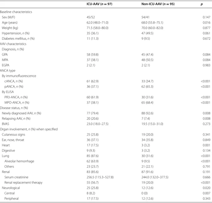
Ninety-seven and ninety-five AAV patients with a median follow-up of 2.28 [IQR 0.2–4.7] and 4.18 [IQR 1.7–7.0] years were included in the ICU group and the non-ICU control group, respectively. Characteristics of the ICU and the non-ICU groups and the main AAV manifesta-tions upon admission are detailed in Table 1. Groups were similar regarding gender and AAV subtype, but patients of the ICU group were significantly younger. Newly diag-nosed AAV was significantly more common in the non-ICU group (93%) compared with the non-ICU group (79%). In the ICU group, 39 patients were diagnosed during the ICU stay and 58 were diagnosed before their admission to ICU. Disease activity assessed by BVAS showed no statis-tical difference between groups. Heart, lung and central neurological injuries were more frequent in ICU patients than in non-ICU patients. A notably high rate of alveolar hemorrhage was observed in ICU patients (64 vs. 10% in non-ICU patients), but the renal involvement rate was comparable. ANCA subtypes by immunofluorescence and ELISA were significantly different between groups. Indeed, 62% of ICU patients had PR3-ANCAs, and 68% of non-ICU patients had MPO-ANCAs.
Parameters specific to AAV‑ICU group and assessment of organ support
The median ICU length of stay was 7 [IQR 4.5–17.5] days. Acute respiratory failure (alone or in combination with renal failure) was the main cause for ICU admission and accounted for 80% of ICU admissions. Approximately 70% of patients required respiratory assistance, which was ini-tiated within 48 h following ICU admission in the large
Page 4 of 9 Demiselle et al. Ann. Intensive Care (2017) 7:39
majority of the patients. Acute kidney injury was highly prevalent, with AKIN score ≥1 in more than 90% of patients at the time of admission, and more than half of the patients required renal replacement therapy (RRT) during their ICU stay. RRT was initiated within 48 h after admis-sion in 35% of patients. Vasopressors were required for 25% of the patients. Infection events were reported in 40% of the patients during the ICU stay, with an identified pathogen in 82% of them. These data are summarized in Table 2.
Site and nature of infectious events are detailed in Additional file 1: Table 1.
Immunosuppressive regimens
Ninety-nine percent of patients of the ICU and 98% of non-ICU groups received glucocorticoids for remis-sion induction. Steroid pulses were given to 95 of the 97 (98%) patients in the ICU group, and to 80 of the 95 (84%) patients in the non-ICU group (p < 0.001). Glu-cocorticoids were combined with cyclophosphamide in 83 (85.6%) ICU patients and in 78 (82.1%) non-ICU patients (p = 0.514). PE was more frequently used in the ICU group than in the non-ICU group (n = 48, 54% vs.
n = 23, 24%, respectively, p < 0.001).
Table 1 Baseline characteristics of the ICU and non-ICU ANCA-associated vasculitis groups
ICU‑AAV (n = 97) Non‑ICU‑AAV (n = 95) p Baseline characteristics Sex (M/F) 45/52 54/41 0.147 Age (years) 62.0 (48.0–71.0) 68.0 (55.8–75.1) 0.016 Weight (kg) 71.5 (58.0–80.0) 70.0 (60.0–82.0) 0.877 Hypertension, n (%) 35 (36.1) 47 (49.5) 0.061 Diabetes mellitus, n (%) 11 (11.3) 9 (9.5) 0.672 AAV characteristics Diagnosis, n (%) GPA 58 (59.8) 45 (47.4) 0.084 MPA 37 (38.1) 48 (50.5) 0.084 EGPA 2 (2.1) 2 (2.1) 0.983 ANCA type By immunofluorescence cANCA, n (%) 61 (62.9) 33 (34.7) <0.001 pANCA, n (%) 36 (37.1) 62 (65.3) <0.001 By ELISA PR3‑ANCA, n (%) 60 (61.9) 30 (31.6) <0.001 MPO‑ANCA, n (%) 37 (38.1) 65 (68.4) <0.001 Disease status, n (%)
Newly diagnosed AAV, n (%) 77 (79.4) 88 (92.6) 0.008
Relapsing AAV, n (%) 20 (20.6) 7 (7.4) 0.008
BVAS 23.0 (18.0–27.5) 19.5 (15.0–31.0) 0.273
Organ involvement, n (%) when specified
Cutaneous signs 25 (25.8) 19 (20.0) 0.341
Ear, nose, throat 36 (37.1) 34 (35.8) 0.849
Heart 17 (17.5) 3 (3.2) 0.001 Digestive 9 (9.3) 3 (3.2) 0.134 Lung 85 (87.6) 30 (31.6) <0.001 Alveolar hemorrhage 62 (63.9) 9 (9.5) <0.001 Others 23 (23.7) 21 (22.1) 0.791 Renal 83 (85.6) 87 (91.6) 0.191 Serum creatinine 256.5 (115.3–527.8) 244.0 (132.0–377.5) 0.666
Renal replacement therapy 55 (56.7) 19 (20.0) <0.001
Neurological 25 (25.8) 12 (12.6) 0.020
Central 8 (8.2) 0 (0) 0.007
The most common induction immunosuppressive regi-men administered to the ICU group was a combination of glucocorticoids and cyclophosphamide used in 41 patients (42.3%) and a combination of glucocorticoids, cyclophosphamide and PE used in 42 patients (43.3%). In the non-ICU group, glucocorticoids and cyclophospha-mide were the most frequent induction regimen (58.9% of patients). Data detailing immunosuppressive regimen used in ICU and non-ICU-AAV patients, and their tim-ing accordtim-ing to ICU admission, are outlined in Addi-tional file 2: Table 2.
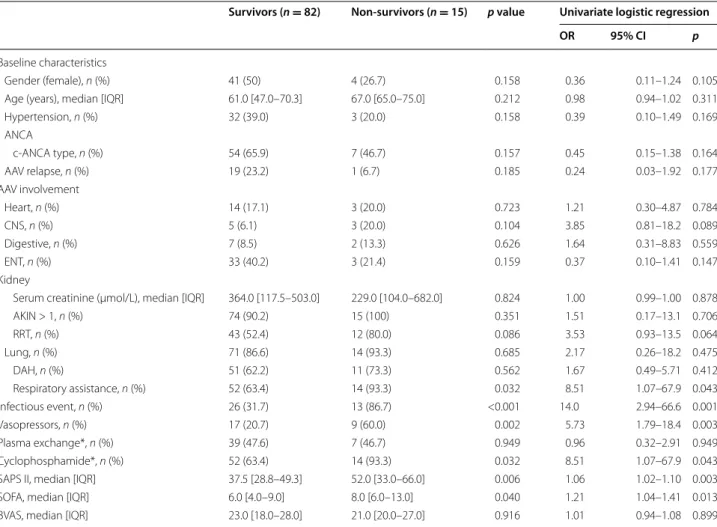
Mortality and predictors of ICU mortality
Fifteen patients (15.5%) died during the ICU stay. SAPS II and ICU SOFA scores were significantly higher in non-survivors compared to non-survivors. The need for mechani-cal ventilation (invasive or not) and vasopressors was more frequent in the non-survivor group. The require-ment of RRT tended to be higher in the non-survivor group, but remained statistically not significant. Moreo-ver, infectious events during ICU stays were significantly more prevalent in non-survivors. Non-surviving patients received cyclophosphamide more frequently than surviv-ing patients. We did not observe any difference between survivors and non-survivors according to the timing of immunosuppressive treatment, including cyclophospha-mide, with respect to ICU admission (data not shown). These data are summarized in Table 3. In a multivariate logistic analysis, cyclophosphamide was no longer asso-ciated with mortality after adjustment on SAPS II or occurrence of infection events (Additional file 3: Table 3).
The cause of ICU death was attributed to refractory vasculitis manifestations in 6 (40%) patients (DAH in 5 patients, digestive involvement in 1), to multiple organ failure likely due to sepsis in 5 (33%) patients and to neu-rologic causes in 4 (27%) patients, including 3 cerebral hemorrhage while receiving anticoagulation for extracor-poreal membrane oxygenation.
Long‑term outcomes of ICU‑AAV patients
Whether ICU stay can impact the prognosis of AAV patients had not been yet analyzed. Regarding hospi-tal morhospi-tality, we observed that ICU-AAV patients had a poorer survival rate compared to non-ICU-AAV patients (Fig. 1a). Given that most deaths in the ICU group occurred soon after ICU admission, we next analyzed the long-term mortality of patients that survived to the first hospital stay (ICU or non-ICU patients). By this way, we were able to observe that the long-term mortality of AAV patients who survived to the first hospital stay was no longer different between the ICU and the non-ICU group (Fig. 1b). We also analyzed the renal outcome of groups. Long-term renal outcome was available for 67 ICU-AAV patients out of 82 and for all non-ICU-AAV patients. Survival analysis showed that renal survival was not sig-nificantly different between ICU and non-ICU patients after 1 year of follow-up (Fig. 1c).
Discussion
In the present work, we described 97 patients who required ICU admission at AAV diagnosis or relapse. Lung involvement (notably, DAH) was the prominent cause for ICU admission, 70% of the patients requir-ing mechanical ventilation. Fifty percent needed RRT and 25% needed vasopressors. Comparison with a
Table 2 Characteristics and supportive therapies used with ICU-AAV patients
* Diagnosed < or >48 h after ICU admission **Among patients that experienced infection
Length of stay (days) 7.0 (4.5–17.5)
Reasons for ICU admission, n (%)
Respiratory failure 44 (45.4)
And renal failure 23 (23.7)
And neurological failure 1 (1.0)
Acute renal failure 17 (17.5)
And neurological failure 4 (4.1)
Neurological failure 4 (4.1)
Heart failure 3 (3.1)
Hemorrhagic shock 1 (1.0)
SOFA (at admission) 6 (4.0–9.0)
SAPS II 39.0 (31.0–51.0)
Respiratory assistance, n (%) Mechanical ventilation
Invasive or/and noninvasive 66 (68.0) Within 48 h of admission 58 (59.8) Noninvasive ventilation only 19 (19.6)
Invasive ventilation only 36 (37.1)
Noninvasive and invasive ventilation 11 (11.3) Length of respiratory assistance (days) 10.0 (5.5–18.5)
PaO2/FiO2 92.0 (58.8–182.0)
Kidney involvement
Serum creatinine at admission, (μmol/L) 256.5 (115.3–527.8) Maximum serum creatinine in ICU, (μmol/L) 348.0 (160.0–673.0)
AKIN score ≥1, n (%) 89 (91.8)
Renal replacement therapy, n (%) 55 (56.7)
Within 48 h of admission 34 (35.1)
Hemodynamic assistance
Vasopressive amines, n (%) 26 (26.8)
Within 48 h 25 (25.8)
Length of treatment (days) 6.0 (3–11.5) Infectious events
Early/late* 29/10
Lung infection, n (%) 29 (74.4)**
Other sites, n (%) 10 (25.6)**
Page 6 of 9 Demiselle et al. Ann. Intensive Care (2017) 7:39
control group of AAV patients admitted to the nephrol-ogy department showed no difference in overall score of disease activity (BVAS), but ICU patients were younger and more likely to have PR3-ANCAs and GPA. A vast
majority of ICU patients and non-ICU patients received an induction regimen with corticosteroid and cyclophos-phamide, and ICU patients received PE more frequently. One-year mortality rate was higher in ICU patients due
Table 3 Comparison between survivor and non-survivor ICU-AAV patients and univariate logistic regression analysis for ICU mortality
OR odd ratio, CI confidence interval, ANCA anti-neutrophil cytoplasmic antibodies, c-ANCA cytoplasmic ANCA, AAV ANCA-associated vasculitis, CNS central nervous system, ENT ear nose throat, AKIN acute kidney injury network score, RRT renal replacement therapy, DAH diffuse alveolar hemorrhage
* Started before or during the ICU stay
Survivors (n = 82) Non‑survivors (n = 15) p value Univariate logistic regression
OR 95% CI p
Baseline characteristics
Gender (female), n (%) 41 (50) 4 (26.7) 0.158 0.36 0.11–1.24 0.105
Age (years), median [IQR] 61.0 [47.0–70.3] 67.0 [65.0–75.0] 0.212 0.98 0.94–1.02 0.311
Hypertension, n (%) 32 (39.0) 3 (20.0) 0.158 0.39 0.10–1.49 0.169 ANCA c‑ANCA type, n (%) 54 (65.9) 7 (46.7) 0.157 0.45 0.15–1.38 0.164 AAV relapse, n (%) 19 (23.2) 1 (6.7) 0.185 0.24 0.03–1.92 0.177 AAV involvement Heart, n (%) 14 (17.1) 3 (20.0) 0.723 1.21 0.30–4.87 0.784 CNS, n (%) 5 (6.1) 3 (20.0) 0.104 3.85 0.81–18.2 0.089 Digestive, n (%) 7 (8.5) 2 (13.3) 0.626 1.64 0.31–8.83 0.559 ENT, n (%) 33 (40.2) 3 (21.4) 0.159 0.37 0.10–1.41 0.147 Kidney
Serum creatinine (µmol/L), median [IQR] 364.0 [117.5–503.0] 229.0 [104.0–682.0] 0.824 1.00 0.99–1.00 0.878
AKIN > 1, n (%) 74 (90.2) 15 (100) 0.351 1.51 0.17–13.1 0.706 RRT, n (%) 43 (52.4) 12 (80.0) 0.086 3.53 0.93–13.5 0.064 Lung, n (%) 71 (86.6) 14 (93.3) 0.685 2.17 0.26–18.2 0.475 DAH, n (%) 51 (62.2) 11 (73.3) 0.562 1.67 0.49–5.71 0.412 Respiratory assistance, n (%) 52 (63.4) 14 (93.3) 0.032 8.51 1.07–67.9 0.043 Infectious event, n (%) 26 (31.7) 13 (86.7) <0.001 14.0 2.94–66.6 0.001 Vasopressors, n (%) 17 (20.7) 9 (60.0) 0.002 5.73 1.79–18.4 0.003 Plasma exchange*, n (%) 39 (47.6) 7 (46.7) 0.949 0.96 0.32–2.91 0.949 Cyclophosphamide*, n (%) 52 (63.4) 14 (93.3) 0.032 8.51 1.07–67.9 0.043
SAPS II, median [IQR] 37.5 [28.8–49.3] 52.0 [33.0–66.0] 0.006 1.06 1.02–1.10 0.003
SOFA, median [IQR] 6.0 [4.0–9.0] 8.0 [6.0–13.0] 0.040 1.21 1.04–1.41 0.013
BVAS, median [IQR] 23.0 [18.0–28.0] 21.0 [20.0–27.0] 0.916 1.01 0.94–1.08 0.899
Fig. 1 a Survival of ICU and non‑ICU‑AAV patients and b survival of patients who survived to the first hospital stay. c Renal survival of ICU and non‑
to high in-ICU fatality (15.5%) but, interestingly, in ICU survivors, 1-year survival after initial hospital admission was no different from non-ICU patients.
Several studies have already reported AAV patients admitted to ICU. Most included AAV patients admitted to ICU for reasons related to vasculitis manifestations and reasons unrelated to the same—such as our results cannot be readily compared. However, some of our results are in line with these reports, showing a majority of GPA among ICU-AAVs [20, 21, 30], and a high preva-lence of DAH [20, 21, 24]. Previous studies have reported very variable rates of respiratory assistance (31–57%) [20, 23, 24, 31] and of RRT use (20–80%) [20, 23, 24], and variable mortality rates from 0 to 33% probably related to the heterogeneity of included patients [20, 23].
However, this study is the first to report on any long-term outcome of AAV patients with active disease after an ICU stay or to assess the impact of ICU stay in com-parison with AAV patients initially admitted to non-ICU wards. Indeed, besides observing that ICU admission for acute organ dysfunction requiring organ support is associated with poor outcome, we thought that the main question was to determine the association between ini-tial disease severity, invasive therapeutic procedures, and long-term outcome. Given that kidney involvement is a major prognostic factor in AAV [11], we estimated that AAV patients with kidney involvement constituted a pertinent control group. Unsurprisingly, a high death rate was observed with ICU patients at the early phase of the disease, but the observation that long-term outcome (both mortality and renal survival) in ICU survivors is no different from non-ICU patients is a key observation and deserves discussion. First, it can be noted that ICU patients were younger and more frequently had GPA compared with MPA, two factors related to better out-come in previous studies outside the ICU [32, 33]. The fact that GPA onset is usually earlier than MPA onset may explain the lower age and the greater frequency of patients with relapsing in the ICU group [33]. Due to the low death rate after the initial stay, no multivariate analy-sis to determine the association between ICU admission or not, AAV type and age with outcome was attempted. Beyond these limitations, our observation tends to show that initial disease severity, once adequately controlled by the induction regimen, is not associated with adverse long-term outcome and does not indicate a more severe disease. From an ICU point of view, it should be noted that this observation is at stake with several reports showing prolonged excess risk of death in ICU survivors in comparison with non-ICU patients [34]. Although the number of patients in our study limits the interpreta-tion of this result, it favors the hypothesis that prolonged mortality in ICU patients is related to baseline conditions
(here: the AAV disease) rather than to acute episodes [35].
Notwithstanding the relatively good prognosis with ICU survivors, ICU mortality is a concern that should be addressed. Intensity of acute organ dysfunction assessed through acute severity scores (SOFA, SAPS II and hence organ support requirement), cyclophosphamide and infection were associated with ICU death. In contrast, BVAS did not appear suitable for predicting short-term mortality of ICU-AAV patients [20]. Despite limita-tion of statistical analysis, cyclophosphamide did not appear as a risk factor for mortality after adjustment on SAPS II and infection occurrence. Although association between organ dysfunction and mortality is trivial in the ICU setting, the association between cyclophosphamide, infection and mortality is certainly not straightforward. Indeed, most infections were nosocomial infections in patients receiving mechanical ventilation, and the attrib-utable mortality of such infections has been debated [36,
37]. Rather, it may represent an indicator of overall sever-ity associated with an immunosuppressive state related to the condition which led to ICU admission (DAH and AAV), consequences of ICU care (tracheal intubation), and immunosuppressive treatments. Analysis of cause of death brings up additional relevant information, as infec-tion causes and disease activity both represent the two major conditions associated with death. Finally, from our dataset, no straightforward message can be established for the purpose of determining whether ICU patients with AAV require a higher or lower level of immunosup-pression, if this concept has any meaning.
Improved ICU outcome may come from more refined and individualized induction regimen. It has been proven that PE has benefited AAV patients with severe renal involvement (i.e., creatinine >500 μmol/L) when used in replacement of methylprednisolone boluses in AAV induction treatment, allowing the achievement of higher rates of renal recovery [11]. Moreover, evidence from clinical practice and retrospective studies also supports PE effectiveness in patients with DAH-related AAV [5,
12, 38]. However, our study does not show a clear-cut impact of PE on survival, this observation being clearly limited by the design of this study and the dataset. It should be noted that rituximab, which appeared as a novel option in both induction and maintenance regi-mens [14, 15] of AAV [39], was rarely used in our study. This may be explained by the date range of the study which ended in 2012, whereby rituximab was not able to pass into clinical practice. Given the limited data related to the use of rituximab in patients with severe forms of AAV including patient with DHA, The French Vasculitis Study Group recommended to use cyclophosphamide as a first-line treatment to induce remission [40]. However,
Page 8 of 9 Demiselle et al. Ann. Intensive Care (2017) 7:39
recently, Cartin-Ceba et al. [41] suggested in a retrospec-tive analysis that rituximab may be superior to cyclo-phosphamide to achieve remission at 6 months in AAV patients with DHA. We observed a high rate of infectious events, a majority of deaths related to DAH or sepsis in our study and an increased rate of cyclophosphamide use in non-survivors. Whether using rituximab in replace-ment of cyclophosphamide in this specific population may improve prognosis merits to be considered.
Our study undeniably has several limitations, start-ing with its retrospective design and restriction to AAV patients with ANCA positivity. Despite all efforts to be as exhaustive as possible, some data may have been missed. Given the very low prevalence of ICU admission for active AAV, a prospective study does not seem eas-ily conceivable. We believe that the multicentric and con-trolled design of our study has contributed to limitation of center-dependent bias and to significant expansion of ICU-AAV-related knowledge, especially with regard to long-term prognosis of these patients.
Conclusion
Acute respiratory failure due to DAH is the most com-mon vasculitis manifestation which puts AAV patients in the ICU. Despite a high early ICU mortality rate, patients who survive to ICU show comparable long-term mor-tality and renal prognosis compared to non-ICU-AAV patients.
Abbreviations
AAV: anti‑neutrophil cytoplasmic antibodies‑associated vasculitis; ANCA: anti‑ neutrophil cytoplasmic antibodies; AKIN: acute kidney injury network score; BVAS: Birmingham vasculitis activity score; DAH: diffuse alveolar hemorrhage; EGPA: eosinophilic granulomatosis with polyangiitis; ELISA: enzyme‑linked immunosorbent assay; GPA: granulomatosis with polyangiitis; ICU: intensive care unit; MPA: microscopic polyangiitis; MPO: myeloperoxidase; PaO2/FiO2: ratio of partial pressure of arterial oxygen over inspired‑fraction of oxygen; PE: plasma exchange; PR‑3: proteinase‑3; RRT: renal replacement therapy; SAPS II: simplified acute physiology score II; SOFA: sequential organ failure assessment.
Authors’ contributions
JD and JA participated in conducting the study, collecting the data for the study and writing the manuscript. FB participated in the formulation of the study and conducting it, and collecting the data and revision of the manu‑ script. PG, SG, DDC, JD, SB, DT, LP, JL, CG, NC, GG, FF, RR, JLD, JBH, PG, PFD, AL, PEB and FM participated in collecting the data for the study and revision of the manuscript. LG participated in the formulation of the study and collecting data for it, and revision of the manuscript. NL and JFA participated in the formulation of the study and conducting it, and collecting the data for it, and in the writing of the manuscript. All authors read and approved the final manuscript.
Additional files
Additional file 1: Table 1. Infectious events in ICU‑AAV group.
Additional file 2: Table 2. Induction immunosuppressive regimens of the ICU and non‑ICU‑AAV patients.
Additional file 3: Table 3. Multivariate logistic analysis for ICU mortality.
Author details
1 Département de Réanimation Médicale et de Médecine Hyperbare, Centre Hospitalier Universitaire, 4 rue Larrey, 49933 Angers Cedex 9, France. 2 Néphrol‑ ogie‑Dialyse‑Transplantation, CHU Angers, 4 rue Larrey, 49933 Angers Cedex 9, France. 3 Service de Néphrologie et Immunologie Clinique, CHRU Tours, Tours, France. 4 Medical Intensive Care Unit, Rouen University Hospital, Rouen, France. 5 Service de Réanimation Médicale, CHU de Caen, Avenue de la Côte de Nacre, CS 30001, 14033 Caen Cedex 9, France. 6 Medical Intensive Care Unit, Archet 1 University Hospital, Route de St Antoine, CS 23079, 06202 Nice, France. 7 Medi‑ cal Intensive Care Unit, Amiens University Medical Center, 80054 Amiens, Cedex 1, France. 8 Service de Médecine Intensive Adulte et Centre des Brûlés, Centre Hospitalier Universitaire Vaudois, Lausanne, Switzerland. 9 Service de Réanima‑ tion Médicale, Hôpital Pontchaillou, CHU Rennes, 2 rue Henri Le Guilloux, 35033 Rennes Cedex, France. 10 Medical Intensive Care Unit, Hôtel‑Dieu, Univer‑ sity Hospital of Nantes, 30 bd Jean Monnet, 44093 Nantes, France. 11 UMR 1064, Inserm, 30 bd Jean Monnet, 44093 Nantes, France. 12 Service de Reanima‑ tion Medico‑Chirurgicale, Centre Hospitalier du Mans, 194 Avenue Rubillard, 72037 Le Mans, France. 13 Service de Réanimation Médicale, Hôpital Cochin, Paris, France. 14 Réanimation, Centre de Réanimation Polyvalente, Hôpital Roger Salengro, CHRU de Lille, Lille, France. 15 Service de Réanimation Médicale, CHU de Poitiers, Poitiers, France. 16 Service de Réanimation Médicale, Hôpital Euro‑ péen Georges Pompidou, Paris, France. 17 Service de Réanimation Médicale, Nouvel Hôpital Civil, Hôpitaux Universitaires de Strasbourg, Strasbourg, France. 18 EA 7293, Fédération de Médecine Translationnelle de Strasbourg (FMTS), Fac‑ ulté de Médecine, Université de Strasbourg, Strasbourg, France. 19 APHP, Hôpital Tenon, Urgences Néphrologiques et Transplantation Rénale, Paris, France. 20 Service de Réanimation Polyvalente, Hôpital Bretonneau, Tours, France. 21 Service de Réanimation Médicale Polyvalente, CHU Gabriel Montpied, 58 rue Montalembert, 63000 Clermont‑Ferrand, France. 22 Service de Réanima‑ tion Médicale, CHU de Nancy Hôpital Central, 29 Avenue de Lattre de Tassigny, 54035 Nancy Cedex, France. 23 Département de Médecine Interne, Assistance Public des Hôpitaux de Paris, Hôpital Cochin, Paris, France.
Competing interests
The authors declare that they have no competing interests.
Availability of data and materials
All relevant data are within the paper.
Ethics approval and consent to participate
The Institutional Ethics Committees of the Angers University Hospital and Lausanne Hospital approved the study protocol (N°2013/21 and N°164/14, respectively).
Publisher’s Note
Springer Nature remains neutral with regard to jurisdictional claims in pub‑ lished maps and institutional affiliations.
Received: 25 November 2016 Accepted: 23 March 2017
References
1. Jennette JC, Falk RJ. Small‑vessel vasculitis. N Engl J Med. 1997;337:1512– 23. doi:10.1056/NEJM199711203372106.
2. Jennette JC, Falk RJ, Hu P, Xiao H. Pathogenesis of antineutrophil cyto‑ plasmic autoantibody‑associated small‑vessel vasculitis. Annu Rev Pathol. 2013;8:139–60. doi:10.1146/annurev‑pathol‑011811‑132453.
3. Jennette JC, Falk RJ, Bacon PA, et al. 2012 revised International Chapel Hill consensus conference nomenclature of vasculitides. Arthritis Rheum. 2013;65:1–11. doi:10.1002/art.37715.
4. Rabe C, Appenrodt B, Hoff C, et al. Severe respiratory failure due to diffuse alveolar hemorrhage: clinical characteristics and outcome of intensive care. J Crit Care. 2010;25:230–5. doi:10.1016/j.jcrc.2009.04.009. 5. West S, Arulkumaran N, Ind PW, Pusey CD. Diffuse alveolar haemorrhage
in ANCA‑associated vasculitis. Intern Med Tokyo Jpn. 2013;52:5–13. 6. Thickett DR, Richter AG, Nathani N, et al. Pulmonary manifestations of
anti‑neutrophil cytoplasmic antibody (ANCA)‑positive vasculitis. Rheuma‑ tol Oxf Engl. 2006;45:261–8. doi:10.1093/rheumatology/kei217.
7. Basu N, Watts R, Bajema I, et al. EULAR points to consider in the develop‑ ment of classification and diagnostic criteria in systemic vasculitis. Ann Rheum Dis. 2010;69:1744–50. doi:10.1136/ard.2009.119032.
8. Lionaki S, Blyth ER, Hogan SL, et al. Classification of antineutrophil cytoplasmic autoantibody vasculitides: the role of antineutrophil cyto‑ plasmic autoantibody specificity for myeloperoxidase or proteinase 3 in disease recognition and prognosis. Arthritis Rheum. 2012;64:3452–62. doi:10.1002/art.34562.
9. Kallenberg CGM, Heeringa P, Stegeman CA. Mechanisms of disease: pathogenesis and treatment of ANCA‑associated vasculitides. Nat Clin Pract Rheumatol. 2006;2:661–70. doi:10.1038/ncprheum0355. 10. Yates M, Watts RA, Bajema IM, et al. EULAR/ERA‑EDTA recommendations
for the management of ANCA‑associated vasculitis. Ann Rheum Dis. 2016;75:1583–94. doi:10.1136/annrheumdis‑2016‑209133.
11. Jayne DRW, Gaskin G, Rasmussen N, et al. Randomized trial of plasma exchange or high‑dosage methylprednisolone as adjunctive therapy for severe renal vasculitis. J Am Soc Nephrol. 2007;18:2180–8. doi:10.1681/ ASN.2007010090.
12. Klemmer PJ, Chalermskulrat W, Reif MS, et al. Plasmapheresis therapy for diffuse alveolar hemorrhage in patients with small‑vessel vasculitis. Am J Kidney Dis Off J Natl Kidney Found. 2003;42:1149–53.
13. Specks U, Merkel PA, Seo P, et al. Efficacy of remission‑induction regi‑ mens for ANCA‑associated vasculitis. N Engl J Med. 2013;369:417–27. doi:10.1056/NEJMoa1213277.
14. Stone JH, Merkel PA, Spiera R, et al. Rituximab versus cyclophospha‑ mide for ANCA‑associated vasculitis. N Engl J Med. 2010;363:221–32. doi:10.1056/NEJMoa0909905.
15. Jones RB, Tervaert JWC, Hauser T, et al. Rituximab versus cyclophospha‑ mide in ANCA‑associated renal vasculitis. N Engl J Med. 2010;363:211–20. doi:10.1056/NEJMoa0909169.
16. Hogan SL, Falk RJ, Chin H, et al. Predictors of relapse and treatment resistance in antineutrophil cytoplasmic antibody‑associated small‑vessel vasculitis. Ann Intern Med. 2005;143:621–31.
17. Pagnoux C, Hogan SL, Chin H, et al. Predictors of treatment resistance and relapse in antineutrophil cytoplasmic antibody‑associated small‑vessel vasculitis: comparison of two independent cohorts. Arthritis Rheum. 2008;58:2908–18. doi:10.1002/art.23800.
18. Flossmann O, Berden A, de Groot K, et al. Long‑term patient survival in ANCA‑associated vasculitis. Ann Rheum Dis. 2011;70:488–94. doi:10.1136/ ard.2010.137778.
19. Guillevin L, Pagnoux C, Seror R, et al. The five‑factor Score revisited: assessment of prognoses of systemic necrotizing vasculitides based on the French Vasculitis Study Group (FVSG) cohort. Medicine (Baltimore). 2011;90:19–27. doi:10.1097/MD.0b013e318205a4c6.
20. Frausova D, Brejnikova M, Hruskova Z, et al. Outcome of thirty patients with ANCA‑associated renal vasculitis admitted to the intensive care unit. Ren Fail. 2008;30:890–5. doi:10.1080/08860220802353892.
21. Khan SA. Outcome of patients with small‑vessel vasculitis admitted to a medical ICU. Chest. 2007;131:972. doi:10.1378/chest.06‑2464. 22. Cruz BA. Prognosis and outcome of 26 patients with systemic necrotiz‑
ing vasculitis admitted to the intensive care unit. Rheumatology. 2003;42:1183–8. doi:10.1093/rheumatology/keg322.
23. Monti S, Montecucco C, Pieropan S, et al. Life‑threatening onset of sys‑ temic vasculitis requiring intensive care unit admission: a case series. Clin Exp Rheumatol. 2015;33:S126–31.
24. Burkhardt O, Köhnlein T, Wrenger E, et al. Predicting outcome and survival in patients with Wegener’s granulomatosis treated on the intensive care unit. Scand J Rheumatol. 2007;36:119–24. doi:10.1080/03009740600958611.
25. Watts R, Lane S, Hanslik T, et al. Development and validation of a consensus methodology for the classification of the ANCA‑associated vasculitides and polyarteritis nodosa for epidemiological studies. Ann Rheum Dis. 2007;66:222–7. doi:10.1136/ard.2006.054593.
26. Luqmani RA, Bacon PA, Moots RJ, et al. Birmingham vasculitis activity score (BVAS) in systemic necrotizing vasculitis. QJM Mon J Assoc Physi‑ cians. 1994;87:671–8.
27. Vincent JL, Moreno R, Takala J, et al. The SOFA (Sepsis‑related Organ Fail‑ ure Assessment) score to describe organ dysfunction/failure. On behalf of the Working Group on Sepsis‑Related Problems of the European Society of Intensive Care Medicine. Intensive Care Med. 1996;22:707–10. 28. Le Gall JR, Lemeshow S, Saulnier F. A new simplified acute physiology
score (SAPS II) based on a European/North American multicenter study. JAMA. 1993;270:2957–63.
29. Mehta RL, Kellum JA, Shah SV, et al. Acute kidney injury network: report of an initiative to improve outcomes in acute kidney injury. Crit Care Lond Engl. 2007;11:R31. doi:10.1186/cc5713.
30. Kimmoun A, Baux E, Das V, et al. Outcomes of patients admitted to intensive care units for acute manifestation of small‑vessel vasculi‑ tis: a multicenter, retrospective study. Crit Care. 2015. doi:10.1186/ s13054‑016‑1189‑5.
31. Dumas G, Géri G, Montlahuc C, et al. Outcomes in critically Ill patients with systemic rheumatic disease: a multicenter study. Chest J. 2015;148:927. doi:10.1378/chest.14‑3098.
32. Hilhorst M, van Paassen P, Tervaert JWC, Limburg Renal Registry. Protein‑ ase 3‑ANCA vasculitis versus myeloperoxidase‑ANCA vasculitis. J Am Soc Nephrol JASN. 2015;26:2314–27. doi:10.1681/ASN.2014090903. 33. Franssen C, Gans R, Kallenberg C, et al. Disease spectrum of patients with
antineutrophil cytoplasmic autoantibodies of defined specificity: distinct differences between patients with anti‑proteinase 3 and anti‑myeloper‑ oxidase autoantibodies. J Intern Med. 1998;244:209–16.
34. Wunsch H, Guerra C, Barnato AE, et al. Three‑year outcomes for Medicare beneficiaries who survive intensive care. JAMA. 2010;303:849–56. doi:10.1001/jama.2010.216.
35. Iwashyna TJ, Netzer G, Langa KM, Cigolle C. Spurious inferences about long‑term outcomes: the case of severe sepsis and geriatric conditions. Am J Respir Crit Care Med. 2012;185:835–41. doi:10.1164/ rccm.201109‑1660OC.
36. Mitharwal SM, Yaddanapudi S, Bhardwaj N, et al. Intensive care unit‑acquired infections in a tertiary care hospital: an epidemiologic survey and influence on patient outcomes. Am J Infect Control. 2016;. doi:10.1016/j.ajic.2016.01.021.
37. Van Vught LA, Klein Klouwenberg PMC, Spitoni C, et al. Incidence, risk factors, and attributable mortality of secondary infections in the intensive care unit after admission for sepsis. JAMA. 2016;315:1469. doi:10.1001/ jama.2016.2691.
38. Beck L, Bomback AS, Choi MJ, et al. KDOQI US commentary on the 2012 KDIGO clinical practice guideline for glomerulonephritis. Am J Kidney Dis Off J Natl Kidney Found. 2013;62:403–41. doi:10.1053/j.ajkd.2013.06.002. 39. Pagnoux C, Guillevin L, French Vasculitis Study Group, MAINRITSAN inves‑
tigators. Rituximab or azathioprine maintenance in ANCA‑associated vasculitis. N Engl J Med. 2015;372:386–7. doi:10.1056/NEJMc1414728. 40. Charles P, Bienvenu B, Bonnotte B, et al. Rituximab: recommendations of
the French Vasculitis Study Group (FVSG) for induction and maintenance treatments of adult, antineutrophil cytoplasm antibody‑associated necrotizing vasculitides. Presse Medicale. 1983;42:1317–30. doi:10.1016/j. lpm.2013.08.003.
41. Cartin‑Ceba R, Diaz‑Caballero L, Al‑Qadi MO, et al. Diffuse alveolar hemor‑ rhage secondary to antineutrophil cytoplasmic antibody‑associated vasculitis: predictors of respiratory failure and clinical outcomes. Arthritis Rheumatol. 2016;68:1467–76. doi:10.1002/art.39562.