On: 06 November 2013, At: 23:27
Publisher: Taylor & Francis
Informa Ltd Registered in England and Wales Registered Number: 1072954 Registered office: Mortimer House,
37-41 Mortimer Street, London W1T 3JH, UK
Journal of Vertebrate Paleontology
Publication details, including instructions for authors and subscription information:
http://www.tandfonline.com/loi/ujvp20
Braincase of Panphagia protos (Dinosauria,
Sauropodomorpha)
Ricardo N. Martínez
a, José A. Haro
a& Cecilia Apaldetti
ab aInstituto y Museo de Ciencias Naturales, Universidad Nacional de San Juan , 5400 , San
Juan , Argentina
b
Consejo Nacional de Investigaciones Científicas y Técnicas , Buenos Aires , Argentina
Published online: 08 Oct 2013.
To cite this article: Ricardo N. Martínez , José A. Haro & Cecilia Apaldetti (2012) Braincase of Panphagia protos (Dinosauria,
Sauropodomorpha), Journal of Vertebrate Paleontology, 32:sup1, 70-82, DOI: 10.1080/02724634.2013.819009
To link to this article:
http://dx.doi.org/10.1080/02724634.2013.819009
PLEASE SCROLL DOWN FOR ARTICLE
Taylor & Francis makes every effort to ensure the accuracy of all the information (the “Content”) contained
in the publications on our platform. However, Taylor & Francis, our agents, and our licensors make no
representations or warranties whatsoever as to the accuracy, completeness, or suitability for any purpose of the
Content. Any opinions and views expressed in this publication are the opinions and views of the authors, and
are not the views of or endorsed by Taylor & Francis. The accuracy of the Content should not be relied upon and
should be independently verified with primary sources of information. Taylor and Francis shall not be liable for
any losses, actions, claims, proceedings, demands, costs, expenses, damages, and other liabilities whatsoever
or howsoever caused arising directly or indirectly in connection with, in relation to or arising out of the use of
the Content.
This article may be used for research, teaching, and private study purposes. Any substantial or systematic
reproduction, redistribution, reselling, loan, sub-licensing, systematic supply, or distribution in any
form to anyone is expressly forbidden. Terms & Conditions of access and use can be found at http://
www.tandfonline.com/page/terms-and-conditions
ABSTRACT—We describe a partial braincase of the basal sauropodomorph Panphagia protos from the Upper Triassic (mid-Carnian) horizons of the Ischigualasto Formation. The disarticulated braincase from a subadult individual includes one frontal, both parietals, one prootic, and the supraoccipital. The frontal is longer anteroposteriorly than it is wide transversely, has a small anterolateral process, and an elongate oval depression for the olfactory bulb. The supraoccipital is broader transversely than it is deep dorsoventrally and lacks a pronounced median nuchal eminence. Some braincase features that characterize more derived basal sauropodomorphs are not present in Panphagia, including a broader frontal and reduced anterior tympanic and floccular recesses. Panphagia appears to represent an early stage in the evolution of sauropodomorph dinosaurs.
RESUMEN—Describimos la caja craneana parcial del sauropodomorfo basal Panphagia protos proveniente de horizontes del Tri ´asico superior (Carniano medio) de la Formaci ´on Ischigualasto. La caja craneana desarticulada es de un individuo sub-adulto e incluye un frontal, dos parietales, un pro ´otico y el supraoccipital. El frontal es m ´as largo anteroposteriormente que ancho transversalmente, tiene un peque ˜no proceso anterolateral y una depresi ´on alargada oval para el bulbo olfatorio. El supraoccipital es transversalmente m ´as ancho que dorsoventralmente alto y carece de una eminencia nucal media pronunciada. Algunas de las caracter´ısticas que caracterizan los neurocr ´aneos de sauropodomorfos basales m ´as derivados no est ´an presentes en Panphagia, incluyendo el frontal ancho y la reducci ´on de las cavidades timp ´anica anterior y flocular. Panphagia parece representar una etapa temprana en la evoluci ´on de los dinosaurios sauropodomorfos.
INTRODUCTION
The earliest and most basal sauropodomorphs now include taxa known from relatively complete skeletons, such as Eorap-tor (Sereno et al., 1993, 2013; Mart´ınez et al., 2011), Panphagia (Mart´ınez and Alcober, 2009), Saturnalia (Langer et al., 1999, 2007; Langer, 2003), and Pampadromaeus (Cabreira et al., 2011). Taken together, these genera document well the external mor-phology of the skull and postcranial skeleton. What is conspic-uously absent in this material, however, is detailed information on the braincase. Only a few isolated elements of the braincase have been found in Eoraptor (Sereno et al., 2013) and Panphagia (Mart´ınez and Alcober, 2009), and these have not yet been de-scribed in detail.
Discovered in 2006 in the mid-Carnian horizons of the Is-chigualasto Formation, the basal sauropodomorph Panphagia protos is known from a single specimen that preserves sev-eral disarticulated cranial elements that include portions of the braincase (PVSJ 874; Mart´ınez and Alcober, 2009). These bones are well preserved and include the frontal, parietal, supraoccip-ital, and prootic. We describe these bones in detail below and compare them with other basal dinosaurs and more advanced sauropodomorphs.
The braincase anatomy is well documented in several basal sauropodomorph genera of Late Triassic (post-Carnian) or Early Jurassic age, including Thecodontosaurus (YPM 2192), Efraasia, Plateosaurus, Massospondylus, Anchisaurus, and Melanorosaurus (Galton, 1984, 1985; Kermack, 1984; Galton and Bakker, 1985; Gow, 1990; Benton et al., 2000; Sues et al., 2004; Yates, 2003, 2004, 2007; Fedak and Galton, 2007). Comparative information on the braincase in the earliest and most basal theropods, by contrast, is poorly known. In the well-preserved skull of Her-rerasaurus, the neurocranial elements are obscured laterally, dor-sally, and anteriorly by dermal bones of the skull roof (Sereno
*Corresponding author.
et al., 1993; Sereno and Novas, 1994), and computed tomographic scans of the specimen failed to resolve significant internal de-tail. The braincase is not known in other herrerasaurids, in-cluding Staurikosaurus (Colbert, 1970; Bittencourt and Kellner, 2009), Chindesaurus (Long and Murry, 1995), and Sanjuansaurus (Alcober and Mart´ınez, 2010). In the contemporaneous (mid-Carnian) theropod Eodromaeus (Mart´ınez et al., 2011) and some-what younger (Norian) Tawa (Nesbitt et al., 2009), more of the braincase is preserved and exposed but has yet to be de-scribed. Comparative information on the braincase of basal or-nithischians is fairly limited as well. Despite excellent preserva-tion of the skull in the basal ornithischians Lesothosaurus and Heterodontosaurus, the braincase is poorly exposed and has not been described from computed tomographic imaging (Sereno, 1991, 2012; Norman et al., 2011). The braincase of Lesothosaurus is the better known of these two ornithischians and has been figured in lateral view with sutures indicated (Sereno, 1991:fig. 13A, B). The sidewall of the braincase in Heterodontosaurus is not exposed, and reconstructions of it do not indicate sutures (Norman et al., 2011:fig. 15). Immediate dinosaur outgroups, such as the silesaurids, also lack detailed comparative informa-tion on the braincase. Although the disarticulated braincase in Silesaurus has been reconstructed in multiple views, there is lit-tle accompanying descriptive information and no way to discern preserved from reconstructed morphology (Dzik, 2003:fig. 7). The external and internal morphology of braincase have not been described.
In sum, an outline of braincase evolution in basal sauropodomorphs and in basal dinosaurs in general has yet to be formulated. At present, we cannot recognize with confi-dence primitive versus derived conditions in the neurocranium at various levels within Dinosauria. We work toward that goal with the detailed documentation of portions of the braincase in the basal sauropodomorph Panphagia. Braincase characters have been incorporated into basal sauropodomorph phylogenies (e.g., Yates, 2003, 2004, 2007) and are likely to garner an increasing presence in future datasets.
70
MATERIALS AND METHODS
Materials
Skull bones studied include the left frontal, both parietals, right prootic, and supraoccipital of the holotype of Panphagia protos (PVSJ 874). These disarticulated bones are fully prepared and ex-posed in all views.
Orientation and Terminology
Because the bones were found in isolation, we assumed an ori-entation in our figures consistent with that in the intact braincases of closely related taxa (e.g., Adeopapposaurus; Mart´ınez, 2009). We employed traditional, or ‘Romerian,’ directional terms over veterinarian alternatives (Wilson, 2006). ‘Anterior’ and ‘posterior’ are employed rather than ‘rostral,’ ‘cranial,’ or ‘caudal.’ When ver-nacular terms in English were not available or widely used, we em-ployed appropriate anatomical terms from the Nomina Anatomica Avium (Baumel et al., 1993).
We followed the phylogenetic definitions proposed by Sereno (2005). Sauropodomorpha, for example, in this work is regarded as a stem-based taxon defined as the most inclusive clade contain-ing Saltasaurus loricatus but not Passer domesticus or Triceratops horridus. With this formulation, the definition does not depend on the monophyly of other suprageneric taxa (e.g., Saurischia, Prosauropoda).
Institutional Abbreviations—PVSJ, Museo de Ciencias
Natu-rales, Universidad Nacional de San Juan, San Juan, Argentina;
YPM, Yale Peabody Museum, Yale University, New Haven,
Con-necticut, U.S.A.
DESCRIPTION
Skull Roof
Frontal—The frontal is anteroposteriorly longer than it is
trans-versely broad (Fig. 1). Its maximum length is greater than twice its maximum width across the posterolateral processes, and the posterior end is transversely wider than the anterior end (Fig. 1A, B). The transversely broad sutural surface for the nasal faces dor-sally and is located medial to the sutural surface for the prefrontal. These nasal and prefrontal lay adjacent to one another on the frontal, although a short anterior process of the frontal separates them (Fig. 2A). Although the medial portion of the anterior end of the frontal is broken away, it appears to have extended to a point along the midline.
The sutural surface for the prefrontal, which is exposed in dor-sal and lateral views, is transversely narrow (Fig. 1A, C). In dordor-sal view, the articular surface for the prefrontal is well marked both anteriorly and posteriorly, the posterior tip of which lies along the orbital rim rather than on the dorsal surface of the frontal (Fig. 1A).
Most of the medial edge of the frontal forms a straight, verti-cal margin for its opposite (Fig. 1A, B), although there is a shal-low medial sulcus bounded ventrally by a short shelf (Fig. 1D). More posteriorly, the medial edge is convex and then curves slightly away from the midline (Fig. 1A, B), which appears to be a natural asymmetry between left and right frontals. The por-tion of the orbital border formed by the frontal is proporpor-tion- proportion-ally long, forming just over 50% of the total length of the bone (Fig. 1A).
A ‘V’-shaped notch for articulation with the postorbital and the arcuate rim of the supratemporal fossa are sharply defined (Fig. 1A). Medial to the slot articulation for the postorbital and within the supratemporal fossa, a subtriangular process projects posterolaterally (Fig. 1A). This tip of this process likely marks the sutural triple junction between the frontal, postorbital, and
pari-etal (Fig. 2A). The posterior edge of the frontal is marked by su-tural contact with other bones and likely contributed little if any to the anterior margin of the supratemporal fenestra (Fig. 2A). The posterior edge of the frontal is arcuate in dorsal view, curving anterolaterally rather than following a transverse course (Fig. 3).
The ventral surface of the frontal has two distinct surfaces separated by a medially convex ridge, or crista cranii (Fig. 1B). In Panphagia, the crista cranii is developed as a pair of paral-lel ridges rather than a single ridge (Figs. 1B, 3A), as occurs in at least in the posterior part of the crista cranii in Pantydraco (Galton and Kermack, 2010:fig. 4D; Fig. 3B). The surface between these ridges is flat along the anterior one-third of its extension and slightly concave in its posterior two-thirds in Panphagia.
The orbital roof is relatively narrow transversely, expanding in width slightly posteriorly. The rim of the orbit is gently upturned, and the orbital surface faces ventrolaterally (Fig. 1C, D). The sur-face medial to the crista cranii is transversely concave and forms part of the roof of the nasal cavity (cavum nasi; Baumel and Wit-mer, 1993). An elongate oval fossa accommodated the olfactory bulb (Figs. 1B, 3A). Medial to this fossa, a longitudinal trough may have housed the olfactory nerve (Fig. 1B) as proposed previ-ously in Coelophysis (Raath, 1977:fig. 5F). A ridge continues ante-riorly and may have supported cartilage of the nasal cavity, judging from hypothesized conchal homologies (Witmer, 1995a) and the condition in the squamate Ctenosaura (Oelrich, 1956).
Anterolateral to the fossa for the olfactory bulb, another de-pression is present and may represent a portion of the roof of the cavum nasi (Fig. 3A; Majungasaurus, Tyrannosaurus; Witmer and Ridgely, 2008:figs. 5A, 6A, E, G). Surfaces medial and lateral to the ridge likely represent parts of the roof of the cavum nasi. The olfactory nerve may have reached the olfactory bulb more laterally than hypothesized for Coelophysis (Raath, 1977:fig. 21), either on the anterolateral aspect of the bulb as in Tyrannosaurus (Witmer and Ridgely, 2008:fig. 6B) or more later-ally as in crocodilians (Shiino, 1914).
The posterior expansion of the frontal presents a shallow fossa cranii anterioris (sensu Baumel and Witmer, 1993; Fig. 1B) for the telencephalon, which occupies the entire width of the frontal en-docranial surface (Fig. 3A). The enen-docranial surface represents half the width of the frontal at the posterior end of the bone.
The sutural area for the laterosphenoid is not well defined (Fig. 1B). A small, anteroposteriorly elongated depression is present medial to the posterior end of the crista cranii, which may represent an articular surface for the head of the laterosphe-noid. The orbitosphenoid-frontal contact has been suggested in this location in Plateosaurus (Galton, 1984), although in spec-imens of Massospondylus (Gow, 1990:fig. 3A) and Allosaurus (McClelland, 1990:fig. 11) the orbitosphenoid does not contact the frontal.
The relatively long proportions of the frontal of Panphagia, in which its maximum length is greater than two times the maxi-mum width at the posterolateral process (Figs. 1A, B, 2A), are similar to those found in Silesaurus (Dzik, 2003:fig. 6D), Eo-raptor (PVSJ 512), Pantydraco (Yates, 2003:fig. 4A), and Coelo-physis (Raath, 1977:figs. 3B, 5F) and differ from the relatively wider frontals of Lesothosaurus (Sereno, 1991:figs. 11B, 12B), Her-rerasaurus (Sereno and Novas, 1994:figs. 1C, 7B, 8B; Fig. 2B), and many basal sauropodomorphs (Fig. 2C, D). The proportion-ally long orbital border formed by the frontal is similar to that in Herrerasaurus (Fig. 2B), Coelophysis (Raath, 1977:fig. 5B, F), Silesaurus (Dzik, 2003:fig. 6D), and some basal sauropodomorphs (Fig. 2D; also Pantydraco, Kermack, 1984:fig. 4; Galton and Kermack, 2010:fig. 4A–C; Massospondylus, Yates, 2003:fig. 2C, 4A; Sues et al., 2004:fig. 3A). The frontal contribution to the orbital border is proportionately shorter in Lesothosaurus
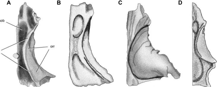
FIGURE 1. Left frontal of Panphagia protos (PVSJ 874) in dorsal (A), ventral (B), lateral (C), and medial (D) views. Abbreviations: an, articu-lar surface for the nasal; aos, articuarticu-lar surface for the orbitosphenoid; ap, anterior process; apo, ar-ticular surface for the postorbital; aprf, arar-ticular surface for the prefrontal; fosob, fossa for the ol-factory bulb; orm, orbital margin; orr, orbital roof; plpr, posterolateral process; ri, ridge; sh, shelf; stf, supratemporal fossa; su, sulcus. Scale bar equals 1 cm.
FIGURE 2. Dorsal view of partial skull roof of the basal dinosaurs Panphagia (A),
Her-rerasaurus (B), Plateosaurus (C), and Adeopap-posaurus (D). The supratemporal fossa is
shaded gray. Modified from Sereno and No-vas, 1994 (B); Galton, 1984 (C); and Mart´ınez, 2009 (D). Abbreviations: ap, anterior process; f, frontal; l, lacrimal; n, nasal; p, parietal; pf, pre-frontal; po, postorbital; stf, supratemporal fossa. The drawings are not in the same scale.
FIGURE 3. Ventral view of the frontal of the basal dinosaurs Panphagia (A), Pantydraco (B), Plateosaurus (C), and Coelophysis (D). Modified from Galton and Kermack, 2010 (B); Galton, 1984 (C); and Raath, 1977 (D). Abbreviations: fosob, fossa for the olfactory bulb; orr, orbital roof; ri, ridge. The drawings are not in the same scale.
(Sereno, 1991:fig. 11B) and other basal sauropodomorphs such a Plateosaurus (Fig. 2C), Melanorosaurus (Yates, 2007:figs. 3A, B, 7), and Lufengosaurus (Barrett et al., 2005:fig. 3A, B). The transversely narrow sutural surface for the prefrontal (Figs. 1A, C, 2A) is similar to that in Pantydraco (Galton and Kermack, 2010:fig. 4B) and narrower than that in Melanorosaurus (Yates, 2007:figs. 3A, B, 7), Adeopapposaurus (Fig. 2D), and Riojasaurus (PVSJ 849). The acute ‘V’-shaped notch for articulation with the postorbital (Fig. 1A) is similar to that in Eoraptor (Langer, 2004), Pantydraco (Yates, 2003), Plateosaurus (Fig. 2C), and Coeloph-ysis (Raath, 1977:fig. 5B). The articular notch is shorter in Her-rerasaurus (Fig. 2B), is intermediate in length in Lesothosaurus (Sereno, 1991:fig. 11B), and is ‘U’-shaped and broader in Het-erodontosaurus (Norman et al., 2011:fig. 6A). The exclusion of the frontal from the margin of the supratemporal fenestra resem-bles the condition in Herrerasaurus (Fig. 2B), Adeopapposaurus (Fig. 2D), Megapnosaurus kayentakatae (Rowe, 1989:fig. 1C, D), and Lesothosaurus (Sereno, 1991:fig. 11B). In Heterodontosaurus (Norman et al., 2011:fig. 1A) and Plateosaurus (Fig. 2C), in con-trast, the frontal has a concave posterior margin that contributes to the supratemporal fenestra.
Parietal—The parietal, which is best preserved on the left side
(Fig. 4), forms a broad arch over the braincase. The posterolat-eral ramus extends posterolatposterolat-erally as a thin lamina between the squamosal and paroccipital processes. In dorsal view, the medial half of the parietal is flat and relatively broad transversely. A low sinusoidal ridge forms the medial margin of the supratemporal fossa, which diverges anteriorly and joins the dorsal border of the posterolateral ramus posteriorly (Fig. 4A, C). The sutural surface for the postorbital anterolaterally has suffered some breakage and is not clearly preserved.
The anterior border of the main body of the parietal is con-vex, with the lateral extremity of the border more posteriorly set than its medial part. The medial one-half of this border is serrated, suggesting an interdigitating suture with the frontal. An articular depression for the frontal is present on the lateral one-half of the anterior margin. The base of the posterolateral ramus is triangular in cross-section, and the ramus is flattened distally and twists into a vertical plane (Fig. 4). The
anterolat-eral surface of the ramus has a depression dorsally limited by a ridge for articulation with the parietal process of the squamosal (Fig. 4F).
The posteromedial surface of the posterolateral ramus is dorsoventrally concave proximally and flattens distally (Fig. 4E). The posteroventral border of the ramus is developed into a con-vex prominence near its base, which forms the ventral border of the posteromedial concavity of the ramus (Fig. 4B, E). This promi-nence is dorsal to a foramen completed ventrally by a notch on the supraoccipital for the vena occipitalis externa (= caudal middle cerebral vein; sensu Witmer and Ridgely, 2008). A weakly differ-entiated anteroposteriorly short surface for articulation with the laterosphenoid is present ventrally at the anterolateral end of the parietal (Fig. 4B).
A deep ‘C’-shaped sulcus that is convex posteriorly is present on the ventral surface of the parietal (Fig. 4B). It contacts the lateral border of the parietal and lies adjacent to a ventrolater-ally facing notch (Fig. 4B). Mediventrolater-ally, the ‘C’-shaped sulcus ap-proaches the midline and then turns anterolaterally, ending in a narrow and deep anteroposteriorly elongate fossa, which likely contacted the sinus occipitalis (Fig. 4B). A series of small grooves, presumably for smaller blood vessels, emerge from the anterior aspect of the elongate fossa. Additional very shallow vascular grooves are present on the ventral surface of the parietal. The ‘C’-shaped sulcus is contacted on the posterior side of its lateral portion by a posterolaterally oriented short, wide, and shallow trough (Fig. 4B). This trough is located on the ventral surface of the prominence of the posteroventral border of the posterolateral ramus. In this depression fits a convex anterodorsolateral surface of the supraoccipital located immediately anterior to the notch for the vena occipitalis externa. If modeled on the encephalic venous system in modern birds (Baumel, 1993:figs. 12, 13B) and basal di-apsids (O’Donoghue, 1920), the posterior part of the sinus petro-sus caudalis was probably located within the short posterolateral trough formed by the parietal and supraoccipital. This sinus likely reached the ‘C’-shaped sulcus anteriorly. At this point, it would have diverged along each branch of the ‘C.’ The medial branch probably ended in the sinus occipitalis. This sinus is interpreted here as the homologue of a more anterior section of the sinus
FIGURE 4. Left and right parietals of Panphagia protos (PVSJ 874). Left parietal in dorsal (A) and ventral (B) views. Right parietal in dorsal (C) view. Left parietal in anterior (D), posterior (E), and anterolateral (F) views. Abbreviations: alr, anterolateral ramus; als, articular surface for the laterosphenoid; em, eminence; fos, fossa; plr, posterolateral ramus; ri, ridge; si, sinus; stf, supratemporal fossa; su, sulcus. Scale bar equals 1 cm.
petrosus caudalis (Baumel, 1993:figs. 12, 13). The lateral branch would have exited the cavum cranii through the notch in the lat-eral border of the parietal and possibly one of the notches on the posterodorsal border of the prootic (see below). The parietal and supraoccipital are separated in the midline by a parietal fenestra.
Comparing the parietal of Panphagia in dorsal view with that in other basal dinosaurs, the medial margin of its supratemporal fossa diverges away from the midline anteriorly (Fig. 4A, C), as in Herrerasaurus (Sereno and Novas, 1994:figs. 4, 7B, 8B), Eo-raptor (PVSJ 512), Adeopapposaurus (Mart´ınez, 2009:figs. 2A, 3B, 5B), and Plateosaurus (Galton, 1984:fig. 3C). This dif-fers from the parasagittal orientation in the basal theropods Coelophysis (Raath, 1977:fig. 5B) and Megapnosaurus kayen-takatae (Tykoski, 1998:figs. 3, 5). The sinus connecting the pos-terior petrosal sinus and extracranial space is clearly present among non-avian reptiles (as the ‘vena parietalis’; Bruner, 1907; O’Donoghue, 1920; Romer, 1956). Osteological correlates of this sinus have been identified in dinosaurs (Galton, 1989; Galton and Kermack, 2010), including Archaeopteryx (Dom´ınguez Alonso et al., 2004), but it has not been reported in Recent birds (e.g., Pearson, 1972; Baumel, 1993; Seldmayr, 2002). The parietal and supraoccipital are separated in the midline by a postparietal fenestra, as in Eoraptor and in most basal sauropodomorphs (e.g., Melanorosaurus, Yates, 2007; Coloradisaurus, Bonaparte, 1978; Anchisaurus, Plateosaurus, Massospondylus, and Adeopap-posaurus, Mart´ınez, 2009:fig. 3B; Riojasaurus, Bonaparte and Pumares, 1995; Sarahsaurus, Rowe et al., 2010; Mussaurus, Pol and Powell, 2007). There is no dorsal opening between these bones in the basal neotheropods Megapnosaurus kayentakatae (Tykoski,
1998:figs. 3, 5) and Majungasaurus (Sampson and Witmer, 2007) or in the ornithischian Heterodontosaurus (Norman et al., 2011).
Braincase
Supraoccipital—We describe the supraoccipital with its
princi-pal external surface facing posterodorsally (Figs. 5, 6, 7A), as it is positioned in an intact braincase of Plateosaurus (Galton, 1984). In posterior and posterodorsal views (Figs. 5A, 6A), the supraoccip-ital has an elliptical shape that is broader transversely than deep dorsoventrally, as in most basal sauropodomorphs. The dorsome-dian or nuchal region of the supraoccipital between the notches of the vena occipitalis externa is shallow. There are no ventrolateral processes as occur in Pantydraco (Galton and Kermack, 2010), and the nuchal region is developed only as a low median eminence (Figs. 5A, C, 6A). Shallow fossae are present to either side of the median eminence (Fig. 6A). The notches for the vena occipitalis externa are not simple slots but rather have depth and a pocket-like form (Figs. 5E, 6A). The passage through the supraoccipital is oblique to the occipital surface (Figs. 5A, C, D, F, 6A), as ap-parently is also the case in a juvenile specimen of Massospondylus (Gow, 1990:fig. 6B). The posteroventral border of the supraoccipi-tal exhibits five concavities; the medial one corresponds to a sharp edge that represents the dorsal border of the foramen magnum (Fig. 6A). The supraoccipital contribution to the foramen mag-num is proportionally narrow compared with the rest of the ven-tral contour of the bone.
In posterodorsal view, the anterodorsal contour of the supraoccipital between the vena occipitalis externa notches is
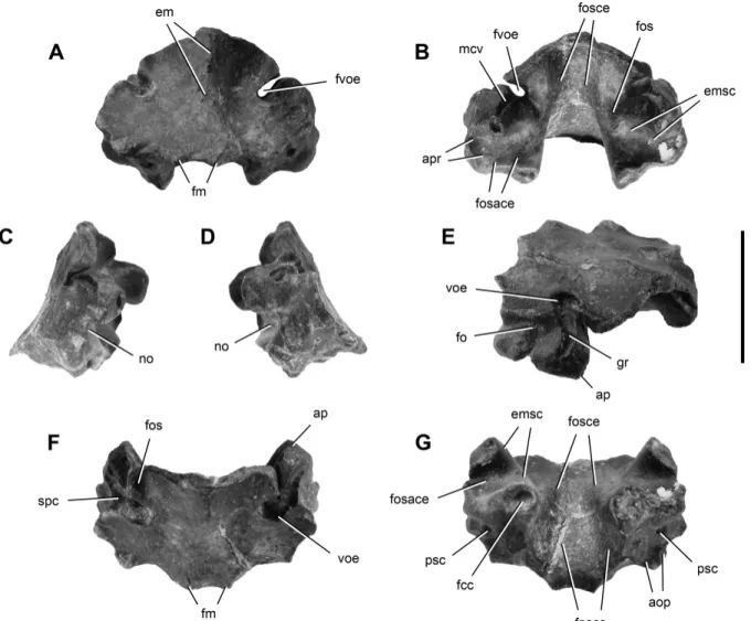
FIGURE 5. Supraoccipital of Panphagia protos (PVSJ 874) in posterior (A), anterior (B), right lateral (C), left lateral (D), posterodorsolateral (E), dorsal (F), and ventral (G) views. Anterior is towards the top of the page in F and G. Abbreviations: aop, articular surface for the exoccipital-opisthotic; ap, articular surface for the parietal; apr, articular surface for the prootic; em, eminence; emsc, eminentia canalis semicicularis; fcc, foramen crus commune; fm, foramen magnum; fo, foramen; fos, fossa; fosace, fossa auriculae cerebelli; fosce, fossa cerebelli; fvoe, foramen for the vena occipitalis externa; gr, groove; mcv, medial cerebral vein; no, notch; psc, posterior semicircular canal; spc, sinus petrosus caudalis; voe, vena occipitalis externa. Scale bar equals 1 cm.
smoothly arched, with only a small median prominence (Figs. 5A, 6A). The lateral and posteroventral articular surfaces for the exoccipital-opisthotic are angled at nearly 90◦ in posterior view (Fig. 6A), in contrast to the straight posterodorsal border in Lesothosaurus (Thulborn, 1970:fig. 1D; Sereno, 1991:fig. 11C) and Melanorosaurus (Yates, 2007:figs. 4A, B, 8). The lateral margins of the supraoccipital are gently everted in posterior view (Fig. 5C–E, G).
A groove is present in the notch of the vena occipitalis externa (Fig. 5E), which turns sharply in the ventromedial region of the fossa, as has been described for Massospondylus (Gow, 1990). Both extremities of the groove are anterolaterally oriented and converge posteromedially in the deepest part of the notch, where they form the aforementioned sharp turn. The lateral-most part of the groove (Fig. 5C, E) may represent the homologue of the avian sinus petrosus caudalis, which also left a mark in the parietal. This part of the groove is exposed dorsally on the ventral aspect of the notch (Figs. 5F, 6A). At its lateral extremity, the groove bifurcates on the left side (Fig. 5D) but remains single on the right
side (Fig. 5C, E). The medial portion of the groove may have accommodated the trigeminal-anterior semicircular vein system (Seldmayr, 2002), which corresponds with various veins in birds (vena nervi trigemini, sinus petrosus anterioris, vena semicircu-laris anterioris; Baumel, 1993) and the vena cerebralis media in non-avian reptiles (O’Donoghue, 1920; Romer, 1956). The sulcus of the trigeminal-anterior semicircular vein system continues on the intracranial surface of the supraoccipital in Panphagia, where it extends across the eminentia canalis semicircularis from the notch of the vena occipitalis externa towards the prootic (Figs. 5B, 6C). In posterodorsal and dorsal views, this portion of the groove is almost completely hidden by the parts of the supraoccipital immediately dorsal to the notch (Figs. 5F, 6A), as in Massospondylus (Gow, 1990:fig. 6A). Two conspicuous foramina open near the ‘turn’ of the groove within the pocket, the first located near the union with the lateral channel and the second located near the union with the intracranial channel (Fig. 5E).
The fossa cerebelli is located on the intracranial surface of the supraoccipital and housed the sinus occipitalis and cerebellum
FIGURE 6. Supraoccipital of Panphagia protos (PVSJ 874) in posterodorsal (A), anteroventral (B), anterodorsal (C), and posteroventral (D) views. Anterior is towards the top of the page in A and B. Abbreviations: aop, articular surface for the exoccipital-opisthotic; ap, articular sur-face for the parietal; apr, articular sursur-face for the prootic; ascf, anterior semicircular channel fora-men; ccf, crus commune forafora-men; em, eminence; fm, foramen magnum; fos, fossa; fosace, fossa au-riculae cerebelli; fosce, fossa cerebelli; mcv, me-dial cerebral vein; no, notch; psc, posterior semi-circular canal; voe, vena occipitalis externa. Scale bar equals 1 cm.
(Figs. 5B, G, 6B). A small, dorsoventrally elongate fossa ex-tends ventromedially from the notch to the dorsal edge of the fossa auriculae cerebelli (Figs. 5B, 6B). The supraoccipital por-tion of the fossa auriculae cerebelli is deep, surrounded by the eminentia canalis semicircularis, an anterolaterally oriented con-vexity (Figs. 5B, 6B). The posterodorsal portion of the canalis semicircularis anterioris and the crus osseum commune are en-closed in this swelling (Evans and Martin, 1993).
The sutural articulation for the exoccipital-opisthotic is com-posed by two rugose surfaces, one facing ventrolaterally (Figs. 5A, C, D, G, 6D) and the other laterally (Fig. 5C, D). The ventrolat-eral surface is approximately square in shape and presents on its lateral region the foramen for the canalis semicircularis caudalis (Fig. 6D). The lateral surface is subrectangular with a dorsoven-trally extended fossa (Fig. 5D, E). This surface and its fossa are
anterodorsally limited by a sulcus housing the sinus petrosus cau-dalis (Fig. 5C, D). The sulcus, which is located on a prominence on the lateral surface of the supraoccipital, lies adjacent to an articu-lar surface for the parietal.
Another ‘C’-shaped articular surface faces anteroventrally and exposes in cross-section the eminentia canalis semicircularis (Fig. 6B). The ventral extremity bears the foramen for the crus osseum commune (Fig. 6B) and a notch (Figs. 5C, D, 6B), which probably formed the dorsal part of the auricular foramen (Seldmayr, 2002). The lack of fit between the ‘C’-shaped articular surface and the prootic suggests that there might have been intervening cartilage. Alternatively, the articulation may have accommodated both the prootic (posteriorly) and exoccipital-opisthotic, as in other non-avian dinosaurs (e.g., Hypsilophodon; Galton, 1974:fig. 9B, C). The foramen for the crus osseum
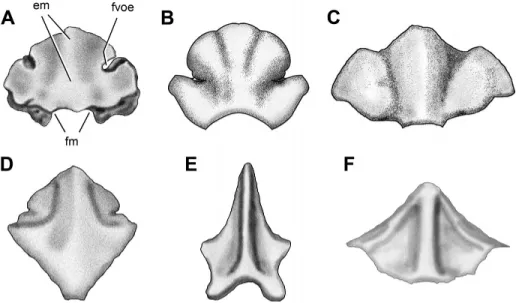
FIGURE 7. Posterior view of the supraoc-cipital of the basal dinosaurs Panphagia (A),
Adeopapposaurus (B), Thecodontosaurus
(YPM 2192) (C), Melanorosaurus (D),
Het-erodontosaurus (E), and Herrerasaurus (F).
Modified from Mart´ınez, 2009 (B); Benton et al., 2000 (C); Yates, 2007 (D); Norman et al., 2011 (E); and Sereno and Novas, 1994 (F). Abbreviations: em, eminence; fm, foramen magnum; fvoe, foramen for the vena occipitalis externa. The drawings are not in the same scale.
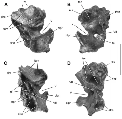
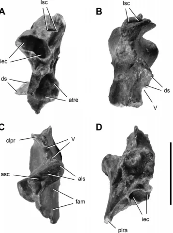
FIGURE 8. Right prootic of Panphagia protos (PVSJ 874) in lateral (A), medial (B), postero-lateral (C), and posterior (D) views. Abbrevia-tions: atre, anterior tympanic recess; clpr, clinoid process; crpr, crista prootica; fac, fossa auriculae cerebelli; fai, fossa acustica interna; fam, fossa for the adductor mandibulae; gr, groove; iec, inner ear cavity; no, notch; plra, posterolateral ramus; sce, semicircular canal eminence; stgr, stapedial groove; V, VII, openings for cranial nerves V and VII. Scale bar equals 1 cm.
commune is slightly larger than the foramina for the canales semicirculares ossei anterioris and caudalis.
Comparing the proportionately broad supraoccipital of Pan-phagia with that in other basal dinosaurs, it is similar to that in most basal sauropodomorphs, such as Efraasia, An-chisaurus, Adeopapposaurus (Fig. 7B), and Thecodontosaurus (Fig. 7C). The supraoccipital has subequal proportions in Sile-saurus (Dzik, 2003) and most basal dinosaurs such as Her-rerasaurus (Fig. 7F), Lesothosaurus (Thulborn, 1970; Knoll, 2002a), and Melanorosaurus (Fig. 7D). The supraoccipital appears to be deeper than broad in Heterodontosaurus (Fig. 7E). A shallow median nuchal eminence (Figs. 5A, 6A, 7A) is similar to that in other basal sauropodomorphs, such as the The-codontosaurus (Fig. 7C), Eoraptor (PVSJ 512), and Adeopap-posaurus (Fig. 7B). The nuchal eminence is more prominent in Melanorosaurus (Fig. 7D) and other sauropodomorphs (Pla-teosaurus, Galton, 1984:fig. 4D; Massospondylus, Hini´c, 2002). In the basal theropods Herrerasaurus (Fig. 7F), Megapnosaurus kayentakatae (Tykoski, 1998:fig. 9A, B), and Majungasaurus (Sampson and Witmer, 2007:fig. 14G), in contrast, the eminence is very prominent. The narrow contribution of the supraoccipital to the foramen magnum is also present in Herrerasaurus (Sereno and Novas, 1994:figs. 1G, 7C, 8C; Fig. 7F) and Melanorosaurus (Yates, 2007:fig. 8), in contrast to the slightly broader condi-tion in Lesothosaurus (Sereno, 1991:figs. 11C, 12C) and the much broader condition present in Adeopapposaurus (Fig. 7B) and Heterodontosaurus (Norman et al., 2011:fig. 14). The fora-men for the crus osseum commune is slightly larger than the foramina for the canales semicirculares ossei anterioris and
cau-dalis is present in a juvenile Massospondylus specimen (Gow, 1990:fig. 6A). This condition contrasts with the wide vestibu-lum present in the supraoccipital of Pantydraco (Galton and Kermack, 2010:fig. 8A), Hypsilophodon (Galton, 1974:fig. 9B, C), and the basal archosauromorph Prolacerta (Evans, 1986). The large fossa cerebelli (= floccular recess) of Panphagia, which con-tinues in the prootic (Figs. 5B, G, 6B), is absent in most sauropods.
Prootic—We describe the prootic, which is preserved on the
right side, with its long axis positioned at 45◦ to the horizon-tal (Figs. 8, 9). The prootic has two fossae dorsal and ventral to a notch and groove for cranial nerve V visible in lateral view (Fig. 5A). The lateral surface of the dorsal region is concave and ventrally delimited by a thick and curved ridge. In other dinosaurs, this fossa has been interpreted either as the recessus tympanicus dorsalis (Baumel and Witmer, 1993; Witmer, 1997) or a surface for attachment of the M. adductor mandibularis externus profun-dus (Vanden Berge and Zweers, 1993; Holliday, 2009).
The undulating dorsal margin of the prootic, which contacted the parietal, has two convexities separated by three concavities. The posterior concavity (Fig. 8A) may represent the ventral bor-der of the opening for the vena parietalis, as suggested in Pla-teosaurus (Huene, 1926:pl. 1, fig. 10; Galton and Kermack, 2010), or a pneumatic opening, as suggested in Megapnosaurus kayen-takatae (Tykoski, 1998:fig. 10B). A pair of grooves converge pos-teroventrally just above the notch for cranial nerve V (Fig. 8A). The anteroventral one likely represents the path of the vena nervi trigemini, as in Pantydraco (Galton and Kermack, 2010). In Mas-sospondylus (Gow, 1990:fig. 6A, B) and Adeopapposaurus (PVSJ 568), the groove leads to a notch on the anterior border.
teroventrally to a subrectangular prominence and adjacent trough (Fig. 8A). This depressed area probably represents the path of the most proximal section of the mandibularis branch of cranial nerve V. The posterior border of this surface forms the anterior border of the external foramen for cranial nerve VII (Fig. 8A). Ventral to the trigeminal notch and groove is a smooth, trapezoidal surface for origin of the M. protractor pterygoidei et quadrati (Vanden Berge and Zweers, 1993) (Fig. 8A).
The external opening of cranial nerve VII is located on the crista prootica, slightly ventral to the opening for cranial nerve V (Fig. 8A). The oval foramen is bounded by sharp ridges with protuberances partially separating the foramen into posterodor-sal and anteroventral portions (Fig. 8A, C). The smaller, pos-terodorsal portion would have lodged the nervus hyomandibularis (Dubbeldam, 1993), a branch of cranial nerve VII, the anteroven-tral portion of which may represent the exit of the nervus palatinus (Fig. 8A). The limiting ridges continue posterodorsally beyond the foramen, bounding a narrow sulcus that would have housed the most proximal portion of the nervus hyomandibularis.
A large anterior tympanic recess is located posteromedially to, and overlapped by, an attachment surface for the M. protractor pterygoidei et quadrati and ventrally to the foramen for cranial nerve VII. This fossa is broad, deep, and approximately circular in posterior view (Fig. 8D). Posterior to the foramen for cranial nerve VII and the crista prootica, the lateral surface of the prootic is reduced to a narrow area (Fig. 8A). This surface corresponds to a wall laterally overlapping the anterior border of the fenestra vestibuli (Baumel and Witmer, 1993; fenestra ovalis, sensu Romer, 1956). From the dorsal region of this area, a short sulcus extends posteriorly just medial and approximately parallel to the narrower sulcus for the nervus hyomandibularis (Fig. 8C).
In medial view, the dorsal part of the prootic bears the an-teroventral portion of the fossa auriculae cerebelli (Fig. 8B). This fossa is anterodorsally limited by the eminentia canalis semicir-cularis (Fig. 8B) and ventrally limited by the medial wall of the auditory bulla (Oelrich, 1956). The auditory bulla is a bony cap-sule surrounding the vestibular recess and is notably inflated to-wards the cavum cranii (Fig. 8B), as in Pantydraco (Galton and Kermack, 2010:fig. 8F). Immediately anteroventral to the audi-tory bulla, there is a wide fossa acustica interna (Baumel and Wit-mer, 1993) into which open two foramina, one immediately dor-sal to the other (Fig. 8B). The smaller, dordor-sal foramen is located on the ventral border of the auditory bulla and provided passage to the anterior ramus (Dubbeldam, 1993) of the nervus vestibulo-cochlearis (cranial nerve VIII), which opens into the anterior am-pullar recess (Oelrich, 1956). The larger, ventral foramen provided passage to the undivided root of the nervus facialis (cranial nerve VII). Anterior to the notch for the nervus trigeminus (cranial nerve V) on the medial aspect of the clinoid process, the prootic presents a medially directed, fan-shaped lamina, which forms the dorsolateral part of the dorsum sellae (Baumel and Witmer, 1993) (Fig. 8B, D). The dorsomedial edge of this transverse lamina is sharp and convex (Fig. 8D) and likely did not contact its paired opposite except at its ventral extremity. The ventral border has a ventromedially directed notch (Fig. 8D) that forms the dorsolat-eral portion of the nervus abducens (cranial nerve VI). Anterior
FIGURE 9. Right prootic of Panphagia protos (PVSJ 874) in posteroven-tral (A), anterior (B), dorsal (C), and venposteroven-tral (D) views. Abbreviations: als, articular surface for the laterosphenoid; asc, anterior semicircular canal; atre, anterior tympanic recess; clpr, clinoid process; ds, dorsum sellae; fam, fossa for the adductor mandibulae; iec, inner ear cavity; lsc, lateral semi-circular canal; plra, posterolateral ramus; V, opening for cranial nerve V. Scale bar equals 1 cm.
to this lamina, the medial surface of the prootic faces the fossa hypophyseos (Fig. 8B).
In dorsal view, the anterodorsal surface of the prootic is ap-proximately rectangular (Fig. 9C). This anterolaterally elongate surface has a foramen for the canalis semicircularis anterioris and several narrow sulci (Fig. 9C). It articulates with the posterome-dial portion of the supraoccipital and anterolateral portion of the laterosphenoid.
The posteromedial surface of the prootic has a broad, rugose articular surface for the exoccipital-opisthotic dorsolateral to the vestibule (Fig. 8B, D). It houses the foramen for the canalis semi-circularis lateralis (Evans and Martin, 1993) (Fig. 8D). The walls of the anterior portion of the canalis semicircularis lateralis, there-fore, are completely surrounded by the prootic. In Pantydraco, in contrast, the prootic does not form the medial wall of this canal (fide Galton and Kermack, 2010). In Panphagia, this foramen is much larger than the preserved foramina for the canalis semicir-cularis anterioris and crus osseum commune. Posterolateral to this foramen, striae are present that parallel the long axis of the pro-cess (Fig. 8B, D).
The anteroventral surface of the prootic (Fig. 9D) has an ar-ticular surface for the anterior portion of the parabasisphenoid (sensu Gower, 2002). The smooth, concave articular surface is
subtriangular and has a straight lateral border. It forms the ven-tral edge of the attachment area for the M. protractor pterygoidei et quadrati. The strongly concave posteroventral border forms the ventral edge of the recessus tympanicus anterioris. The medial border forms the ventral edge of the endocranial surface of the prootic, which is slightly convex in ventral view (Fig. 9D). The pos-teromedial corner of the surface is anteroposteriorly elongated, medial to the anterior tympanic recess. The posterior surface of the prootic bears a rugose sutural area, most likely for the paraba-sisphenoid (Fig. 8D). In lateral view, the subquadrate sutural sur-face is concave and is marked by a small, conical fossa (Fig. 8D).
A spacious inner ear cavity opens between the posteriorly facing articular surfaces for the exoccipital-opisthotic and parabasisphenoid (Fig. 8D). The cavity is subconical, narrowing anteriorly (Fig. 8D). Within the cavity, a slight constriction differentiates the vestibulum posteriorly from the smaller ampulla ossea lateralis anteriorly (sensu Evans and Martin, 1993). In posterior view, the vestibular recess is subelliptical, with its long axis oriented dorsomedially (Fig. 8D). On the ventromedial surface of the ampulla ossea lateralis, a foramen is present for the anterior ramus of the nervus vestibulocochlearis (cranial nerve VIII) or perhaps only the nervus ampullaris lateralis (Dubbeldam, 1993). The foramen for the canalis semicircularis lateralis opens dorsolaterally on the ampulla ossea lateralis. Anterior view of the ampulla ossea anterioris is obscured by matrix. A small notch for the ramus caudalis (sensu Dubbeldam, 1993) of the nervus vestibulocochlearis (cranial nerve VIII) is present on the poste-rior border of the vestibular recess (Fig. 8B), as in Plateosaurus (Galton, 1985:fig. 7G). A notch on the posterior border of the lateral wall of the vestibular recess forms the anterior margin of the fenestra vestibuli (Fig. 8B, C). A posteroventrally directed depression, the recessus columellae (sensu Baumel and Witmer, 1993) (Figs. 8C, D, 9D), is present dorsolateral to the fenestra vestibule and continues posterolaterally to join the stapedial groove (sensu Sampson and Witmer, 2007) on the ventral aspect of the posterolateral process (Figs. 8D, 9D). The stapedial groove likely continued distally onto the paroccipital process of the opisthotic.
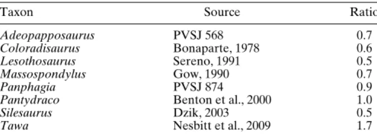
Comparing the prootic of Panphagia with that in other basal dinosaurs, the surface for the origin of the M. protractor ptery-goidei et quadrati, which corresponds to the lateral surface of the prootic ventral to the notch for the cranial nerve V, is antero-posteriorly wider than dorsoventrally high. The ratio between the length of this area along the long axis of the prootic to the length along its orthogonal axis in the sagittal plane is greater than in Silesaurus, Lesothosaurus, and more derived sauropodomorphs, but less than in the basal theropod Tawa (Table 1). The op-posing borders of the notch for cranial nerve V are nearly par-allel in lateral and medial views (Fig. 8A, B) in Panphagia, which is similar to that in Massospondylus (Gow, 1990:fig. 6A) and Plateosaurus (Galton, 1984:fig. 4B). In other non-avian
di-TABLE 1. Ratio of the length of surface for the origin of the M.
pro-tractor pterygoidei et quadrati along the long axis of the prootic and along its orthogonal axis in the sagittal plane in several dinosauriforms.
Taxon Source Ratio
Adeopapposaurus PVSJ 568 0.7
Coloradisaurus Bonaparte, 1978 0.6
Lesothosaurus Sereno, 1991 0.5
Massospondylus Gow, 1990 0.7
Panphagia PVSJ 874 0.9
Pantydraco Benton et al., 2000 1.0
Silesaurus Dzik, 2003 0.5
Tawa Nesbitt et al., 2009 1.7
nosaurs and immediate outgroups, the borders of the notch either converge anterodorsally (Silesaurus, Dzik, 2003:fig. 7A, B; Coelophysis, Raath, 1985:fig. 1A; Melanorosaurus, Yates, 2007:fig. 11A, B) or diverge (Pantydraco, Galton and Kermack, 2010:fig. 8D, E, G; Yates, 2003:fig. 7A, B). The external opening of cranial nerve VII on the crista prootica is situated somewhat ventral to the level of cranial nerve V (Fig. 8A), a topology found in many basal sauropodomorphs (e.g., Adeopap-posaurus, Mart´ınez, 2009:fig. 10C; Pantydraco, Galton and Kermack, 2010:fig. 8E; Massospondylus, Gow, 1990:fig. 6A, B) and basal theropods (e.g., Coelophysis, Raath, 1985:fig. 1A). The an-terior tympanic recess of Panphagia (Fig. 8D) is similar to that in neotheropods (Witmer, 1997) and larger than that in some other basal sauropodomorphs (e.g., Adeopapposaurus; PVSJ 568). Sauropods appear to lack the anterior tympanic recess altogether.
DISCUSSION
Specimen Maturity
The subadult status of the holotype of Panphagia is inferred from the lack of fusion of the preserved braincase elements, open neurocentral sutures in presacral vertebrae, and the open scapulocoracoid suture (Brochu, 1995; Irmis, 2007; Mart´ınez and Alcober, 2009). Some features of the preserved braincase ele-ments, in addition, are consistent with subadult status, such as the absence of a precise articular match between the supraoccipital and prootic. The edges of these bones may have yet to fully ossify near their sutural junction (DeBeer, 1937). The proportionately large size of the notches in the supraoccipital for the vena occip-italis externa resembles the condition in subadult Massospondy-lus (Gow, 1990; for adult form, see Sues et al., 2004). The medial edge of the parietals in Panphagia suggests that there was a me-dian foramen, which has been cited as an indication of immaturity in ornithischians (Knoll, 2002b). Pol and Powell (2007) and oth-ers have noted, however, that many adult sauropodomorphs retain this foramen.
Soft-Tissue Considerations
The fossa on the lateral surface of the prootic has been alter-natively interpreted either as a muscular attachment site (Raath, 1985; Holliday, 2009:fig. 5A–C) or a pneumatic feature related to the recessus tympanicus dorsalis (Witmer, 1997). Rauhut (2004) considered both interpretations equally plausible. The shape and position of the fossa resembles the attachment site for the M. ad-ductor mandibularis externus profundus in Ctenosaura (Oelrich, 1956). This interpretation may be more reasonable, given the gen-eral absence of prominent pneumatic features (Smith et al., 2007). On the other hand, the M. adductor mandibularis externus pro-fundus does not attach to the prootic in extant archosaurs (Holli-day and Witmer, 2007), which also have extensive pneumatic fea-tures in this region. In the light of this bracket of extant outgroups (Witmer, 1995b), we regard the pneumatic hypothesis as the more likely.
Comparisons
The braincase is the source of little character evidence at basal nodes within Dinosauria (e.g., frontal contribution to the supratemporal fenestra [Gauthier, 1986]; depth of the occipital wing of the parietal [Wilson and Sereno, 1998]; position of foram-ina for mid-cerebral vein on occiput [Yates, 2003]). The paucity of characters reflects the limited information available on the brain-case in many basal dinosaurs. When preserved, the brainbrain-case is only rarely exposed, and isolated braincase elements that offer a
The nuchal crest is low in Panphagia, as in Thecodontosaurus (YPM 2192), Adeopapposaurus, and Riojasaurus. Given the larger body size of some of these taxa, this character may be size inde-pendent. A more prominent crest characterizes most theropods, ornithischians, and the basal dinosauromorph Silesaurus.
The deeply incised notches in the supraoccipital for the vena occipitalis externa resemble the condition in many basal sauropodomorphs (e.g., Plateosaurus, Massospondylus, Adeopap-posaurus, Melanorosaurus), but in contrast to the condition in theropods (e.g., Herrerasaurus, Megapnosaurus kayen-takatae, Coelophysis) and ornithischians (e.g., Lesothosaurus, Heterodontosaurus). A deeply notched supraoccipital as in sauropodomorphs may be apomorphic, although deep notches are also present in the dinosaurian outgroup Silesaurus (Dzik, 2003).
The attachment area on the prootic for the M. protractor ptery-goidei et quadrati has an elongate shape in Panphagia, simi-lar to that in Thecodontosaurus (YPM 2192). The shape of this feature, however, has a complex distribution among dinosaurs. In Silesaurus, the basal ornithischian Lesothosaurus, and many sauropodomorphs (e.g., Adeopapposaurus, Coloradisaurus, Rio-jasaurus, Massospondylus), the attachment area is short, but in the basal theropod Tawa it is slightly longer than in Panphagia.
Plesiomorphic Characters within Sauropodomorpha—The
frontal of Panphagia has a characteristic shape: it is elongate (length more than twice width at the level of the posterolateral process), with its anterior tip located closer to the midline than to the lateral border of the bone (Fig. 2A). Most basal dinosaurs (e.g., Lesothosaurus, Herrerasaurus, Coelophysis) including basal-most sauropodomorphs (e.g., Eoraptor, Pantydraco, Pampadromaeus) have a frontal of this form. In more derived sauropodomorphs, in contrast, the frontal is broader than long and the anterior tip is closer to the lateral border of the bone (e.g., Plateosaurus, Massospondylus, Adeopapposaurus, Melanorosaurus).
The large anterior tympanic and floccular recesses in Panpha-gia (Figs. 5B, G, 6B, 8D) are similar to those in neotheropods (Witmer, 1997). In at least some basal sauropodomorphs, such as Adeopapposaurus, these recesses are smaller, and they ap-pear to be absent in sauropods. Both of these features sug-gest that Eoraptor and a few close relatives may constitute stem sauropodomorphs at or near the base of the clade.
CONCLUSIONS
The disarticulated braincase elements of a subadult speci-men of Panphagia protos provide new information on the mor-phology of the braincase in an early sauropodomorph dinosaur. A few features of the braincase link Panphagia with other sauropodomorphs, and a few other features suggest that Panpha-gia may be more primitive than basal sauropodomorphs such as Plateosaurus or Massospondylus. The former includes a propor-tionately elongate frontal, and the latter includes the presence of anterior tympanic and floccular recesses. The significance of these features will be easier to assess when additional information on the braincase is available for other basal dinosaurs.
tance with the figures.
LITERATURE CITED
Alcober, O. A., and R. N. Martinez. 2010. A new herrerasaurid (Dinosauria, Saurischia) from the Upper Triassic Ischigualasto For-mation of northwestern Argentina. ZooKeys 63:55–81.
Barrett, P. M., P. Upchurch, and X.-L. Wang. 2005. Cranial osteology of
Lufengosaurus huenei Young (Dinosauria: Prosauropoda) from the
Lower Jurassic of Yunnan, People’s Republic of China. Journal of Vertebrate Paleontology 25:806–822.
Baumel, J. J. 1993. Systema cardiovasculare; pp. 407–475 in J. J. Baumel, A. S. King, J. E. Breazile, H. E. Evans, and J. C. Vanden Berge (eds.), Handbook of Avian Anatomy: Nomina Anatomica Avium, second edition. Publications of the Nutall Ornithological Club 23, Cambridge, Massachusetts.
Baumel, J. J., and L. M. Witmer. 1993. Osteologia. pp. 45–132 in J. J. Baumel, A. S. King, J. E. Breazile, H. E. Evans, and J. C. Van-den Berge (eds.), Handbook of Avian Anatomy: Nomina Anatom-ica Avium, second edition. PublAnatom-ications of the Nutall OrnithologAnatom-ical Club 23, Cambridge, Massachusetts.
Baumel, J. J., A. S. King, J. E. Breazile, H. E. Evans, and J. C. Vanden Berge. 1993. Handbook of Avian Anatomy: Nomina Anatomica Avium, second edition. Publications of the Nutall Or-nithological Club 23, Cambridge, Massachusetts, 779 pp.
Benton, M. J., L. Juul, G. W. Storrs, and P. M. Galton. 2000. Anatomy and systematics of the prosauropod dinosaur Thecodontosaurus antiquus from the Upper Triassic of southwest England. Journal of Vertebrate Paleontology 20:77–10.
Bittencourt, J. S., and A. W. A. Kellner. 2009. The anatomy and phyloge-netic position of the Triassic dinosaur Staurikosaurus pricei Colbert, 1970. Zootaxa 2079:1–56.
Bonaparte, J. F. 1978. Coloradia brevis n. g. et n. sp. (Saurischia -Prosauropoda), dinosaurio Plateosauridae de la Formaci ´on Los Col-orados, Tri ´asico superior de La Rioja, Argentina. Ameghiniana 15:327–332.
Bonaparte, J. F., and J. A. Pumares. 1995. Notas sobre el primer cr ´aneo de Riojasaurus incertus (Dinosauria, Prosaur ´opoda, Melanosauridae) del Tri ´asico superior de La Rioja. Ameghiniana 32:341–349. Brochu, C. A. 1995. Heterochrony in the crocodylian scapulocoracoid.
Journal of Herpetology 29:464–468.
Bruner, H. L. 1907. On the cephalic veins and sinuses of reptiles, with a description of a mechanism for raising the venous blood-pressure in the head. American Journal of Anatomy 7:1–117.
Cabreira, S. F., C. L. Schultz, J. S. Bittencourt, M. B. Soares, D. C. Fortier, L. R. Silva, and M. C. Langer. 2011. New stem-sauropodomorph (Di-nosauria, Saurischia) from the Triassic of Brazil. Naturwissenschaften 938:1035–1040.
Colbert, E. H. 1970. A saurischian dinosaur from the Triassic of Brazil. American Museum Novitates 2405:1–60.
De Beer, G. 1937. The Development of the Vertebrate Skull. Clarendon Press, Oxford, U.K., 552 pp.
Dom´ınguez Alonso, P., A. C. Milner, R. A. Ketcham, M. J. Cookson, and T. B. Rowe. 2004. The avian nature of the brain and inner ear of
Archaeopteryx. Nature 430:666–669.
Dubbeldam, J. B. 1993. Systema nervosum periphericum; pp. 555–584 in J. J. Baumel, A. S. King, J. E. Breazile, H. E. Evans, and J. C. Van-den Berge (eds.), Handbook of Avian Anatomy: Nomina Anatom-ica Avium, second edition. PublAnatom-ications of the Nutall OrnithologAnatom-ical Club 23, Cambridge, Massachusetts.
Dzik, J. 2003. A beaked herbivorous archosaur with dinosaur affinities from the early Late Triassic of Poland. Journal of Vertebrate Pale-ontology 23:556–574.
Evans, S. E. 1986. The braincase of Prolacerta broomi (Reptilia, Trias-sic). Neues Jahrbuch f ¨ur Geologie und Pal ¨aontologie, Abhandlungen 173:181–200.
Evans, H. E., and G. R. Martin. 1993. Organa sensuum [Organa sensoria]; pp. 585–611 in J. J. Baumel, A. S. King, J. E. Breazile, H. E. Evans, and J. C. Vanden Berge (eds.), Handbook of Avian Anatomy: Nom-ina Anatomica Avium, second edition. Publications of the Nutall Or-nithological Club 23, Cambridge, Massachusetts.
Fedak, T. J., and P. M. Galton. 2007. New information on the braincase and skull of Anchisaurus polyzelus (Lower Jurassic, Connecticut, USA; Saurischia: Sauropodomorpha): implications for sauropodomorph systematics. Special Papers in Palaeontology 77:245–260.
Galton, P. M. 1974. The ornithischian dinosaur Hypsilophodon from the Wealden of the Isle of Wight. Bulletin of the British Museum (Natu-ral History), Geology 25:1–152.
Galton, P. M. 1984. Cranial anatomy of the prosauropod dinosaur
Pla-teosaurus from the Knollenmergel (Middle Keuper, Upper
Trias-sic) of Germany. I. Two complete skulls from Trossingen W ¨urtt. with comments on the diet. Geologica et Palaeontologica 18:139– 171.
Galton, P. M. 1985. Cranial anatomy of the prosauropod dinosaur
Pla-teosaurus from the Knollenmergel (Middle Keuper, Upper
Trias-sic) of Germany. II. All the cranial material and details of soft-part anatomy. Geologica et Palaeontologica 19:119–159.
Galton, P. M. 1989. Cranial and endocranial casts from ornithopod di-nosaurs of the families Dryosauridae and Hypsilophodontidae (Rep-tilia: Ornithischia). Geologica et Palaeontologica 23:217–239. Galton, P. M., and R. T. Bakker. 1985. The cranial anatomy of the
prosauropod dinosaur ‘Efraasia diagnostica’, a juvenile individual of Sellosaurus gracilis from the Upper Triassic of Nordw ¨urtemberg, West Germany. Stuttgarter Beitr ¨age zur Naturkunde, Serie B (Ge-ologie und Pal ¨aont(Ge-ologie) 117:1–15.
Galton, P. M., and D. Kermack. 2010. The anatomy of Pantydraco caducus, a very basal sauropodomorph dinosaur from the Rhaetian (Upper Triassic) of South Wales, UK. Revue de Pal ´eobiologie 29:341–404. Gauthier, J. A. 1986. Saurischian monophyly and the origin of birds.
Mem-oirs of the California Academy of Sciences 8:1–55.
Gow, C. E. 1990. Morphology and growth of the Massospondylus brain-case (Dinosauria, Prosauropoda). Palaeontologia africana 27:59–75. Gower, D. J. 2002. Braincase evolution in suchian archosaurs (Reptilia:
Diapsida): evidence from the rauisuchian Batrachotomus
kupferzel-lensis. Zoological Journal of the Linnean Society 136:49–76.
Hini´c, S. 2002. Cranial osteology of Massospondylus carinatus Owen, 1854 and its implications for prosauropod phylogeny. M.S. thesis, Univer-sity of Toronto, Toronto, Ontario, Canada, 163 pp.
Holliday, C. M. 2009. New insights into dinosaur jaw muscle anatomy. The Anatomical Record 292:1246–1265.
Holliday, C. M., and L. M. Witmer. 2007. Archosaur adductor chamber evolution: integration of musculoskeletal and topological criteria in jaw muscle homology. Journal of Morphology 268:457–484. Huene, F. von. 1926. Vollst ¨andige Osteologie eines Plateosauriden aus
dem schw ¨abischen Keuper. Geologische und Palaeontologische Ab-handlungen, Neue Folge 15:139–179.
Irmis, R. B. 2007. Axial skeleton ontogeny in the Parasuchia (Archosauria: Pseudosuchia) and its implications for ontogenetic determination in archosaurs. Journal of Vertebrate Paleontology 27:350–361. Kermack, D. 1984. New prosauropod material from South Wales.
Zoolog-ical Journal of the Linnean Society of London 82:101–117.
Knoll, F. 2002a. New skull of Lesothosaurus (Dinosauria: Ornithischia) from the Upper Elliot Formation (Lower Jurassic) of southern Africa. Geobios 35:595–603.
Knoll, F. 2002b. Nearly complete skull of Lesothosaurus (Dinosauria: Or-nithischia) from the Upper Elliot Formation (Lower Jurassic: Het-tangian) of Lesotho. Journal of Vertebrate Paleontology 22:238–243. Langer, M. C. 2003. The pelvic and hind limb anatomy of the stem-sauropodomorph Saturnalia tupiniquim (Late Triassic, Brazil). PaleoBios 23:1–30.
Langer, M. C. 2004. Basal Saurischia; pp. 25–46 in D. B. Weishampel, P. Dodson, and H. Osm ´olska (eds.), The Dinosauria, second edition. University of California Press, Berkeley, California.
Langer, M. C., M. A. G. Franca, and S. Gabriel. 2007. The pectoral gir-dle and forelimb anatomy of the stem-sauropodomorph Saturnalia
tupiniquim (Upper Triassic, Brazil). Special Papers in Palaeontology
77:113–137.
Langer, M. C., F. Abdala, M. Richter, and M. J. Benton. 1999. A sauropodomorph dinosaur from the Upper Triassic (Carnian) of southern Brazil. Comptes Rendus de l’Academie des Sciences, Paris 329:511–517.
Long, R. A., and P. A. Murry. 1995. Late Triassic (Carnian and Norian) tetrapods from the southwestern United States. Bulletin of the New Mexico Museum of Natural History and Science 4:1–254.
Mart´ınez, R. N. 2009. Adeopapposaurus mognai, gen. et sp. nov. (Di-nosauria: Sauropodomorpha), with comments on adaptations of basal Sauropodomorpha. Journal of Vertebrate Paleontology 29:142–164. Mart´ınez, R. N., and O. A. Alcober. 2009. A basal sauropodomorph
(Dinosauria: Saurischia) from the Ischigualasto Formation (Triassic, Carnian) and the early evolution of Sauropodomorpha. PLoS ONE 4:e4397. doi: 4310.1371/journal.pone.0004397.
Mart´ınez, R. N., P. C. Sereno, O. A. Alcober, C. E. Colombi, P. R. Renne, I. P. Monta ˜nez, and B. S. Currie. 2011. A basal dinosaur from the dawn of the dinosaur era in southwestern Pangaea. Science 331:206–210.
McClelland, B. K., 1990. Anatomy and kinesis of the Allosaurus skull. M.S. thesis, Texas Tech University, Lubbock, Texas, 122 pp.
Nesbitt, S. J., N. D. Smith, R. B. Irmis, A. H. Turner, A. Downs, and M. A. Norell. 2009. A complete skeleton of a Late Triassic saurischian and the early evolution of dinosaurs. Science 326:1530–1533.
Norman, D. B., A. W. Crompton, R. J. Butler, L. B. Porro, and A. L. Charig. 2011. The Lower Jurassic ornithischian dinosaur
Heterodon-tosaurus tucki Crompton & Charig, 1962: cranial anatomy, functional
morphology, taxonomy, and relationships. Zoological Journal of the Linnean Society 163:1–182.
O’Donoghue, C. H. 1920. The blood vascular system of the tuatara,
Sphen-odon punctatus. Philosophical Transactions of the Royal Society of
London, Series B 210:175–252.
Oelrich, T. M. 1956. The anatomy of the head of Ctenosaura pectinata (Iguanidae). Miscellaneous Publications of the Museum of Zoology, University of Michigan 94:1–122.
Pearson, R. 1972. The Avian Brain. Academic Press, London and New York, 658 pp.
Pol, D., and J. E. Powell. 2007. Skull anatomy of Mussaurus patagonicus (Dinosauria: Sauropodomorpha) from the Late Triassic of Patagonia. Historical Biology 19:125–144.
Raath, M. A. 1977. The anatomy of the Triassic theropod Syntarsus
rhode-siensis (Saurischia: Podokesauridae) and a consideration of its
biol-ogy. Ph.D. dissertation, Rhodes University, Salisbury, Rhodesia, 233 pp.
Raath, M. A. 1985. The theropod Syntarsus and its bearings on the origin of birds; pp. 219–227 in M. K. Hecht, J. H. Ostrom, G. Viohl, and P. Wellnhofer (eds.), The Beginning of Birds: Proceedings of the In-ternational Archaeopteryx Conference, Eichstatt. Freunde des Jura-Museums, Eichstatt, 382 pp.
Rauhut, O. W. M. 2004. Braincase structure of the Middle Juras-sic theropod Piatnitzkysaurus. Canadian Journal of Earth Sciences 41:1109–1122.
Romer, A. S. 1956. Osteology of the Reptiles. University of Chicago Press, Chicago, Illinois, 772 pp.
Rowe, T. R. 1989. A new species of the theropod dinosaur Syntarsus from the Early Jurassic Kayenta Formation of Arizona. Journal of Verte-brate Paleontology 9:125–136.
Rowe, T. B., H.-D. Sues, and R. R. Reisz. 2010. Dispersal and diver-sity in the earliest North American sauropodomorph dinosaurs, with a description of a new taxon. Proceedings of the Royal Society B 278:1044–1053.
Sampson, S. D., and L. M. Witmer. 2007. Craniofacial anatomy of
Ma-jungasaurus crenatissimus (Theropoda: Abelisauridae) from the Late
Cretaceous of Madagascar. Memoir of the Society of Vertebrate Paleontology 8:32–102.
Sedlmayr, J. C. 2002. Anatomy, evolution, and functional significance of cephalic vasculature in Archosauria. Ph.D. dissertation, Ohio Uni-versity, Athens, Ohio, 398 pp.
Sereno, P. C. 1991. Lesothosaurus, “fabrosaurids,” and the early evolution of the Ornithischia. Journal of Vertebrate Paleontology 11:168–197.
Eoraptor lunensis (Dinosauria, Sauropodomorpha). pp. 83–179 in P. Sereno (ed.), Basal sauropodomorphs and the vertebrate fossil record of the Ischigualasto Formation (Late Triassic: Carnian–Norian) of Argentina. Society of Vertebrate Paleontology Memoir 12.
Shiino, K. 1914. Das Chondrocranium von Crocodylus porosus mit Ber ¨ucksichtigung der Gehirnnerven und Kopfgef ¨asse. Anatomische Hefte 50:257–381.
Smith, N. D., P. J. Makovicky, E. R. Hammer, and P. J. Currie. 2007. Os-teology of Cryolophosaurus ellioti (Dinosauria: Theropoda) from the Early Jurassic of Antarctica and implications for early theropod evo-lution. Zoological Journal of the Linnean Society 151:377–421. Sues, H.-D., R. R. Reisz, S. Hinic, and M. A. Raath. 2004. On the skull of
Massospondylus carinatus Owen, 1854 (Dinosauria:
Sauropodomor-pha) from the Elliot and Clarens formations (Lower Jurassic) of South Africa. Annals of the Carnegie Museum 73:239–257.
Thulborn, R. A. 1970. The skull of Fabrosaurus australis, a Triassic or-nithischian dinosaur. Palaeontology 13:414–432.
Tykoski, R. S. 1998. The osteology of Syntarsus kayentakatae and its impli-cations for ceratosaurid phylogeny. M.S. thesis, University of Texas at Austin, Austin, Texas, 217 pp.
Vanden Berge, J. C., and G. A. Zweers. 1993. Myologia; pp. 189–250 in J. J. Baumel, A. S. King, J. E. Breazile, H. E. Evans, and J. C. Vanden Berge (eds.), Handbook of Avian Anatomy: Nomina Anatomica Avium, second edition. Publications of the Nutall Or-nithological Club 23, Cambridge, Massachusetts.
chosaurs (birds and crocodilians), with special reference to paranasal pneumaticity and nasal conchae. Journal of Morphology 225:269– 327.
Witmer, L. M. 1997. Craniofacial air sinus systems; pp. 151–159 in P. J. Currie and K. Padian (eds.), Encyclopedia of Dinosaurs. Academic Press, New York.
Witmer, L. M., and R. C. Ridgely. 2008. The paranasal air sinuses of preda-tory and armored dinosaurs (Archosauria: Theropoda and Anky-losauria) and their contribution to cephalic structure. The Anatom-ical Record 291:1362–1388.
Yates, A. M. 2003. A new species of the primitive dinosaur
Thecodon-tosaurus (Saurischia: Sauropodomorpha) and its implications for the
systematics of early dinosaurs. Journal of Systematic Palaeontology 1:1–42.
Yates, A. M. 2004. Anchisaurus polyzelus (Hitchcock): the smallest known sauropod dinosaur and the evolution of gigantism amongst sauropodomorph dinosaurs. Postilla 230:1–58.
Yates, A. M. 2007. The first complete skull of the Triassic dinosaur
Melanorosaurus haughtoni (Sauropodomorpha: Anchisauria).
Spe-cial Papers in Palaeontology 77:9–55.
Submitted December 31, 2012; revisions received May 30, 2013; accepted June 20, 2013.
Handling editor: Jeffrey Wilson.