R E S E A R C H A R T I C L E
Open Access
A toothed turtle from the Late Jurassic of
China and the global biogeographic history
of turtles
Walter G. Joyce
1*, Márton Rabi
2,3*, James M. Clark
4and Xing Xu
5Abstract
Background: Turtles (Testudinata) are a successful lineage of vertebrates with about 350 extant species that inhabit all major oceans and landmasses with tropical to temperate climates. The rich fossil record of turtles documents the adaptation of various sub-lineages to a broad range of habitat preferences, but a synthetic biogeographic model is still lacking for the group.
Results: We herein describe a new species of fossil turtle from the Late Jurassic of Xinjiang, China, Sichuanchelys palatodentata sp. nov., that is highly unusual by plesiomorphically exhibiting palatal teeth. Phylogenetic analysis places the Late Jurassic Sichuanchelys palatodentata in a clade with the Late Cretaceous Mongolochelys efremovi outside crown group Testudines thereby establishing the prolonged presence of a previously unrecognized clade of turtles in Asia, herein named Sichuanchelyidae. In contrast to previous hypotheses, M. efremovi and Kallokibotion bajazidi are not found within Meiolaniformes, a clade that is here reinterpreted as being restricted to Gondwana. Conclusions: A revision of the global distribution of fossil and recent turtle reveals that the three primary lineages of derived, aquatic turtles, including the crown, Paracryptodira, Pan-Pleurodira, and Pan-Cryptodira can be traced back to the Middle Jurassic of Euramerica, Gondwana, and Asia, respectively, which resulted from the primary break up of Pangaea at that time. The two primary lineages of Pleurodira, Pan-Pelomedusoides and Pan-Chelidae, can similarly be traced back to the Cretaceous of northern and southern Gondwana, respectively, which were separated from one another by a large desert zone during that time. The primary divergence of crown turtles was therefore driven by vicariance to the primary freshwater aquatic habitat of these lineages. The temporally persistent lineages of basal turtles, Helochelydridae, Meiolaniformes, Sichuanchelyidae, can similarly be traced back to the Late Mesozoic of Euramerica, southern Gondwana, and Asia. Given the ambiguous phylogenetic relationships of these three lineages, it is unclear if their diversification was driven by vicariance as well, or if they display a vicariance-like pattern. The clean, primary signal apparent among early turtles is secondarily obliterated throughout the Late Cretaceous to Recent by extensive dispersal of continental turtles and by multiple invasions of marine habitats. Keywords: Testudinata, Sichuanchelyidae, Helochelydridae, Meiolaniformes, Sichuanchelys palatodentata, Jurassic, Xinjiang, China, Phylogeny, Biogeography
* Correspondence:walter.joyce@unifr.ch;iszkenderun@gmail.com 1
Department of Geosciences, University of Fribourg, Chemin de Musée 6, 1700 Fribourg, Switzerland
2Department of Geosciences, University of Tübingen, Hölderlinstrasse 12, 72074 Tübingen, Germany
Full list of author information is available at the end of the article
© The Author(s). 2016 Open Access This article is distributed under the terms of the Creative Commons Attribution 4.0 International License (http://creativecommons.org/licenses/by/4.0/), which permits unrestricted use, distribution, and reproduction in any medium, provided you give appropriate credit to the original author(s) and the source, provide a link to the Creative Commons license, and indicate if changes were made. The Creative Commons Public Domain Dedication waiver (http://creativecommons.org/publicdomain/zero/1.0/) applies to the data made available in this article, unless otherwise stated.
Background
Turtles (Testudinata), the clade arising from the first Amni-ote with a fully formed turtle shell (sensu [1]), currently in-habit all major landmasses with tropical to temperate climates [2]. The clade has an excellent, though often poorly studied fossil record that reaches back to the Late Triassic [3]. Turtles are therefore ideal model organisms to investigate global biogeographic patterns as their evolution-ary history coincides with the break-up of the supercontin-ent Pangaea and the secondary assembly of the continsupercontin-ents as seen today. The last 25 years of research using computer assisted cladistic analyses have retrieved an increasingly congruous picture of turtle evolution [4–12] but synthetic biogeographic analyses that include fossil taxa are still rare, noncomprehensive, and either failed to retrieve meaningful global patterns [6, 13] or concentrated on the primary clades of crown-cryptodires [14].
Here we present a new basal turtle, Sichuanchelys palato-dentatan. sp., from the Late Jurassic of Xinjiang Uygur Au-tonomous Region, China that is not only unusual for displaying residual palatal teeth, but also has important im-plications for the global paleobiogeography of turtles. The primary goals of this contribution are therefore to provide a comprehensive description of the new taxon and to re-evaluate the global biogeographic history of the group. The surprising result of this study is that the early evolution of turtles was purely driven by vicariance through the early break-up of Pangaea in the Mesozoic, but that this crisp bio-geographic signal was later obscured through profuse dis-persal and the invasion of the marine realm.
Results
Systematic paleontology
TESTUDINATA Klein, 1760 [15].
SICHUANCHELYIDAE Tong et al., 2012 [16]. SICHUANCHELYSYe and Pi, 1997 [17]. Sichuanchelys palatodentatasp. nov.
Nomenclatural acts
This published work and the nomenclatural acts it con-tains have been registered in Zoobank. The LSID for this publication is urn:lsid:zoobank.org:pub:0CB0FE99-2C9E-4953-A688-777D4D71BC37, that of the new species presented herein urn:lsid:zoobank.org:act:B5C8E6B6-C0DA-4BD8-B20F-D9B638AE8491.
Etymology
In reference to the presence of palatal teeth. The species epithet is here formed and used explicitly as a noun in apposition and therefore does not have a gender [18].
Holotype
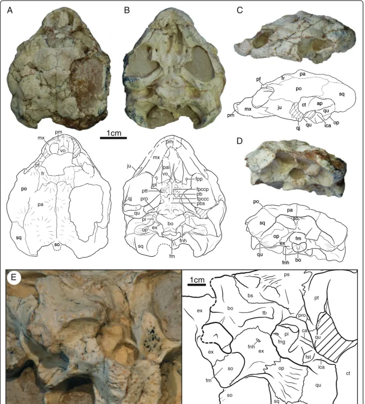
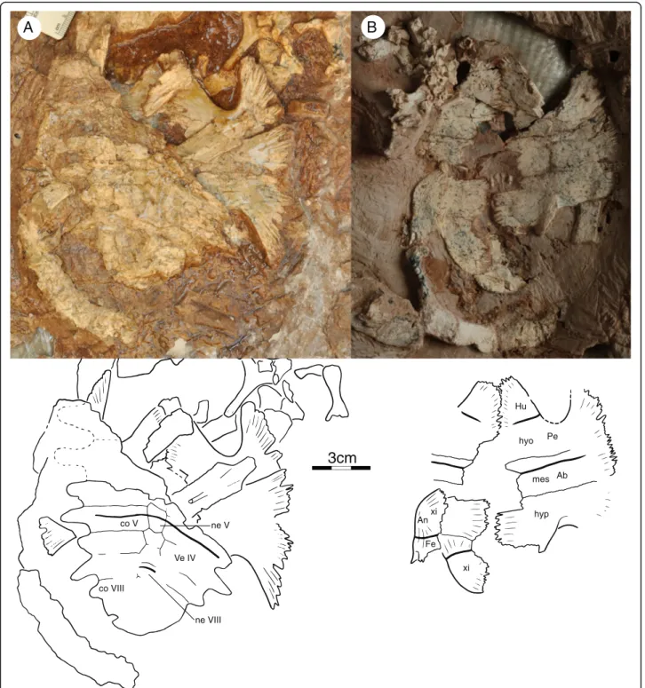
IVPP V18093 (Figs. 1, 3, 4 and 7a), a partial skeleton of a subadult individual consisting of a near complete,
slightly crushed skull, missing much of the dorsal skull roofing, complete mandible visible in ventral view, right hyoid, near complete shell lacking the right half of the carapace and most of the pygal region, at least 22 caudals in partial articulation, disarticulated left scapula and coracoid, isolated left pubis, partial right or left manus, including carpals, phalanges, and unguals, left femur, and possible left tibia and fibula. The midline plastron length, excluding epiplastra and entoplastron, is ca. 14 cm. The carapace of this individual is estimated to have had a midline length of ca. 23 cm.
Referred material
IVPP V18101–V18103, three poorly preserved cara-paces, previously described under the name ?Sichuan-chelyssp. [19]. Of these, IVPP V18102 is the largest and perhaps corresponds to an adult.
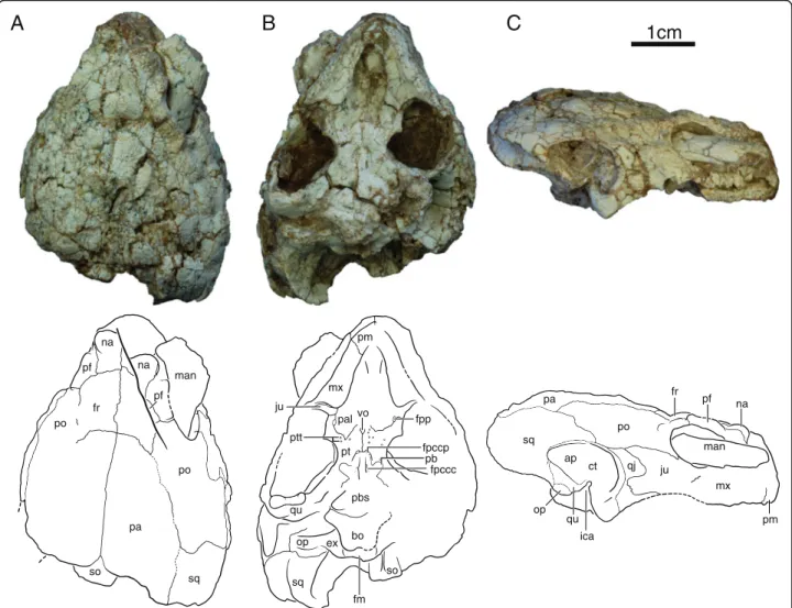
IVPP V18094 (Figs. 2, 6 and 7b), partial skeleton of a subadult individual that includes a near complete skull crushed along the sagittal axis, left jaw ramus, the dam-aged anterior plastral lobe, five disarticulated cervical vertebrae, right scapula, crushed left scapulocoracoid, and right humerus. Mid-plastral length, excluding epi-and entoplastron, estimated to be 14 cm by comparison to IVPP V18093.
IVPP V18095 (Fig. 5a), partial skeleton of a subadult consisting of heavily eroded carapace, near complete plastron lacking epiplastra and entoplastron, 2 cervical vertebrae, approximately 20 disarticulated caudals, iso-lated scapula, and both humeri. Mid-plastral length, ex-cluding epi- and entoplastron, ca. 11 cm.
IVPP V18096 (Figs. 5b and 7c), partial skeleton of a subadult that includes posterior portion of dorsal skull roof, disarticulated carapace consisting of at least 11 cos-tals and five peripherals, near complete plastron lacking the epiplastra and entoplastron, at least two isolated cervicals and two isolated caudals, isolated scapula and ischium, left humerus, femur and tibia, and various un-identified distal limb bones. Mid-plastral length, exclud-ing epiplastra and entoplastron, ca. 11 cm.
IVPP V18097 (not figured), highly fragmentary skel-eton of a presumably adult individual consisting of heav-ily fragmented, partial skull comprised of nasal region, left cheek region, both prootics and partial quadrates, and partial basisphenoid, basioccipital, and exoccipital, near complete, but fragmented mandible, left lateral third of shell missing the nuchal and pygal region, at least two isolated caudals, and various unidentifiable fragments of long bones. Mid-plastral length, excluding epiplastra and entoplastron, ca. 20–25 cm.
Locality and horizon
All specimens herein referred to the new taxon were collected from the early Upper Jurassic (Oxfordian)
1cm
A
C
pmB
D
mx pal fpp pt ptt pb bo qu ju qj op ex fpccp fm sq fnh pi fpccc vo pro po sq so pf vo pm mx pm mx ju po qu ct ap ica qu sq fr pa pf pa sq op ex fm bo so qu fnh po qj op pbs fr pa po sq so pm mx ju po qu ct ap ica qu sq fr pa pf pa sq op ex fm bo so qu fnh po qj op1cm
bo pi fnh ex bs so so ps pro pt ex fng op qu sq qu fst ca fm ex tb ct icaE
Fig. 1 Skull of IVPP V18093, holotype, Sichuanchelys palatodentata n. sp., Late Jurassic (Oxfordian), Shishugou Formation, Wucaiwan, Xinjiang, China, in dorsal (a), ventral (b), left lateral (c), posterior (d), and oblique view focused on the basicranial region (e). Hatch marks indicate damaged areas. Abbreviations: ap = antrum postoticum; bo = basioccipital; bs = basisphenoid; ca = columella auris; ct = cavum tympani; ex = exoccipital; fm = foramen magnum; fng = foramen nervi glossopharyngei; fnh = foramen nervi hypoglossi; fpccc = foramen posterius canalis carotici cerebralis; fpccp = foramen posterius canalis carotici palatinum; fpp = foramen palatinum posterius; fr = frontal; fst = foramen stapedio-temporalis; ica = incisura columella auris; ju = jugal; mx = maxilla; op = opisthotic; pa = parietal; pal = palatine; pb = processus basipterygoideus; pbs = parabasisphenoid; pf = prefrontal; pi = processus interfenestralis; pm = premaxilla; po = postorbital; pro = prootic; ps = parasphenoid; pt = pterygoid; ptt = pterygoid teeth; qj = quadratojugal; qu = quadrate; so = supraoccipital; sq = squamosal; tb = tuberculum basioccipitale; vo = vomer
upper part of the Shishugou Formation at the Wucaiwan Locality in Xinjiang Uygur Autonomous Region, China (see [20] for map). The holotype and referred specimens IVPP V18094–18096 (see below) were found in close as-sociation to one another, along with nearly complete, ar-ticulated skeletons of a squamate and a shartegosuchid crocodyliform. IVPP V18097 was recovered 1.2 km to the north of the type locality and IVPP V18102 an add-itional 2.2 km northward. The type locality is positioned between two tuffs (T-2 and T-BW of [20]) and can thereby be dated securely to the early Oxfordian. V18102 and V18097 were recovered from sediments slightly higher in the formation, just above the T-BW tuff dated 159.7+/-0.3 million years ago, but still thought to be Oxfordian in age considering locally calculated sedimentation rates. Precise locality information is
unavailable for V18101 and V18103 within Wucaiwan, but they are likely from the upper part of the Shishugou Formation, and therefore Oxfordian as well.
Diagnosis
Sichuanchelys palatodentata sp. nov. can be diagnosed as a representative of Sichuanchelys by the following, unique combination of shell characters: broad nuchal emargination delimited by peripheral II, vertebral scutes broader than long, marginal restricted to peripherals, liga-mentous bridge, broad plastron, one pair of fully devel-oped mesoplastra, short midline contact of epiplastra, and anteroposteriorly short extragular scutes. Sichuanchelys palatodentatais differentiated from S. chowi by a consist-ent contact of vertebral I with marginal II, by being larger, and, perhaps, by retaining a central plastral fontanelle
1cm
A
B
C
fr pa po sq so pf na man na pf po pm mx pal fpp pt ptt pb bo qu ju op ex fpccp fm sq vo so pm mx ju po ct ap ica sq fr pa qj man qu op na pf fpccc pbsFig. 2 Skull of IVPP V18094, Sichuanchelys palatodentata n. sp., Late Jurassic (Oxfordian), Shishugou Formation, Wucaiwan, Xinjiang, China, in dorsal (a), ventral (b), and right lateral view (c). Abbreviations: ap = antrum postoticum; bo = basioccipital; ct = cavum tympani; ex = exoccipital; fm = foramen magnum; fpccc = foramen posterius canalis carotici cerebralis; fpccp = foramen posterius canalis carotici palatinum; fpp = foramen palatinum posterius; fr = frontal; ica = incisura columella auris; ju = jugal; man = mandible; mx = maxilla; na = nasal; op = opisthotic; pa = parietal; pal = palatine; pb = processus basipterygoideus; pbs = parabasisphenoid; pf = prefrontal; pm = premaxilla; po = postorbital; pt = pterygoid; ptt = pterygoid teeth; qj = quadratojugal; qu = quadrate; so = supraoccipital; sq = squamosal; vo = vomer
when reaching maturity. Using cranial characters, S. pala-todentatacan be diagnosed to be closer to extant turtles than Proganochelys quenstedti Baur, 1887 [21] by having a fused basicranial joint, an anteriorly shifted canalis stapedio-temporalis, and by lacking vomerine and palatine teeth, but more basal relative to extant turtles by posses-sing pterygoid teeth, visible remnants of the basipterygoid process, and a prootic that is visible in ventral view. Sichuanchelys palatodentata can be distinguished from Kayentachelys aprix Gaffney et al., 1987 [22], the only other known turtle with this combination of three plesio-morphic characters, by the exclusion of the frontals from the orbit, an elongated jugal that nearly contacts the quad-rate posteriorly, posteriorly extended squamosals, and a closed interpterygoid vacuity and formed foramina poster-ius canalis carotici palatinum.
Comments
The new material we present herein from the Late Juras-sic of Xinjiang Province overlaps in overall shape and shell texture with that of previously described material Sichuanchelys chowifrom the Middle Jurassic of Sichuan Province and it is therefore not surprising that initial finds from Xinjiang were identified as Sichuanchelys sp. [19]. The available material of Sichuanchelys chowi only consists of shells [16] and has not yet been described in detail. We are therefore not able to make detailed com-parisons, although sufficient insights are available to dis-tinguish a new taxon. Given the lack of character information for S. chowi, our phylogenetic analysis (see Methods below) is furthermore not able to rigorously resolve the relationships within Sichuanchelyidae. As we do not favor naming a new genus based on poor charac-ter evidence, we here place the new species in Sichuan-chelys, but note that future analyses may not resolve this taxon to be monophyletic.
Phylogenetic nomenclature
We generally follow previously established phylogenetic nomenclature [23–26]. In addition, we herein phylogenet-ically redefine the name Sichuanchelyidae Tong et al., 2012 [16] as referring to the clade that includes all turtles more closely related to Sichuanchelys chowi Ye and Pi, 1997 [17] than to Meiolania platyceps Owen, 1886 [27], Helochelydra nopcsai Lapparent de Broin and Murelaga, 1999 [28], or any extant turtle. The name Mongolochelyi-dae“Sukhanov and Pozdnjakov, In Press” (as provided in [29]) is not used herein, because Sukhanov and Pozdnja-kov, In Press never appeared in print and because the name was otherwise never formally designated as a new family group taxon [30–32]. This name is therefore not available for nomenclatural consideration [18]. We simi-larly define the name Helochelydridae Nopcsa 1928 [33] as referring to the clade that includes all turtles more
closely related to Helochelydra nopcsai than to Meiolania platyceps, Sichuanchelys chowi, or any extant turtle. The rule of priority also applies to names within the family group [18] and we therefore disregard Solemydidae Lap-parent de Broin and Murelaga 1996 [34] since it is the jun-ior synonym of Helochelydridae.
Description Skull
We herein utilize previously established terminology for cranial anatomy [35] with recent amendments [11] in regards to the carotid circulation.
At least four skulls are present in varying degrees of preservation. The skull of IVPP V18093 shows the least amount of distortion, particularly in ventral view, but much of the dorsal surface is missing and crushed (Fig. 1). The skull of IVPP V18094 is the most complete, but the surface is heavily fractured and the shape is greatly distorted by shearing. In particular, the right or-bital region is shifted to the posterior, the left ear is de-formed, and the entire skull is slightly crushed along the sagittal plane (Fig. 2). Only the posterior margin of the skull roof is preserved in IVPP V18096 and only frag-ments are associated with IVPP V18097 (not figured). This description is therefore based mostly on IVPP V18093 and IVPP V18094.
The skull is relatively low, the external nares are conflu-ent, the orbits face laterally, and there are no signs of tem-poral emargination, lacrimals, or supratemtem-porals. The parietals and squamosals form an incipient ‘collar’ (i.e., a posterior expansion of the dorsal skull roof) that protrudes posterior beyond the regular margin of the skull. The entire skull roof is decorated by numerous protuberances that we interpret as evidence for cranial scales, but well-defined sulci are not apparent. The frontals, parietals, and postor-bitals combined are decorated by at least one unpaired and four paired scales (Fig. 1a), but we refrain from homologiz-ing them until the skull roof is better understood for this taxon. Particularly well-developed horn-like protuberances are present around the dorsal margin of the orbit.
Nasal The nasal is a relatively large, rectangular element that forms the dorsal margin of the external nares and contacts the maxilla ventrally, the prefrontals posterolat-erally, the frontal posteriorly, and its counterpart medi-ally (Fig. 2). The anterior margin is decorated by a prominent, bulbous protrusion. The full outline of the external nares is not preserved in any specimen, but the well-preserved ventral margin of IVPP V18093 demon-strates that the external nares were not subdivided by the premaxillae.
Prefrontal The dorsal plate of the prefrontal is slightly smaller than that of the nasal. The prefrontal forms the
anterodorsal portion of the rim of the orbit and contacts the maxilla ventrally, the nasal anteromedially, the frontal posteromedially, and the postorbital posteriorly (Figs. 1 and 2). The dorsal plate is decorated by bulbous protrusions, particularly along the margin of the orbit. The descending plate of the prefrontal forms the anter-ior wall of the orbit and contacts the vomer, palatine, and maxilla ventrally. The foramen orbito-nasale is lo-cated at the contact between the prefrontal, vomer, and maxilla and is reduced to the size of a pinhole. The out-line of the sulcus olfactorius is not preserved.
Frontal The frontal is a sub-triangular element that does not contribute to the orbit and that contacts the nasals along a slightly oblique suture anteriorly, the pre-frontal anterolaterally, the postorbital laterally, the par-ietal along a heavily interdigitated suture posteriorly, and its counterpart medially (Figs. 1 and 2).
Parietal The parietal is the largest element on the dorsal skull roof. It contacts the frontal anteriorly, the postorbital anterolaterally, and squamosal posterolaterally, the supraoccipital posteriorly, and its counterpart along the midline (Figs. 1 and 2). The parietals combined form a midline scale protrusion in their anterior third, a pair of anterior scale protrusions together with the frontals, a pair of posterior anteroposteriorly elongate scale protrusions in their posterior third, and a pair of scale protrusions along the suture with the postorbital. The inferior process of the parietal is not preserved in any specimen and its extent and possible contacts are therefore not known.
Jugal The anterior portion of the lateral plate of the jugal is best preserved in IVPP V18093, whereas the pos-terior portion is best preserved in IVPP V18094. The jugal forms the posteroventral rim of the orbit, contacts the maxilla anteroventrally, the postorbital anterodor-sally, and the quadratojugal posteriorly (Figs. 1 and 2). The posterior portion of the jugal is split into ventral and dorsal processes that surround much of the lateral exposure of the quadratojugal. The ventral process nearly contacts the quadrate. The medial plate of the jugal contacts the maxilla and palatine within the orbit and additionally contacts the pterygoid within the lower temporal fossa.
Quadratojugal The lateral exposure of the quadratoju-gal is greatly reduced by the juquadratoju-gal (Figs. 1 and 2). The quadratojugal contacts the jugal anteriorly and frame the anterior rim of the cavum tympani.
Squamosal The dorsal exposure of the squamosal con-tacts the postorbital anteriorly, the parietal medially, and frames the posterodorsal portion of the cavum tympani
together with the quadrate (Figs. 1 and 2). The squamo-sals form distinct posteromedial protrusions that form a ‘collar’ together with the parietals that is intermediate between the condition seen in most turtles and the extreme collar apparent in Mongolochelys efremovi. In posterior view, the squamosal broadly contacts the paroccipital process of the opisthotic. The posterolateral aspects of the squamosal are decorated by fine striations.
PostorbitalThe dorsal exposure of the postorbital is only slightly smaller than that of the parietal (Figs. 1 and 2). The postorbital forms the posterior margin of the orbit, contacts the prefrontal and frontal anterolaterally, broadly contacts the parietal medially, the squamosal posteriorly, and the jugal, quadratojugal, and quadrate ventrolaterally. The dorsal surface is decorated by a distinct scale protru-sion along the posteromedial rim of the orbit and a broad scale protrusion along the posteromedial contact with the parietal. A descending process is absent.
Premaxilla The premaxillae are paired and contribute to the ventral margin of the external nares and the an-terior portion of the labial ridge and the triturating sur-face (Figs. 1 and 2). The premaxilla contacts the maxilla posterolaterally and the vomer posteriorly. A pair of small prepalatine foramina pierce the premaxilla in dor-sal view at mid-length, but exit in ventral view at the contact with the vomer.
MaxillaIn lateral view, the maxilla forms the anteroven-tral aspects of the orbit, and contacts the premaxilla an-teriorly, the nasal and prefrontal dorsally, and the jugal posteriorly (Figs. 1 and 2). Within the orbit, the maxilla contacts the prefrontal anteriorly, the palatine medially, and the jugal posteriorly. In ventral view, the maxilla forms the majority of the lingual margin and the rela-tively broad, flat triturating surface and contacts the pre-maxilla anteromedially, the palatine medially, and the jugal posteriorly. The labial ridge is straight in lateral view and the palatine and jugal do not contribute to the triturating surface. A small contact with the vomer is perhaps present just posterior to the contact with the premaxilla.
Vomer The vomer is an elongate, toothless, and unpaired element best preserved in IVPP V18093 (Fig. 1b). The an-terior third of this bone is flat and unusually wide, the intermediate third is decorated by a distinct ridge, and the posterior third is narrow and flat. The vomer contacts the premaxilla anteriorly, the palatine laterally, and the ptery-goid posteriorly. A minute anterolateral contact may per-haps be present with the maxilla. An anterolateral contact is apparent with the prefrontal in dorsal view.
Palatine The palatine is a flat element that lacks teeth, forms much of the roof of the primary palate, and frames the internal narial opening anteriorly and contributes to the medial aspects of the small foramen palatinum poster-ius (Figs. 1 and 2). The palatine contacts the vomer medi-ally, the pterygoid posteriorly, and the maxilla laterally. In dorsal view, the palatine forms much of the floor of the orbit and contacts the prefrontal anteriorly, the maxilla anterolaterally, and the jugal posterolaterally.
Quadrate In lateral view, the quadrate forms a well-developed, kidney-shaped cavum tympani and contacts the quadratojugal anteriorly, the postorbital anterodor-sally, and the squamosal posterodorsally (Figs. 1 and 2). The region posterior to the incisura columella auris is greatly inflated. In most turtles this area is laterally cov-ered by bone to form the antrum postoticum, but in Sichuanchelys palatodentatamost of this cavity remains laterally open. This condition is otherwise only seen in Mongolochelys efremovi. The quadrate does not fully en-circle the anterior opening of the antrum, instead the dorsal portion is formed by the squamosal. The incisura columella is clearly incised into the posterior aspect of the quadrate, but remains open towards the posterior. The articular processes are low and face anteroventrally.
In ventral view, the quadrate contacts the pterygoid medial to the articular processes. Posterior to the inci-sura columella auris, the quadrate has a broad poster-omedial contact with the distinct paroccipital process of the opisthotic and a posterior contact with the squamosal. The anteromedial contacts of the quadrate within the upper temporal fossa are not visible in available specimens.
IVPP V18097 is the only specimen to partially pre-serve the quadrate in dorsal view. The specimen is too fragmentary to demonstrate the presence of a processus trochlearis oticum, but the dorsal surface of the quadrate is nevertheless decorated by a roughened surface similar to that developed in Mongolochelys efremovi. This roughened surface likely indicates the former presence of a cartilaginous cap in this region and can be seen as evidence of an otic trochlear mechanism [8].
Epipterygoid The epipterygoids, if present, are fully obscured by sediment and their morphology therefore cannot be discerned.
PterygoidThe ventral surface of each pterygoid in IVPP V18093 is decorated by about a dozen circular structures that are arranged in a V-shaped pattern with its apex posteriorly (Figs. 1 and 2). About half of these structures consist of a ring of dense tissue and can therefore be interpreted safely as palatal teeth. All but one of these teeth broke near their base. The remaining circular
structures are shallow depressions lacking any evidence of enamel and therefore represent facets from which palatal teeth dislodged either pre or post mortem. The ventral surfaces of the pterygoids of IVPP V18094 are poorly preserved, but a number of pterygoid teeth can be identified here as well. There is no evidence for palatal teeth on the vomer and the palatines.
The anterior branch of the pterygoid has a small con-tact with the jugal anterolaterally and with the vomer anteromedially. In addition, it contributes to the lateral margin of the foramen palatinum posterius, contacts the palatine anteriorly, and broadly contacts its counterpart along the midline. The external pterygoid process is clearly developed, has a small posterior projection, and a small, but distinct vertical plate. The pterygoid has a short, but clear sutural contact with the parabasisphe-noid (sensu [36]) along the midline. The interpterygoid vacuity is therefore closed and the palatine artery enters the skull through a distinct foramen posterius canalis carotici palatinum formed by the pterygoid and the basisphenoid. The pterygoid furthermore has a sutural articulation with the parabasisphenoid posterior to the basipterygoid process and fully surrounds the basiptery-goid process. The posterior half of the pterybasiptery-goid contacts the prootic posteriorly and the quadrate poster-olaterally and only partially floors the cranio-quadrate space. The prootic therefore remains exposed in ventral view (Fig. 1e). Much of the cavum acustico-jugulare is open in ventral view due to the short posterior process of the pterygoid failing to reach the basioccipital or the exoccipital. The quadrate process forms a thin lamina of bone that partially floors the incipient canalis cavernosus in ventral view.
Supraoccipital In posterior view, the supraoccipital forms the dorsal margin of the foramen magnum and contacts the exoccipitals ventrally (Figs. 1 and 2). The supraoccipital crista is short and likely did not protrude significantly beyond the level of the occipital process. The distal tip of the crista is expanded into a horizontal shelf that is partially visible in dorsal view behind the pa-rietals. However, the shelf does not contribute directly to the dorsal roofing of the skull. The ventrolateral contacts of the supraoccipital within the upper temporal fossa are not preserved, beyond the posterolateral contact with the opisthotic.
Exoccipital The exoccipital forms the lateral margin of the foramen magnum, contacts the supraoccipital dor-sally, the opisthotic laterally, and the basioccipital med-ioventrally (Figs. 1 and 2). The occipital condyle is damaged in all specimens, but it is nevertheless apparent that the exoccipital contributed to the dorsolateral por-tion of the condyle. The exoccipital forms a bony wall
that defines the posterior border of the recessus scalae tympani and that is pierced by a single hypoglossal for-amen, which is oriented slightly to the anterior and thereby easily overlooked in posterior view. A notch at the lateral margin of this wall may either be a second, partially developed hypoglossal foramen or a partially developed posterior jugular foramen.
Basioccipital The basioccipital contacts the basisphe-noid anteriorly along a deeply concave suture, the exoc-cipital dorsally, and forms the ventral rim of the foramen magnum (Figs. 1 and 2). The occipital condyle is damaged in all specimens, but it is apparent that it is situated dorsal to the ventral surface of the basioccipital and that the basioccipital forms the central portion of the process. The ventral surface of the basioccipital forms a broad depression together with the posterior portion of the basisphenoid that is defined laterally by a single pair of distinct tubercles.
ProoticThe prootic is only partially covered by the ptery-goid ventrally and is therefore visible in ventral view, where it contacts the pterygoid laterally, the parabasisphe-noid medially, and roofs the incipient canalis cavernosus (Figs. 1 and 2). Matrix and the dorsal skull roof obscure the contacts of the prootic within the temporal fossa in IVPP V18093 and V18094. In IVPP V18097 the prootic contacts the quadrate laterally and ventrally, contributes to the foramen stapedio-temporale and the trigeminal for-amen, but not to the trochlear process.
Opisthotic The paroccipital process is a conspicuous, vertically oriented extension of the opisthotic that has a sutural articulation with the quadrate and squamosal lat-erally, is decorated distally by fine striations, and is par-tially visible in lateral view (Figs. 1 and 2). In posterior view, the opisthotic otherwise has a broad dorsolateral contact with the squamosal, and broad ventromedial contact with the exoccipital, and a short lateral contact with the supraoccipital. Within the upper temporal fossa, the opisthotic contacts the quadrate laterally and the supraoccipital medially, but a possible contribution to the margin of the foramen stapedio-temporale is ob-scured in all skulls. The processus interfenestralis is ex-posed in ventral view in the form of a well-developed, slender process that is oriented anteroventrally. It has a ventral expansion, and possibly contacts the basioccipi-tal, but does not contribute to the ventral surface of the skull. The processus interfenestralis forms the posterior rim of the fenestra ovalis, forms the anterior wall of the recessus scalae tympani, and constricts the perilymphatic fenestra to the size of the foramen nervi hypoglossi.
Parabasisphenoid The parabasisphenoid consists of the basisphenoid and the parasphenoid [36]. Its dual compos-ition is best revealed at its posterior end, where it is possible to discern the parasphenoid as a thin lamina that only par-tially overlaps the basisphenoid in this region (Fig. 1e).
The anterior half of the parabasisphenoid forms a broad and rounded midline ridge (Figs. 1 and 2). This ridge has a sutural contact with the pterygoid anteriorly, thereby fully closing the medial portion of the interpter-ygoid vacuity. The remaining, lateral portions of the interpterygoid vacuity are reduced to form a pair of for-amina posterius canalis carotici palatinum, which are sit-uated on both sides of the elevated midline ridge. Further to the posterior, a pair of foramina posterius canalis carotici cerebralis pierces the parabasisphenoid at the level of the basipterygoid process, again along the sides of the elevated midline ridge. The basipterygoid processes of the parabasisphenoid are distinct, rounded lobes that are oriented in a ventrolateral angle and that are firmly sutured to the pterygoid.
The posterior half of the parabasisphenoid is signifi-cantly broader than the anterior half and has a sutural contact with the prootic laterally and a posterior convex contact with the basioccipital posteriorly (Figs. 1 and 2). The central ridge of the anterior half expands along the posterior half of the parabasisphenoid to cover a triangular area that spans nearly the full breadth of the bone and that is punctured by a pair of large, deep pits. Coarse, anteroposteriorly-oriented ridges decorate all elevated portions of the triangular area. There is no sign of a vidian foramen on the ventral surface of the parabasisphe-noid. The parabasisphenoid of Sichuanchelys palatoden-tataresembles that of Mongolochelys efremovi in its bony contacts, the development of the posteriorly expanded and ventrally decorated medial ridge, the placement of the carotid foramina, the presence of a pair of pits, and the presence of clearly developed basipterygoid processes. Columella Auris The columellae auris are preserved on both sides of the skull in IVPP V18093, but both ends still remain in matrix (Figs. 1 and 2). It is nevertheless clear that the columella auris is a slender element that fills the fenestra ovalis medially and is attached to the tympanic membrane laterally.
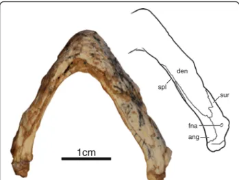
Mandible IVPP V18093 and IVPP V18094 are the only specimens to preserve their mandible, however, only the ventral side of the left ramus of IVPP V18093 is well preserved, whereas the rest remains embedded in sedi-ment (Fig. 3). The mandibular rami are relatively narrow and lack expanded triturating surfaces. The dentaries are fused along the midline and form the majority of the mandible. The dentaries contact the surangulars poster-iorly along an interdigitated suture, the angulars
posteroventrally, and the splenials ventrally. The splenial is an elongate, flat element that forms the ventromedial aspects of the rami. It has an elongate posteromedial contact with the angular and an elongate lateral contact with the dentary that closely approaches the symphysis. The surangular is pierced by a small foramen nervi auri-culotemporalis. The articulars cap the posterior ends of the rami. The lateral view of the left ramus of IVPP V18094 reveals that the lower jaws were massive relative to the slender ones of coeval xinjiangchelyids [11, 19, 25] and that the coronoid process was relatively high. The right ramus of this specimen is lodged into the orbit and does not reveal any additional details.
Carapace
All four specimens preserve at least parts of the cara-pace, but IVPP V18093 (Fig. 4a) and IVPP V18095 are the most informative (Fig. 5a). The original outline of the shell is somewhat unclear, as all specimens show evi-dence of distortion. However, despite this distortion, IVPP V18093 demonstrates that the shell had rather par-allel lateral margins, that a distinct anterior shoulder was formed by peripheral II, and that a broad nuchal emargi-nation was present. The sulci are deeply incised and delin-eate slightly convex scutes with week growth rings. The shell otherwise lacks any apparent shell sculpturing. In general shape, the shell resembles that of extant wood tur-tles, such as the extant emydid Glyptemys insculpta [2]. The shell bones are thin, about 1.5 mm thick in most parts of the shell, with the exception of the axillary and inguinal notches, which are about 3 mm thick. The shells of these presumed subadult individuals have carapacial, central, and posterior plastral fontanelles.
Nuchal The nuchal is best preserved in IVPP V18093 (Fig. 4a). It appears to be strongly curved, but this is largely due to plastic deformation of the left side of that carapace. The nuchal is a broad, trapezoidal bone that broadly con-tacts peripheral I along an oblique suture anterolaterally and costal I and neural I posteriorly. The anterior margin is approximately two thirds of the posterior length of the element. The nuchal forms a broad anterior emargination together with peripherals I and II, but plastic deformation makes it appear deeper than it likely was during life. Neurals The anterior portion of the neural series is pre-served in IVPP V18093 (Fig. 4a), whereas the posterior portion is preserved in IVPP V18095 (Fig. 5a). Neural I is the longest element of the series, contacts the nuchal anteriorly, neural II posteriorly, and costals I and II lat-erally. Neural I is intersected by the vertebral I/II sulcus. Neural II is approximately two thirds the length of neural I. A short contact of neural III with costal II pre-vents neural II from contacting costal III and gives neural II a rectangular outline. Neural III is only partially preserved. Given that this element has a short anterolat-eral contact with costal II and tapers posteriorly, we pre-sume that it had a hexagonal outline. Furthermore, the posteriorly tapering outline of neural III indicates that neural IV had an anterolateral contact with costal III. Only the posterior portion of neural IV is preserved as the most anterior element of IVPP V18095. It lacks a postero-lateral contact with costal V and therefore had a hexagonal outline. Neural V is the most posterior element to have an elongate hexagonal outline with short anterior sides. It is intersected by the vertebral III/IV sulcus. Neurals VI, VII, and VIII are greatly reduced in their anteroposterior length but are not significantly narrower than the more anterior elements. They therefore form isometric hexa-gons. Neural VIII has a broad posterior contact with the most anterior suprapygal element. The neural formula can be summarized as 6-4-6-6-6-6-6-6.
Costals Costals I–VI are well preserved in IVPP V18093 (Fig. 4a), whereas IVPP V18095 preserves costals IV– VIII (Fig. 5a). The anterior costals were likely oriented to the anterior, but the exaggerated anterior orientation seen in IVPP V18093 is due to plastic deformation. As in most turtles, the posterior costals have a slight orien-tation to the posterior, as is apparent from IVPP V18095. Costal I only contacts neural I medially, whereas costal II contacts neurals I–III. All remaining costals contact two neurals medially. Costal I contacts the nuchal and peripherals I–II anteriorly and periph-erals III–IV laterally. The detailed lateral contacts of the remaining costals with the peripherals are obscured by deformation, but it is apparent that the costals articulate with the peripherals via free ribs and that small costal
1cm den sur ang spl fna
Fig. 3 Mandible of IVPP V18093, holotype, Sichuanchelys palatodentata n. sp., Late Jurassic (Oxfordian), Shishugou Formation at Wucaiwan, Xinjiang, China, in ventral view. Abbreviations: ang = angular; den = dentary; fna = foramen nervi auriculotemporalis; spl = splenial; sur = surangular
fontanelles were retained in smaller individuals, but were closed in larger individuals. The free rib ends are better developed in IVPP V18095, a smaller specimen, indicat-ing that the costal fontanelles were larger in juveniles.
Peripherals The contacts and morphology of the anter-ior ten peripherals are best preserved in IVPP V18093 (Fig. 4a). The posterior peripherals are present in IVPP V18095 (Fig. 5a), but poor preservation obscures their
3cm
A
B
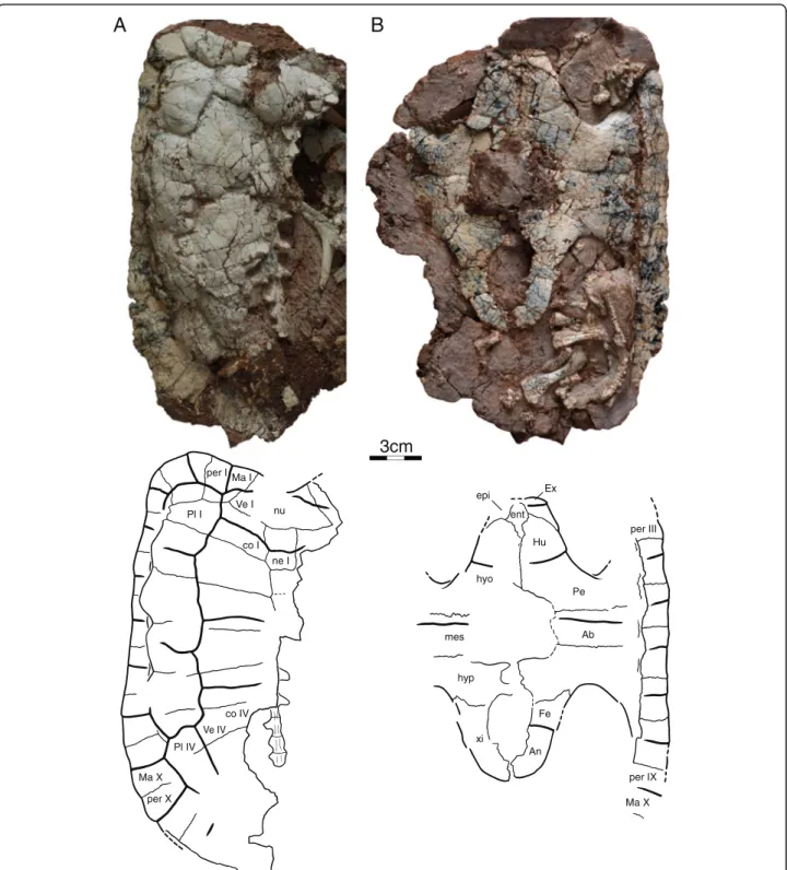
nu ne I co I co IV per I per X Ma I Ma X Pl I Pl IV Ve I Ve IV xi hyp hyo mes ent epi Ex Hu Pe Ab Fe An per IX Ma X per IIIFig. 4 Shell of IVPP V18093, holotype, Sichuanchelys palatodentata n. sp., Late Jurassic (Oxfordian), Shishugou Formation, Wucaiwan, Xinjiang, China, in dorsal (a) and ventral (b) view. Abbreviations: Ab = abdominal scute; An = anal scute; co = costal; ent = entoplastron; epi = epiplastron; Ex = extragular scute; Fe = femoral scute; Hu = humeral scute; hyo = hyoplastron; hyp = hypoplastron; Ma = marginal scute; mes = mesoplastron; ne = neural; nu = nuchal; Pe = pectoral scute; per = peripheral; Pl = pleural scute; Ve = vertebral scute; xi = xiphiplastron
sutures. The number of peripherals is therefore un-known. Peripheral I is wedge-shaped, but nevertheless retains a broad, posterior contact with costal I. Periph-erals I–II and XIII–X are flat elements with broad dorsal exposure. By contrast, peripherals III–VII are elongate
elements with only minor dorsal exposure. The ventral view of the bridge region is not sufficiently preserved in any specimen and the ventral contacts of the peripherals with the plastron are unclear. The contacts with the cos-tals are discussed above.
Fig. 5 Sichuanchelys palatodentata n. sp., Late Jurassic (Oxfordian), Shishugou Formation, Wucaiwan, Xinjiang, China. Shell of IVPP V18095 in dorsal view (a) and that of IVPP V18096 in ventral view (b). Abbreviations: An = anal scute; co = costal; Fe = femoral scute; Hu = humeral scute; hyo = hyoplastron; hypo = hypoplastron; mes = mesoplastron; ne = neural; Pe = pectoral scute; Ve = vertebral scute; xi = xiphiplastron
Pygal and suprapygals The pygal region is poorly pre-served in all specimens and no significant details can be discerned.
Carapacial scutes The surface of IVPP V18093 is deco-rated by wide and distinct carapacial scutes that allow asserting the presence of at least four vertebral scutes, four pleural scutes, and ten marginal scutes (Fig. 4a). IVPP V18095 furthermore confirms the presence of a fifth vertebral scute (Fig. 5a). The likely presence of a cervical cannot be confirmed.
The vertebral series consists of at least five elements, of which the anterior four are approximately equal in width. All vertebrals are about twice as wide as the pleurals. Vertebral I has a lenticular to octagonal shape and is there-fore anteroposteriorly longer along the midline than at its lateral margins. Vertebral I has a broad anterior contact with marginal I, a short anterolateral contact with marginal II, a broad lateral contact with pleural I, and a broad posterior contact with vertebral II. An anterior contact with the cer-vical was likely present as well. Vertebrals II–IV are roughly hexagonal elements that contact two pleurals each laterally. Vertebral II has the outline of a butterfly that thereby par-tially surrounds vertebral I. Vertebrals II and IV and notably larger than vertebrals I and III.
Each pleurals contact two vertebrals medially (Fig. 4a). Pleural I is barred from contacting marginal I through a contact of vertebral I with vertebral II. It otherwise con-tacts marginals II–V laterally. Pleural II likely concon-tacts marginals V–VII, pleural III contacts marginals VII–IX, and pleural IV contacts at least marginals IX–XI. The remaining contacts of the marginals with the plastral scutes are unclear.
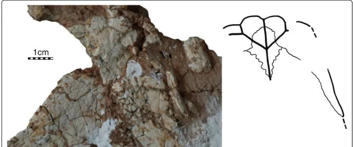
Plastron
The plastron of IVPP V18093 is near complete, but there is some damage to the anterior margin and the right bridge (Fig. 4b). The plastron of IVPP V18094 pre-serves the entoplastron best, but otherwise only consists of part of the anterior plastron lobe (Fig. 6). The plastra of V18095 and V18096 (Fig. 5) only consists of the hyo-, meso-, hypo-, and xiphiplastra. There are no meaningful visceral views of the plastron. Most plastral scutes are relatively indistinct.
Plastral bones The epiplastra are relatively large ele-ments that form the margins of the anterior half of the an-terior plastral lobe (Fig. 6). The anan-terior, rectangular part of the epiplastra contacts the entoplastron posterome-dially, and the hyoplastron posteriorly, and has a short midline contact with its counterpart. The posterior half of the epiplastron is a notably elongate, triangular posterior process that frames the anterolateral portions of the hyoplastra, similar to the process seen in Mongolochelys
efremovi. The contacts with the hyoplastra are blunt and the epiplastra therefore easily dislocate from the anterior plastral lobe after decomposition. The anterior margin of the plastron is oriented transversely, but it is decorated by four broad lobes that correspond to the gular and extragu-lar scutes. The margin, however, is not thickened. A dis-tinct articular scar along the anterolateral margin of the hyoplastron in partial specimens confirms presence of a small contact between the epiplastra and hyoplastra.
The anterior portion of the entoplastron contacts the epiplastra anterolaterally but does not contribute to the anterior plastral margins (Fig. 6). The posterior portion is broadly covered by the hyoplastra in ventral view and the full extension of this element therefore remains unclear.
The remaining part of the plastron is formed by a large pair of hyoplastra, a pair of mesoplastra, a pair of hypoplastra, and a pair of xiphiplastra (Figs. 4, 5 and 6). The mesoplastra are well-developed, rectangular in shape, show no sign of narrowing medially, but do not contact one another due to the presence of a medial plastral fontanelle in all subadult specimens. The plas-tron is not preserved in the largest, presumable adult specimens and it therefore remains unclear if this fonta-nelle closes during ontogeny. The posterior plastral lobe is similar in dimensions to the anterior plastral lobe and does not exhibit an anal notch.
The sutural margins of the hyo- hypo-, and xiphiplastra are finely digitated (Figs. 4, 5 and 6). The detailed quality of the bridge articulation is unclear, but the lack of blunt su-tures combined with the presence of finely fingered mar-gins indicates that the bridge appears to have been ligamentous. The lateral margins of the plastron are too ir-regular or damaged to allow identifying the presence of musk duct foramina. The hyoplastron and hypoplastron form well-developed axillary and inguinal buttresses, re-spectively. The distal ends of the buttresses are not pre-served in any specimen, but it is apparent that the anterior buttress ended anterior to peripheral IV (and therefore may have inserted in peripherals I, II, or III) and that the poster-ior buttresses ended posterposter-ior to peripheral VII (and there-fore may have inserted in peripheral VIII to XI).
The hyoplastra meet broadly along their posterior half thereby leaving a narrow, triangular gap for the ento-plastron. A clear central plastron fontanelle is formed by the hyo-, meso-, and hypoplastra that fully separates the mesoplastra along the midline, but it remains unclear if this is a juvenile feature, as the plastron is not known for any of the skeletally mature individuals. A second midline plastral fontanelle is also present between the hypo- and xiphiplastra.
Plastral scutes The anterior plastral lobe of IVPP V18094 clearly reveals that a pair of gulars and extragu-lars are present (Fig. 6). The guextragu-lars are triangular scutes
that produce clear lobes from the anterior margin of the plastron. The gulars contact the extragulars laterally, the humerals posterolaterally, and one another along the midline and cover the anterior half of the entoplastron. The extragulars are mediolaterally elongate elements that cap the anterolateral margin of the plastron. The extragulars contact the gulars medially and the humerals posteriorly, but do not contact one another medially and are restricted to the epiplastra.
The humeral/pectoral sulcus is transverse, straight, and situated midway along the hyoplastron (Figs. 4 and 5). If the remaining portion of the sulcus were to continue trans-versely, it would not intersect with the entoplastron. The medial portion of the pectoral/abdominal sulcus is also ori-ented transversely on the anterior third of the mesoplastra and enters the anterior third of the central fontanelle in IVPP V18095 and IVPP V18096. The abdominal/femoral sulcus cannot be found in any specimen, but likely was present, as in all turtles. The femoral/anal sulcus originates approximately at the anterior quarter of the xiphiplastral margin and curves anteromedially from there. The medial portion of the sulcus enters the central fontanelle and does not cross the hypoplastral/xiphiplastra suture.
Scute sulci are poorly preserved in the bridge region of all specimens and it is therefore unclear if and how many inframarginals are present. Sichuanchelys chowi has four pairs of inframarginals [16, 17].
Vertebral column
Cervical vertebrae and ribsA number of disarticulated cervicals are preserved associated with specimens IVPP V18094 – V18096, but preservation is generally poor (Fig. 7b). The cervicals are typical of basal turtles in
being relatively short, but high. It is unclear if cervical ribs are present, as no cervical ribs were found and the centra are too damaged to preserve parapophyses. Only a single, concave articular facet is preserved among the centra and it is therefore unclear if formed cervical artic-ulations were present. Transverse processes are relatively long and centrally located.
Dorsal and sacral vertebrae and ribs The dorsal and sacral vertebrae and ribs are either covered by sediment or too poorly preserved to allow any meaningful observations. Caudal vertebrae and ribs At least 22 caudal vertebrae are preserved with specimen IVPP V18093 (Fig. 7a, c), about 20 with IVPP V18095, and numerous isolated ones with IVPP V18096, but preservation is, once again, generally poor. Transverse processes are distinct along the anterior processes, but become increasingly smaller towards the posterior and are absent in the posterior half of the caudal column. The entire caudal column ap-pears to have chevrons, as is evidenced by clear articular sites along the anterior half of the column and minute chevrons in articulation with the posterior caudals of IVPP V18093. The articular surfaces of only a few cau-dals are visible, but some appear to be amphicoelous, whereas others are slightly opisthocoelous. The great size of the basal caudals is consistent with the tail having been long (i.e., at least as long as the carapacial length).
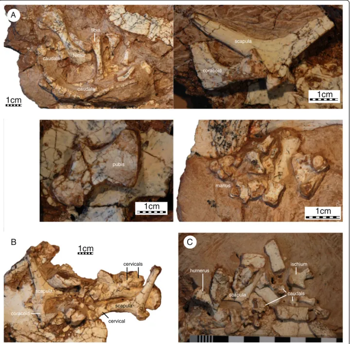
Girdles and limbs
All specimens preserve remains of the shoulder girdle, but the elements are universally crushed and/or encased in sediment, making it impossible to observe all aspects
1cm
Fig. 6 Sichuanchelys palatodentata n. sp., Late Jurassic (Oxfordian), Shishugou Formation, Wucaiwan, Xinjiang, China. Detail of the anterior plastral lobe of IVPP V18094. Abbreviations: ent = entoplastron; epi = epiplastron; Ex = extragular scute; Gu = gular scute. The scale is metric
(Fig. 7). It is nevertheless apparent that the scapulocoracoid is a slender triradiate complex that lacks a coracoid for-amen and that the glenoid is not fused in any specimen. The scapular process is rounded distally and is only slightly longer than the acromion process. Only a minor bony lam-ina is developed between the dorsal process and the acro-mion, but it is unclear if a bony lamina or ridge runs to the glenoid, as this region is not preserved in any specimen. The distal end of the acromion is not preserved in all views and it is therefore uncertain if it is rounded distally or deco-rated by ridges. The scapula has a distinct neck that offsets
the processes from the glenoid. The angle formed by the dorsal process and acromion is approximately 110°. The coracoid is shorter than the acromion and distally expanded to a broad fan.
Only two isolated pelvic elements are preserved, indi-cating that the acetabulum was not fused in subadult specimens. The isolated pelvic element associated with IVPP V18093 is interpreted as a pubis (Fig. 7a). The pubes have a broad midline contact with one another, the thyroid fenestrae are large, perhaps even confluent, and the epipubic process was not ossified. The isolated
femur tibia caudals caudals scapula coracoid pubis manus scapula coracoid cervical cervicals scapula humerus scapula caudals ischium
A
B
C
1cm
1cm
1cm
1cm
1cm
Fig. 7 Sichuanchelys palatodentata n. sp., Late Jurassic (Oxfordian), Shishugou Formation, Wucaiwan, Xinjiang, China. Details from the postcranial skeleton of IVPP V18093, holotype (a), IVPP V18094 (b), and IVPP V18096 (c). The scale is metric
element associated with IVPP V18096 is interpreted as the ischium (Fig. 7c). The ischia have a broad midline contact and the ischial process is relatively indistinct. The posterior margin of the ischium agrees with that of Mongolochelys efremoviin being poorly emarginated but differs by being much smaller [32].
A number of disarticulated elements are preserved that can be attributed to the limbs, but all material is encased in sediments making it impossible to observe most details (Fig. 7). The humerus is more than twice as long as wide and has a slightly sigmoidal shaft. The medial process is flared outwards and better developed than the ventrolat-erally oriented lateral process. The head is damaged in all specimens and it is therefore uncertain if a shoulder is present. The ectepicondylar canal is open, at least in the subadult specimens that preserve this bone. The ulna and radius could not be identified among the remains. A col-lection of bone is associated in the anterior region of IVPP V18093 that may represented a disarticulated hand, but it is not possible to identify any particular digit and the digital formula therefore remains unknown. The phalan-ges are nevertheless short and robust.
The only preserved femur, tibia, and fibula are too poorly preserved to allow discerning any details, beyond the observation that the femur has a slightly sigmoidal shaft (Fig. 7).
Presence of teeth in Sichuanchelys palatodentata
Sichuanchelys palatodentata n. sp. is striking because of the presence of palatal teeth, but the presence of such teeth is not novel among basal turtles. The Late Triassic Proganochelys quenstedti possesses a full set of palatal teeth that adorn the ventral surfaces of the paired vo-mers, palatines, and pterygoids [37]. Palatal teeth are otherwise known from the Permian proto turtle Eunoto-saurus africanus[38] and the Middle Triassic proto tur-tle Odontochelys semitestacea [39]. This is the basal amniotic condition [40]. The skull of all other known Triassic turtles is either missing or too poorly preserved to allow rigorously assessing the presence of palatal teeth. The gradual loss of teeth was previously docu-mented only by the Early Jurassic Kayentachelys aprix, which clearly lacks vomerine and palatine teeth, but re-tain a reduced count of pterygoid teeth. All more de-rived and younger turtles were thought to lack palatal teeth [22]. The presence of pterygoid teeth in S. palato-dentata extends the plesiomorphic retention of these structures in at least one lineage to the Late Jurassic, but we do not believe that this has any particular functional significance.
Phylogenetic analysis
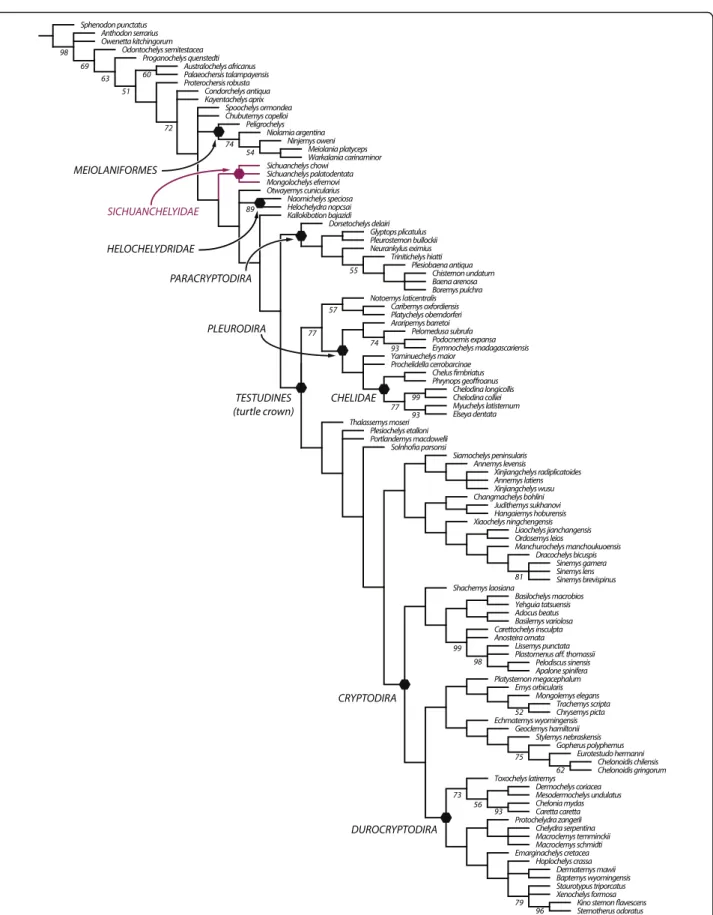
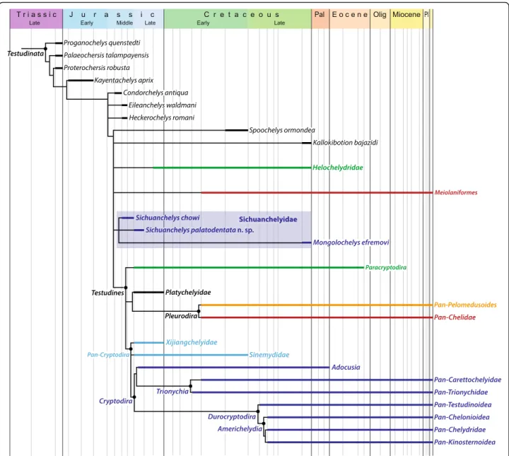
Our parsimony analysis (see Methods below) resulted in 550 most parsimonious trees with 960 steps. Heckerochelys
romani, Eileanchelys waldmani, Indochelys spatulata, Patagoniaemys gasparinae, and Xinjiangchelys junggarensis act as wild-card taxa and were therefore pruned from the consensus tree (Fig. 8). Sichuanchelys palatodentata was re-trieved in a polytomy with Sichuanchelys chowi and Mongolochelys efremovi within the clade Sichuanchelyidae along the stem lineage of turtles. Bootstrap resampling re-veals that support for this group is strong (73 %) if S. chowi is removed from the analysis, likely because this species is incompletely known. Helochelydridae and Kallokibotion bajazidi are placed in successively more crownward posi-tions relative to Sichuanchelyidae. Turtles with the para-cryptodiran carotid circulation are retrieved as monophyletic as the immediate sister of crown Testudines. Meiolaniformes is here restricted to Meiolaniidae and Peligrochelys walshaeand are placed in a more basal pos-ition than Sichuanchelyidae. Spoochelys ormondea and Chubutemys copelloiform a polytomy with Meiolaniformes and the clade consisting of all other more derived taxa. Morphological support for the placement of M. efremovi into a clade with Sichuanchelys palatodentata is high and we are therefore confident in that M. efremovi is not nested within Meiolaniformes as previously proposed (e.g., [26, 41–43]).
Biogeographic analysis
The fossil record of turtles is relatively good and multiple attempts have therefore been made to discern global [6, 13] or regional biogeographic patterns [26, 31, 44–46]. How-ever, new fossils, insights into the paleoecology of fossil turtles, and novel phylogenetic hypotheses allow us to synthesize a global biogeographic model that reveals that the diversification of turtles was primarily driven by vicariance caused by the breakup of Pangaea. We demonstrate below that this pattern is apparent at two successive phylogenetic levels. Given that some parts of the turtle tree remain controversial, in particular the in-clusiveness of Pan-Cryptodira and the interrelationships of sichuanchelyids, helochelydrids, and meiolaniforms [6–12, 25, 26, 41–43, 47], we attempt to present a model that is relatively immune to future changes in the understanding of phylogenetic patterns by highlighting the distinct evolu-tionary history of seven clades of turtles. These conflicting signals are reflected in the composite topology we utilize herein (Fig. 9), which combines the result of previous stud-ies with our current strict consensus tree (see Phylogenetic analysis above). The monophyly of each clade is discussed below and phylogenetic ambiguities are highlighted.
The taxonomic identity of the fossils utilized herein is not controversial, as we only employ specimens that exhibit clear, apomorphic characters. The vast majority of inform-ative fossils is fragmentary, however, and has therefore not yet been integrated into global phylogenetic analyses. We therefore refrain for the moment from providing a
Fig. 8 A strict consensus tree of 550 most parsimonious trees with 960 steps resulting from phylogenetic analysis. 5 wildcard taxa were pruned from the consensus
probabilistic model of historical biogeography and rather present a narrative account based on all available data.
There has been full consensus over the course of the last 100 years that extant turtles can be grouped into two clades, Pleurodira and Cryptodira, but the vast majority of fossil taxa were traditionally shoehorned into this dichot-omy and turtles were therefore thought to lack a substan-tial stem lineage. All species-level phylogenies of the last decade [6–12, 25, 26, 41–43, 47] have converged upon the novel conclusion that the stem lineage leading to the crown is populated by a diverse assemblage of fossil turtles that inhabited all continents from the Triassic to the Pleis-tocene. All conflicting hypotheses [4–7] have been shown to converge upon this result through minor modifications,
in particular the addition of characters, taxa, or new speci-mens [48, 49]. Although a certain amount of ecological plasticity is apparent, the basal stem turtle lineage is domi-nated by terrestrial forms, whereas crown turtles and their immediate sister groups are dominated by freshwater aquatic forms [50]. We herein discuss the parallel diversi-fication of derived, aquatic turtles and basal, terrestrial turtles separately for convenience and highlight important developments that occurred in parallel.
It is important to emphasize that the strong biogeo-graphic signal we discuss herein only emerges once all littoral to marine clades are omitted from consideration, as these obscure the continental pattern that otherwise emerges based on freshwater aquatic and terrestrial
Fig. 9 A composite phylogenetic consensus of turtle relationships highlighting the most important clades discussed in the text and their stratigraphic distribution as derived from the inclusion of fragmentary material. To aid understanding the text, internal relationships and nodes are only provided within Pan-Cryptodira. Support for all clades or polytomies is provided in the text (see Biogeographic analysis)
forms alone. The littoral to marine groups we identify are listed further below under dispersal.
The biogeography of derived, freshwater aquatic turtles
The vast majority of recent molecular and morphological studies (see [14, 47] for most recent summary) support the monophyly of the primary clades that make up crown Tes-tudines: Pan-Cryptodira and Pan-Pleurodira, which in turn is comprised of Pan-Chelidae and Pan-Pelomedusoides. The fossil record furthermore reveals the presence of an-other clade that diverged near the base of the crown group: Paracryptodira (e.g. [8, 10, 42]). Our review of the fossil rec-ord indicates that these four clades can be traced back to four distinct biogeographic areas in the Late Jurassic to Early Cretaceous. The monophyly of each group and their biographic distribution is discussed below.
Pan-Chelidae
The monophyly of crown Chelidae has never been con-troversial. Chelids are freshwater aquatic turtles that today occur throughout South America and Australasia [2] (Fig. 11d), but its total group, Pan-Chelidae, was re-stricted, without exception, to Australasia and the southern half of South America for most of its history (Fig. 10a) and only invaded the northern half of South America during the Neogene [44]. The oldest known fossils referable to the chelid lineage are from the Aptian/Albian of Argentina [51, 52] and the Albian of Australia [53] and the group therefore lacks an apparent center of origin (contra [13, 54]). The known distribution of pan-chelids predicts the former presence and extinction of the group on Antarctica, as a transoceanic dispersal event is highly unlikely between South America and Australia. The original distribution of pan-chelids was therefore restricted to southern South America, Australia, and presumably Antarctica, a landmass previously termed “Southern Gondwana” [44]. The early history of the group is consistent with a vicariant origin of South American versus Australian chelids in the Early Cretaceous, as predicted by molecular phylogenies [55, 56] and molecular calibration studies [57], but contrast with morphological data [58]. Rigorous phylogenetic ana-lysis of all Cretaceous representatives is needed to further test this hypothesis.
Pan-Pelomedusoides
In contrast to chelids, pelomedusoids today occur through-out Africa, Madagascar, and the northern half of Sthrough-outh America [2] (Fig. 11d). Although various littoral to marine representatives helped this group of turtles to achieve a near-global distribution during much of the Cretaceous and Tertiary [59, 60] through a dizzying array of marine disper-sal events (see below), the freshwater aquatic representa-tives of this clade were consistently restricted to northern South America, Africa, Madagascar, and India throughout
their evolutionary history [59, 60] (Fig. 10a), a landmass named “Northern Gondwana” [44]. Throughout the mid-Cretaceous numerous stem-pelomedusoid “species pairs” are apparent between northern South America and Africa that highlight the faunal ties between these continents (e.g., Araripemydidae, Cearachelyini, and Euraxemydidae [59, 61]), but focused analysis will be necessary to infer vicariance as the direct cause of speciation within these clades (see also [62]). There currently is no evidence that the primary split of Pelomedusoides into the pelomedusid and podocnemidid lineages was caused by vicariance, as the fossil record of freshwater aquatic pan-podocnemidids is broadly distributed across Northern Gondwana [60]. Unambiguous pan-pelomedusids, by contrast, are only known from the Neogene of Africa [57].
There has been much debate whether the unusual distri-bution of extant podocnemidids in South America and Madagascar [2] is, among others, the result of vicariance [63], differential extinction within a formerly widespread group [64, 65], dispersal from Africa to Madagascar [66], or a mixture of vicariance and dispersal across Antarctica [67]. One reason why this conundrum remains unresolved is because there is no agreement as to the phylogenetic rela-tionships among the three primary lineages of extant podocnemidids (i.e., Erymnochelys madagascariensis, Pelto-cephalus dumerilianus, and Podocnemis spp.) and because competing topologies imply different biogeographic histor-ies. This problem is only further compounded by the lack of fossil forms that unambiguously represent the stem line-ages of the Malagasy Erymnochelys madagascariensis and the South American Peltocephalus dumerilianus, which may perhaps reach back into the Cretaceous. Until the fos-sil record provides a more definitive answer, we here refrain from supporting any particular biogeographic scenario. However, we feel that the complete lack of fossil podocne-midid turtles in Southern Gondwana [60, 68] make disper-sal from South America to Madagascar via Southern Gondwana highly unlikely.
The primary distribution of pan-pelomedusoids in North-ern Gondwana and pan-chelids in SouthNorth-ern Gondwana is best interpreted as the result of a vicariance event, as previ-ously proposed [44], and this event must have occurred prior to the Barremian [69]. A previous study [69] specu-lated that vicariance was driven by a volcanic event that is documented by large volcanic fields in southern Brazil, but we do not think this to be likely, as this volcanic event only lasted about one million years [70]. As an alternative, we speculate that the subtropical desert zone that crossed the southern portion of Gondwana during much of the late Mesozoic [71–73] persistently divided the freshwater habi-tats of early pleurodires into a larger northern and a smaller southern range. This desert zone apparently influenced the biogeographic distribution of other groups of organisms, in-cluding dinosaurs [74].
Paracryptodira
Although the phylogenetic relationships of Paracryptodira relative to Pleurodira and Cryptodira remains unresolved, there is broad agreement that the group is monophyletic, not situated within crown Cryptodira or crown Pleurodira, and that their freshwater aquatic habitat preferences are a derived character shared with crown turtles [7, 8, 10, 11, 25, 26, 42, 47, 75]. This clade is comprised of a diverse assemblage of medium-sized turtles that were re-stricted, without exception, to Euramerica (Europe + North America) throughout their evolutionary history (Fig. 10a) and that provide evidence for the close bio-geographic relationships of these land masses, as previously noted [6]. The oldest known paracryptodires are known from Upper Jurassic deposits in both North America and
Europe [31, 46, 76], but isolated finds extend the range to the Middle Jurassic of Europe [77]. This biogeographic area was fully separated from Asia by the Turgai Strait and from Gondwana by the Central Atlantic and the Tethys for much of the Late Jurassic and Early Cretaceous [78]. Paracrypto-dires were particularly diverse throughout the Late Cretaceous and Paleogene [76, 79–81], but the group went extinct prior to the Oligocene [82]. The paracryptodiran clade Baenidae is restricted to the Early Cretaceous to Paleogene of western North America (Laramidia), but there is no reason to interpret this as evidence for vicariance (contra [6]), as Baenidae lacks a sister group on a nearby landmass. The currently accepted sister of Baenidae, Pleurosternidae, instead shows a broad distribution across Euramerica.
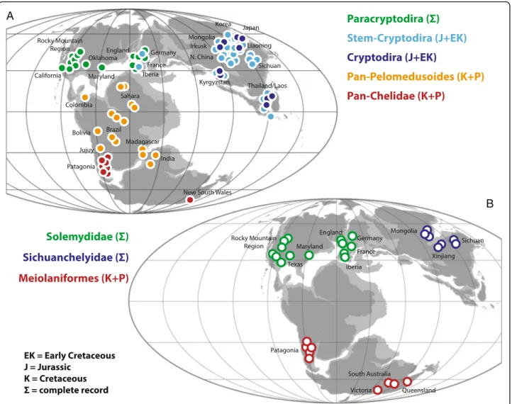
Fig. 10 The vicariant origin of the primary clades of turtles. a the early or complete fossil record of the clades Pan-Chelidae, Pan-Pelomedusoides, Paracryptodira, Pan-Cryptodira and Cryptodira imposed upon a paleogeographic reconstruction for the Late Jurassic (modified from [142]) highlighting the biogeographic areas of southern Gondwana, northern Gondwana, Euramerica and Asia, respectively. b the complete fossil record of the clades Meiolaniformes, Helochelydridae, and Sichuanchelyidae imposed upon the paleogeographic reconstruction for the Late Jurassic (modified from [142]) illustrating the biogeographic areas of southern Gondwana, Euramerica and Asia, respectively
Pan-Cryptodira
The composition of the total group of Cryptodira, i.e., Pan-Cryptodira, is currently one of the most controver-sial subjects in turtle phylogeny. Although the broad sample of basal, terrestrial forms discussed below has been removed from the cryptodiran stem group with confidence (see above), there is still much uncertainty regarding a similarly broad sample of freshwater aquatic forms [7, 8, 10, 11, 25, 26, 42, 47, 75], in particular xin-jiangchelyids, sinemydids, and macrobaenids (sensu [11]). The character evidence that places xinjiangchelyids and sinemydids along the phylogenetic stem of Cryptodira is quite convincing, because these taxa document the step-wise acquisition of cryptodiran characters throughout the
Middle to Late Jurassic. The character evidence is particu-larly strong in the basicranial region and these turtles have therefore been collectively united with crown cryptodires in the clade Eucryptodira [83]. Yet, pleurodires have been rou-tinely recovered deep within Eucryptodira (e.g., as sister to Testudinoidea) (e.g., [8, 11, 25, 47]), although the latest study demonstrated that this signal is an analytical artifact [12]. The extremely rich Middle Jurassic to Late Jurassic fossil record of freshwater aquatic pan-cryptodires is re-stricted to Asia [29, 84, 85], with the exception of marine plesiochelyids and eurysternids found in the Late Jurassic of Europe [31, 46] (Fig. 10a). It was not before the Early Cretaceous that isolated freshwater eucryptodire taxa dispersed into Europe [86].
Fig. 11 The biogeographic history of derived turtles following their primary origin through dispersal. a dispersal during the Cretaceous; b dispersal during the Paleogene; c dispersal during the Neogene; d the current distribution of turtle clades. Shaded areas highlight the distribution of turtle clades at the beginning of a particular time bin as inferred from the fossil record (see Fig. 10). Arrows highlight paths of freshwater aquatic or terrestrial dispersal. For simplicity, all island taxa and groups adapted to coastal and marine settings are disregarded. Abbreviations: Ad = Adocusia; Ca = (Pan)-Carettochelyidae; Cd = (Pan)-Chelydridae; Ch = Chelidae; Co = Compsemys; Em = Emydidae; Ge = (Pan)-Geoemydidae; Ki = Kinosternidae; Ma = Macrobaenidae; Si = Sinemydidae; Td = (Pan)-Testudinidae; Tr = (Pan)-Trionychidae