DOI 10.1007/s00221-014-3843-y
REsEaRch aRtIclE
Very early processing of emotional words revealed
in temporoparietal junctions of both hemispheres by EEG
and TMS
Vincent Rochas · Tonia A. Rihs · Nadia Rosenberg · Theodor Landis · Christoph M. Michel
Received: 27 august 2013 / accepted: 10 January 2014 / Published online: 5 February 2014 © springer-Verlag Berlin heidelberg 2014
Introduction
since the seminal works of Broca (1861) and Wernicke (1874), the left cerebral hemisphere in right handers is known to be dominant for language processing. however, the study of split-brain patients with surgically discon-nected hemispheres (sperry 1961; Gazzaniga and sperry
1967; Gazzaniga and hillyard 1971; Gazzaniga 2005 for a review) and the study of healthy subjects with lateral-ised stimulus presentations (for a review see springer and Deutsch 2004) leave no doubt that the right hemisphere also plays a considerable role in language processing. More recently, neuropsychological studies, lesion-based studies, and imagery studies (stroobant et al. 2009; lidzba et al.
2011; Mohr et al. 2007) revealed the participation of the right hemisphere in various features of language process-ing—e.g. intonation (Ross et al. 1988; Pell 2007), concrete-ness (Papagno et al. 2009), or context-related language (Diaz and hogstrom 2011). Most classical is the relation of the right hemisphere with the emotional dimension of language processing. the special role that emotional words play with respect to cerebral laterality has been recently summarised (landis 2006).
the present study aims to assess language lateralisation and the interaction between the two hemispheres in rela-tion to the treatment of the emorela-tional content of the words. It is based on earlier work from our group (Ortigue et al.
2004) that used a lexical decision task with a simultane-ous bilateral presentation of letter strings. In this previsimultane-ous work, event-related potential (ERP) analysis showed a dis-tinct scalp topography for emotional words presented in the right visual field (RVF), indicative of a specific intracranial generator configuration for these words. the well-known emotional word advantage was accompanied by increased activation of right lateral occipital brain areas at around Abstract We investigate the contribution of both
hemi-spheres in a lateralised lexical decision paradigm with emotional and neutral words in healthy volunteers. In a first experiment, high-density EEG analysis using source imag-ing methods revealed early specific participation of the temporoparietal junctions (tPJ) in both hemispheres for the detection of words. then, in an event-related transcra-nial magnetic stimulation experiment with the same task, the disruption of left or right tPJ compared with a control stimulation over the vertex showed a slowing that is more pronounced when words are emotional and presented in the left visual field (lVF). this indicates that interference with both left and right tPJ results in impaired processing of words that were presented to the lVF. In addition, these results point to a specific cooperative contribution of the right hemisphere in the processing of words with emotional content compared with neutral words at very early stages. Results from the two experiments can be integrated in a brain-based spatiotemporal model of the early detection of written words.
Keywords lexical decision · Word detection · Emotion · lateralisation · Right hemisphere · EEG · source imaging · tMs
V. Rochas (*) · t. a. Rihs · N. Rosenberg · t. landis · c. M. Michel
Functional Brain Mapping laboratory, Department of Fundamental Neuroscience, cMU, University of Geneva, Rue Michel-servet 1, 1206 Geneva, switzerland
100–140 ms. however, somewhat unexpectedly, this effect was only found for emotional words presented to the RVF and not to left visual field (lVF), despite the enhanced per-formance for emotional words in both conditions.
Derived from the study of Ortigue et al. (2004), the pre-sent study does not only better control the stimulus mate-rial and improve the analysis of the ERPs, but also assesses the early contribution of the right hemisphere to emotional word processing by applying transcranial magnetic stimu-lation (tMs). We performed a first experiment where we tracked the early brain processes of real word detection by high-density electroencephalography (256-channel EEG) using statistical analysis of the ERPs in the sensor space (voltage topography) and source space [electrical source imaging (EsI)] (Michel and Murray 2011). In the second experiment on an independent group of participants, event-related tMs was alternatively delivered in two regions that were identified in the first experiment to be recruited in these early stages of word processing—the left and right temporoparietal junctions (tPJ). the time window of stimulation was also selected on the basis of the first experiment. Behavioural measurements after interruption of these areas allowed to evaluate their implication in word processing.
the combination of these two time-resolved brain imagery methods allows a comprehensive understand-ing of particular actors and processes of the network in a given cognitive task. Our findings are then discussed and included in a functional spatiotemporal model of early pro-cessing of written words and emotionality.
Materials and methods study 1 (EEG)
Participants
Eighteen healthy volunteers participated in this study approved by the Ethical committee of the University hos-pital of Geneva, switzerland. Included participants were young adult males, right-handed, aged from 20 to 32 years (mean age = 24.8; sD = 3.5). all had normal or corrected-to-normal visual acuity and French as their mother tongue. None had experienced psychiatric or neurological disease, or any language difficulties (e.g. dyslexia) in their past. Participants were paid for their participation (30 chF/h). Experiments were performed with the informed and overt consent of each participant.
three participants were excluded after the training ses-sion and before any EEG recording, due to low detection rates in at least one condition. In addition, the recordings of two participants had bad impedances or muscular artefacts
during the EEG acquisition and were also excluded from further analysis. the analyses for study 1 were thus con-ducted with thirteen participants.
Stimuli and procedure
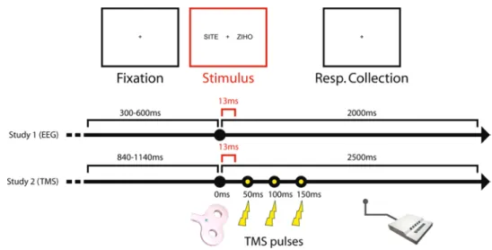
a lexical decision task was used in a simultaneous bilat-eral presentation. this is a revised version of the task pre-viously investigated in our laboratory (Ortigue et al. 2004; Mohr et al. 2005). On the screen, at 1 meter from the partici-pant, two strings of letters were displayed at the same time on each side of a central fixation cross, in the lVF and in the RVF. letter strings represented either a real French word or a pronounceable pseudo-word. two kinds of words were used: neutral and emotional words. this resulted in five possible stimulus conditions: a neutral word in the lVF, a neutral word in the RVF, an emotional word in the lVF, an emotional word in the RVF (all paired with a pseudo-word in the opposite side), or two pseudo-words. all letter strings were constituted of four black letters displayed in upper case (1.1 cm high and 4.2 cm length) on a white background. the letter strings were presented from 2.00° to 4.85° of visual angle from the fixation point on each side. thus, the two-let-ter strings were simultaneously displayed in distinct horizon-tal visual hemifields. the presentation time was 13 ms. Dur-ing the experiment, the participants were asked to respond during the 2 s following the stimulus if they had seen a word or not. If yes, they had to press a button with the hand on the side on which they detected the word. No response was required if they did not detect a word on either side. the inter-stimulus interval was randomly jittered between 2,300 and 2,600 ms. a sample trial is shown in Fig. 1.
Four-letter substantives with highest frequencies (occur-rences by million determined from a written database of text book material and an oral database of movie sub-title material) were taken from the freely accessible and well-controlled database for the French language “lex-ique 3.55” (New et al. 2001; see also http://www.lexique. org). Independent participants (n = 12) rated these words for familiarity, concreteness, imageability, emotional neu-trality, and emotionality on a 1–9 likert scale. Only four-letter words with high familiarity, low concreteness, and low imageability were kept. the 16 words with highest neutral and lowest emotional ratings were chosen for the neutral category (e.g. the French word “voie”), while the reverse—i.e. highest emotional, lowest neutral ratings— were included in the emotional category (e.g. the word “peur”). the selected words were matched with pronounce-able pseudo-words with identical patterns of consonants and vowels with four letters. Pseudo-words with spelling close to familiar foreign language words (commonly Eng-lish and German) were excluded to avoid any interaction in potential multilingual participants. t tests on the selected
words showed that neutral and emotional words did not differ in terms of frequency (respective averages 92.25 vs. 95.44; with a t = −0.73 and p = 0.89), familiarity (7.40 vs. 7.80; t = −1.88 and p = 0.07), concreteness (3.7 vs. 3.28;
t = 0.85 and p = 0.40), or imageability (4.63 vs. 3.94;
t = −1.24 and p = 0.23).
the task was divided into 8 blocks (4 min each) of 96 trials fully randomised for each block and for each partici-pant. the total number of repetitions was 128 for neutral words in the lVF, 128 for neutral words in the RVF, 128 for emotional words in the lVF, 128 for emotional words in the RVF, and 256 for two pseudo-words in both hemifields.
to avoid learning effects during the EEG recording due to the repeated presentation of the words, the subject prac-tised the task in four blocks the day before the EEG record-ing. Before each EEG session, the whole set of words were presented individually in a central position below the fixa-tion point and the participants performed a simple lexical decision task without EEG recordings. this learning ses-sion was performed to further avoid learning effects during task performance.
Behavioural data
average accuracies and median reaction times (Rt) were calculated for each condition in each subject. these behav-ioural data were plotted with their standard errors in bar graphs (see Fig. 2). the statistical analysis of the behav-ioural data was performed to replicate previous studies and was thus based on the prediction that words are better detected when presented in the RVF than to the lVF (Young et al. 1980; Graves et al. 1981; Iacoboni and Zaidel 1996; Babkoff et al. 1997) and that emotional words are better detected than neutral words (landis, 2006; schacht and sommer 2009; Kissler and Koessler 2011; skrandies 2013). Missed events and false alarms were also computed in order to detect potential outlier subjects (not represented).
EEG data acquisition and preanalysis
high-density EEG data were collected during 8 blocks of the task. the system used for acquisition was a 256-channel (agcl carbon-fibre-coated electrodes) hydrocel Geodesic sensor Net in association with Net station EEG acquisition software (Electrical Geodesics, Inc., Eugene, OR, Usa). Recordings were referenced to the vertex (cz) and sampled with a 16-bit analog-to-digital converter at 1,000 hz with a Dc removal. all electrode impedances were kept below 30 kΩ.
Offline analyses of the EEG were conducted using the “cartool” software (D. Brunet, Functional Brain Map-ping laboratory and cIBM, Geneva, switzerland; http:// sites.google.com/site/fbmlab/cartool). the signals of peripheral electrodes (on the cheeks and the nape) were omitted from the 256 channels, and the remaining 204 channels were submitted to further analysis. the EEG of these remaining channels was bandpass filtered from 0.16 to 40 hz and converted to the average reference. the con-tinuous EEG was visually inspected, and only epochs without eye blinks or muscular artefacts and with correct responses were taken into account. the number of aver-aged epochs was counterbalanced between the four condi-tions for each participant—i.e. we randomly picked a num-ber of epochs determined by the condition with the lowest number of accepted epochs for each individual (average number of accepted epochs 57.2 ± 6.4 sE). therefore, for a given subject, the different conditions included compa-rable signal-to-noise ratios. the remaining epochs were averaged by condition from 0 to 300 ms after the onset of the stimulus to reveal the event-related potential (ERP) for each participant. For each individual, artefactual electrode signals were interpolated using a spherical spline interpo-lation (Perrin et al. 1987). all individual ERP’s were rea-ligned on the peak of the global field power of the P100 component determined once for each subject to reduce Fig. 1 schematic chronology in
milliseconds (ms) of a trial for the presentation of one pair of letter strings in the lexical deci-sion task in study 1 (EEG) and study 2 (tMs)
the inter-individual variability of the time for visual infor-mation transfer from the retina to primary visual cortex (Woody 1967; Morand et al. 2000). Realignment has been
−27 up to +30 ms from the original onset of the
stimu-lus. as visual information does not reach the primary visual cortex before 50 ms (alexander and Wright 2006; thorpe
2009), this realignment on the first milliseconds does not impede the analysis of cognitive cortical processes but reduces their temporal smearing. Finally, ERPs were down-sampled to 500 hz before statistical analysis.
ERP’s topographic ANOVA
since the aim of this first experiment was to determine when and where in the brain the different stimuli were pro-cessed to guide the subsequent tMs study, the analysis of the high-density ERPs was entirely based on topographical analysis of the scalp potential fields and on EsI. In contrast to the conventional waveform analysis, the topographic analysis gives non-ambiguous and reference-free informa-tion about changes in the underlying generators in time and between conditions (Michel et al. 2009; Michel and Mur-ray 2011; Murray et al. 2008).
the first step of the analysis consisted in a topographic analysis of variance (T-aNOVa) based on paired permuta-tion tests with entire topographies as dependent variables (Murray et al. 2008; Koenig et al. 2013). this analysis was performed with the freely available software randomisation graphical user interface (RaGU; Koenig et al. 2011), which allows for multivariate T-aNOVas. the program calculates the global field power of the normalised difference maps between conditions for each subject, also called Global Dissimilarity (lehmann and skrandies 1980; Murray et al.
2008). the Dissimilarity is calculated repeatedly after ran-dom shuffling the condition assignments of the maps across subjects. this permutation statistic enables us to compare the real distribution of the Map Dissimilarity against the random distribution. the significance of the effect is then determined by the percent of randomly obtained values of the Dissimilarity that are larger than the real value. these
T-aNOVas were performed for each individual time point from 50 to 250 ms post-stimulus in a two-by-two within-subject factorial design (p value < 0.05; 5,000 repetitions); for words detected in the lVF versus in the RVF (lateral-ity); and for detected neutral words versus emotional words (Emotionality).
Electrical source imaging (ESI)
In order to estimate the location of current generators in the brain, the local auto-regressive average (laURa) algo-rithm, a distributed linear inverse solution comprising bio-physical laws as constraints (Grave de Peralta Menendez
et al. 2001, 2004; Michel et al. 2004 for review), was used. source estimations were computed from ERPs of each par-ticipant for each condition according to the factors Emo-tionality and laterality using the cartool software in a space of 3,005 solution points restricted to the grey matter of the MNI brain (spinelli et al. 2000; Brunet et al. 2011). For each participant and each condition, a normalisation of the estimated activity values was performed by z-score transformation of the activity at each solution point for a given time point. the transformation in z-scores homog-enised inter-individual contrasts by removing the back-ground noise and reducing extreme values. Using these
z-scores, as dependent variables, time-point-by-time-point aNOVas were computed for each solution point (p < 0.01 for at least five consecutive time frames corresponding to 10 ms and with a size restriction criterion of five neigh-boured solution points) with the stEN toolbox (stEN toolbox developed by J.-F. Knebel, Functional Electrical Neuroimaging laboratory, lausanne, switzerland. www.u nil.ch/fenl/sten). laterality and Emotionality of the words were taken as within-subject factors. Post hoc analyses, consisting in t tests on each solution point for each time frame, were computed to define the direction of the signifi-cant differences established by the aNOVa.
study 2 (tMs)
Participants
thirteen healthy volunteers participated in this study approved by the Ethical committee of the University hospital of Geneva, switzerland. these participants did not take part in study 1 (EEG). they were young adult males, right-handed, aged from 21 to 33 years old (mean age = 24.54; sD = 4.14). all had normal or corrected-to-normal visual acuity, French as their mother tongue and reported no language impairments (e.g. dyslexia). None had experienced psychiatric or neurological disease or unexplained loss of consciousness, and none reported ing experienced any epileptic episodes in their past or hav-ing a history of epileptic events in their family. the experi-ment was performed with the informed and overt consent of each participant. Participants were paid for their par-ticipation (30 chF/h) and were free to withdraw from the study at any time.
Results of three participants were excluded from the anal-ysis due to outlier detection levels or a high level of false alarms. two participants had high levels of false alarms (wrong detection in pseudo-words condition) in all three conditions of tMs, indicating that they had difficulties dis-criminating words from pseudo-words or were not focused on the task. In both cases, the false alarm levels were con-sidered as outliers according to the mean plus or minus
two standard deviation criterion. One other participant had extremely high levels of average detection, which also met the criteria for outlier exclusion—i.e. 2 standard deviations above the group mean. the analyses for the study 2 (tMs) were thus conducted on a sample of ten participants.
Stimuli and procedure
the same simultaneous bilateral lexical decision task as in study 1 (EEG) was used with the same stimuli presented in the same manner and with the same instructions. the only difference with the task from study 1 was that the inter-stimulus interval was randomly jittered from 3,340 to 3,640 ms—i.e. an extended period to avoid cumulative effects of tMs between trials. Responses of the partici-pants were collected during 2,500 ms (Fig. 1).
the task was divided into blocks of 48 trials, freely randomised for each block and for each participant. Eight neutral words in the lVF, eight neutral words in the RVF, eight emotional words in the lVF, eight emotional words in the RVF, and 16 pairs of pseudo-words were presented per block. the day before the tMs experiment, participants were asked to perform four blocks as behavioural meas-urement and training session without any tMs involve-ment. then, on the following day, they had to execute three blocks of the task while tMs was released over each site of stimulation (3 blocks × 3 sites of stimulation). In addition, each word was first displayed as single word in a central position below the fixation point and subjects performed a simple word detection task. this was done to avoid learn-ing effects durlearn-ing the task due to the repeated presentation of a word.
Transcranial magnetic stimulation parameters
the tMs parameters were in accordance with the current safety guidelines of tMs use (Rossi et al. 2009). a Mag-stim Rapid2 with a double 70-mm figure-of-eight coil (the
Magstim company ltd, Whitland, Wales, UK) was used for magnetic stimulation. tMs was performed as an event-related paradigm to interact transiently with cortical areas of interest during the task. a train of three bipolar pulses at 20 hz was released 50 ms after each onset of the stim-ulus—i.e. the pulses were delivered 50, 100, and 150 ms post-stimulus (Fig. 1). hence, the tMs pulses are deliv-ered after the display of the letter strings, thus avoiding alerting effects on perception due to sound or skin sensa-tions induced by tMs. the strength of stimulation was set individually to 90 % of the resting motor threshold (RMt) intensity—i.e. the intensity, which generates a finger move-ment for more than half of the motor cortex stimulations. Reduced intensity is known to produce a more focal effect (Wagner et al. 2009). In addition, a moderate intensity
of stimulation limits participant discomfort and muscle contraction.
Both the time period and the sites of stimulation were based on the results of the EEG study (see “EEG-based tMs target choice”). On a different day prior to the tMs sessions, the participants underwent a t1-weighted struc-tural MRI scan. the target sites for tMs stimulation were determined based on individual structural MRI on the anterior bend of the angular gyrus—Brodmann area 39, in the middle of the tPJ of both hemispheres. the aver-age talairach coordinates of the actual targets across par-ticipants were −58.9; −54; 25.8 for the left angular gyrus (laG) and 58.2; −50.7; 27.8 for the right angular gyrus (RaG). the vertex (0; −25; 68) was used as an active con-trol site. a frameless neuronavigation system combining the neuronavigation module of BrainVoyagerQx (BrainIn-novation, Maastricht, the Netherlands) with the ultrasound cMs20 measuring system for navigation (Zebris Gmbh, tübingen, Germany) and based on individual structural MRI was used to place and to maintain the coil over the considered site for every single task block. the three dif-ferent locations of the stimulation were alternated follow-ing a sequence of nine blocks in a sfollow-ingle-blind design; sequenced as a/b/c/a/b/c/a/b/c. the stimulation sites were randomly attributed to a, b, or c between participants. For the two lateralised positions, the direction of the currents flowing through the coil at the junction of the two wings was oriented from superior medial to inferior lateral. In the vertex condition, the currents were oriented forward.
Analysis of behavioural data
average accuracy and median Rt of correct detections were calculated for each stimulus condition and each tMs condition in each subject. these behavioural data were computed for conditions with real words using a three-way aNOVa with the within-subject factors: tMs—i.e. site of stimulation: laG versus RaG versus vertex, laterality— i.e. side of presentation: lVF versus RVF, and Emotional-ity—i.e. valence of word: neutral versus emotional. Missed events and false alarms were also computed in order to detect potential outlier subjects (not represented).
the accuracy and median Rt for the laG and RaG stimulations were individually normalised by the behav-ioural variables obtained for the control vertex stimulation as follows:
the variables thus obtained were vertex normalised indi-ces (VNI). In the case of accuracy, a positive VNI reflects an increase in detection performance caused by tMs over laG or RaG compared with accuracy obtained after
Vertex normalized index = (variable LAG or RAG
vertex stimulation. concerning Rt, a positive VNI indi-cates a slowing of the response speed. this normalisation reduces unspecific side effects of tMs that could be due to peripheral sensations as well as the alerting proper-ties of the loud click sound of the tMs stimulation (see also Romei et al. 2011; Rochas et al. 2013). Note that this index is unit-free.
the VNI for accuracy and median Rt were computed using a three-way aNOVa (2 × 2 × 2) with the within-subject factors: tMs—i.e. site of the tMs: laG versus RaG, Emotionality—i.e. valence of the word: neutral ver-sus emotional, and laterality—i.e. side of presentation: lVF versus RVF.
Results study 1 (EEG)
Behavioural results
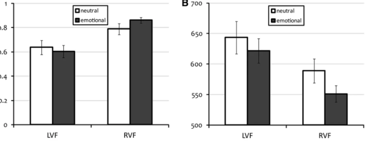
Figure 2 indicates the mean and standard error of the accu-racy and median Rt in the EEG study. In view of our analysis, the results for both accuracy and median Rt pro-viding two tests for each effect, a p value < 0.025 critical level was chosen to maintain an overall p value < 0.05 error rate. Greenhouse–Geisser corrections were employed for all effects. as can be seen in Fig. 2, both the expected RVF advantage [accuracy: F(1,12) = 12.82; p value = 0.004 and Rt: F(1,12) = 6.28; p value = 0.028 n.s.] and the expected
emotional word advantage [accuracy: F(1,12) = 0.49; p value = 0.50 n.s. and Rt: F(1,12) = 17.09; p value = 0.001]
were present. an interaction between emotional word advan-tage and visual field was observed [accuracy: F(1,12) = 31.67;
p value < 0.001 and Rt: F(1,12) = 1.46; p value = 0.25 n.s.].
ERP’s topographic ANOVA results
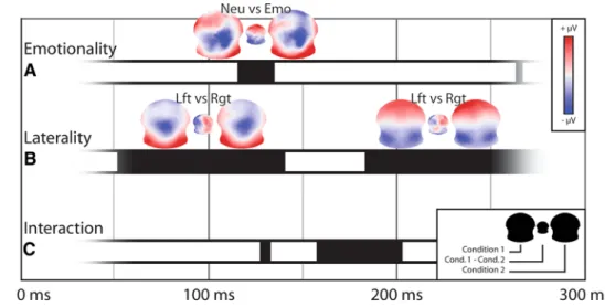
the time-point-by-time-point T-aNOVa revealed signifi-cant differences for the factor Emotionality from 116 to 134 ms after the stimulus onset (Fig. 3a). the difference map indicated increased positivity over anterior electrodes for neutral words and increased positivity for emotional words over posterior electrodes for this time window. Based on the global field power of the grand-mean ERPs (not represented), this time period falls in the transition between the P100 component and the subsequent N170-like component.
the comparison for the factor laterality showed dif-ferences in a large time window, starting just 52 ms after stimulus presentation and covering the entire P100 component—from 52 to 140 ms (Fig. 3b). In this period, the difference map between the lVF and RVF conditions showed a clear left–right polarity inversion with relative stronger right positivity when the real word was detected in the lVF and the pseudo-word in the RVF, and a left positivity in the reversed condition. the corresponding averaged maps during this time period, resembling the visual P100 topography, had left- and right-side maxima of positive potentials that were inverted between the two conditions, with more positivity over the right pos-terior electrodes for lVF words and more positivity over left posterior electrodes for RVF words. the two condi-tions differed again in a second time window starting at 184 ms—from 184 to more than 250 ms. this time, the difference map showed inverted polarities over frontal electrodes with more positivity over right frontal elec-trodes for words in the lVF and more left frontal positiv-ity for words in the RVF.
there were interactions between the two factors lasting less than 10 ms at 130 ms, then more durably from 158 to
Fig. 2 averages and standard errors of the individual accuracies (a) and median Rt in ms (b) for the four stimulus conditions with words (left
202 ms (Fig. 3c). Interactions signified that differences of map configurations for the Emotional factor are dependent on the difference according to the side of presentation; in other words, the differences of processing between neutral and emotional words were dependent on the visual field in which they were presented. the scheme of the interaction opposes any real word in the RVF and neutral words in any visual field against words in the lVF and emotional words; this indicates that words in the RVF share similar brain states with neutral words, while words in the lVF show a similar response to emotional words.
critically though, the chronology of these effects sug-gests that map topographies differed first as a function of the side of presentation of the word (50 ms post-onset), then as a function of the emotional content of the words, and finally with an interaction of these two factors after 150 ms. From 210 ms onwards, the only remaining differ-ences were for the side of word presentation.
Electrical source imaging (ESI) results
the analysis in the inverse space focused on those time periods that revealed significant topographic differences in the analysis of the scalp maps—i.e. around the P100 com-ponent and the N170-like comcom-ponent.
considering the first period of interest—i.e. from 70 to 134 ms, the analyses of variances on the z-scored cur-rent density values revealed differences for the main fac-tor laterality similarly to the scalp map analysis. these differences were found mainly in the right Precuneus— Brodmann area 7 (Fig. 4a), and somewhat weaker in the right inferior parietal lobule and RaG—Brodmann area 39, and in the left superior and middle temporal gyri—Brod-mann areas 21, 22, 41 and 42. Post hoc analysis revealed a stronger activity in these areas for words detected in the lVF as compared to the RVF.
concerning the second time period of interest—i.e. from 130 to 210 ms, the analysis of variance revealed differences for both main factors laterality and Emotionality and for the interaction between the two factors. the first differences were found for the factor Emotionality in the very beginning of the period from 132 to 156 ms, in a large cortical area from the right inferior to the superior parietal lobule—Brodmann areas 7, 19, 39 and 40. Post hoc analyses revealed stronger activ-ity for emotional words than for neutral words in these areas (Fig. 4b). the factor laterality differed from 148 ms onwards. From 148 to 170 ms, the differences were found first in the right supramarginal gyrus—Brodmann area 40, and in the left post-central gyrus. afterwards, from 172 to 200 ms, dif-ferences were found in an area connecting the posterior part of the right middle temporal gyrus, the RaG, and the ante-rior part of the right extrastriate cortex—Brodmann areas 19, 37 and 39, in the left Insula—Brodmann area 13, and in the left inferior frontal gyrus. all these areas of divergence were found to be more active for the detection of words presented in the lVF than in the RVF, the most relevant area being the right middle temporal gyrus (Fig. 4c). Finally, the aNOVa revealed interaction effects at the end of the period from 172 to 206 ms, with differences located in the left precuneus and in the posterior part of the left middle temporal gyrus—Brod-mann area 21 (not represented). the interaction was mainly due to stronger activity for emotional words detected in the lVF than neutral words detected in the RVF.
study 2 (tMs)
EEG‑based TMS target choice
the targets of the tMs and the timing of stimulation were both based on the results of the EEG study. as described in the EEG results section above, the main differences identified by the z-score statistics of EsI were recurrently Fig. 3 topographic aNOVa
comparisons of data accord-ing to the emotional nature (emotionality) or the side of presentation (laterality) of words and their interaction across time from 50 to 250 ms after the onset of the stimulus. Black solid rectangles depict time points of significance (p < 0.05), and the topographic maps (μV) show the maps aver-aged for these time periods for the corresponding conditions as well as the difference maps between them (n = 13)
located in the right temporoparietal area during the periods of interest from 70 to 200 ms (Fig. 4). More specifically, from the beginning of significance of the Emotionality fac-tor at 132 ms, these differences in the right hemisphere were found on a triangle delineated by the posterior part of the right middle temporal gyrus, the right superior parietal lobule and the anterior part of the right extrastri-ate cortex. therefore, the right temporoparietal junction was chosen as a target area for tMs and more precisely in the angular gyrus. Interestingly, according to the aver-ages of the z-scores of EsI (not represented) and following the initial activation of the primary visual cortices, the left tPJ was consistently activated in any condition while the right was more activated by emotional and lVF words—as suggested by the statistical analysis in EsI. consequently, we decided to test the implication of the two hemispheres by stimulating two homologous areas in the tMs study. these bilateral activations were present from around 70 ms. We chose to initiate the stimulation at 50 ms in order to disturb the focused area before the reception of information—i.e. the transfer from the primary visual cor-tex to the ipsilateral tPJ—and to curtail the propagation to other areas.
Behavioural data and vertex normalised index
the mean and standard error of the accuracy and median Rt are resumed in table 1. the volunteers did not report any difficulties executing the task during tMs, and accu-racy was above chance (>0.33). In view of our analysis, the results for both accuracy and median Rt providing two tests for each effect, a p value < 0.025 critical level was chosen to maintain an overall p value < 0.05 error rate.
Greenhouse–Geisser corrections were employed for all effects. as in study 1, the data demonstrated a replication of the well-known advantage for the detection of words in Fig. 4 Graphical representations on horizontal sections of MNI
tem-plate brain of the significant differences (p < 0.01 for at least 10 ms) from aNOVa of the z-scored current density values according to the factors Emotionality (Neu/Emo) and laterality (lVF/RVF), weighted by the t values from post hoc analyses and averaged over time peri-ods. Warm colours depict differences in favour of the first group
(Neutral or lVF), and cold colours depict differences in favour of the second group (Emotional or RVF). Brain sections were represented in neurological convention (“left is left”). For an easier reading of the obtained differences, graphical representations were condensed on the maximal difference averaged on time periods of interest based on the topographical results (n = 13)
Table 1 averages and standard errors of the individual accuracies
and median Rt for the four stimulus conditions with words (left or right visual field neutral word, and left or right visual field emotional word) and the three conditions of tMs (vertex, left aG, and right aG) in study 2 (tMs) (n = 10)
Neutral Emotional
left Right left Right
accuracy Vertex average 0.479 0.783 0.525 0.825 sE 0.048 0.046 0.054 0.037 laG average 0.446 0.763 0.454 0.813 sE 0.057 0.035 0.029 0.020 RaG average 0.471 0.813 0.488 0.833 sE 0.049 0.043 0.055 0.032 Median Rt Vertex average 799.6 651.8 727.9 624.2 sE 32.0 37.3 40.4 37.2 laG average 782.0 678.7 735.4 637.7 sE 45.4 45.7 39.2 47.9 RaG average 773.5 677.9 778.7 629.9 sE 51.3 49.9 39.5 45.0
the RVF compared with the lVF [accuracy: F(1,9) = 55.26;
p value < 0.0001 and Rt: F(1,9) = 22.77; p value = 0.001] and for emotional words compared with neutral words [accuracy: F(1,9) = 0.96; p value = 0.95 n.s. and Rt:
F(1,9) = 10.13; p value = 0.01]. Neither an interaction nor an effect of the factor tMs was found.
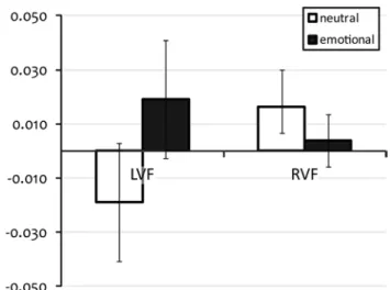
the mean and standard error of VNI for accuracy and median Rt are summarised in table 2. In view of our analysis, the results for both accuracy and median Rt providing two tests for each effect, a p value < 0.025 critical level was chosen to maintain an overall p value < 0.05 error rate. Greenhouse–Geisser corrections were employed for all effects. Results of the aNOVa with the VNI of accuracy did not show independent effects of a factor or any interaction. the results for the aNOVa with the VNI of the Rt also did not show any independent effects of the different factors—i.e. location of tMs, lat-erality (side of presentation) or Emotionality. however, there was an interaction between the factors laterality and Emotionality [F(1,9) = 8.02; p = 0.02]. While the
lim-ited statistical power provided by the n = 10 sample size precludes follow-up post hoc statistical tests of this inter-action, the basis for the interaction appears from Fig. 5, the detection of emotional words presented to the lVF was more slowed by tMs in comparison with the detec-tion of neutral words, while the reverse was true for words presented to the RVF.
Discussion
Performances in word detection change from one indi-vidual to another (levy et al. 2010) and as a function of the stimuli or their environment. Differences of the type of words and the conditions in which they are presented lead to multiple detection strategies reflected also by differences in performance (Riba et al. 2010). the location of the word in the visual field will define by which cerebral hemisphere the visual information is initially treated. the brief simulta-neous bilateral presentation used in our experiments allows a single-letter string to be initially projected to its contralat-eral target hemisphere as segregated information since it excludes reflexive saccades during these very short presen-tation times. While this dissociation at the entry stage might appear unnatural, the distinction between hemispheres for word detection constitutes an interest for the understanding of the functional lateralisation and the balance between the two hemispheres in the case of challenging situations in the healthy brain or following brain injuries such as stroke. the other factor tested in the present study was the emotionality of the words. Emotionality is known to lead to an advan-tage in detection compared with neutral conditions and to interfere with the processing of word reading (schacht and sommer 2009; Kissler and Koessler 2011; skrandies
2013). since a lexical analysis would be assumed to start before a word can be characterised as emotional, this early differentiation for emotional word detection is surprising. the current study confirmed and refined the previous find-ings on the specificity of processing of emotional words and the role of the right hemisphere in lexical decision Table 2 averages and standard errors of the vertex normalised index
(VNI) of accuracy and median reaction times (Rt) for tMs over left and right angular gyri (aG) for the four stimuli conditions with words (left or right visual field neutral word, and left or right visual field emotional word) in the study 2
Positive VNI indicates augmented accuracy or slowed Rt compared with vertex stimulation (n = 10)
Neutral Emotional
left Right left Right
VNI accuracy laG average −0.051 −0.009 −0.053 −0.005 sE 0.053 0.028 0.045 0.020 RaG average 0.039 0.029 0.003 0.011 sE 0.064 0.019 0.055 0.022 VNI median Rt laG average −0.015 0.017 0.005 0.006 sE 0.019 0.013 0.022 0.015 RaG average −0.023 0.015 0.034 0.001 sE 0.028 0.016 0.026 0.007
Fig. 5 averages and standard errors of the vertex normalised index
(VNI) of median reaction times (Rt) merged between left and right tMs for the four stimulus conditions with words (left or right visual field neutral word, and left or right visual field emotional word) in study 2. a positive VNI indicates slowed Rt compared with vertex stimulation (n = 10)
(Ortigue et al. 2004) by further analyses with new algo-rithms and extended them by showing that tMs during these early latencies over the temporoparietal areas selec-tively influences emotional word processing. In addition, it revealed a cooperative engagement of these homologous areas. Our results on specific early bilateral processing of written words will be integrated in a spatiotemporal model of cerebral mechanisms.
Early bilateral processing of words
the behavioural data show that the participants recognised the words—accuracies were above chance in all conditions for the two studies (Fig. 2; table 1), denoting non-sublimi-nal phenomena in spite of the rapid presentation of the stim-uli. Moreover, behavioural results in the two studies show that our paradigm was in accordance with the well-known advantage for the detection of words presented to the RVF in the right-handed male subject (Young et al. 1980; Graves et al. 1981; Iacoboni and Zaidel 1996; Babkoff et al. 1997) and the advantage for the detection of emotional words compared with neutral words (landis 2006). For any trial, each visual hemifield, and therefore each opposite hemi-sphere, receives a four-letter string flashed exactly in the same fashion. the only remaining difference is the sig-nificance of these letters—i.e. they are a pseudo-word or a real word, neutral, or emotional. Words are detected better and faster when they are presented in the RVF com-pared with the lVF, and they are also detected faster when they are emotional. these behavioural results reflect the electrophysiological signals. In spite of apparent similar global topographic maps and electric source images in all conditions—a certain similarity is to be expected when physical parameters are identical between conditions—the inter-condition contrasts stand out with obvious differ-ences. the cerebral processing is already different at early stages, during the P100 component, according to the side of presentation of a word and according to its emotional content. these results agree with a rapid access to the lexi-cal dimension after stimulus onset reported in the previous literature (scott et al. 2009; hauk et al. 2009; segalowitz and Zheng 2009; skrandies 2013). these differences occur, however, much earlier than the classically reported time for letter string processing around 150 ms (Grainger and holcomb 2009; Pylkkanen and Marantz 2003). Moreover, the divergences found in our bilateral lexical decision stud-ies in EEG and tMs argue for a specific detection at early stages in each hemisphere. these early latencies argue for an extremely fast start of the discrimination process before the crossing of the information to the opposite hemisphere is likely. similar early differences in the ERPs between 80 and 150 ms depending on the connotative meaning of visu-ally presented words have been described by skrandies in
adults as well as in children (skrandies 2013). the fact that in our study the words are very short and familiar plays a probable role in the swiftness of processing. Dissimilarities between the detection of a word in the lVF versus in the RVF shown by topographic analysis are mainly reflected as differences in left–right lateralisation of the polarities (Fig. 3b) during the two time windows of interest—i.e. P100 and N170. More than a simple left–right inversion of the pattern of activations in the brain, the aNOVa on the
z-scored current density values explains this persistent dis-similarity in topography with additional generators initially located in the right precuneus with a supplementary acti-vation for words detected in the lVF (Fig. 4a), in addition to the standard network activated by the detection of words in the RVF. While the P100 mainly corresponds to the pro-cessing of visual information in the visual cortex, these results argue for differences that are due to concomitant preprocessing of word material in areas beyond the primary cortex. the precuneus has functional connections with 1) the visual cortex from its posterior part along the parieto-occipital fissure and 2) the inferior parietal lobule and the superior temporal sulcus from its central part near the pre-cuneal sulcus (Margulies et al. 2009; Zhang and li 2012). the expected activation of the visual cortex by words flashed in both hemifields is followed by the activation of the inferior parietal lobule and the superior temporal sul-cus for further steps of word processing. Indeed, the diver-gences are then located in the areas adjacent to the occipital lobe in particular with the right tPJ around 130 ms and the right middle temporal gyrus at 170 ms (Fig. 4b, c). again, the results indicate supplementary activations in the right hemisphere, respectively, for the detection of emotional words and words presented in the lVF.
a delayed but superimposed processing of the emotionality the assumption of a lexical detection process in the right hemisphere for words presented to the lVF would not have been validated by the isolated analysis of the lateral-ity factor in the EEG study. Indeed the observed difference between the two conditions, lVF versus RVF, at a given time could be solely due to a detection of a word in the RVF by the left hemisphere versus a non-detection in the other. however, the results from the aNOVa on the z-scored cur-rent density values indicate functional activations of the right hemisphere. Moreover, the early divergences in the process-ing of emotionality (Fig. 3a, b) and the interactions involv-ing the factors Emotionality and laterality point to an actual early detection of words also in the lVF and different detec-tions in both hemispheres. the tMs results argue in favour of a cooperative network between both hemispheres that is balanced according to the two factors. specific effects for emotional words in the lVF point to a semantic treatment.
compared with the laterality factor, the differences due to the factor Emotionality in the EEG study are more focused in time and appear a little later at 116 ms in the topography analyses (Fig. 3a)—orthogonally to what was observed in the results for the factor laterality. Results from the aNOVa on z-scores of current density values show brief but strongly significant differences for the factor Emotionality between the P100 and the N170-like compo-nents, from 132 ms onwards, with a maximum of increased activity for the detection of emotional words compared with neutral words in the right tPJ (Fig. 4b). hence, the emotional differentiation might thus occur downstream of the primary visual cortices. Interestingly, the tPJ is con-nected with the precuneus and also with the primary visual cortex (Richardson et al. 2011; Zhang and li 2012).
additionally, the slight emotional advantage in detection in the RVF in study 1 (Fig. 2) could be due to the observed bilateral implication of the tPJs. Indeed, RVF emotional words could be processed in the left hemisphere as they come from the RVF and by an extra processor in the right hemisphere for their Emotionality (Fig. 4b). therefore, the two sites involved in RVF emotional word process-ing might lead to a redundant lexical treatment and reduce missed events while only the right side is involved in the processing of emotional words from the lVF in the condi-tions of our experiment—i.e. with short, abstract, and very familiar words. Interestingly, the absence of a significant advantage in Rt for these emotional words coming from the RVF (table 1) is in accordance with a model of dou-ble processing in the left and the right hemispheres. the bilateral processing of these words would be better but not necessarily faster due to multiple exchanges between brain areas.
Functional interaction between the conditions
Obviously the brain computes words in the lVF or the RVF as well as neutral or emotional words in a different way. Nevertheless, the interactions found in the EEG and tMs results during the first 250 ms of processing suggest that word detection varies as a function of Emotionality and laterality in a dependent and similar way and indicate a difference but not a clear distinction. More precisely, the processing of words in the lVF is related to the processing of emotional words, while the detection of neutral words and of words presented in the RVF would also converge to similar brain states. a possible reason could be that the treatments of these two characteristics interact in the pro-cessing of word detection because they share similar struc-tures or networks—as their disruption by short-burst tMs tends to assert. In the case of regular word detection, some unavoidable relays may be used. the interactions would be generated by interdependent collisions (interactions)
in these common neural substrates. as developed hereaf-ter, the tPJ regions might be at the centre of a possible common network. as tMs is released from 50 to 150 ms, the physiological functions of the target area and possibly its connected areas are degraded during this time period and the following decades of milliseconds (Valero-cabré et al. 2007). tMs applied over the left or RaG does not disturb the key tendencies of the behaviour of the par-ticipants. the detection is still better for words presented in the RVF compared with lVF, and emotional words are detected faster than neutral words regardless of the tMs conditions (table 1). however, compared to what is observed for the vertex stimulation, tMs modifies the Rt when applied over the left or RaG according to the fac-tors laterality and Emotionality (table 2). Nevertheless, as the slowing is not the same for all categories of stimuli, the implication of the angular gyri is stimulus-dependent. Indeed, the behavioural perturbation mediated by the dis-ruption of the angular gyri in the first 200 ms is an interac-tion indicating that emointerac-tional word detecinterac-tion in the lVF is more impaired by tMs in comparison with the detec-tion of neutral words. this is supported by the difference between neutral and emotional word detection for the lVF, while the detection of words in the RVF shows a weaker difference (Fig. 5). In this sense, the detection of words in the lVF would have a more weight in the interaction found in the aNOVa of VNI for median Rt. the tMs evidence indicates that the processing of emotional words in the lVF can be impaired by acting on bi-hemispheric targets of the tPJs, namely the angular gyri. Both tPJs might have cooperative functional roles in word detec-tion as already suggested by differences found in both hemispheres in the EsI analyses. Notably, previous stud-ies using non-repetitive tMs showed rapid contralateral propagations of tMs-evoked activations to the homolo-gous area in the motor cortex (Ilmoniemi et al. 1997; Parks et al. 2012; casali et al. 2013; Ragazzoni et al. 2013), the premotor cortex (Massimini et al. 2005) as well as the visual cortex (Ilmoniemi et al. 1997). although all these studies used single-pulse stimulation protocols, a trans-callosal propagation with our triple-pulse stimulation that would lead to the observed absence of distinction between left and right tMs cannot be fully ruled out. the right side that is specialised for emotional processing could thus also be disturbed by the stimulation of the left side. In this case, the effect of the tMs of the left tPJ could be due to the impairment of the activations found in the EsI analy-ses in the right tPJ. While differences between tMs over the left or the right tPJs did not reach statistical signifi-cance, the stronger slowing of the detection of emotional words from the lVF due to the disruption of the right tPJ (highest VNI for median Rt in table 2) could result from a main contribution of this structure in the network activated
by the detection of emotional words and words in the lVF as revealed concordantly in the EsI analyses (Fig. 4). this last point would indeed argue in favour of an effect of tMs over the left tPJ mediated by the propagation of dis-turbance to the right. Further combined tMs–EEG studies during task execution are needed to fully clarify the mech-anisms underlying the observed effects.
a left–right balance
the results from the EEG and tMs experiments argue for a variable implication of both hemispheres in the detection of written words balanced by the conditional involvement of the right hemisphere according to different conditions. Indeed, the main areas of divergence in the EEG analysis are found in the right hemisphere—in or close to the right tPJ—across both time periods of interest. the tMs results confirm the conditional implication of the right tPJ and suggest a possible cooperation with the left homologous tPJ. thus, the right hemisphere would be more active for the detection of emotional words and words presented in the lVF. the areas thereby involved in the right hemisphere would be part of the process in addition to the regular net-work, which is located in the left hemisphere. conversely, the balance would be all the more in favour of the left hem-isphere for neutral words and words coming from the RVF, with a reduced intervention of the right hemisphere during early stages of word processing.
the recruitment of the right hemisphere observed for emotional words is in accordance with an important part of the literature demonstrating the role of the right hemisphere in emotional functions in general (from Gainotti 1972,
2005 with the “right hemisphere hypothesis” to the adapted version of the “valence hypothesis” of Davidson 2001 for the comprehension of emotional information) and more interestingly in the processing of emotional words (lan-dis 2006). Numerous studies were conducted in aphasic patients showing an advantage for emotional word process-ing with a lesion of the left hemisphere (landis et al. 1982,
1983; Reuterskiöld 1991) or specific emotion processing impairments with a right hemispheric lesion (Borod et al.
1992; lalande et al. 1992; cicero et al. 1999). In addition to the left lateralised language network, the processing of word-related emotionality is also found in the right hemi-sphere in healthy participants—e.g. Ortigue et al. 2004; Frühholz et al. 2011; Ponz et al. 2013. Our results argue for supplementary processing performed by the right hemi-sphere at very early stages. Moreover, this lexical emotion processing in the right hemisphere is dependent on the con-nection with the left hemisphere. this interhemispheric cooperation could explain paradoxical left-sided processing of emotionality in words observed in some studies (Kissler et al. 2007; herbert et al. 2008).
the role of the tPJ
the tPJs could be a part of the networks as either a path-way or a processor or both. the fact that the tMs-induced disturbance also includes Emotionality supports an advanced treatment of words in both tPJs or at least in the related upstream areas, and would disagree with a simple pathway of the information that does not involve word pro-cessing. the left tPJ is known to be implicated in language comprehension (for review Price 2012). Delimited parts of the tPJ have shown their contribution for specific aspects of visual word processing, such as the supramarginal gyrus in the phonologic dimension of visual word recognition (stoeckel et al. 2009), the angular gyrus in response to semantic versus phonologic aspects (Binder et al. 1999), or in multimodal processing (Bonner et al. 2013), while the posterior superior temporal gyrus is rather implicated in word sound processing (Buchsbaum et al. 2001). a bilat-eral involvement is depicted in the stG notably for speech intonation processing, and the right temporal lobe is also more engaged in speech processing during sentence and context comprehension (Vigneau et al. 2011; Diaz and hogstrom 2011). Bilateral activation of the angular gyri and superior temporal gyri is found in word reading com-pared to non-words (Kuchinke et al. 2005) or angular gyri alone in high-frequency word reading (Graves et al. 2010). In the present work, the two tPJs engage a real coop-eration, which is conditioned by the stimulus features to achieve word reading. the right hemisphere is involved for lVF words and emotional words implying a bilateral pro-cessing with an interaction between the two hemispheres as shown by interaction in the EsI results and suggested by tMs. accordingly, Vigneau et al. (2011) also found an implication of the right hemisphere in an inter-hemispheric manner in association with left homologous areas.
spatiotemporal model
Based on our studies, a spatiotemporal model may be proposed that would explain the path of information pro-cessing in four steps. the information from each visual hemifield reaches each primary visual cortex separately 50 ms after the stimulus display as revealed by the aver-aged z-scores of EsI. From here, divergences are observed in the processing of word detection with maximal diver-gences around 100 ms in the right precuneus and slightly in the right inferior parietal lobules according to laterality. average activities in z-scores of EsI are already found in both tPJs before 100 ms. as a first step, a precursor of the lexical decision involving the classification of a letter string as a word—or not—is elicited in the receptor hemisphere from 50 to 130 ms. activity is concentrated around the superior temporal sulci with diverging processing in terms
of laterality in the right supramarginal gyrus from 160 ms onwards and then in the superior temporal gyrus—i.e. higher activity is observed for words detected in the lVF. In the meantime, a precursor of a semantic analysis of the let-ter strings has to be initiated to enable further computation of their emotional characteristics. the emotional detection appears to start with first differentiations between neutral and emotional words at 130 ms. In this second step, while the tPJs are already engaged in lateralised processing, the influence of Emotionality emerges concomitantly from 110 to 150 ms. the superior part of the right tPJ shows a spe-cific treatment of emotionality from 132 to 156 ms with a higher activity found in the inferior parietal lobule for emo-tional words. at this point, the processing seems to be spe-cific in each hemisphere while also integrating information from interhemispheric communication. Indeed, the activa-tion suggests an inter-hemispheric crossover of informa-tion—i.e. for emotional words from the RVF. From there, the interaction between the two factors appears at 130 ms and then from 160 to 210 ms. the interaction could indi-cate a conflict between the processing of different factors in common areas of the two hemispheres from precuneus to temporal lobes via inferior parietal lobules. the interac-tion found in the tMs study tends to confirm the scenario of this bilateral communication with the contribution of the left and right tPJ. From 210 ms onwards, the differ-ences were found for laterality only in the right tPJ and in the primary visual cortex, followed by activations in the primary visual cortex and the left middle temporal gyrus (not related to the “Results” section). this could imply further computations that are related to the motor response required by the task; however, the EsI would rather sug-gest a feedback process with activations and differences located in primary visual areas, which might enable inte-gration for further semantic processing. From this point onwards, the processing would eventually continue in the left hemisphere.
Conclusions
In conclusion, our results show that lexical detection pro-cessing starts very early in both hemispheres. a bilateral network is required for the detection of words according to the side of presentation and their emotional content. More-over, analysis of slowed Rt in the tMs experiment indi-cates that interference with both left and right tPJ results in impaired processing of emotional words that were pre-sented to the lVF. Our research confirms the hypothesis of an early cooperation between the two hemispheres condi-tioned by word features. the complementarity between the left and the right side of the brain together with their inter-action in word processing constitutes a critical point in the
understanding of reading in healthy subjects but also in the case of recovery from injury.
Acknowledgments this research was supported by the swiss
National science Foundation: Grant No. 320030_132967 to theodor landis and Grant No. 310030_132952 to christoph M. Michel. the tMs equipment and the cartool software are supported by the center for Biomedical Imaging (cIBM), Geneva and lausanne, switzerland. the cartool software was developed by Denis Brunet from the center for Biomedical Imaging (cIBM), Geneva and lausanne, switzer-land. http://sites.google.com/site/fbmlab/cartool. the stEN toolbox (http://www.unil.ch/fenl/sten) was programmed by Jean-François Knebel, from the Functional Electrical Neuroimaging laboratory, lausanne, switzerland, and is supported by the center for Biomedical Imaging (cIBM) of Geneva and lausanne and by the National center of competence in Research project “sYNaPsY—the synaptic Bases of Mental Disease”; Project No. 51aU40_125759. the authors would like to thank alexis hervais-adelman who assisted us with the analysis of selecting a balanced number of epochs between conditions in study 1 (EEG). special thanks go to Gregor thut who helped to initiate this project and provided precious input and expertise during all stages of the project.
References
alexander DM, Wright JJ (2006) the maximum range and timing of excitatory contextual modulation in monkey primary visual cor-tex. Vis Neurosci 23:721–728
Babkoff h, Faust M, lavidor M (1997) lexical decision, visual hemi-field and angle of orientation. Neuropsychologia 35(4):487–495 Binder JR, Frost Ja, hammeke ta, Bellgowan PsF, Rao sM, cox
RW (1999) conceptual processing during the conscious resting state: a functional MRI study. J cogn Neurosci 11(1):80–93 Bonner MF, Peelle JE, cook Pa, Grossman M (2013) heteromodal
con-ceptual processing in the angular gyrus. NeuroImage 71:175–186 Borod Jc, andelman F, Obler lK, tweedy JR, Welkowitz J (1992)
Right hemisphere specialization for the identification of emo-tional words and sentences: evidence from stroke patients. Neu-ropsychologia 30:827–844
Broca P (1861) Remarques sur le siège de la faculté du langage articulé, suivies d’une observation d’aphémie (perte de la parole). Bulletins de la société anatomique de Paris 6:330–357
Brunet D, Murray MM, Michel cM (2011) spatiotemporal analy-sis of multichannel EEG: caRtOOl. comput Intell Neurosci 2011:813870
Buchsbaum BR, hickok G, humphries c (2001) Role of left posterior superior temporal gyrus in phonological processing for speech perception and production. cogn sci 25(5):663–678
casali aG, Gosseries O, Rosanova M, Boly M, sarasso s, casali KR, casarotto s, Bruno M-a, laureys s, tononi G, Massimini M (2013) a theoretically based index of consciousness independent of sensory processing and behavior. sci transl Med 5(198):198ra105 cicero Ba, Borod Jc, santschi c, Erhan hM, Obler lK, agosti RM,
Welkowitz J, Grunwald Is (1999) Emotional versus nonemo-tional lexical perception in patients with right and left brain dam-age. Neuropsychiatry Neuropsychol Behav Neurol 12:255–264 Davidson RJ (2001) Prefrontal and amygdala contributions to
emo-tion and affective style. In: Gainotti G (ed) handbook of neu-ropsychology, emotional behaviour and its disorders, vol 5, 2nd edn. Elsevier, amsterdam, pp 111–112
Diaz Mt, hogstrom lJ (2011) the influence of context on hemi-spheric recruitment during metaphor processing. J cogn Neurosci 23(11):3586–3597
Frühholz s, Jellinghaus a, herrmann M (2011) time course of implicit processing and explicit processing of emotional faces and emotional words. Biol Psychol 87(2):265–274
Gainotti G (1972) Emotional behavior and hemispheric side of the lesion. cortex 8(1):41–55
Gainotti G (2005) Emotions, unconscious processes and the right hemisphere. Neuro-Psychoanal 7:71–81
Gazzaniga Ms (2005) Forty-five years of split-brain research and still going strong. Nat Rev Neurosci 6(8):653–659
Gazzaniga Ms, hillyard sa (1971) language and speech capacity of the right hemisphere. Neuropsychologia 9:273–280
Gazzaniga Ms, sperry RW (1967) language after section of the cer-ebral commissures. Brain 90:131–148
Grainger J, holcomb PJ (2009) Watching the word go by: on the time-course of component processes in visual word recognition. lang linguist compass 3(1):128–156
Grave de Peralta Menendez R, Gonzalez andino s, lantz G, Michel cM, landis t (2001) Noninvasive localization of electromag-netic epileptic activity I Method descriptions and simulations. Brain topogr 14(2):131–137
Grave de Peralta Menendez R, Murray MM, Michel cM, Martuzzi R, Gonzalez andino sl (2004) Electrical neuroimaging based on biophysical constraints. Neuroimage 21(2):527–539
Graves R, landis t, Goodglass h (1981) laterality and sex differ-ences for visual recognition of emotional and non-emotional words. Neuropsychologia 19(1):95–102
Graves WW, Desai R, humphries c, seidenberg Ms, Binder JR (2010) Neural systems for reading aloud: a multiparametric approach. cereb cortex 20(8):1799–1815
hauk O, Pulvermuller F, Ford M, Marslen-Wilson WD, Davis Mh (2009) can I have a quick word? Early electrophysiological manifestations of psycholinguistic processes revealed by event-related regression analysis of the EEG. Biol Psychol 80(1):64–74 herbert c, Junghöfer M, Kissler J (2008) Event related potentials to
emotional adjectives during reading. Psychophysiology 45:487–498 Iacoboni M, Zaidel E (1996) hemispheric independence in word rec-ognition: evidence from unilateral and bilateral presentations. Brain lang 53(1):121–140
Ilmoniemi RJ, Virtanen J, Ruohonen J, Karhu J, aronen hJ, Näätänen R, Katila t (1997) Neuronal responses to magnetic stimula-tion reveal cortical reactivity and connectivity. Neuroreport 8(16):3537–3540
Kissler J, Koessler s (2011) Emotionally positive stimuli facilitate lexical decisions—an ERP study. Biol Psychol 86(3):254–264 Kissler J, herbert c, Peyk P, Junghöfer M (2007) Buzzwords. Early
cortical responses to emotional words during reading. Psychol sci 18:475–480
Koenig t, Kottlow M, stein M, Melie-García l (2011) Ragu: a free tool for the analysis of EEG and MEG event-related scalp field data using global randomization statistics. comput Intell Neurosci 2011:938925
Koenig t, stein M, Grieder M, Kottlow M (2013) a tutorial on data-driven methods for statistically assessing ERP topographies. Brain topogr (Epub 29 aug 2013)
Kuchinke l, Jacobs aM, Grubich c, Võ Ml, conrad M, herrmann M (2005) Incidental effects of emotional valence in single word processing: an fMRI study. Neuroimage 28(4):1022–1032 lalande s, Braun cM, charlebois N, Whitaker ha (1992) Effects of
right and left hemisphere cerebrovascular lesions on discrimi-nation of prosodic and semantic aspects of affect in sentences. Brain lang 42(2):165–186
landis t (2006) Emotional words: what’s so different from just words? cortex 42(1):170–179
landis t, Graves R, Goodglass h (1982) aphasic reading and writ-ing: possible evidence for right hemisphere participation. cortex 18:105–112
landis t, Regard M, Graves R, Goodglass h (1983) semantic paralexia: a release of right hemisphere function from left hemi-spheric control? Neuropsychologia 21:359–364
lehmann D, skrandies W (1980) Reference-free identification of components of checkerboard-evoked multichannel potential fields. Electroencephogr clin Neurophysiol 48:609–621
levy t, Walsh V, lavidor M (2010) Dorsal stream modulation of vis-ual word recognition in skilled readers. Vis Res 50(9):883–888 lidzba K, schwilling E, Grodd W, Krägeloh-Mann I, Wilke M (2011)
language comprehension vs. language production: age effects on fMRI activation. Brain lang 119(1):6–15
Margulies Ds, Vincent Jl, Kelly c, lohmann G, Uddin lQ, Biswal BB, Villringer a, castellanos FX, Milham MP, Petridesi M (2009) Precuneus shares intrinsic functional architecture in humans and monkeys. Proc Natl acad sci 106(47):20069–20074 Massimini M, Ferrarelli F, huber R, Esser sK, singh h, tononi G
(2005) Breakdown of cortical effective connectivity during sleep. science 309(5744):2228–2232
Michel cM, Murray MM (2011) towards the utilization of EEG as a brain imaging tool. Neuroimage 61:371–385
Michel cM, Murray MM, lantz G, Gonzalez s, spinelli l, Grave de Peralta R (2004) EEG source imaging. clin Neurophysiol 115(10):2195–2222
Michel cM, Koenig t, Brandeis D, Gianotti lRR, Wackermann J (2009) Electrical neuroimaging. cambridge University Press, cambridge
Mohr c, Michel cM, lantz G, Ortigue s, Viaud-Delmon I, landis t (2005) Brain state-dependent functional hemispheric specializa-tion in men but not in women. cereb cortex 15:1451–1458 Mohr B, Endrass t, hauk O, Pulvermüller F (2007) ERP correlates
of the bilateral redundancy gain for words. Neuropsychologia 45(9):2114–2124
Morand s, thut G, Grave de Peralta R, clarke s, Khateb a, landis t, Michel cM (2000) Electrophysiological evidence for fast visual processing through the human koniocellular pathway when stim-uli move. cereb cortex 10:817–825
Murray MM, Brunet D, Michel cM (2008) topographic ERP analy-ses: a step-by-step tutorial review. Brain topogr 20:249–264 New B, Pallier c, Ferrand l, Matos R (2001) Une base de données
lexicales du français contemporain sur internet: lEXIQUE. l’année Psychologique 101:447–462
Ortigue s, Michel cM, Murray MM, Mohr c, carbonnel s, landis t (2004) Electrical neuroimaging reveals early generator modula-tion to emomodula-tional words. Neuroimage 21(4):1242–1251
Papagno c, Fogliata a, catricalà E, Miniussi c (2009) the lexical processing of abstract and concrete nouns. Brain Res 1263:78–86 Parks Na, Maclin El, low Ka, Beck DM, Fabiani M, Gratton G
(2012) Examining cortical dynamics and connectivity with simul-taneous single-pulse transcranial magnetic stimulation and fast optical imaging. Neuroimage 59(3):2504–2510
Pell MD (2007) Reduced sensitivity to prosodic attitudes in adults with focal right hemisphere brain damage. Brain lang 101(1):64–79
Perrin F, Pernier J, Bertrand O, Giard Mh, Echallier JF (1987) Map-ping of scalp potentials by surface spline interpolation. Electroen-cephalogr clin Neurophysiol 66:75–81
Ponz a, Montant M, liegeois-chauvel c, silva c, Braun M, Jacobs aM, Ziegler Jc (2013) Emotion processing in words: a test of the neural re-use hypothesis using surface and intracranial EEG. soc cogn affect Neurosci. doi:10.1093/scan/nst034. (advance online publication)
Price cJ (2012) a review and synthesis of the first 20 years of PEt and fMRI studies of heard speech, spoken language and reading. Neuroimage 62(2):816–847
Pylkkanen l, Marantz a (2003) tracking the time course of word rec-ognition with MEG. trends cogn sci 7(5):187–189