REVIEW
Matricellular protein CCN1/CYR61: a new player
in inflammation and leukocyte trafficking
Yalin Emre&Beat A Imhof
Received: 31 January 2014 / Accepted: 19 February 2014 / Published online: 18 March 2014 # Springer-Verlag Berlin Heidelberg 2014
Abstract Cystein-rich protein 61 (CYR61/CCN1) is a com-ponent of the extracellular matrix, which is produced and secreted by several cell types including endothelial cells, fibroblasts and smooth muscle cells. CCN1 has been impli-cated in leukocyte migration and the inflammatory process, but it is also involved in cardiovascular development and carcinogenesis. It exerts its functions through binding to tiple integrins present in many different cell types. This mul-tiplicity in function is now known to contribute to the diverse array of cellular processes it can regulate. The expression of CCN1 is tightly regulated by cytokines and growth factors. However, CCN1 can directly modulate cell adhesion and migratory processes whilst simultaneously regulating the pro-duction of other cytokines and chemokines through paracrine and autocrine feedback loops. This complex functionality of CCN1 has highlighted the pivotal role this molecule can play in regulating the immunosurveillance process. Furthermore, CCN1 has now emerged as an important partner when targeting components of the infectious or chronic inflamma-tory disease processes such as atherosclerosis or rheumatoid arthritis. This review will focus on CYR61/CCN1 and its ability to control the migration of leukocytes, the production of cytokines and cell proliferation or senescence at the site of inflammation.
Keywords CCN1 . CYR61 . Extracellular matrix . Leukocyte migration . Inflammation
Abbreviations
CCN1/CYR61 Cysteine-rich protein 61 ECM Extracellular matrix FLS Fibroblast-like synoviocytes LPA Lysophosphatidic acid MMP Metalloproteinase RA Rheumatoid arthritis
VSMC Vascular smooth muscle cells
Introduction
The immune system consists of a heterogeneous cell popula-tion that shares the ability to rapidly respond to tissue and organ injury. Research in immunology is mainly focused on the roles of inflammatory cytokines and chemokines, antigen-ic and co-stimulation signals or receptors for pathogen-associated molecular patterns. However, the extracellular ma-trix (ECM) is emerging as an essential partner modulating the course of inflammation. Aside from providing a structural support for cell adhesion, the ECM influences various aspects of cellular life not only in physiological conditions but also in pathological conditions [1,2]. Immune cells infiltrating the site of inflammation secrete not only cytokines but also pro-teases, such as matrix metalloproteinases (MMP), which mod-ify the synthesis of ECM by tissue resident cells or their cleavage. Consequently, modified ECM can alter cell migra-tion, activamigra-tion, differentiation and survival. For example, cleavage of type I collagen by MMPs enables a chemotactic activity analogous to CXC-chemokine ligand 8 (CXCL8) on neutrophils during lung inflammation [3]. ECM compounds such as fibronectin, heparin sulphate or hyaluronan, have been This article is a contribution to the special issue on New paradigms in
leukocyte trafficking, lessons for therapeutics - Guest Editors: F. W. Luscinskas and B. A. Imhof
Y. Emre (*)
:
B. A. ImhofDepartment of Pathology and Immunology, Centre Médical Universitaire, University of Geneva, 1 rue Michel Servet, 1211 Genève 4, Switzerland
e-mail: yalinemre@gmail.com B. A. Imhof
well studied over many years; however, the CCN family of proteins has not been as well characterised for their role in the inflammatory response [4].
Named after the first three members, cysteine-rich protein 61 (CYR61; CCN1), connective tissue growth factor (CTGF; CCN2) and nephroblastoma overexpressed protein (NOV; CCN3), the CCN family comprises six secreted proteins that share a similar modular structure consisting of four domains exhibiting sequence homologies to insulin-like growth factor b i n d i n g p r o t e i n , v o n Wi l l e b r a n d f a c t o r t y p e C , thrombospondin type I and a cysteine knot motif [4, 5]. CCN proteins were initially identified as immediate-early genes whose secretion was induced by mitogenic factors [4–6]. More recently, studies revealed that these matricellular proteins play critical roles in cardiovascular development, inflammation, injury repair and cancer. Their functionality is mediated via specific integrin binding and heparin sulphate proteoglycans, which in turn can activate signalling pathways responsible for the regulation of cell adhesion, migration, proliferation or senescence. In addition, CCN binds growth factors (BMP, TGFβ) or ECM-associated proteins such as laminin, thus bridging cells and extracellular ligands (Fig.1) [4,5,7].
In this review, we will focus on CYR61/CCN1 and how it is implicated in many different pathways of the immune and inflammatory process.
Expression and regulation of CCN1 under homeostasis Under homeostatic conditions, matricellular protein CYR61/ CCN1 is expressed at low levels in most adult tissues by endothelial cells, fibroblasts and vascular smooth muscle cells
(VSMC). In addition, intracellular flow cytometry studies revealed constitutive expression of CCN1 in human leuko-cytes circulating in the blood [8]. No expression was reported in developing murine thymocytes [9]. During embryonic de-velopment, the importance of CCN1 for the cardiovascular system has been demonstrated by the phenotype of Ccn1-deficent mice, which are embryonic lethal because of cardiovascular defects [10] and deficiencies in promot-ing angiogenesis [11, 12].
Expression and regulation under pathological conditions Although expressed at low levels during steady-state homeostasis, it is remarkable how CCN1 expression is induced by serum growth factors, cytokines and envi-ronmental stress.
Rheumatoid arthritis
Rheumatoid arthritis (RA) is a chronic inflammatory disease that principally manifests in the synovial joints. Overexpression of CCN1 protein has been reported in fibroblast-like synoviocytes (FLS), with elevated levels ob-served in synovial fluid samples from RA patients as com-pared with healthy controls and patients suffering from oste-oarthritis [13]. Similarly, CCN1 mRNA was strongly in-creased in lymphoblastoid B cell lines derived from RA-discordant monozygotic twins [13,14], being one of the three most overexpressed genes [14]. Interestingly, mRNA and protein expression of CCN1 is inhibited by TNFα in chondrocytes [15–17], whereas it is strongly induced in pri-mary human osteoblasts and U2OS cells by TNFα, oncostatin M and the pro-inflammatory cytokines IL6 and IL1β [16,17]. However, TNFα was shown to have no effect on CCN1 expression in FLS. The expression of CCN1 in FLS is induced by IL17 in a p38 MAPK and NF-κB-dependent manner and suppressed by simvastatin treatment [16–19]. These findings strongly suggest that CCN1 plays a key role in inflammation and chronic inflammatory disease.
Bacterial and viral infection
Lysophosphatidic acid (LPA) is a bioactive lysophospholipid that can bind to specific G protein-coupled receptors termed LPA receptors. These interactions have been shown promote morphological changes and cell proliferation/survival. Production of LPA is a common feature of many bacteria. In vitro infection of primary murine epithelial cells or a human epithelial cell line with the Gram-negative bacteria Yersinia enterocolitica led to increased mRNA expression of CCN1 through the activation of LPA receptors [20, 21]. Similar results have also been obtained using Gram-negative bacteria Fig. 1 Schematic representation of CCNs. CCNs share a modular
struc-ture consisting of four domains exhibiting sequence homologies to insu-lin-like growth factor binding protein (IGFBP), von Willebrand factor type C (vWF), thrombospondin type I (TSP) and a cysteine knot motif (CK). They bridge cells to ECM compounds by binding cell surface integrins and heparin sulphate proteoglycans (HSPG)
Escherichia coli and Pseudomonas aeruginosa and Gram-positive bacteria Enterococcus faecalis or Staphylococcus aureus [21]. Induction of CCN1 protein has also been shown to occur in primary murine hepatocytes stimulated with LPS in vitro through the TLR4/MyD88/AP-1 pathway and hepa-tocytes of LPS-stimulated normal and obese mice [22]. Consistent with these observations, increased levels of CCN1 have been reported in the blood of patients with sepsis, compared to healthy controls [8].
Comparably, in vitro infection of human epithelial cell lines with poliovirus, hepatitis C virus [23] and coxsackievirus B3 [24] or in vivo with oncolytic HSV-1 in rats [25] also led to upregulation of CCN1 expres-sion in epithelial cells. The induction of CCN1 follow-ing coxsackievirus B3 infection occurs through the ac-tivation of JNK, which was induced by virus replication and viral protein expression in infected epithelial cells [24]. Interestingly, inhibition of CCN1 expression with shRNA reduces coxsackievirus B3 growth [24] whilst CCN1 treatment reduced HSV-1-derived oncolytic virus replication and cytotoxicity [26].
These studies have strongly highlighted the induction of CCN1 as a common host response to bacterial or viral infection.
Vascular injury
Protein and mRNA levels of CCN1 are increased in the plasma of patients with giant cell myocarditis [8] and cardiac biopsies of patients with dilated cardiomyopathy, respectively [27]. This was also observed in a mouse model of myocardial infarction [28] and diabetic retinopathy [29]. In the context of diabetic retinopathy, CCN1 is processed by MMP2 and MMP14 [30–32], where different domains are observed to exhibit proangiogenic or antiangiogenic activities [32]. The expression of CCN1 was upregulated in arterial smooth mus-cle walls during restenosis following balloon angioplasty in rat carotid artery, as part of ECM remodelling resulting from vascular injury [33]. Furthermore, CCN1 was shown to be highly expressed, via angiotensin 2, in aortic arteriosclerotic plaques of ApoE-deficient mice and in human arteriosclerotic lesions [34–37].
Colon inflammation
A role for angiogenesis and angiogenic factors is established in inflammatory bowel diseases. In this context, increased levels of CCN1 were measured in the colons of patients with Crohn's disease and ulcerative colitis and in colons of mice treated with dextran sodium sulphate to induce inflammatory colitis [38].
Receptors of CCN1
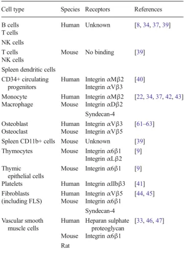
Matricellular CCN1 has the ability to mediate cell adhe-sion, mainly through binding specific integrins although it does not contain the canonical RGD sequence (Fig.1 and Table 1). This has been reported not only for human T cells, B cells, natural killer (NK) cells or monocytes in the blood [8, 34, 37, 39] but also for CD34+ circulating progenitors and platelets [40, 41]. Some discrepancies have been observed between human and mouse cells. Hence, in mouse, CCN1 binds to CD11b+ cells and B cells but not T cells or NK cells [39]. Regarding CD34+ progenitors, interactions occur through αMβ2 and αVβ3 [40]. Interactions with monocytes and macrophages are mediated by integrin αMβ2, αDβ2 and syndecan-4 [22,
34, 37, 42, 43], whilst integrin α6β1 and αLβ2 are important for thymocytes [9]. For mesenchymal cell pop-ulations, integrins αVβ5, α6β1 and syndecan-4 support CCN1 binding to fibroblasts [44,45] whilst heparan sul-phate proteoglycans and integrinα6β1 are implicated for vascular smooth muscle cells [33, 46, 47] and only integrin α6β1 for thymic epithelial cells [9].
Table 1 Receptors of CCN1
Cell type Species Receptors References B cells Human Unknown [8,34,37,39] T cells
NK cells
T cells Mouse No binding [39] NK cells
Spleen dendritic cells CD34+ circulating
progenitors
Human IntegrinαMβ2 [40] IntegrinαVβ3
Monocyte Human IntegrinαMβ2 [22,34,37,42,43] Macrophage Mouse IntegrinαDβ2
Syndecan-4
Osteoblast Human IntegrinαVβ3 [61–63] Osteoclast Mouse IntegrinαVβ5
Spleen CD11b+ cells Mouse Unknown [39] Thymocytes Mouse Integrinα6β1 [9]
IntegrinαLβ2 Thymic
epithelial cells
Mouse Integrinα6β1 [9] Platelets Human IntegrinαIIbβ3 [41] Fibroblasts Human IntegrinαVβ5 [44,45] (including FLS) Mouse Integrinα6β1
Syndecan-4 Vascular smooth
muscle cells
Human Heparan sulphate proteoglycan
[33,46,47] Mouse Integrinα6β1
Role of CCN1 in cytokine production
In diabetic retinopathy, many defects are associated with localised inflammation. The upregulation of CCN1 leads to increased expression of MCP-1 in chorioretinal vascular en-dothelial cells through theαVβ3/PI3K/Akt signalling path-way [48]. In the same way, CCN1 treatment stimulates the production of IL6 in FLS of RA patients via the αvβ5 integrin/Akt/NF-κB pathway [18], promoting Th17 cell dif-ferentiation in vitro. Interestingly, targeting CCN1 with blocking antibodies has been shown to reduce IL6 levels and a Th17 response in a collagen-induced arthritis model of RA [18]. In addition, CCN1 has been observed to be increased in the synovial fluid of RA patients, which is known to promote the proliferation and survival of FLS [13]. CCN1 also promotes CCL2 expression in osteoblasts [16], skin fibroblasts [49] and renal tubular epithelial cell [50]. In murine macrophages, treatment with CCN1 induced transcriptional changes characteristic of M1 macrophages, including the up-regulation of TNFα, IL1α, IL1β, IL6, IL12b, MIP1α, MCP3 and IP10 in an NF-κB-dependent manner [42]. In summary, these studies have shown that CCN1 plays a diverse role in promoting cytokine production and may be a key player in the inflammatory response and chronic disease.
Role of CCN1 in adhesion and migration
CCN1 supports the adhesion of a variety of cell types includ-ing monocytes [34,37], macrophages [42], developing thy-mocytes and thymic epithelial cells [9], human blood leuko-cytes [8], fibroblasts [45,51], vascular smooth muscle cells [52] or endothelial cells [53].
As well as mediating cell adhesion to multiple cell types, CCN1 has the ability to promote cell migration events of endothelial cells [32,38,53,54], cancer cells [5,55,56] and vascular smooth muscle cells [33]. CCN1 acts as a chemoattractant for lymphocytes, monocytes and murine macrophages in multiple in vitro studies [8, 22, 39]. Consistent with these observations, CCN1 had no effect on integrin density or chemokine receptor expression in human monocyte and lymphocyte populations [8]. It activates the PI3K/Akt and p38 signalling pathways and actin polymerisa-tion [8] while murine macrophage chemotaxis occurs via MEK/ERK signalling [22]. A role for CCN1 in disease has been confirmed in numerous animal studies, CCN1 protein treatment or overexpression in the liver exacerbates hepatic inflammation and macrophage infiltration in high-fat diet mice compared to normal fed mice [22]. Similarly, the impor-tance of CCN1 in kidney fibrosis was demonstrated in a model of unilateral ureteral obstruction surgery in mice [50]. In this model, treatment with anti-CCN1 antibodies reduced kidney fibrosis by decreasing macrophage infiltration [50].
Finally, simvastatin treatment inhibits CCN1 expression and CCN1-dependent infiltration of macrophages and CCN1+ osteoblasts and slows down the progression of disease in the mouse model of collagen-induced arthritis [16,17,19].
The chemotactic activity of CCN1, secreted by vascular endothelial growth factor (VEGF)-stimulated osteoblasts, reg-ulates the formation of capillary-like sprouts by endothelial cells in vitro and promotes angiogenic processes in vivo [54]. CCN1 attracts endothelial cells and promotes angiogenesis in the context of bone fracture in mouse models, thereby con-tributing to the fracture healing process [54]. In addition, CCN1 promotes the recruitment and the differentiation of circulating CD34+progenitors to endothelial cells, hinting at a role in cardiovascular regeneration [40].
Consistent with observations using chemokines, prolonged exposure to CCN1 inhibits the migration of spleen macro-phages, T cells and monocytes [8,39]. Actually CCN1 abol-ishes their chemotactic response to CCL2 or SDF1 [8,39] by downregulating PI3K, p38 and Akt signalling [8]. A regula-tory role for CCN1 in immune cell migration was also dem-onstrated in inflammatory cardiomyopathy. In experimental autoimmune myocarditis, systemic CCN1 overexpression in-hibits the migration of circulating immune cells, without af-fecting cardiac chemokine or chemokine receptor expression thereby ameliorating the disease process and reducing disease scores [39].
Role of CCN1 in cell proliferation and survival Vascular endothelium
CCN1 promotes cell survival and tubule formation in human umbilical vein endothelial cells. Interestingly, proliferation can be mediated through the integrinα6β1 in the unactivated state, whereas VEGF stimulation is required to activate the integrinαVβ3 for proliferation and adhesion/migration mech-anisms of endothelial cells [53]. A truncated form of CCN1 consisting of the insulin-like growth factor binding protein and von Willebrand factor type C domains has been shown to exhibit proangiogenic properties on retinal endothelial cells, whilst the presence of the thrombospondin type I domain to the previous variant suppressed cell growth [32].
As mentioned above, CCN1 can regulate the recruitment and differentiation of CD34+progenitor cells, which are im-portant in cardiovascular tissue regeneration [40]. Furthermore, treatment with CCN1 or supernatants from CCN1-stimulated human CD34+ cells has been shown to promote the proliferation of endothelial cells and enhance endothelial proliferation and neovascularization [40].
VSMC proliferation is characteristic of many vascular diseases, such as atherosclerosis. Forced expression of FOXO3a in VSMC decreased their viability through
inhibition of CCN1 in a rat balloon carotid arterial injury model [52].
Rheumatoid arthritis
In addition to sustaining a Th17 response in RA [18], the high levels of CCN1 detected in synovial fluid and tissue are responsible for the proliferation of FLS in RA patients. Moreover, CCN1 also protects FLS from apoptosis by main-taining Bcl-2 expression in FLS [13].
Thymic epithelial cells and thymus function
In the thymus, not only thymocytes are undergoing massive proliferation but also thymic epithelial cells (TEC) too. TEC are the main stromal cell populations of the thymic microen-vironment that provide key signals to developing thymocytes. Alteration in TEC architecture is characteristic of age-associated thymic involution or cytoablative treatments [57,
58]. In vitro, CCN1 treatment of foetal thymic lobes favours the expansion of thymic stroma by promoting the proliferation of TEC through integrinα6β1/Akt axis. In vivo, the overex-pression of CCN1 in thymic stroma increases the production of T cells via expansion of the TEC compartment. Thereby additional space is available for the recruitment and hosting of circulating hematopoietic progenitors and their development into T cells [9]. It is important to mention that CCN1 does not affect the proliferation and development of thymocytes per se.
Role of CCN1 in cell death and senescence
In total opposition to its proliferative activities, CCN1 can also contribute to senescence or cell death induction, which has been evidenced in several models.
In TNFα-resistant primary human fibroblasts, CCN1 un-masks the cytotoxic activity of TNFα. In this way, mice expressing a mutant Ccn1 are resistant to TNFα-induced apoptosis in vivo [44]. During the healing process of cutane-ous wounds, senescent fibroblasts accumulated in granulation tissues, where CCN1 was strongly expressed [49]. CCN1 induced the expression of antifibrotic genes in fibroblasts, thus limiting fibrosis during tissue repair. Induction of senes-cence by CCN1 occurred through binding to integrinα6β1 and heparan sulphate proteoglycans, activating p53 and Rac1/Nox1 signalling. As a consequence, mice bearing a mutant Ccn1 gene exhibited an exacerbated fibrosis [45]. Comparably, treatment of muscle progenitor cells with CCN1 hampered their proliferative potential through the in-crease of p53 and p16Ink4A levels but without affecting the myogenic marker myoD [59].
Fibrosis is also observed in liver injuries. Upon carbon tetrachloride intoxication or bile duct ligation, CCN1 was
induced, promoting senescence of hepatic stellate cells and portal fibroblasts [60]. In this model, integrinα6β1 and the Rac1/Nox1 pathway also played a regulatory role [60], as described in cutaneous wound healing [45]. Consequently, mice with hepatocyte-specific Ccn1-deletion displayed aggra-vated fibrosis due to a lack of cellular senescence [60].
Conclusion
The molecule CCN1 can exhibit diverse and different func-tions based on its modular structure and its ability to bind different integrins, thereby implicating it in distinct and com-plex processes. It is therefore remarkable that CCN1 expres-sion is induced by proinflammatory cytokines while promot-ing itself the production of cytokines and chemokines. Additionally, locally produced CCN1 supports immune cell trafficking not only by both attracting and immobilising im-mune cells but also by driving differentiation by turning macrophages into M1-type cells. However, despite its upreg-ulation upon viral or bacterial infections, the multiple roles of CCN1 have not been investigated in detail for these condi-tions. Finally, the expression of CCN1 in thymic stromal cells and its ability to improve thymus size arise interesting per-spectives for studies in lymph nodes and bone marrow, two immune organs, which share structural similarities with the thymus and the setup of an appropriate immune response.
Acknowledgments This work was supported by EMBO and Fondation Machaon (to Y.E.) and SNSF and Oncosuisse (31003AB_135701 and KFS 2914-02-2012 to B.A.I.).
Conflict of interest The authors declare that they have no competing financial interests.
References
1. Sorokin L (2010) The impact of the extracellular matrix on inflam-mation. Nat Rev Immunol 10:712–723
2. Verollet C, Charriere GM, Labrousse A, Cougoule C, Le Cabec V et al (2011) Extracellular proteolysis in macrophage migration: losing grip for a breakthrough. Eur J Immunol 41:2805–2813
3. Weathington NM, van Houwelingen AH, Noerager BD, Jackson PL, Kraneveld AD et al (2006) A novel peptide CXCR ligand derived from extracellular matrix degradation during airway inflammation. Nat Med 12:317–323
4. Jun JI, Lau LF (2011) Taking aim at the extracellular matrix: CCN proteins as emerging therapeutic targets. Nat Rev Drug Discov 10: 945–963
5. Lau LF (2011) CCN1/CYR61: the very model of a modern matricellular protein. Cell Mol Life Sci 68:3149–3163
6. Lau LF, Nathans D (1987) Expression of a set of growth-related immediate early genes in BALB/c 3 T3 cells: coordinate regulation with c-fos or c-myc. Proc Natl Acad Sci U S A 84:1182–1186
7. Kular L, Pakradouni J, Kitabgi P, Laurent M, Martinerie C (2011) The CCN family: a new class of inflammation modulators? Biochimie 93:377–388
8. Lobel M, Bauer S, Meisel C, Eisenreich A, Kudernatsch R et al (2012) CCN1: a novel inflammation-regulated biphasic immune cell migration modulator. Cell Mol Life Sci 69:3101–3113
9. Emre Y, Irla M, Dunand-Sauthier I, Ballet R, Meguenani M et al (2013) Thymic epithelial cell expansion through matricellular protein CYR61 boosts progenitor homing and T-cell output. Nat Commun 4:2842 10. Mo FE, Muntean AG, Chen CC, Stolz DB, Watkins SC et al (2002)
CYR61 (CCN1) is essential for placental development and vascular integrity. Mol Cell Biol 22:8709–8720
11. Kireeva ML, Mo FE, Yang GP, Lau LF (1996) Cyr61, a product of a growth factor-inducible immediate-early gene, promotes cell prolif-eration, migration, and adhesion. Mol Cell Biol 16:1326–1334 12. Babic AM, Kireeva ML, Kolesnikova TV, Lau LF (1998) CYR61, a
product of a growth factor-inducible immediate early gene, promotes angiogenesis and tumor growth. Proc Natl Acad Sci U S A 95:6355– 6360
13. Zhang Q, Wu J, Cao Q, Xiao L, Wang L et al (2009) A critical role of Cyr61 in interleukin-17-dependent proliferation of fibroblast-like synoviocytes in rheumatoid arthritis. Arthritis Rheum 60:3602–3612 14. Haas CS, Creighton CJ, Pi X, Maine I, Koch AE et al (2006) Identification of genes modulated in rheumatoid arthritis using com-plementary DNA microarray analysis of lymphoblastoid B cell lines from disease-discordant monozygotic twins. Arthritis Rheum 54: 2047–2060
15. Moritani NH, Kubota S, Sugahara T, Takigawa M (2005) Comparable response of ccn1 with ccn2 genes upon arthritis: an in vitro evaluation with a human chondrocytic cell line stimulated by a set of cytokines. Cell Commun Signal 3:6
16. Kok SH, Hou KL, Hong CY, Wang JS, Liang PC et al (2011) Simvastatin inhibits cytokine-stimulated Cyr61 expression in osteo-blastic cells: a therapeutic benefit for arthritis. Arthritis Rheum 63: 1010–1020
17. Kok SH, Lin LD, Hou KL, Hong CY, Chang CC et al (2013) Simvastatin inhibits cysteine-rich protein 61 expression in rheuma-toid arthritis synovial fibroblasts through the regulation of sirtuin-1/ FoxO3a signaling. Arthritis Rheum 65:639–649
18. Lin J, Zhou Z, Huo R, Xiao L, Ouyang G et al (2012) Cyr61 induces IL-6 production by fibroblast-like synoviocytes promoting Th17 differentiation in rheumatoid arthritis. J Immunol 188:5776–5784 19. Lin LD, Lin SK, Chao YL, Kok SH, Hong CYet al (2013) Simvastatin
suppresses osteoblastic expression of Cyr61 and progression of apical periodontitis through enhancement of the transcription factor Forkhead/winged helix box protein O3a. J Endod 39:619–625 20. Bohn E, Muller S, Lauber J, Geffers R, Speer N et al (2004) Gene
expression patterns of epithelial cells modulated by pathogenicity factors of Yersinia enterocolitica. Cell Microbiol 6:129–141 21. Wiedmaier N, Muller S, Koberle M, Manncke B, Krejci J et al (2008)
Bacteria induce CTGF and CYR61 expression in epithelial cells in a lysophosphatidic acid receptor-dependent manner. Int J Med Microbiol 298:231–243
22. Bian Z, Peng Y, You Z, Wang Q, Miao Q et al (2013) CCN1 expression in hepatocytes contributes to macrophage infiltration in nonalcoholic fatty liver disease in mice. J Lipid Res 54:44–54 23. Johannes G, Carter MS, Eisen MB, Brown PO, Sarnow P (1999)
Identification of eukaryotic mRNAs that are translated at reduced cap binding complex eIF4F concentrations using a cDNA microarray. Proc Natl Acad Sci U S A 96:13118–13123
24. Kim SM, Park JH, Chung SK, Kim JY, Hwang HY et al (2004) Coxsackievirus B3 infection induces cyr61 activation via JNK to mediate cell death. J Virol 78:13479–13488
25. Kurozumi K, Hardcastle J, Thakur R, Shroll J, Nowicki M et al (2008) Oncolytic HSV-1 infection of tumors induces angiogenesis and upregulates CYR61. Mol Ther 16:1382–1391
26. Haseley A, Boone S, Wojton J, Yu L, Yoo JY et al (2012) Extracellular matrix protein CCN1 limits oncolytic efficacy in glio-ma. Cancer Res 72:1353–1362
27. Wittchen F, Suckau L, Witt H, Skurk C, Lassner D et al (2007) Genomic expression profiling of human inflammatory cardiomyop-athy (DCMi) suggests novel therapeutic targets. J Mol Med (Berl) 85: 257–271
28. Hilfiker-Kleiner D, Kaminski K, Kaminska A, Fuchs M, Klein G et al (2004) Regulation of proangiogenic factor CCN1 in cardiac muscle: impact of ischemia, pressure overload, and neurohumoral activation. Circulation 109:2227–2233
29. Hughes JM, Kuiper EJ, Klaassen I, Canning P, Stitt AW et al (2007) Advanced glycation end products cause increased CCN family and extracellular matrix gene expression in the diabetic rodent retina. Diabetologia 50:1089–1098
30. Dean RA, Butler GS, Hamma-Kourbali Y, Delbe J, Brigstock DR et al (2007) Identification of candidate angiogenic inhibitors proc-essed by matrix metalloproteinase 2 (MMP-2) in cell-based proteo-mic screens: disruption of vascular endothelial growth factor (VEGF)/heparin affin regulatory peptide (pleiotrophin) and VEGF/connective tissue growth factor angiogenic inhibitory com-plexes by MMP-2 proteolysis. Mol Cell Biol 27:8454–8465 3 1 . B u t l e r G S , D e a n R A , Ta m E M , O v e r a l l C M ( 2 0 0 8 )
Pharmacoproteomics of a metalloproteinase hydroxamate inhibitor in breast cancer cells: dynamics of membrane type 1 matrix metalloproteinase-mediated membrane protein shedding. Mol Cell Biol 28:4896–4914
32. Choi J, Lin A, Shrier E, Lau LF, Grant MB et al (2013) Degradome products of the matricellular protein CCN1 as modulators of patho-logical angiogenesis in the retina. J Biol Chem 288:23075–23089 33. Grzeszkiewicz TM, Lindner V, Chen N, Lam SC, Lau LF (2002) The
angiogenic factor cysteine-rich 61 (CYR61, CCN1) supports vascu-lar smooth muscle cell adhesion and stimulates chemotaxis through integrin alpha (6) beta (1) and cell surface heparan sulfate proteogly-cans. Endocrinology 143:1441–1450
34. Schober JM, Chen N, Grzeszkiewicz TM, Jovanovic I, Emeson EE et al (2002) Identification of integrin alpha (M) beta (2) as an adhesion receptor on peripheral blood monocytes for Cyr61 (CCN1) and connective tissue growth factor (CCN2): immediate-early gene products expressed in atherosclerotic lesions. Blood 99: 4457–4465
35. Hilfiker A, Hilfiker-Kleiner D, Fuchs M, Kaminski K, Lichtenberg A et al (2002) Expression of CYR61, an angiogenic immediate early gene, in arteriosclerosis and its regulation by angiotensin II. Circulation 106:254–260
36. Sigala F, Georgopoulos S, Papalambros E, Chasiotis D, Vourliotakis G et al (2006) Heregulin, cysteine rich-61 and matrix metalloprotein-ase 9 expression in human carotid atherosclerotic plaques: relation-ship with clinical data. Eur J Vasc Endovasc Surg 32:238–245 37. Schober JM, Lau LF, Ugarova TP, Lam SC (2003) Identification of a
novel integrin alphaMbeta2 binding site in CCN1 (CYR61), a matricellular protein expressed in healing wounds and atherosclerotic lesions. J Biol Chem 278:25808–25815
38. Koon HW, Zhao D, Xu H, Bowe C, Moss A et al (2008) Substance P-mediated expression of the pro-angiogenic factor CCN1 modulates the course of colitis. Am J Pathol 173:400–410
39. Rother M, Krohn S, Kania G, Vanhoutte D, Eisenreich A et al (2010) Matricellular signaling molecule CCN1 attenuates experimental au-toimmune myocarditis by acting as a novel immune cell migration modulator. Circulation 122:2688–2698
40. Grote K, Salguero G, Ballmaier M, Dangers M, Drexler H et al (2007) The angiogenic factor CCN1 promotes adhesion and migra-tion of circulating CD34+ progenitor cells: potential role in angio-genesis and endothelial regeneration. Blood 110:877–885
41. Jedsadayanmata A, Chen CC, Kireeva ML, Lau LF, Lam SC (1999) Activation-dependent adhesion of human platelets to Cyr61 and
Fisp12/mouse connective tissue growth factor is mediated through integrin alpha (IIb) beta (3). J Biol Chem 274:24321–24327 42. Bai T, Chen CC, Lau LF (2010) Matricellular protein CCN1 activates
a proinflammatory genetic program in murine macrophages. J Immunol 184:3223–3232
43. Yakubenko VP, Yadav SP, Ugarova TP (2006) Integrin alphaDbeta2, an adhesion receptor up-regulated on macrophage foam cells, ex-hibits multiligand-binding properties. Blood 107:1643–1650 44. Chen CC, Young JL, Monzon RI, Chen N, Todorovic V et al (2007)
Cytotoxicity of TNFalpha is regulated by integrin-mediated matrix signaling. EMBO J 26:1257–1267
45. Jun JI, Lau LF (2010) The matricellular protein CCN1 induces fibroblast senescence and restricts fibrosis in cutaneous wound healing. Nat Cell Biol 12:676–685
46. Wu DD, Zhang F, Hao F, Chun J, Xu X, et al. (2013) Matricellular protein Cyr61 bridges lysophosphatidic acid and integrin pathways leading to cell migration. J Biol Chem.
47. Matsumae H, Yoshida Y, Ono K, Togi K, Inoue K et al (2008) CCN1 knockdown suppresses neointimal hyperplasia in a rat artery balloon injury model. Arterioscler Thromb Vasc Biol 28:1077–1083 48. You JJ, Yang CH, Yang CM, Chen MS (2014) Cyr61 induces the
expression of monocyte chemoattractant protein-1 via the integrin alphanubeta3, FAK, PI3K/Akt, and NF-kappaB pathways in retinal vascular endothelial cells. Cell Signal 26:133–140
49. Chen CC, Mo FE, Lau LF (2001) The angiogenic factor Cyr61 activates a genetic program for wound healing in human skin fibro-blasts. J Biol Chem 276:47329–47337
50. Lai CF, Chen YM, Chiang WC, Lin SL, Kuo ML et al (2013) Cysteine-rich protein 61 plays a proinflammatory role in obstructive kidney fibrosis. PLoS One 8:e56481
51. Chen CC, Chen N, Lau LF (2001) The angiogenic factors Cyr61 and connective tissue growth factor induce adhesive signaling in primary human skin fibroblasts. J Biol Chem 276:10443–10452
52. Lee HY, Chung JW, Youn SW, Kim JY, Park KW et al (2007) Forkhead transcription factor FOXO3a is a negative regulator of angiogenic immediate early gene CYR61, leading to inhibition of vascular smooth muscle cell proliferation and neointimal hyperplasia. Circ Res 100:372–380
53. Leu SJ, Lam SC, Lau LF (2002) Pro-angiogenic activities of CYR61 (CCN1) mediated through integrins alphavbeta3 and alpha6beta1 in
human umbilical vein endothelial cells. J Biol Chem 277:46248– 46255
54. Athanasopoulos AN, Schneider D, Keiper T, Alt V, Pendurthi UR et al (2007) Vascular endothelial growth factor (VEGF)-induced up-regulation of CCN1 in osteoblasts mediates proangiogenic activities in endothelial cells and promotes fracture healing. J Biol Chem 282: 26746–26753
55. Nguyen N, Kuliopulos A, Graham RA, Covic L (2006) Tumor-derived Cyr61(CCN1) promotes stromal matrix metalloproteinase-1 production and protease-activated receptor 1-dependent migration of breast cancer cells. Cancer Res 66:2658–2665
56. Jim Leu SJ, Sung JS, Huang ML, Chen MY, Tsai TW (2013) A novel anti-CCN1 monoclonal antibody suppresses Rac-dependent cytoskeletal reorganization and migratory activities in breast cancer cells. Biochem Biophys Res Commun 434: 885–891
57. Manley NR, Richie ER, Blackburn CC, Condie BG, Sage J (2011) Structure and function of the thymic microenvironment. Front Biosci (Landmark Ed) 16:2461–2477
58. Kyewski B, Klein L (2006) A central role for central tolerance. Annu Rev Immunol 24:571–606
59. Du J, Klein JD, Hassounah F, Zhang J, Zhang C, et al. (2013) Aging increases CCN1 expression leading to muscle senescence. Am J Physiol Cell Physiol.
60. Kim KH, Chen CC, Monzon RI, Lau LF (2013) Matricellular protein CCN1 promotes regression of liver fibrosis through induction of cellular senescence in hepatic myofibroblasts. Mol Cell Biol 33: 2078–2090
61. Crockett JC, Schutze N, Tosh D, Jatzke S, Duthie A et al (2007) The matricellular protein CYR61 inhibits osteoclastogenesis by a mech-anism independent of alphavbeta3 and alphavbeta5. Endocrinology 148:5761–5768
62. Su JL, Chiou J, Tang CH, Zhao M, Tsai CH et al (2010) CYR61 regulates BMP-2-dependent osteoblast differentiation through the {alpha} v {beta} 3 integrin/integrin-linked kinase/ERK pathway. J Biol Chem 285:31325–31336
63. Schutze N, Schenk R, Fiedler J, Mattes T, Jakob F et al (2007) CYR61/CCN1 and WISP3/CCN6 are chemoattractive ligands for human multipotent mesenchymal stroma cells. BMC Cell Biol 8:45