HAL Id: hal-00374512
https://hal.archives-ouvertes.fr/hal-00374512
Submitted on 8 Apr 2009HAL is a multi-disciplinary open access archive for the deposit and dissemination of sci-entific research documents, whether they are pub-lished or not. The documents may come from teaching and research institutions in France or abroad, or from public or private research centers.
L’archive ouverte pluridisciplinaire HAL, est destinée au dépôt et à la diffusion de documents scientifiques de niveau recherche, publiés ou non, émanant des établissements d’enseignement et de recherche français ou étrangers, des laboratoires publics ou privés.
Direct repair of a synthetic 5S-configured spore
photoproduct by a spore photoproduct lyase.
Marcus G. Friedel, Olivier Berteau, Carsten J Pieck, Mohamed Atta, Sandrine
Ollagnier-De-Choudens, Marc Fontecave, Thomas Carell
To cite this version:
Marcus G. Friedel, Olivier Berteau, Carsten J Pieck, Mohamed Atta, Sandrine Ollagnier-De-Choudens, et al.. Direct repair of a synthetic 5S-configured spore photoproduct by a spore photoproduct lyase.. Chemical Communications, Royal Society of Chemistry, 2006, pp.445. �10.1039/b514103f�. �hal-00374512�
The DNA-Repair Enzyme “Spore Photoproduct Lyase“ Repairs The
Interstrand 5S-Configured Spore Photoproduct**
Marcus G. Friedel, Olivier Berteau, Carsten Pieck, Mohamed Atta, Sandrine Ollagnier-de-Choudens, Marc Fontecave and Thomas Carell*
[*]
Dipl.-Chem. Marcus G. Friedel, Dipl.-Ing. (FH) Carsten Pieck and Prof. Dr. Thomas Carell
Department of Chemistry
Ludwig Maximilians University Munich Butenandtstrasse 5-13, Haus F
D-81377 München
Fax: (+49)089-2180 77756
E-mail: Thomas.Carell@cup.uni-muenchen.de
Dr. Olivier Berteau, Dr. Mohamed Atta, Dr. Sandrine Ollagnier-de-Choudens and Prof. Dr. Marc Fontecave
Laboratoire de Chimie et Biochimie des Centres Rédox Biologiques UMR CEA-CNRS-Université Joseph Fourier n°5047
CEA Grenoble, DRDC-CB 17 Avenue des Martyrs 38054 Grenoble Cedex 9 E-mail: mfontecave@cea.fr
[**]
This work was supported by the Deutsche Forschungsgemeinschaft, the Fonds der Chemischen Industrie and by the Commissariat à l’Energie Atomique. (O. Berteau fellowship)
Bacteria of the Bacillus and Clostridium species form metabolically dormant endospores in response to nutrient depletion. Spores are entirely different compared to the vegetative cell, which allows them to be resistant to conditions such as toxic chemicals, heat or desiccation. Spores are in addition stable over extreme periods of time.[1] One of the most striking feature is their 50 fold increased resistance to 254 nm UV-light.[2] UV irradiation of cells induces in DNA the formation of a variety of mutagenic UV lesions such as cyclobutane pyrimidine dimers[3] and (6-4) photoadducts.[4] The DNA in spores, however, has a very different UV-photochemistry.[5, 6] Only small amounts of the standard DNA lesions are formed. Instead the irradiation gives rise to a unique spore photoproduct lesion.[1] The completely different reactivity is currently explained with the unusual packing of DNA in spores, which seems to allow storage of the genetic information even under harshest conditions over time. The DNA in spores is strongly dehydrated and tightly bound to small acid soluble proteins (SASP’s).[7, 8] These conditions seem to induce an unusual DNA structure, which upon UV-irradiation gives rise to this novel, for the spore lethal, spore photoproduct shown in Scheme 1.[9, 10]
O N HN O O OR N NH O O O RO O O N HN O O OR HN N O RO O O
O Spore DNA+ UV-light
O N HN O O OR N NH O O O RO O O N HN O O OR N O RO O SP-lyase SP-lyase H H H HN O O O O O O C5 C5' C6' C6 H C1'H SP-lyase
Scheme 1. Formation and repair of the spore photoproduct DNA lesion in UV-irradiated spores. R: It is today unknown whether the lesion is a crosslink (R = H) or an intrastrand lesion with R than being a central phosphodiester group as in standard DNA. SP-lyase = spore photoproduct lyase.
Today neither the mechanism of spore photoproduct formation nor the stereochemistry at C5 (see Scheme 1 for numbering) of the spore DNA lesion is known. In addition, it is at this point not clear if the spore photoproduct is formed in an intrastrand reaction, which would produce a lesion with a central phosphodiester group or if it exists as an interstrand crosslink. The latter situation would give a lesion which lacks the phosphordiester group. Recently Cadet and Douki concluded, based on HPLC-MS/MS data obtained from irradiated and fully digested dehydrated model DNA, that the spore photoproduct may be formed in a significant yield as an interstrand crosslink lesion.[11]
Revival of spores requires efficient repair of the unusual spore photo product. Repair is performed with an enigmatic repair enzyme called spore photoproduct lyase (SP-lyase).[12-15] Sequence comparisons, preliminary spectroscopic studies, and a recent
labelling experiment provided evidence that the SP-lyase is a member of the radical-SAM enzyme family.[16] SP-lyase thus is a (4Fe-4S) protein catalyzing lesion repair
as depicted in Scheme 1, using a radical based mechanism dependent on S-adenosylmethionine (SAM) as an essential cofactor. [17-20]
So far most of the spore repair investigations were performed with crude DNA substrate obtained by direct irradiation of DNA in the presence of SASP’s. In order to gain understanding of the lesion structure, its mutagenicity and repair it is however essential to synthesize the spore photoproduct lesion both in an intrastrand and interstrand version and to analyze repair with the SP-lyase.[21]
Herein we report the synthesis of the interstrand crosslink version of the spore lesion. The successful synthesis allowed us to perform the first repair experiment with the SP-lyase enzyme using defined substrates. This approach allowed us to clarify that the crosslink version is indeed a substrate for the enzyme. We show in addition, that the enzyme recognizes specifically the 5S-isomer. This is a surprise because previously it was assumed that the dehydrated DNA in spores has an A-like structure,
and that the steric restrictions imposed by this duplex conformation would favour formation of the 5R-isomer.[22]
The synthesis was achieved based on earlier work reported by Begley.[22] The synthesis depicted in Scheme 2 starts with thymidine 1, which is first hydrogenated to give dihydrothymidine 2. Protection of the hydroxyl groups and of the ring imide was performed with triethylsilylchloride (TES-Cl) and 2-(trimethylsilyl)ethoxymethylchloride (SEM-Cl) to give compound 3. A second batch of thymidine was tert-butyldimethylsilyl- (TBDMS) and SEM-protected to 4 and subsequently converted into the bromide 5. Deprotonation of the dihydrothymidine compound 3 with LDA and coupling of the enolate with the allylbromide 5 afforded the methyl linked bis-thymidine compound in form of two diastereoisomers (6a + 6b). Cleavage of the TES and TBDMS groups with tetrabutylammoniumfluoride (TBAF) furnished the SEM-protected diastereoisomers 7a and 7b. Selective cleavage of the TES and TBDMS groups is also possible, which is important for the future synthesis of a phosphoramidite building block required for DNA synthesis. On the stage of 7a / 7b separation of the two compounds was possible by reversed phase HPLC using reversed phase silica gel (120 Å, 3 µm, C8) as the stationary phase. After complete separation, cleavage of the SEM protecting group with SnCl4 gave the required
O HO N HN O O OH 1 Rh/Al2O3, H2 O HO N HN O O OH 2 1) TESCl, imid. 2) SEMCl, DIEA 70% over 3 steps O TESO N N O O OTES 3 SEM 1) TBDMSCl, imid. 2) SEMCl, DIEA 70% O TBDMSO N N O O OTBDMS 4 SEM NBS, DBPO 60% O TBDMSO N N O O OTBDMS 5 SEM Br LDA, -78 °C 60% O N R3N O O OR1 N NR3 O O O OR2 R1O R2O 6: R1=TES, R2=TBDMS, R3=SEM 7: R1=R2=H, R3=SEM 8: R1=R2=R3=H TBAF, THF SnCl4, 0 °C, 75%
Scheme 2. Synthesis of the interstrand crosslink version of the spore photoproduct. TESCl = Triethylsilylchloride, SEMCl = 2-(Trimethylsilyl)ethoxymethylchloride, DIEA = Diisopropyldiethylamine, TBDMSCl = tert-Butyldimethylsilylchloride,
NBS = N-Bromsuccinimide, DBPO = Dibenzoylperoxide, LDA = Lithiumdiisopropyl-amine, TBAF = Tetrabutylammoniumfluoride.
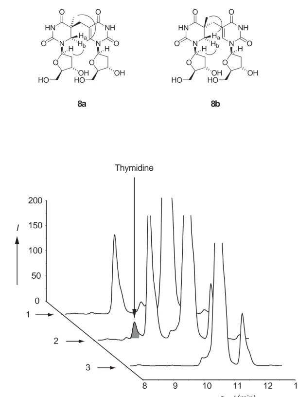
NOESY experiments were performed to assign the stereochemistry. For the 5S-isomer 8a we measured a strong NOE between Hb and C1’-H. Hb features in addition
a strong NOE to the C5 methyl protons. For the 5R-isomer 8b, the measured NOE between Ha and C1’-H is significantly smaller compared to Hb interacting with C1’-H.
Ha however features a strong NOE to the C5 methyl protons. The observed strong
correlations in the NOE experiment are indicated in Figure 1.
In order to investigate whether compounds 8a and 8b would be accepted as a substrate by the enzyme SP-lyase, we have purified the recombinant His-tagged protein from E. coli and prepared it in the active holoform containing a (4Fe-4S) cluster under anaerobic conditions. Details of the procedures will be described elsewhere. Standard reaction mixtures contained 50 µM SP-lyase monomer, 1 mM
SAM, 3 mM dithionite, and 1 mM of either 8a or 8b in 0.1 M Tris HCl buffer, pH 7.0,
containing 5 mM DTT together with 0.2 M KCl. Small samples were removed from the
assay solution and analysed by reversed phase HPLC (C18 column) for the presence of thymidine, the expected product of the reaction. The chromatograms obtained after 3 h of repair of both isomers (trace 1 and 2) and a control assay containing 8a but no enzyme (trace 3) are depicted in Figure 1.
0 50 100 150 200 1 2 3 8 9 10 11 12 13 t(min) I Thymidine b) a) O N HN O O OH N NH O O O OH HO HO Hb Ha H H 8a O N HN O O OH N NH O O O OH HO HO Hb Ha H H 8b
Figure 1. a) Depiction of the two diastereoisomers 5S = 8a and 5R = 8b together with the strong NOE contacts. b) HPLC traces of the enzymatic reaction with 8a (trace 2) and 8b (trace 1). The control experiment with 8a but no enzyme is shown in trace 3. Conditions: 20 min gradient 0 → 28% buffer B (buffer A: 0.1% TFA in water, buffer B: 0.1% TFA in 50% acetonitrile). The additional peaks in the chromatogramm are also
present at the beginning of the reaction and represent other compounds in the reaction mixture.
In all the performed experiments we observe only in the presence of compound 8a and the enzyme a single new peak in the HPLC experiment with a retention time of 9.67 min. This peak appears consequently only in trace 2. Co-injection of thymidine proofed that this peak is caused by the nucleobase T, which was also confirmed by its UV spectrum. The peak does not form without the enzyme (trace 3) in the control experiment. Most importantly, the new peak does also not form in the presence of the 5R-isomer 8b (trace 1) even after 24 h of incubating 8b with the enzyme solution. Increasing the concentration of the enzyme and of the substrate increased the efficiency of the repair of 8a but gave again no detectable thymidine formation in the assay with 8b. These experiments show for the first time that the 5R-isomer is not accepted as a substrate by the enzyme. 8a is in our hands the only and importantly an efficiently accepted substrate paving the way for detailed enzymatic studies not possible so far.
Additional observation require commentation: During the enzymatic reaction SAM is converted into 5’-deoxyadenosine AdoH. We observed continuous formation of AdoH even in the absence of substrate (data not shown) or in the presence of compound
8b. Detection of AdoH is therefore not a proper indicator for SP-lyase activity. These
data are in agreement with the results reported by W. L. Nicholson[17] but differ from those reported by J. Broderick.[18] Whether the discrepancy is due to the fact that different substrates and now defined substrates, are used in these different studies remains to be established.
Lesion repair is well detectable in our system but overall rather slow. The expected very low binding of the lesion outside the DNA environment and the noted extremely high sensitivity of the enzyme is most likely responsible for this fact. This studies represents the first study in which the enzyme SP-lyase was challenged with defined substrates. The main observation is that the enzyme accepts the interstrand crosslink lesions and only the 5S-configured diastereoisomer 8a. This now raises the interesting questions if the spore photoproduct lesion is indeed the 5S crosslink and
now needed to further characterize the lesion and the activity of the repair enzyme in order to gain understanding of how nature stores in spores genetic information over thousands of years.
References
[1] W. L. Nicholson, N. Munakata, G. Horneck, H. J. Melosh, P. Setlow, Microbiol.
Mol. Biol. Rev. 2000, 64, 548-572.
[2] P. Setlow, Environ. Mol. Mutagen. 2001, 38, 97-104.
[3] T. Carell, L. T. Burgdorf, L. Kundu, M. Cichon, Curr. Opin. Chem. Biol. 2001,
5, 491-498.
[4] M. G. Friedel, M. K. Cichon, T. Carell, in CRC Handbook of Organic
Photochemistry and photobiology, 2 ed. (Eds.: W. Horspool, F. Lenci), CRC
Press, Boca Raton, 2003, pp. 1-22.
[5] J. E. Donnellan, R. S. Stafford, Biophys. J. 1968, 8, 17-28.
[6] E. Baillie, G. R. Germaine, W. G. Murell, D. F. Ohye, J. Bacteriol. 1974, 120, 516-523.
[7] P. Setlow, Annu. Rev. Microbiol. 1988, 42, 319-338.
[8] S. C. Mohr, N. V. H. A. Sokolov, C. He, P. Setlow, Proc. Natl. Acad. Sci. USA
1991, 88, 77-81.
[9] J. E. Donnellan, R. B. Setlow, Science 1965, 149, 308-310.
[10] A. J. Varghese, Biochem. Biophys. Res. Comm. 1970, 38, 484-490. [11] T. Douki, G. Laporte, J. Cadet, Nucleic Acids Res. 2003, 31, 3134-3142. [12] N. Munakata, C. S. Rupert, J. Bacteriol. 1972, 111, 192-198.
[13] N. Munakata, C. S. Rupert, Mol. Gen. Genet. 1974, 130, 239-250.
[14] R. Rebeil, Y. B. Sun, L. Chooback, M. Pedraza-Reyes, C. Kinsland, T. P. Begley, W. L. Nicholson, J. Bacteriol. 1998, 180, 4879-4885.
[15] T. A. Slieman, R. Rebeil, W. L. Nicholson, J. Bacteriol. 2000, 182, 6412-6417. [16] J. Cheek, J. B. Broderick, J. Biol. Inorg. Chem. 2001, 6, 209-226.
[17] R. Rebeil, W. L. Nicholson, Proc. Natl. Acad. Sci. USA 2001, 98, 9038-9043. [18] J. Cheek, J. B. Broderick, J. Am. Chem. Soc. 2002, 124, 2860-2861.
[19] J.-D. Guo, Y. Luo, F. Himo, J. Phys. Chem. B 2003, 107, 11188-11192. [20] M. Fontecave, E. Mulliez, S. Ollagnier-de-Choudens, CURRENT OPINION IN
CHEMICAL BIOLOGY 2001, 5, 506-511.
[21] J. Butenandt, L. T. Burgdorf, T. Carell, Synthesis 1999, 1085-1105. [22] S. J. Kim, C. Lester, T. P. Begley, J. Org. Chem. 1995, 60, 6256-6257.