HAL Id: insu-00805857
https://hal-insu.archives-ouvertes.fr/insu-00805857
Submitted on 29 Mar 2013HAL is a multi-disciplinary open access archive for the deposit and dissemination of sci-entific research documents, whether they are pub-lished or not. The documents may come from teaching and research institutions in France or abroad, or from public or private research centers.
L’archive ouverte pluridisciplinaire HAL, est destinée au dépôt et à la diffusion de documents scientifiques de niveau recherche, publiés ou non, émanant des établissements d’enseignement et de recherche français ou étrangers, des laboratoires publics ou privés.
Distributed under a Creative Commons Attribution| 4.0 International License
Synchrotron infrared confocal microscope: Application
to infrared 3D spectral imaging
Frédéric Jamme, B. Lagarde, G. Giuliani, Ga Garcia, Lionel Mercury
To cite this version:
Frédéric Jamme, B. Lagarde, G. Giuliani, Ga Garcia, Lionel Mercury. Synchrotron infrared con-focal microscope: Application to infrared 3D spectral imaging. 11 Conference on Synchrotron Radiation Instrumentationh International C, Jul 2012, Lyon, France. pp.142002, �10.1088/1742-6596/425/14/142002�. �insu-00805857�
Synchrotron infrared confocal microscope:
Application to infrared 3D spectral imaging
F Jamme1, 2, B Lagarde1, A Giuliani1, 2, GA Garcia1 and L Mercury31 Synchrotron SOLEIL, L’Orme des Merisiers, Gif sur Yvette, France 2 INRA, UAR 1008 CEPIA, Rue de la Géraudière, F-44316 Nantes, France
3 Institut des Sciences de la Terre d’Orléans, UMR 7327 CNRS-INSU/ Universités d’Orléans, F-45071 Orléans Cedex, France
E-Mail: [email protected]
Abstract. The brightness of a synchrotron source coupled to an infrared microscope allows
imaging at the so-called diffraction limit. Thus, numerous infrared beamlines around the world have been developed for infrared chemical imaging. Infrared microscopes employ Schwarzschild objectives, which are based on two spherical mirrors centered on a common optical axis. A common drawback of the latter design comes with the central obscuring of the incident beam and the corresponding diffraction effects. However, in this work we are taking advantage of the “shadow” below the primary mirror in order to “vertically” probe our sample and record depth profiling images. We recorded infrared confocal images of liquid water occluded inside a quartz micro-cavity and reconstructed a 3D infrared image based on the OH stretching vibrational bands. Although only a restricted sample size can be analyzed, we demonstrate here the feasibility of a 3D confocal infrared measurement. The chemical profiles have been calculated along the Z-axis of the water inclusion to study its heterogeneity and water-solid interfaces.
1. Introduction
1.1 Infrared microspectroscopy at a synchrotron facility
Since the early 90s, infrared microspectroscopy at synchrotron facilities has been largely developed [1-6]. Nowadays there are many applications in a large number of scientific domains (biology, geology, chemistry, soft matter, high pressure). The brightness of the synchrotron source coupled to infrared microscope allows imaging at the diffraction limit (≈λ/2). Thus, nowadays, 23 infrared beamlines around the world are accessible for infrared chemical imaging at the diffraction limit. More recently with the development of focal plane array (FPA) detectors, few beamlines are now developing infrared synchrotron source extraction in order to perform rapid data acquisition and improve the image resolution by point spread function (PSF) deconvolution [7, 8].
1.2 The Schwarzschild objective
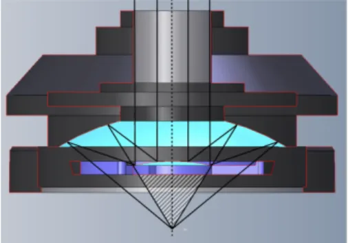
Schwarzschild objectives have a catoptric design based on two spherical mirrors centered on a common optical axis. Schwarzschild objectives are available with finite back conjugates and can be “infinity-corrected” design; see figure 1. Infinity correction refers to the collimation of both visible and infrared beams in the microscope. Therefore, the image of the aperture comes to focus at the focal length of the objective. Spherical aberration, comatic aberration and field curvature are minimized by infinity corrected objectives, which results in a high quality visible image and infrared sampling precision. In addition, the infinity corrected systems are relatively insensitive to optical component insertion into the collimated beam. A common drawback of the design comes with the central obscuring (primary mirror) of the incident beam and the corresponding diffraction effects [5, 6]. However, in this work we are taking advantage of the “shadow” belong the primary mirror in order to “vertically” probe our sample and record depth profiling images.
11th International Conference on Synchrotron Radiation Instrumentation (SRI 2012) IOP Publishing
Figure 1. Schematic of an infinity corrected Schwarzschild type objective. The hatch area represents the “shadow” below the primary mirror (light blue). The working distance of this 32x Schwarzschild infinity corrected objective (Nicolet Reflachromat™) is 7 mm and its numerical aperture (N.A) is 0.65.
1.3 Dual confocal apertures
Several studies have demonstrated that the spatial resolution is controlled by the diffraction specific to the Schwarzschild objectives [1, 5]. The best way to limit the first-order diffraction is to use a dual confocal aperturing optical arrangement. Indeed, it is well known that the implementation of dual apertures at the confocal image provides higher spatial resolution [5]. Thus, by setting up the aperture dimensions equal to the wavelength of interest one can expect to reach a spatial resolution at the diffraction limit. In the work presented, in order to obtain the best performance at 3 µm (3400 cm-1), a dual mask aperture has been used in transmission mode geometry coupled with the high brightness of the synchrotron source (SMIS beamline, SOLEIL synchrotron, Gif sur Yvette, France).
1.4 Vertical profiling
For a restricted sample geometry and transparent sample environment, the infrared confocal microscope is very sensitive to vertical variations along the optic axis (Z-axis). As mentioned previously, Schwarzschild objectives produce a “shadow” caused by the primary mirror of the objective. Therefore, in transmission geometry, shadows are present above and below the specimen placed at the confocal plan. By scanning the sample (Z direction) into the shadows, the infrared beam allows probing the sample along the Z-axis. It is then possible to record 2D images at different vertical positions of the sample to obtain vertical depth profiling of the specimen studied.
2. Experimental set-up
2.1 Sample preparation
Quartz samples hosting water-filled inclusions, synthetized in internally heated pressure vessels (IHPV), were selected for analysis [8]. The hydrothermal synthesis was performed at 300 MPa and 626°C over 4 days. The IHPV was loaded with Au-capsules filled with a piece of quartz, the selected liquid (pure water) and amorphous silica. Hydrothermal healing restores a massive solid containing cavities filled with the liquid present in the synthesis capsules. The water inclusion studied is shown in figure 2. The OH-stretching band of water is very sensitive to minute amount and solid interfaces, and has been closely studied in 2D plane [9].
2.2 Infrared microscopy at the SMIS beamline
Infrared experiments were performed at SOLEIL Synchrotron (Gif sur Yvette, France, SMIS beamline). The SMIS beamline is equipped with a Continuum XL microscope (Thermo Scientific, USA) coupled to a Nicolet 5700 FT-IR spectrometer (Thermo Scientific, USA). The microscope comprises a motorized sample stage and a liquid nitrogen cooled mercury cadmium telluride (MCT-A) detector (50 µm). The microscope operates in confocal mode, using a 32 × infinity corrected Schwarzschild objective (NA= 0.65) and a matching 32 × condenser. All spectra were obtained using a double path single masking aperture of size 6 µm × 6 µm. The spectra were collected in the 6000 – 2000 cm-1 mid-infrared range at a spectral resolution of 8 cm1 with 64 co-added scans. The profiles
11th International Conference on Synchrotron Radiation Instrumentation (SRI 2012) IOP Publishing
Journal of Physics: Conference Series 425 (2013) 142002 doi:10.1088/1742-6596/425/14/142002
were calculated by using the OMNIC software (Thermo Scientific, USA). The term "profile" is used to refer to the integrated absorbance intensity of a specified spectral region for each sample point. The 2D profiles analysis (XZ, YZ and 3D slices) was carried out using Matlab software (Matlab V7.10, MathWorks, USA).
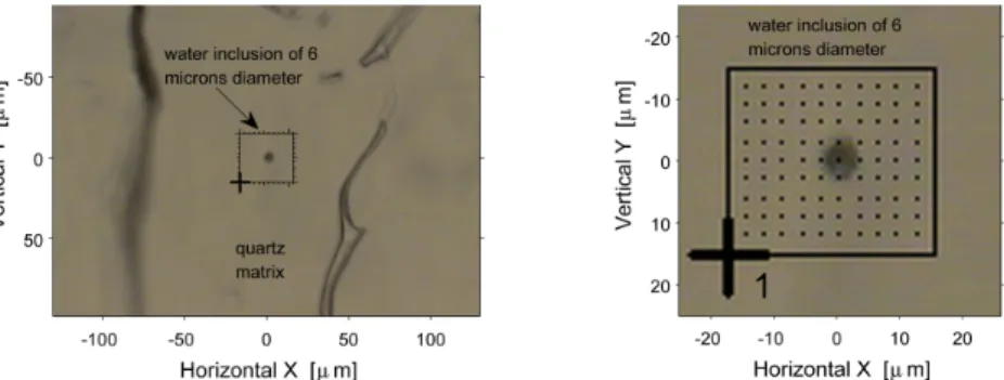
Figure 2. Visible image of a water inclusion (6 µm diameter) in quartz. The mapping area is 36 by 33 µm2 with a step size of 3 µm (map size 12 by 11 points)..
3. Results and discussion
3.1 2D images at fixed Z position
The sample was laterally rastered through the focused infrared beam (Z=0) with spectra collected at each scanning point (3 µm). The confocal point was found using both the visible image and the maximum absorbance intensity of the OH vibration band. The 2D chemical images were produced by area calculation of the OH stretching water band (4000-3000 cm-1) using a linear baseline correction.
3.1 3D stacks of spectral images
Figure 3 shows the infrared profiles (slices) recorded along the Z-axis. The microscope stage allows high precision displacements along the Z-axis. The OMNIC software indicated the position in microns along the X and Y axis. For the Z direction the corresponding value (a.u) given by OMNIC software has been calibrated. Therefore, we recorded 11 infrared maps from -50 to 50 microns with a step size of 10 microns. During the experiment the 32X objective and 32X condenser were kept fixed without the use of the compensation rings. Thus, the sample was scanned along the Z-axis above and below the focal point.
3.2 Vertical resolution
By assuming a negligible first order diffraction effect and no scattering of the incident light, the beam energy distribution at the sample plane can be approximated by a Gaussian beam. Therefore, the Rayleigh range for Gaussian beam propagation is λ/πθ2, with θ the divergence angle, which dominates the Z depth resolution. Thus, the Z depth resolution is estimated to be twice the X, Y plane spatial resolution. The vertical resolution is therefore much poorer than the lateral one.
3.3 2D profiles images (ZY, XY XZ)
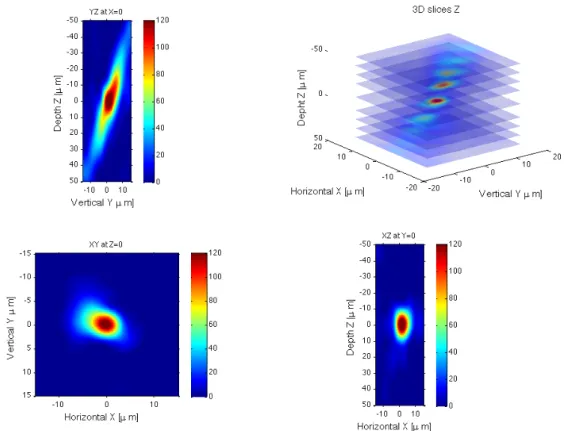
A 3D matrix has been generated in the Matlab software by using all the 2D profiles calculated at each Z position. Therefore, it is possible to plot the 2D profiles of the water inclusion studied. A spline interpolation has been used between each Z plane. One can note the diagonal distortion of the inclusion studied, which could be attributed to an asymmetrical filling of the microscope objective. 4. Conclusion and future horizon
The results presented in this work have shown, for the first time, that it is possible to carry out “confocal” infrared microspectroscopy and obtain vertical profile information. Due to the beam waist
11th International Conference on Synchrotron Radiation Instrumentation (SRI 2012) IOP Publishing
(λ/πθ) only restricted samples can be analyzed; however we demonstrate here the feasibility of 3D infrared measurement by studying a water inclusion in a quartz sample. The calculated profiles show heterogeneity along the Z-axis of the water inclusion, which may represent the geometry of the inclusion but could also be due to infrared beam misalignment.
Figure 3. Absorbance 2D profiles ZY, XY XZ and 3D vertical slices along Z-axis. [1] Martin M, Schade U, Lerch P and Dumas P 2010 Trends In Anal. Chem. 29 453-63 [2] Miller LM, Dumas P 2006 Biochim. Biophys. Acta.1758 846–57
[3] Carr GL 1999 Vib. Spectrosc. 19 53–60
[4] Dumas P, Polack F, Lagarde B, Chubar O, Giorgetta J and Lefrancois S 2006 Infrared Phys.
Tech. 49 52–60
[5] Carr GL 2001 Rev. Sci. Instrum 72 1613
[6] Carr GL, Reffner JA and Williams GP 1995 Rev. Sci. Instrum. 66 1490
[7] Carr GL, Miller LM and Dumas P 2011 Synchrotron Radiation as a Source for Infrared Microspectroscopic Imaging with 2D Multi-Element Detection Biomedical Applications of
Synchrotron Infrared Microspectroscopy vol 11 ed D Moss (Royal Society of Chemistry) pp
226-259
[8] Mattson EC, Nasse MJ, Rak M, Gough KM and Hirschmugl CJ 2012 Anal. Chem. 84 6173-6180
[9] Mercury L, Jamme F, Dumas P 2012 Phys. Chem. Chem. Phys. 14 2864 Acknowledgments
The authors wish to acknowledge Paul Dumas (SMIS Beamline, Synchrotron SOLEIL) and G. Lawrence Carr (NSLS, Brookhaven National Laboratory) for fruitful discussions and encouragement. We thank Bruno Beccard (Thermo Fisher Scientific) for invaluable help and assistance.
11th International Conference on Synchrotron Radiation Instrumentation (SRI 2012) IOP Publishing
Journal of Physics: Conference Series 425 (2013) 142002 doi:10.1088/1742-6596/425/14/142002