Development of Polymer and Lipid Materials
for Enhanced Delivery of Nucleic Acids and Proteins
by
Ahmed Atef Eltoukhy
B.S., Chemical Engineering Stanford University, 2007
SUBMITTED TO THE DEPARTMENT OF BIOLOGICAL ENGINEERING IN PARTIAL FULFILLMENT OF THE REQUIREMENTS FOR THE DEGREE OF
ARCHNES
DOCTOR OF PHILOSOPHY IN BIOLOGICAL ENGINEERING
AT THE
MASSACHUSETTS INSTITUTE OF TECHNOLOGY JUNE 2013
MAASAicHUSrrsTNsTfffE
C V
J(JN
2
7 ;F@ 2013 Massachusetts Institute of Technology. All rights reserved.
Signature of Author:
__-Department of Biological Engineering May 3, 2013
Certified by:
Robert Langer David H. Koch Institute Professor
/7
Thesis SupervisorCertified by:
DanieT .Anderson
S uel A. Goldblith Associate Professor of Chemical Engineering
Thesis Supervisor
Accepted by:
Forest M. White Mitsui Career Development Associate Professor of Biological Engineering Chair, Biological Engineering Graduate Program Committee
This thesis has been examined by a committee of the Biological Engineering Department as follows:
Thesis Committee Chair:
Alfred H
Thesis Supervisor, Committee Member:
Thesis Supervisor, Committee Member:
Ram Sasisekharan Caspary Professor of Biological Engineering
Robert Langer
David H. Koch titute Professor
Daniel G. Anderson
Samuel A. Goldblith Asso e Professor of Chemical Engineering
Committee Member:
Wilso f H IV & Sangeeta N. Bhatia
Development of Polymer and Lipid Materials
for Enhanced Delivery of Nucleic Acids and Proteins
By Ahmed Atef Eltoukhy
SUBMITTED TO THE DEPARTMENT OF BIOLOGICAL ENGINEERING ON MAY 3,2013 IN PARTIAL FULFILLMENT OF THE REQUIREMENTS
FOR THE DEGREE OF DOCTOR OF PHILOSOPHY IN BIOLOGICAL ENGINEERING
ABSTRACT
The development of synthetic vectors enabling efficient intracellular delivery of macromolecular therapeutics such as nucleic acids and proteins could potentially catalyze the clinical translation of many gene and protein-based therapies. However, progress has been hindered by a lack of safe and effective materials and by
insufficient insight into the relationship between key delivery properties and efficacy.
Accordingly, working with a promising class of cationic, degradable gene delivery vectors, poly(-amino ester)s (PBAEs), we develop novel, hydrophobic PBAE terpolymers that display dramatically increased gene delivery potency and nanoparticle stability. We then develop a technique based on size-exclusion chromatography that enables the isolation of well-defined, monodisperse PBAE polymer fractions with greater transfection activities than the starting polymer. This technique also allows us to elucidate the dependence of gene delivery properties on polymer molecular weight (MW). Subsequently, we examine the cellular uptake and trafficking mechanisms of PBAE/DNA polyplexes, and demonstrate that polyplex internalization and transfection depend on a key endo/lysosomal cholesterol transport protein, Niemann-Pick C1 (Npcl).
Finally, working with cationic lipids termed lipidoids, which have shown exceptional potency for the delivery of RNAi therapeutics, we develop these
materials for intracellular delivery of proteins using a simple and novel approach in which nucleic acids serve as a handle for protein encapsulation and delivery.
Preliminary in vivo experiments suggest the potential application of this approach toward lipidoid-mediated delivery of protein-based vaccines.
Taken together, the work presented here advances the development of polymer and lipid materials for the safe and effective intracellular delivery of DNA and protein therapeutics.
ACKNOWLEDGEMENTS
First, I would like to express my deepest appreciation for and utmost gratitude to my thesis advisors, Robert Langer and Daniel Anderson. Bob has been an inspiring role model who has graciously encouraged my personal and professional
development, and Dan has been an outstanding mentor who has offered invaluable guidance and support at every step of my thesis research. I am truly fortunate to have had the opportunity to interact with them, learn from them, and work in their labs.
I would like to thank the other members of my thesis committee, Ram Sasisekharan
and Sangeeta Bhatia. They have provided terrific support and guidance for which I am grateful. I also want to acknowledge the BE department for offering me the opportunity to come to MIT and for providing a stimulating learning environment. Among the tremendous network of scientists comprising the Langer and Anderson labs, I am fortunate to count many collaborators and friends. Among current
labmates, I would particularly like to thank Delai Chen, Gaurav Sahay, Yizhou Dong, Chris Alabi, Omid Veiseh, Jeisa Pelet, Roman Bogorad, Hao Yin, Manos Karagiannis, Rose Kanasty, Omar Khan, James Dahlman, Tim O'Shea, Yi Chen, Patrick Fenton, Beata Chertok, and Janet Zoldan. Among former labmates, I would like to thank Daniel Siegwart, Shan Jiang, Jordan Green, David Nguyen, Avi Schroeder, Seung-Woo Cho, Said Bogatyrev, Hao Cheng, Chris Levins, Kevin Love, Michael Goldberg, Joao Guerreiro, Fan Yang, Nathan Hwang, Kerry Mahon, and Cheol Am Hong.
I would also like to thank the undergraduate students I have mentored, especially
my UROP, Jay Rajan, as well as my 10.29 project team, Danielle Class, Mark Kalinich, and Jefferson Sanchez.
Additionally, I would like to acknowledge my collaborators at Mount Sinai School of Medicine, including Kevin Costa, Kenneth Fish, Irene Turnbull, Amy Rosen
Kontorovich, and Satish Rao. It has been a pleasure to work with them.
I am very grateful for the friends who have enlivened and enriched my experience at
MIT, particularly those from college who ended up in Boston along with me, and those from among my BE classmates, lab, and the Egyptian community at MIT. Last but not least, I would like to thank my family. I am deeply indebted to my mom and dad for their unwavering love and support despite the toll my time in Boston has taken on them. I also want to thank my brother, sister-in-law, and two adorable nieces for their encouragement. I am very fortunate to have in my father and in my brother two close examples of successful scientist-entrepreneurs, and I greatly appreciate the guidance and advice they have provided.
TABLE OF CONTENTS
A BST RA CT... 3
A CK N O W LED G EM ENTS... 4
LIST O F FIGU R ES... 8
LIST O F TA BLES...11
1 INTRO D U CTIO N...12
2 BA CK GRO U N D ... 15
2.1 M OTIVATION FOR GENE THERAPY... ... ... ... 15
2.2 LIM ITATIONS OF VIRAL VECTORS... 20
2.3 BARRIERS TO SYSTEM IC NON-VIRAL GENE DELIVERY... 23
2.3.1 Extracellular barriers... 23
2.3.2 Cellular internalization ... 25
2.3.3 Endosom al escape ... 26
2.3.4 Cytosolic trafficking and nuclear localization... 27
2.3.5 Vector unpacking ... 2 9 2.3.6 Gene expression ... 30
2.4 OVERVIEW OF CURRENT NON-VIRAL VECTORS ... 32
2.4.1 Lipid-based vectors...32
2.4.2 Polym eric vectors ... 35
2.4.3 Cell-penetrating peptides...40
2.4.4 Inorganic nano particles...41
2.5 DEVELOPMENT OF POLY(BETA-AMINO ESTER)S FOR GENE THERAPY... 42
2.5.1 Initial synthesis and characterization ... 43
2.5.2 High-through put screening and selection... 47
2.5.3 End-modification and ligand coating for cell-specific gene delivery... 50
2.5.4 Unsolved challenges ... 54
2.6 REFERENCES... 57
3 DEVELOPMENT OF DEGRADABLE HYDROPHOBIC POLY(BETA-AMINO ESTER) TERPOLYMERS FOR ENHANCED GENE DELIVERY POTENCY AND NANOPARTICLE STA BILITY ... 75
3.1 INTRODUCTION ... 75
3.2 M ATERIALS AND M ETHODS ... 77
3.2.1 M aterials ... 77
3.2.2 Polym er synthesis ... 78
3.2.3 Analytical Gel Permeation Chromatography (GPC)... 78
3.2.4 Preparative GPC/HPLC... 79
3.2.5 NM R...79
3.2.6 Transfection experim ents ... 79
3.2.7 Fluorescence-activated cell sorting (FA CS) ... 80
3.2.8 Dye exclusion assay... 81
3.2.9 Gel electrophoresis... 81
3.2.10 Particle formulation with PEG-lipid at high DNA concentration ... 82
3.2.11 Dynam ic light scattering (DLS) m easurem ents ... 82
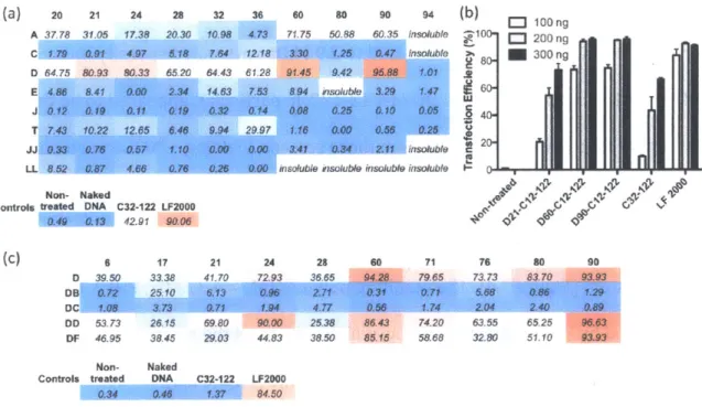
3.3.1 PBAE terpolymer library synthesis and screening... 83
3.3.2 Effect of alkyl side chain content on PBAE polyplex stability and transfection...89
3.3.3 Effect of alkyl side chain content on DNA binding and encapsulation efficiency ...95
3.3.4 Formulation of terpolymer/DNA nanoparticles with PEG-lipid conjugates...97
3 .4 C O N CLU SIO N S...1 0 0 3 .5 R EFEREN CES...1 0 1 4 DEVELOPMENT OF DEGRADABLE HYDROPHOBIC PBAE TERPOLYMERS FOR CELL-SPECIFIC GENE DELIVERY... 103
4 .1 IN T RO D U CT I0 N ... 10 3 4.2 MATERIALS AND METHODS ... 104
4.2.1 Materials...104
4.2.2 Polymer synthesis...104
4.2.3 Analytical Gel Permeation Chromatography (GPC)...105
4.2.4 Transfection experiments ... 105
4.2.5 GFP expression analysis... 106
4.2.6 Luciferase expression analysis... 107
4.2.7 Dynamic light scattering (DLS) measurements... 107
4.2.8 Dye exclusion assay... 108
4.2.9 Na noparticle formulation at high DNA concentration... 108
4.2.10 In vivo transfection experiments... 109
4.2.11 Whole-animal bioluminescence imaging ... 110
4.3 RESULTS AND DISCUSSION...110
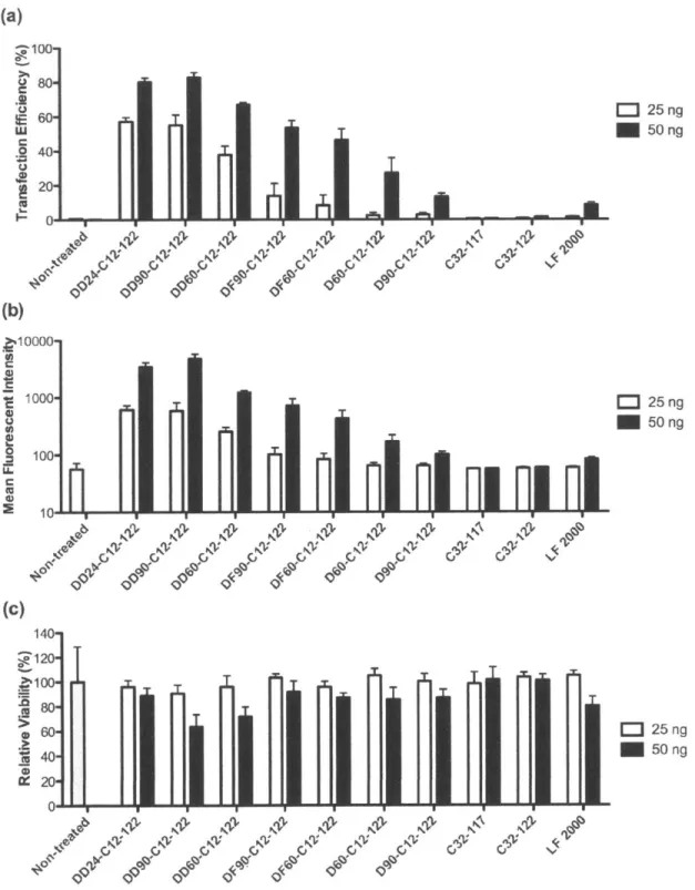
4.3.1 PBAE terpolymer transfection of HUVECs, pMSCs, and NRCMs...110
4.3.2 Combinatorial library of PBAE terpolymers centered on DD24-C12-122 and LL24-C 12 -12 2 ... 11 6 4.3.3 PBAE terpolymer/DNA formulation development and in vivo transfection...122
4 .4 C O N CLU SIO N S ... 1 2 7 5 EFFECT OF MOLECULAR WEIGHT OF AMINE END-MODIFIED POLY(BETA-AMINO ESTER)S ON GENE DELIVERY EFFICIENCY AND TOXICITY... 128
5 .1 IN T R O D U CT IO N ... 1 2 8 5.2 MATERIALS AND METHODS ... 131
5.2 .1 M a teria ls ... 13 1 5.2.2 Polymer synthesis... 132
5.2.3 Analytical size exclusion chromatography (SEC)... 133
5.2.4 in vitro GFP plasmid DNA transfection ... 133
5.2.5 FA CS an a lysis... 134
5.2.6 Dynamic light scattering (DLS) measurements... 135
5.2.7 Dye exclusion assay... 135
5.2.8 Preparative SEC ... 136
5.3 RESULTS AND DISCUSSION ... 137
5.3.1 Synthesis and analytical SEC ofstoichiometric PBAE variants... 137
5.3.2 Plasmid DNA transfection and cytotoxicity ... 139
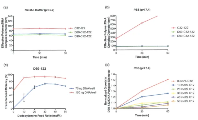
5.3.3 Bio physical characterization of polymer/DNA nanoparticles... 142
5.3.4 Relative DNA binding efficiency... 146
5.3.5 Preparative SEC ... 149
5 .4 C O N C L U SIO N S ... 1 5 4 5 .5 R EFE R E N CES...1 5 5
6 THE CHOLESTEROL TRANSPORTER NIEMANN PICK C1 PLAYS A CRITICAL ROLE IN
DNA INTERNALIZATION AND TRANSFECTION BY POLY(BETA-AMINO ESTER)S... 158
6.1 INTRODUCTION ... 158
6.2 M ATERIALS AN D M ETHODS ... 161
6.2.1 M aterials...161
6.2.2 Poly m er sy nthesis... 161
6.2.3 D NA transfection experim ents... 162
6.2.4 FA CS analysis...164
6.3 RESULTS AND DISCUSSION ... 165
6.3.1 Pharm acological inhibition studies... 165
6.3.2 Studies w ith cell lines varying in Npcl expression... 170
6.4 CONCLUSIONS...181
6.5 R EFERENCES...182
7 NUCLEIC ACID CONJUGATION ENABLES EFFICIENT INTRACELLULAR PROTEIN DELIVERY BY LIPID-BASED NANOPARTICLES ... 188
7.1 INTRODUCTION ... 188
7.2 M ATERIALS AND M ETHODS ... 191
7.2.1 M aterials...191
7.2.2 Protein -oligon ucleotide conjugation ... 192
7.2.3 Microfluidic device formulation of lipid nano particles (LNPs)... 193
7.2.4 In vitro protein transfection ... 194
7.2.5 RP activity assay ... 194
7.2.6 FA S analysis... 195
7 72.7 Gel electrophoresis ... 195
7.2.8 A nim al experim ents... 196
7.3 RESULTS AND DISCUSSION ... 197
7.3.1 Intracellular delivery of horseradish peroxidase ... 197
7.3.2 In tracellular delivery of N eutrA vidin ... 204
7.4 CONCLUSIONS ... 214
7.5 R EFERENCES ... 215
8 CO N CLU SIO N S ... 2 18 8.1 M AIN CONTRIBUTIONS...218
LIST OF FIGURES
FIGURE 2.1 CLINICAL TRIALS OF GENE THERAPY APPROVED BY YEAR ... 16
FIGURE 2.2 INDICATIONS ADDRESSED BY GENE THERAPY CLINICAL TRIALS WORLDWIDE SINCE 1989 ... 17
FIGURE 2.3 VECTORS USED IN GENE THERAPY CLINICAL TRIALS WORLDWIDE SINCE 1989...22
FIGURE 2.4 SCHEMATIC REPRESENTATION OF PLASMID AND MINICIRCLE VECTORS...31
FIGURE 2.5 CHEMICAL STRUCTURES OF CATIONIC AND NEUTRAL LIPIDS COMMONLY USED IN GENE DELIVERY STUDIES ... ,...3 3 FIGURE 2.6 SYNTHESIS OF EPOXIDE-DERIVED LIPIDOIDS... 34
FIGURE 2.7 CHEMICAL STRUCTURES OF SELECTED POLYMERIC GENE VECTORS...36
FIGURE 2.8 SYNTHESIS OF POLY($-AMINO ESTER)S...44
FIGURE 2.9 SYNTHESIS OF AMINE END-MODIFIED PBAEs ... 51
FIGURE 2.10 AGGREGATION OF PBAE POLYPLEXES UNDER PHYSIOLOGICAL CONDITIONS ... 55
FIGURE 2.11 PBA E BATCH-TO-BATCH VARIABILITY ... 56
FIGURE 3.1 SYNTHETIC SCHEME AND MONOMERS FOR A LIBRARY OF HYDROPHOBIC, AMINE END-MODFIED PBAE T ER PO LY M ER S ... 8 4 FIGURE 3.2 DEVELOPMENT OF HYDROPHOBIC PBAE TERPOLYMERS WITH HIGH DNA TRANSFECTION POTENCY...85
FIGURE 3.3 TRANSFECTION PERFORMANCE OF LEAD PBAE TERPOLYMERS AT REDUCED DNA DOSES IN HELA CELLS87 FIGURE 3.4 HPLC/SEC PURIFICATION OF DD24-C12-122 ... 88
FIGURE 3.5 1H NMR SPECTRA OF DD24-12-122 AND MONOMERS... 89
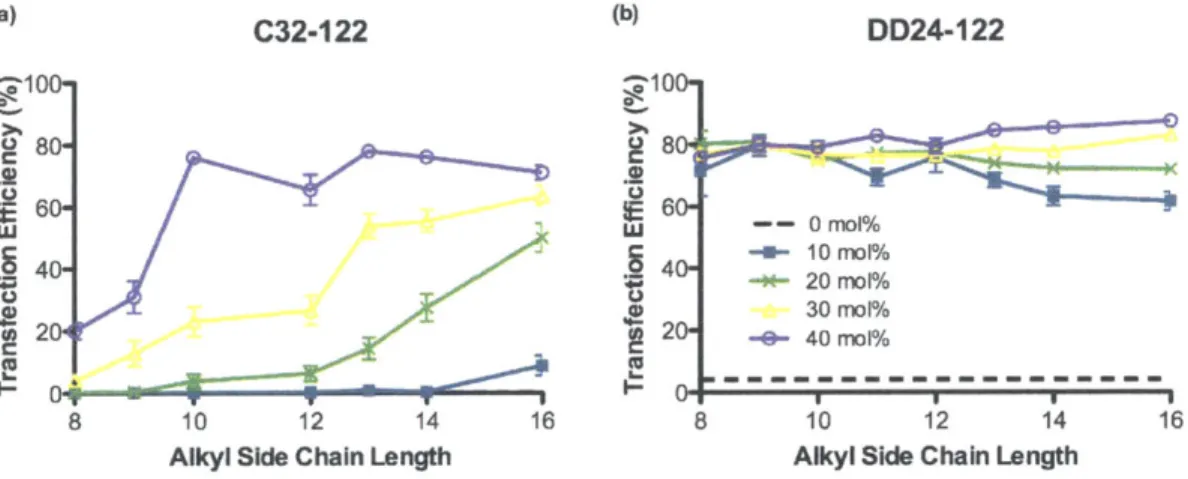
FIGURE 3.6 EFFECT OF ALKYL SIDE CHAIN CONTENT ON PBAE POLYPLEX STABILITY AND TRANSFECTION...90
FIGURE 3.7 EFFECT OF ALKYL SIDE CHAIN LENGTH AND CONTENT ON PBAE TRANSFECTION EFFICIENCY...91
FIGURE 3.8 EFFECT OF AMINE END-MODIFICATION OF PBAE TERPOLYMER TRANSFECTION EFFICIENCY...92
FIGURE 3.9 IDENTIFICATION OF POLYMER SPECIES RESPONSIBLE FOR POLYPLEX STABILITY AND TRANSFECTION PO T E N CY ... 9 4 FIGURE 3.10 RELATIVE ENCAPSULATION EFFICIENCIES OF C32-C12-122 TERPOLYMERS OF VARYING H Y D R O PH O BICITY ... 9 5 FIGURE 3.11 RELATIVE DNA BINDING EFFICIENCIES OF C32-C12-122 TERPOLYMERS OF VARYING HYDROPHOBICITY ... 9 6 FIGURE 3.12 EFFECTS OF PBAE ALKYL SIDE CHAINS (C12) AND THE PRESENCE OF PEG-LIPID CONJUGATE ON NANOPARTICLE FORMULATION STABILITY AND TRANSFECTION EFFICIENCY AT HIGH DNA CONCENTRATION ....98
FIGURE 4.1 SCREENING THE INITIAL PBAE TERPOLYMER LIBRARY FOR GENE TRANSFECTION OF HUVECS...111
FIGURE 4.2 COMPARISON OF PBAE TERPOLYMER LIBRARY SCREEN RESULTS IN HELA CELLS AND HUVECS...112
FIGURE 4.3 INFLUENCE OF GROWTH MEDIUM ON PBAE-MEDIATED TRANSFECTION OF HUVECS AND HELA CELLS 113 FIGURE 4.4 TRANSFECTION OF PORCINE MESENCHYMAL STEM CELLS BY HYDROPHOBIC PBAE TERPOLYMERS...114
FIGURE 4.5 TRANSFECTION OF NEONATAL RAT CARDIOMYOCYTES BY HYDROPHOBIC PBAE TERPOLYMERS...115
FIGURE 4.6 COMBINATORIAL LIBRARY OF 24 PBAE TERPOLYMERS CENTERED ON DD24-C12-122 AND LL24-122 ... 1 1 6 FIGURE 4.7 BIOPHYSICAL PROPERTIES OF THE DD24- AND LL24-FOCUSED COMBINATORIAL LIBRARY...118
FIGURE 4.8 HELA TRANSFECTION EFFICIENCIES OF THE DD24- AND LL24-FOCUSED COMBINATORIAL LIBRARY .... 120
FIGURE 4.9 HUVEC TRANSFECTION EFFICIENCIES OF THE DD2 4- AND LL24-FOCUSED COMBINATORIAL LIBRARY 120 FIGURE 4.10 RAT CORTICAL NEURON TRANSFECTION ACTIVITIES OF THE DD24- AND LL24-FOCUSED COM BINATORIA L LIBRA RY ... 12 1 FIGURE 4.11 HELA TRANSFECTION EFFICIENCIES OF D60-C12-122/DNA NANOPARTICLE FORMULATIONS CONTAINING VARIOUS PEG-LIPID CONJUGATES ... 124
FIGURE 4.12 INTRAPERITONEAL AND INTRAVENOUS GENE DELIVERY IN MICE USING D60-C12-122 AND D90-C12-1 2 2 T ER PO LY M ERS...D90-C12-1 2 5 FIGURE 5.1 SYNTHESIS SCHEME FOR END-MODIFIED POLY(P-AMINO ESTER)S. ... 138 FIGURE 5.2 RELATIONSHIP BETWEEN MwAND C:32 MONOMER MOLAR FEED RATIO FOR AMINE END-MODIFIED PBAES ... 1 3 8
FIGURE 5.3 CORRELATION BETWEEN POLYMER Mw AND GENE TRANSFECTION EFFICIENCY IN HELA CELLS FOR PBAE
STO ICH IO M ETRIC VA RIA NTS...14 0
FIGURE 5.4 CORRELATION BETWEEN POLYMER Mw AND RELATIVE VIABILITY OF HELA CELLS FOLLOWING DNA
TRANSFECTION WITH PBAE STOICHIOMETRIC VARIANTS... 141
FIGURE 5.5 CORRELATION BETWEEN POLYMER Mw AND POLYPLEX DIAMETER FOR PBAE STOICHIOMETRIC VARIANTS ... 1 4 3 FIGURE 5.6 CORRELATION BETWEEN GENE TRANSFECTION EFFICIENCY AND POLYPLEX DIAMETER FOR PBAE STOICHIOM ETRIC VA RIANTS... 144
FIGURE 5.7 CORRELATION BETWEEN POLYMER MwAND -POTENTIAL FOR PBAE STOICHIOMETRIC VARIANTS...145
FIGURE 5.8 CORRELATION BETWEEN POLYMER MW AND -POTENTIAL FOR C32-122 STOICHIOMETRIC VARIANTS IN SODIUM ACETATE BUFFER ... 146
FIGURE 5.9 CORRELATION BETWEEN POLYMER MwAND RELATIVE DNA BINDING FOR PBAE STOICHIOMETRIC V A R IA N T S1... 1 4 7 FIGURE 5.10 CORRELATION BETWEEN GENE TRANSFECTION EFFICIENCY AND RELATIVE DNA BINDING FOR PBAE STOICHIOM ETRIC VARIANTS... 148
FIGURE 5.11 CHROMATOGRAM OF C32 -12 2 ELUTING FROM THE HPLC/SEC COLUMN ... 149
FIGURE 5.12 PREPARATIVE H PLC/SEC ON C32-122...150
FIGURE 5.13 CORRELATION BETWEEN GENE TRANSFECTION EFFICIENCY AND Mw OF C32-122 POLYMER FRACTIONS ISO LA T E D BY S E C...1 5 1 FIGURE 5.14 CORRELATION BETWEEN NANOPARTICLE BIOPHYSICAL PROPERTIES AND POLYMER Mw FOR C32-122 S E C FRA CT ION S ... 1 5 2 FIGURE 5.15 CORRELATION BETWEEN Mw OF C32-122 SEC FRACTIONS AND RELATIVE VIABILITY FOLLOWING GENE TRANSFECTIO N IN H ELA CELLS...153
FIGURE 6.1 SCREEN FOR SMALL MOLECULE-MEDIATED INHIBITION OF C32-122-MEDIATED DNA UPTAKE IN MEFS ... 1 6 6 FIGURE 6.2 C32-122-MEDIATED DNA UPTAKE IN MEFS IN THE PRESENCE OF VARIOUS ENDOCYTIC PATHWAY IN H IB IT O R S... 1 6 8 FIGURE 6.3 U18666A INHIBITS C32-122-MEDIATED DNA TRANSFECTION OF MEFS...170
FIGURE 6.4 NPC1 KNOCKOUT INHIBITS C32-122-MEDIATED DNA TRANSFECTION OF MEFs ... 171
FIGURE 6.5 NPC1 KNOCKOUT INHIBITS C32-122-MEDIATED INTERNALIZATION OF DNA IN MEFS...172
FIGURE 6.6 EFFECTS OF NPC1 KNOCKOUT ON INTERNALIZATION OF DNA IN MEFS FOLLOWING TRANSFECTION WITH C32-122, PEI, AND LIPOFECTAM INE 2000 ... 173
FIGURE 6.7 INCREASING C32-122 TERPOLYMER HYDROPHOBICITY IMPROVES GENE TRANSFECTION POTENCY IN N Pcl-/- M EFS BUT DOES NOT RESCUE INHIBITION... 174
FIGURE 6.8 C32-122-MEDIATED DNA UPTAKE AND TRANSFECTION IN CHO CELL LINES VARYING IN NPC1 EX P R E SSIO N ... 1 7 6 FIGURE 6.9 CONFOCAL MICROSCOPY ANALYSIS OF RELATIVE ENDOCYTIC PATHWAY ACTIVITIES IN NPcJ +/+ AND NPc1 -/ - M EF s ... 1 7 7 FIGURE 6.10 FACS ANALYSIS OF RELATIVE ENDOCYTIC PATHWAY ACTIVITIES IN NPcl+/+ AND NPcl-/- MEFS .... 178
FIGURE 6.11 CO-LOCALIZATION OF INTERNALIZED DNA DELIVERED BY C32-122 WITH MARKERS OF DISTINCT ENDOCYTIC PATHW AYS IN M EFS ... 179
FIGURE 7.1 SCHEME FOR DELIVERY OF PROTEINS BY LIPID-BASED NANOPARTICLES VIA OLIGONUCLEOTIDE C O N JU GA TIO N ...---... 1 9 0 FIGURE 7.2 CHARACTERIZATION OF HRP-DNA OLIGONUCLEOTIDE CONJUGATES ... 198
FIGURE 7.3 SCREEN OF VARIOUS LIPIDOIDS FOR DELIVERY OF ACTIVE HRP-OLIGO CONJUGATES ... 199
FIGURE 7.4 OPTIMIZATION OF LNP FORMULATIONS FOR DELIVERY OF HRP-OLIGO CONJUGATES... 200
FIGURE 7.5 OLIGONUCLEOTIDE CONJUGATION IS REQUIRED FOR EFFECTIVE DELIVERY OF HRP BY LN PS...201
FIGURE 7.6 INTRACELLULAR LOCALIZATION OF HRP-OLIGO CONJUGATES ... 203
FIGURE 7.7 CHARACTERIZATION OF NEUTRAVIDIN-OLIGONUCLEOTIDE CONJUGATES AND LNPS BY GEL ELECT RO PH O R ESIS ... 2 0 5 FIGURE 7.8 OLIGONUCLEOTIDE CONJUGATION IS REQUIRED FOR EFFICIENT DELIVERY OF NAV BY LN PS...206
FIGURE 7.9 UPTAKE OF NAV-OLIGO CONJUGATES IN HELA CELLS ... 207
FIGURE 7.10 BIODISTRIBUTION OF NAV-OLIGO CONJUGATES IN MICE ... 208
FIGURE 7.11 QUANTIFICATION OF NAV-OLIGO LOCALIZATION IN MOUSE SPLEENS... 209
FIGURE 7.13 QUANTIFICATION OF NAV-OLIGO UPTAKE IN DISTINCT IMMUNE CELL POPULATIONS WITHIN THE SPLEEN ... 2 1 2
LIST OF TABLES
TABLE 2.1 SELECTED EXAMPLES OF CELL-PENETRATING PEPTIDES ... 40
TABLE 3.1 MW OF TOP-PERFORMING PBAE TERPOLYMERS ... 88
TABLE 3.2 Z-POTENTIAL MEASUREMENTS OF NANOPARTICLES FORMED FROM C32-122 TERPOLYMERS OF VARYING H Y D R O PH O B ICITY ... ... 9 9
1 INTRODUCTION
The development of synthetic vectors enabling efficient intracellular delivery of macromolecular therapeutics such as nucleic acids and proteins could potentially catalyze the clinical translation of many gene and protein-based therapies. However, progress has been hindered by a lack of safe and effective materials and by
insufficient insight into the relationship between key delivery properties and efficacy. For instance, the biodegradable, cationic polymers known as poly(p-aminoester)s (PBAEs) have shown tremendous potential as non-viral gene delivery carriers in numerous studies, yet several key challenges and questions remain unaddressed, such as poor stability of polymer/DNA nanoparticles under
physiological conditions, batch-to-batch variability in transfection performance, and the lack of mechanistic knowledge of cellular uptake and trafficking pathways. Similarly, lipid-based nanoparticles (LNPs) incorporating cationic lipid materials termed lipidoids have demonstrated excellent in vitro and in vivo efficacy for delivery of oligonucleotides such as short interfering RNA (siRNA), but their application toward intracellular delivery of protein-based therapeutics is impeded
by the wide range of physicochemical properties characterizing these diverse
macromolecules.
Accordingly, the overall objective of this thesis is to develop polymer and lipid-based materials for safe, effective intracellular delivery of gene and protein therapeutics, primarily through the following specific aims:
(1) Development of novel, degradable PBAE polymers with enhanced gene
delivery potency and nanoparticle aggregation resistance
(2) Systematic investigation into the gene delivery properties of PBAE polymers,
particular with respect to the effects of polymer hydrophobicity and
molecular weight, and with respect to the mechanisms employed for cellular
internalization and trafficking
(3) Development of lipid-based nanoparticles for efficient intracellular delivery
of proteins by way of oligonucleotide conjugation
In light of these aims, Chapter 2 of this thesis offers a broad survey outlining
the current clinical prospects for gene therapy, the limitations of viral vectors, the
barriers to non-viral delivery, and the most commonly studied synthetic gene
carriers. It also details the development of poly(P-amino ester)s as gene delivery
materials and presents some of the remaining unresolved challenges and questions
for these polymers. Chapter 3 describes the development of novel, degradable PBAE
terpolymers demonstrating significantly improved gene delivery potency and
nanoparticle stability, and Chapter 4 explores the potential of these polymers for
gene transfection of clinically relevant cell types as well as for in vivo gene delivery.
Chapter 5 details a systematic study of the effect of PBAE molecular weight on gene delivery properties, the results of which elucidate a potential cause of PBAE
batch-to-batch variability. Chapter 6 then reveals new mechanistic insights into the
dependence of PBAE/DNA polyplex internalization and transfection on a key
cholesterol transport protein, Niemann-Pick C1 (Npc1). Subsequently, Chapter 7
delivery with lipid-based nanoparticles via oligonucleotide conjugation. Finally,
Chapter 8 provides a conclusion that summarizes the major findings and
contributions of this thesis and discusses the future outlook for continued
2 BACKGROUND
2.1 MOTIVATION FOR GENE THERAPY
In a broad sense, gene therapy can be defined as the intentional modulation
of gene expression within cells to prevent or treat a pathological process[l'. More
narrowly, this modulation of gene expression is accomplished through the
introduction of exogenous nucleic acids, such as DNA, messenger RNA (mRNA),
small interfering RNA (siRNA), microRNA, or antisense oligonucleotides. In contrast
to many small molecule or protein-based drugs, a singular aspect of DNA
therapeutics is that for many diseases, especially monogenic disorders defined by
inheritance of one mutated gene, the cure - a nucleic acid representing a functional copy of the gene - is plainly manifest. However, because most cells are impermeable to these large, negatively charged macromolecules, a carrier or vector is typically
required to mediate effective intracellular delivery.
This fundamental engineering challenge, which is in equal measures both
exciting and exasperating, largely explains why within the U.S., gene therapy
remains a strictly experimental approach, with no FDA-approved gene therapeutics
yet on the market despite nearly 2,000 clinical trials of gene therapy worldwide
since 1989 (Figure 2.1). Nevertheless, recent clinical progress, including the
approval of the first gene therapeutic for use in Europe, has marked a resurgence of
120 0 100 80 40 I-U 20 z 0
Figure 2.1 Clinical trials of gene therapy approved by year
1,903 clinical trials of gene therapy have been approved worldwide since 1989.
Adapted from Ginn et al.[31
Over the past two decades, gene therapy has been investigated clinically for
the treatment or prevention of a wide range of diseasesl31 (Figure 2.2). Cancer-related diseases comprise nearly two-thirds of the indications addressed by clinical
trials thus far. Gene therapies for cancer treatment generally adhere to one of the
following broad strategies: mutation compensation, immunopotentiation, suicide
gene therapy, oncolytic virotherapy, and radio- or chemo-sensitization[4-51.
Controversially, two cancer gene therapeutics have already been approved for use
in China, even though similar versions have failed to pass clinical development in
the U.S.: Gendicine, a modified adenovirus encoding the human p53 gene, and
H101/Oncorine, a recombinant oncolytic adenovirus targeting p53-deficient cancer
Neurological diseases 2% Healthy volunteers 2% Gene marking 3% Infectious diseases 8% Cardiovascular diseases 8% , Ocular diseases 2% Others 2%
Figure 2.2 Indications addressed by gene therapy clinical trials worldwide since 1989
Adapted from Ginn et al.[31
Within the U.S., Allovectin-7, a locally administered formulation consisting of
a DNA plasmid encoding two immunotherapeutic genes and a cationic lipid delivery
reagent, is currently undergoing a Phase III clinical study for treatment of advanced
metastatic melanoma[71 (ClinicalTrials.gov ID: NCT00395070). Also undergoing a
Phase III clinical trial is ProstAtak, which consists of an adenoviral vector encoding
the herpes thymidine kinase gene, for suicide gene therapy of prostate cancer in
conjunction with a prodrug and radiation therapy (ClinicalTrials.gov ID:
Despite the enormous potential of gene therapy to address monogenic
diseases, this class of disorders represents only -9% of gene therapy clinical trials
to date (Figure 2.2), which include some major successes along with some tragic failures. One of the earliest human trials of gene therapy in the early 1990s involved
the retroviral transfer of adenosine deaminase (ADA) into the T cells of two children with ADA-associated severe combined immunodeficiency disorder (ADA-SCID)[8]. In 2000, the first gene therapy cure was reported in children with X-linked SCID who experienced dramatic clinical improvement following delivery of interleukin-2 receptor gamma chain with a murine retroviral vector[91. Unfortunately, the trial's success was marred two years later when several of these young patients developed leukemia resulting in one death[10' 111. Just a few years earlier, the gene therapy field
had suffered its first major setback with the death of a Jesse Gelsinger, an 18-year-old clinical trial patient who experienced a severe immune response after receiving adenoviral gene therapy for a mild form of ornithine transcarbamylase (OTC) deficiency[21. On the basis of preclinical studies, cystic fibrosis, a severe and fatal lung disease caused by mutations in the CFTR gene, appeared to be a promising early candidate for gene therapy, but clinical trials in humans have so far shown a disappointing lack of efficacy[121.
Nevertheless, one recent triumph has been the approval of Glybera in Europe
for the treatment of lipoprotein lipase (LPL) deficiency[131. This orphan metabolic
disease results from mutation of the LPL gene and is characterized by elevated
levels of blood plasma triglycerides and debilitating bouts of pancreatitis[141. When
viral (AAV) vector encoding an LPL gene construct, has shown efficacy in reducing
plasma triglycerides and rates of pancreatitis [111.
Cardiovascular diseases are the targets of approximately 8% of clinical gene
therapy studies to date, but clinical trial results have so far been marked by low
gene transfer efficacy[161. A promising exception may be Mydicar, an AAV vector
encoding SERCA2a (sarcoplasmic reticulum Ca+2-ATPase), currently under Phase IlIb
study for congestive heart failure (ClinicalTrials.gov ID: NCT01643330). In a Phase
Ila trial, patients treated with the highest dose of Mydicar experienced significant
reduction of major cardiovascular events over a 12-month period compared with
those receiving placebo['71.
Infectious diseases comprise another 8% of indications that have been
addressed for clinical gene therapy trials. One interesting example under Phase I/II
study is a cell-based therapy for HIV/AIDS termed SB-728-T, which consists of
autologous CD4+ T cells genetically modified with a zinc finger nuclease to resist
HIV infection (ClinicalTrials.gov ID: NCT00842634).
Neurological diseases comprise a relatively small proportion of targets of
active gene therapy trials. In a Phase II study, patients with Parkinson's disease
showed some improvement in motor function following subthalmic infusion of
AAV2-GAD, an adeno-associated viral vector encoding glutamic acid decarboxylase (GAD)[ 181, yet its clinical development is in doubt following the bankruptcy of the
trial's corporate sponsor. However, another gene therapeutic for Parkinson's
disease, an AAV vector encoding neurturin, is currently undergoing Phase I/II
With respect to ocular diseases, recent early-phase clinical trials have raised
the prospects for treatment of some forms of inherited blindness, such as Leber's
congenital amaurosis (LCA)[ 201. In a pair of landmark studies, some patients with LCA caused by mutations in the gene encoding an essential retinal pigment
epithelial protein (RPE65) experienced marked improvements in vision following
local injection of AAV vectors delivering the RPE65 gene[21t 221. A follow-up study in
three patients showed efficacy after a second administration to the contralateral
eye[2 3I.
2.2 LIMITATIONS OF VIRAL VECTORS
As suggested above, the key challenge limiting clinical translation of gene
therapeutics is the lack of delivery vectors considered safe and effective. Many of the
aforementioned clinical trials have employed modified viruses such as retroviruses, lentiviruses, adenoviruses, and adeno-associated viruses (AAVs) to deliver genes; in
fact, nearly 70% of gene therapy clinical trials to date have involved viral vectors[31 (Figure 2.3). Although they can be quite efficient and have advanced the field of gene therapy in significant ways, several limitations have been associated with viral vectors, as described below.
(1) Carcinogenesis: Retroviruses and lentiviruses in particular have been
associated with a risk of carcinogenesis[24. The development of leukemia in
children receiving retroviral gene therapy for X-linked SCID has been attributed to a combination of insertional mutagenesis caused by viral
effects[261. Although AAV vectors are generally considered safer due to a very
low probability of integration, and have become more widely used in recent
years, tumorigenesis has been observed in mice treated with this class of
vectors271, and the long-term cancer risk is still unclear[13.
(2) Immune responses: Pre-existing immunity comprises a serious safety concern
for certain classes of viral vectors, especially adenoviruses[28. The death of
Jesse Gelsinger within days of receiving adenoviral gene therapy was blamed
on a severe innate immune response characterized by dramatically elevated
levels of inflammatory cytokines[281. In contrast, AAVs have not been
associated with such severe immune responses, but pre-existing immunity to
many AAV serotypes in significant fractions of the human population can
greatly diminish their efficacy[2 9I. Furthermore, adaptive immune responses,
both humoral and cell-mediated, can prevent the possibility of repeat dosing
and result in the elimination of transduced cells[2 91.
(3) Broad tropism: Some viral serotypes show relatively narrow transduction
specificity, or tropism, for particular cells or tissues, but many others have
broad tropism[30l. This feature may be undesirable if the transgene is
expressed in non-target tissues following systemic delivery.
(4) Limited DNA packaging capacity: AAVs have a DNA cargo capacity of <5 kb;
retroviruses, lentiviruses, and some adenoviruses have packaging capacities
of -8 kb, which may limit the potential for transduction of long
(5) Difficulty of production: Depending on the viral vector, raising the necessary
numbers of vector particles can be challenging, requiring purification
procedures that may be difficult and costly to scale up[321.
Herpes simplex virus
3% Other Lentivirus 3% Adenoviru Poxvirus 23% 5% Adeno-associated virus 5% Lpofection 6% Naked/Plasmid DNA 18%
Figure 2.3 Vectors used in gene therapy clinical trials worldwide since 1989
Adapted from Ginn et al.[3]
Non-viral gene therapy has the potential to address many of these
limitations, particularly with respect to safety. For DNA-based therapeutics,
transgenes are often encoded on plasmids that are episomal and non-integrating, reducing the risk of cancer due to insertional mutagenesis. Moreover, due to the
general lack of pre-existing immunity, synthetic vehicles tend to have comparably
lower immunogenicity and toxicity. These carriers also have the potential to deliver
larger genetic payloads and are typically easier to synthesize.
A number of non-viral gene delivery strategies have been developed so far.
physical methods[33-391, such as gene gun, electroporation, hydrodynamic delivery,
sonoporation, magnetofection, and laser irradiation. Because these techniques are
generally less applicable to systemic gene delivery in humans, a panoply of synthetic
delivery vectors have also been developed including lipids and liposomes40 411,
polymers (linear and branched polymers, dendrimers, and polysaccharides)[4250- ,
polymersomes[5I, cell-penetrating peptides1 52-s4, and inorganic nanoparticles[551.
Traditionally, synthetic vectors have been plagued by low delivery efficiency
relative to viral vectors[451.Whereas viruses have been naturally selected to deliver
their genomes efficiently to mammalian cells, most synthetic vectors have not been
designed to mediate gene delivery past the multiple barriers that confront them,
which are detailed in the next section.
2.3 BARRIERS TO SYSTEMIC NON-VIRAL GENE DELIVERY
2.3.1 Extracellular barriers
For systemic delivery of DNA therapeutics, an initial barrier is the poor
chemical stability of nucleic acids due to the presence of endonucleases in
physiological fluids and the extracellular space. The half-life of plasmid DNA has
been estimated to be 10 min. following intravenous injection in mice[56 and 60 min.
following intratracheal instillation in mouse lungs[571. For this reason, entrapment of
the DNA within a nanoparticulate carrier is desirable in order to provide protection
from nuclease degradation, as well as to improve circulation time.
Polyplexes, for instance, are spherical or toroidal nanoparticulate complexes
of DNA's expanded wormlike chain, representing a loss of entropy, is driven primarily by entropic gain due to the release of counterions upon polymer-DNA binding, which is favored by electrostatic interaction between the cationic polymer and the anionic phosphate backbone[581. Similarly, mixtures of cationic lipids, neutral lipids, and DNA are able to spontaneously assemble into lipoplexes or liposomes characterized by DNA entrapped within lamellar or inverted hexagonal arrangements of lipid bilayers[91. An excess of cationic polymer or lipid promotes enhanced DNA binding and the formation of stable complexes with positively charged surfaces; however, at high salt concentrations, electrostatic repulsion between the positively charged complexes is reduced, making them prone to colloidal instability and aggregation within physiological fluids[60l. In particular,
aggregation of nanoparticles within the blood, either via colloidal instability or via interaction with blood components such as serum proteins and erythrocytes, can
inhibit localization within the desired tissues, induce rapid clearance by circulating macrophages, and even cause embolism within lung capillaries[421.
Selective accumulation of the gene therapeutic at the cell or tissue of interest is another major challenge[611. Passive targeting can be achieved through
modulation of nanoparticle size, charge, shape, or surface chemistry. For instance, systemic gene delivery to liver hepatocytes generally requires particles smaller than
-100-200 nm to traverse the fenestrated endothelium6 2, whereas uptake in macrophages can be achieved with positively charged, spherical particles on the order of 500 nm and above[631. Stabilization of nanoparticles with non-fouling
blood and promote accumulation in tumors with leaky vasculatures, a phenomenon known as the enhanced permeation and retention (EPR) effect[641. Active targeting of synthetic gene vectors has been attempted using numerous ligands including small molecules, vitamins, carbohydrates, peptides, growth factors, antibodies, and aptamers62,s651, but unfortunately there have been relatively few clear
demonstrations of efficacy.
2.3.2 Cellular internalization
Once the nanoparticulate gene carrier is within the vicinity of the target cell, it may adsorb to the cell surface non-specifically through electrostatic interaction with negatively charged proteoglycans[66 . Internalization and endocytosis of the
nanoparticle may then proceed through a variety of mechanisms[671 . Besides phagocytosis for some types of immune cells, these endocytic mechanisms include macropinocytosis, clathrin-dependent endocytosis, caveolae-mediated endocytosis,
and a growing number of clathrin- and caveolae-independent pathways such as RhoA-dependent, Arf6-dependent, Cdc42-dependent, and flotillin-dependent endocytosis[681. For most of the nanoparticles commonly used for non-viral gene
delivery, including poly-L-lysine (PLL) and polyethylenimine (PEI)-based
polyplexes, as well as various lipoplexes and liposomes, evidence exists for the use of multiple endocytic mechanisms[69-771, not all of which may contribute to
successful gene expression[71' 781. In some cases, an active targeting ligand may allow
uptake and endocytosis of the nanoparticle to proceed selectively through one
2.3.3 Endosomal escape
Once internalized, the nanoparticle will generally be located within a
vesicular structure known as an endosome formed from pinching off an
invagination of the membrane[681. Depending on the endocytic mechanism, cargo
within the endosomes may then follow a pathway leading through increasingly
acidified compartments toward enzymatic degradation within lysosomes (pH -4.5),
or may get recycled back to the extracellular space[671. Dissection of these routes has
been accomplished primarily via co-localization of the nanoparticles with certain
protein markers of endocytic trafficking pathways, such as caveolin-1 (caveolae),
Rab5 and EEA1 (early endosomes), Rab7 and ESCRTs (late endosomes), LAMP-1
(lysosomes), and Rab11 (recycling compartments)[801.
Efficient cytosolic localization and gene expression have generally been
assumed to depend on an active mechanism for endosomal escape mediated by the
gene carrier[461. For lipoplexes, two mechanisms have been proposed, one involving
interaction and fusion of the lipid carrier with endosomal membranes, and the other
involving detergent-like destabilization of these membranes[771. In contrast,
polyplexes have been hypothesized to escape via a "proton-sponge" mechanism
hinging on the ability of amines in the polymer with suitable pKa values to buffer
acidic endosomes or lysosomes8 1 821. The polymer's ability to absorb protons
presumably results in increased activity of pH-dependent proton pumps, which in
turn promotes facilitated diffusion and internalization of chloride ions to maintain
electroneutralityle31. This increase in osmolarity is then thought to cause osmotic
of alternative hypotheses, this mechanism has been controversial, with some
reports providing evidence to bolster it[84-861 and others challenging itl87
-8 91.
2.3.4 Cytosolic trafficking and nuclear localization
Microinjection experiments suggest that a key cellular barrier limiting the
efficiency of polymeric gene delivery is the transport of DNA from the cytoplasm to
the nucleus. Working with a mouse cell line deficient in thymidine kinase (TK), Mario Capecchi reported nearly 30 years ago that if he microinjected plasmid DNA encoding TK directly into the nuclei, 50-100% expressed TK activity, as detected by
the incorporation of 3H-thymidine into DNA following autoradiographic analysis[901.
However, in over 1,000 cells receiving cytoplasmic injections of the same plasmid
DNA, no TK activity was detected. The inefficiency of transgene expression following
cytoplasmic microinjection of DNA relative to nuclear microinjection has since been
confirmed by several other groupsl9 1 .
The importance of the nuclear barrier is further highlighted in the
observation that quiescent or slowly dividing cells with intact nuclei are generally
more difficult to transfect than cells that divide rapidly and undergo frequent
breakdown of their nuclear envelopes 921. In one study, transfection efficiency with
lipoplexes and polyplexes was found to be cell-cycle dependent: luciferase
expression was 30- to 500-fold higher in K-562 and HeLa cells transfected during S
or G2 phase compared with cells transfected during G1 phase[931. This effect was
much less pronounced (only a fourfold difference) when the cells were transduced
Both deterministic and stochastic kinetic models of synthetic gene delivery
have identified nuclear uptake as a potential rate-limiting step. In a direct
comparison of the kinetics of intracellular gene transfer by PEI and by an adenoviral vector in the C3A human hepatocellular carcinoma cell line, one of the main
advantages of the adenovirus in its superior gene delivery was its faster rate of nuclear import[941. Similarly, after running stochastic simulations of synthetic gene delivery using PEI as an example, another group suggested that slow nuclear transport greatly reduced overall delivery because plasmids in the cytoplasm are quickly digested by nucleases[951, with an apparent half-life of 50-90 min[961. Due to
their relatively large size (25-80 nm in diameter), the diffusion coefficient of DNA
plasmids in the cytoplasm is estimated to be small, on the order of 10- or 104
[tm 2/s, and it has been observed for HeLa cells that DNA longer than 2,000 bp
undergoes little or no diffusion in the cell cytoplasm[971. Even so, polyplexes have
been seen to accumulate quickly in the perinuclear region apparently via motor-protein driven transport on microtubules[981.
The nuclear envelope features a double membrane studded with
-3,000-5,000 nuclear pore complexes (NPCs) that serve as conduits between the nuclear
and cytoplasmic compartments. Transport through the NPCs is selective, not only due to the small diameter of the pore (-25 nm), but also the presence of
hydrophobic Phe-Gly (FG) sequence motifs in nucleoporin proteins lining the channel[991. Molecules with a molecular weight less than -40 kDa can enter the nucleus passively, but larger molecules require active transport[1001.
Several groups have tried co-opting endogenous mechanisms for nuclear
trafficking of proteins by directly attaching a nuclear localization signal (NLS)
peptide to plasmid DNA or to the vector; however, these efforts have yielded mixed
results likely attributable to confounding variables such as the conjugation method,
the site of attachment, and the number of NLS peptides attached, as well as masking
of the positively-charged NLS through its association with the negatively-charged
DNA backbone[10 1-1031. An alternative strategy relies on the presence of a so-called
DNA nuclear targeting sequence (DTS) within the plasmid to enhance nuclear
localization, presumably by binding to newly synthesized transcription factors in
the cytoplasm that facilitate shuttling to the nucleus [92, 1041. The evidence in support
of such a strategy has also been mixed[1051.
2.3.5 Vector unpacking
Vector unpacking is generally assumed to be necessary for DNA release and
gene expression, but the extent to which suboptimal dissociation affects gene
delivery is not fully clear. For lipoplexes, it has been proposed that fusion of the
cationic lipid with endosomal membrane lipids facilitates not only endosomal
escape but also DNA release[106,1071. Polyplexes, meanwhile, have been observed to
localize to the nucleus intact where they presumably undergo dissociation[108 1091.
For certain polyplexes, mechanistic studies have implicated slow vector unpacking
as an explanation for decreased transfection efficiency["0-1121.A recent report
suggests that lipoplex-delivered plasmids are nearly 10-fold more efficiently
than polyplex-delivered plasmids, a potential consequence of incomplete polyplex dissociation within the nucleus[113.
2.3.6 Gene expression
For DNA therapeutics, expression of the transgene and production of the protein of interest constitute the final barrier, which has sometimes been neglected despite its significant contribution to the overall transfection efficacy of a gene carrier. Plasmids are routinely used as expression vectors in non-viral gene therapy studies due to the relative ease of construction and amplification. The choice of enhancer/promoter combination has a tremendous impact on both the level and duration of transgene expression. Viral enhancers and promoters derived from cytomegalovirus (CMV), respiratory syncytial virus (RSV), and simian virus 40 (SV40) are frequently used to achieve high-level expression in a wide variety of mammalian cell types and tissues, but this expression is typically short-lived[1 41.
Constitutive mammalian promoters such as the human polyubiquitin C (UbC) and the elongation factor 1-a (EF1-a) promoters have been observed to result in more
persistent expression1" 51. Tissue-specific promoters such as the a-fetoprotein
enhancer/albumin promoter for expression within the liver[1161 offer the possibility
of enhanced safety by minimizing off-target transgene expression. Numerous cis-acting sequences including various polyadenylation signals[1171, introns[117-1181, and
scaffold/matrix attachment regions (S/MAR) [1191 have been reported to increase the
level and persistence of transgene expression. DNA size and topology have been shown to affect gene expression efficiency, with small, covalently closed circular (ccc) plasmids mediating greater transgene expression than large or linearized
constructs[12 01. Highly supercoiled plasmid DNA is often stated to be preferable for transfection over relaxed forms, although there is evidence to contradict this
assumption[1211. bacterial enhancer backbone antibiotic resistance r gene plasmid DNA transgene minicircle ori DNA
Figure 2.4 Schematic representation of plasmid and minicircle vectors
Recently, compact DNA vectors known as minicircles have been developed
that demonstrate superior levels and duration of gene expression relative to
full-length DNA plasmids[12 2
-12 6
1. The major distinction between the two constructs is
that minicircles lack a bacterial backbone, consisting of an origin of replication and
an antibiotic resistance gene, necessary for propagation in bacteria, while retaining
only the mammalian expression cassette (Figure 2.4). Like plasmids, minicircles are
non-integrating and characterized by transient expression. However, minicircles
present at least four key advantages over plasmids: (1) increased potency due to the
elimination of features that are unnecessary for therapeutic gene expression; (2)
lower risk of TLR9-mediated immunogenicity owing to the reduction of
unmethylated CpG motifs[1 2 7
,12 8
compact DNA cargo; and (4) significantly greater level and potential for duration of
gene expression[1291. This last advantage has been hypothesized to result from the
absence of the bacterial backbone, which may induce gene silencing through the
formation of repressive heterochromatin[1301.
In an effort to promote longer-term expression, a number of systems for
transgene integration have been developed, including transposition systems based
on the recombinases phiC31[13 11, piggyBac[1321, and Sleeping Beauty[13 3
1. However,
the safety of these integrating systems with respect to unwanted side effects as a
result of transgene insertion has not yet been established[1 .
2.4 OVERVIEW OF CURRENT NON-VIRAL VECTORS
To address these formidable barriers, myriad natural and synthetic materials
have been explored as non-viral vectors for safe and effective gene delivery. A brief
survey of the most commonly studied materials has been provided below.
2.4.1 Lipid-based vectors
Lipid-based vectors are among the most widely used and most clinically advanced non-viral gene carriers. Fraley et al. first showed in 1980 that liposomes composed of the phospholipid phosphatidylserine could entrap and deliver SV40
DNA to CV-1 monkey kidney cells[13 41.More efficient transfection was obtained in 1987 by Felgner et al., who demonstrated that the synthetic cationic lipid DOTMA
spontaneously formed small, uniform liposomes capable of efficient encapsulation
and delivery of DNA to various mammalian cell lines[1351. Cationic lipids such as DOTMA are characterized structurally by three components: a cationic headgroup, a
hydrophobic tail, and a linking group between these domains[411. DOSPA, DOTAP,
DMRIE, and DC-Cholesterol feature particular modifications of these three domains
and are examples of cationic lipids that have been applied for liposomal gene
delivery[1071 (Figure 2.5). Neutral lipids, such as the fusogenic phospholipid DOPE or the membrane component cholesterol, have been included in liposomal
formulations as "helper lipids" to enhance transfection activity and nanoparticle
stabilityl401. Pairs of cationic lipids and helper lipids are widely available as
commercial reagents for in vitro transfection, with one example being
Lipofectamine, a combination of DOSPA and DOPE[4 1
1. I 0 \/O -N O+H 3N "--N NoN H2' +H2N H DOTMA DOSPA NH 3+ 0 00 DOTAP DMRIE H H H H HO H H Cholesterol DC-Cholesterol o o + OO NN+NOOQO IH3N**-R o- 0 o- 0 DSPC DOPE
Figure 2.5 Chemical structures of cationic and neutral lipids commonly used in gene delivery studies
Although some traditional liposomes including DC-Chol/DOPE and DMRIE/DOPE have been applied clinically for gene therapy[411, their delivery
efficacy has generally been low due to poor stability and rapid clearance, and more recently, lipid formulations have been developed that show greater clinical
potential[1361. So-called stable nucleic acid-lipid particles (SNALPs), or lipid-based nanoparticles (LNPs), typically comprise at least four components including a synthetic cationic amino lipid, a phospholipid such as DSPC, a PEG-lipid conjugate, and cholesterol; these components are combined in an organic solvent such as ethanol and then mixed with nucleic acids in an acidic aqueous buffer to form stable nanoparticles[401. The most promising amino lipid materials, such as DLinDMA-based ionizable lipids[1371 and cationic lipid-like molecules termed lipidoids[138,1391
(Figure 2.6), have been developed primarily for LNP-mediated delivery of RNAi therapeutics and are currently under clinical investigation[1 40,1411. Interestingly, the pKa of the ionizable DLinDMA-based lipids has been found to correlate tightly with their hepatic gene silencing activity in mice, with an optimal pKa range of 6.2-6.5[1421.
R R 90*C HO>. OH R +H2N" NH2 N -NH 3 eq. 1eq. OH R
Figure 2.6 Synthesis of epoxide-derived lipidoids
Adapted from Love et al.[1391
In general, although they have been used to achieve systemic DNA delivery to subcutaneous mouse tumor xenografts[1431, such LNPs appear to have less potency
poorer relative entrapment of the larger DNA cargo. An additional limitation is that
cationic lipids have been observed to stimulate immune or anti-inflammatory
responses[144 .
2.4.2 Polymeric vectors
The extraordinary chemical diversity of polymers has enabled a plethora of
biomaterials applications, most notably within the fields of tissue engineering and
the controlled release and delivery of drugs. Selected examples of polymers that
have been frequently studied as gene delivery vectors are highlighted below.
2.4.2.1. Poly(L-lysine)
Poly(L-lysine) (PLL) is a homopolypeptide of the basic amino acid lysine, the
side chain of which terminates in a primary E-amine with a pKa of ~9.3-9.5[145].
Polylysine's capacity to condense DNA has been known since at least the 1960s[146,
1471. Pioneering studies in the late 1980s indicated that PLL conjugated to the
asialoorosomucoid glycoprotein could potentially be applied toward non-viral
liver-targeted gene deliveryl1 48-1 4 9
]. In general, in the absence of a lysosomal disruption
agent such as chloroquine, PLL has relatively poor transfection activity, presumably
because its amine groups tend to be positively charged at physiological pH and
therefore have low capacity for endosomal buffering and lysis[41]. Moreover,
unmodified PLL demonstrates fairly significant in vitro cytotoxicity[150l. Numerous
modified variants of PLL with enhanced gene delivery properties have been
reported[14s. One prominent example includes the grafting of histidine's imidazole
endosomal buffering capacity[151l. Another example is the synthesis of a degradable
polyester analog of PLL, poly[a-(4-aminobutyl)-L-glycolic acid] (PAGA), which is
characterized by improved transfection activity and reduced cytotoxicity[15 21.
NH H2N N NH2 n NH NH2 ---- HN N -, N N H H
poly(L-lysine) linear polyethylenimine branched polyethylenimine
OOOH OH
O n /t 0
n 0 OH O OH 0
NH2 NH2 HN O
poly[a-(4-aminobutyl)-L-glycollc acid] poly[2-(dlmethylamlno) ethyl methacrylate] chitosan
N- -N" NH HN N OH O <O O H .. HO 0 O N N-,"N fH -NH,- Nl H O NH NH OH N OH O N - O s H N g N H NO H OH NH 1 -NH OH
0
5
il 0
0H0
,N H 0 OH HN 0 NH Hpolyamidoamine (PAMAM) dendrimer p-cyclodextrin-containing polycation
Figure 2.7 Chemical structures of selected polymeric gene vectors
2.4.2.2. Polyethylenimine
Polyethylenimine (PEI) and its variants have become the most widely used
polymeric materials for gene delivery. With a nitrogen atom at every third position
values, a characteristic which is postulated to aid in condensation of DNA and
endosomal escape[1531. PEI's gene transfection activity in vitro was first
demonstrated in 1995 by Behr and colleagues[811. Soon after, it was shown that the
transfection efficiency and cytotoxicity of PEI strongly depend on its structural
properties, especially with respect to molecular weight[154 (MW) and the linear
versus branched forms[155-1561. In mice, intravenous injection of PEI/DNA polyplexes
has been observed to afford gene transfection in the lungs[157-1591, perhaps as a
result of nanoparticle aggregates accumulating within pulmonary capillaries[601.
Several groups have also reported tumor gene delivery in mice using PEI/DNA
polyplexes[161,1621. In humans, PEI is under clinical study for local gene therapy of
bladder cancer (ClinicalTrials.gov ID: NCT00595088) and pancreatic cancer
(ClinicalTrials.gov ID: NCT01413087). Nonetheless, because PEI is well known to
induce significant cytotoxicity[163,1641, modifications to PEI have been extensively
investigated, with prominent examples including block copolymers of PEG and PEI
to improve stability and biocompatibility[165,1661, degradable disulfide-crosslinked
PEls to reduce toxicity[1671, and alkylated PEI to increase potency[168,1691.
2.4.2.3. Poly[(2-dimethylamino) ethyl methacrylate]
Poly [(2 -dimethylamino) ethyl methacrylate] (pDMAE MA) is a water-soluble
cationic polymer first investigated for gene delivery by Hennink and coworkers[170,
1711. Effective DNA condensation and gene transfer require relatively high MW (>300 kDa) pDMAEMA[1721. Although these polymers have been evaluated in rodents for
systemic in vivo gene delivery[173,1741, additional studies are needed to ameliorate
2.4.2.4. Carbohydrate-based polymers
A number of promising natural and synthetic carbohydrate-based polymers
are currently under study for gene delivery applications. One of the oldest chemical methods of transfection, developed in the late 1960s, involves the use of cationic diethylaminoethyl (DEAE)-modified dextran[1761; however, this material is mainly
limited to in vitro gene transfer for biological studies. A carbohydrate-based
material more actively being researched is chitosan, a polysaccharide derivative of chitin composed of cationic glucosamine and N-acetyl glucosamine units joined by
P(1,4)
glycosidic linkages. A relatively biocompatible, biodegradable, andinexpensive material, unmodified chitosan has relatively low transfection activity, but modified versions of chitosan have been synthesized with greater gene delivery
potential[1 7 7-1781. Chitosan's mucoadhesive and permeability-enhancing properties
have enabled its use for certain oral gene delivery applications[1791, such as
immunization of mice against peanut antigen-induced anaphylactic responses[1801. Produced from the enzymatic degradation of starch, cyclodextrins (CDs) are cyclic oligosaccharides of glucose linked by a(1-4) glycosidic bonds, with six-,
seven-, or eight-membered glucose rings respectively defining the a-seven-,
P-,
or y-variants. Cyclodextrin's membrane-permeabilizing properties have been exploited for genetransfection through the synthesis of CD-based polymers and through the grafting of
CDs to existing gene delivery materials[1811. Davis and colleagues synthesized novel,
linear P-CD-containing polycations and described the effects of a variety of
structural modifications on gene transfection activity[182 1 8 41. Although they are
![Figure 2.3 Vectors used in gene therapy clinical trials worldwide since 1989 Adapted from Ginn et al.[ 3 ]](https://thumb-eu.123doks.com/thumbv2/123doknet/14734015.573732/22.918.172.623.291.625/figure-vectors-therapy-clinical-trials-worldwide-adapted-ginn.webp)