Publisher’s version / Version de l'éditeur:
Proceedings of the National Academy of Sciences, 114, 25, pp. 6551-6556,
2017-06-05
READ THESE TERMS AND CONDITIONS CAREFULLY BEFORE USING THIS WEBSITE. https://nrc-publications.canada.ca/eng/copyright
Vous avez des questions? Nous pouvons vous aider. Pour communiquer directement avec un auteur, consultez la
première page de la revue dans laquelle son article a été publié afin de trouver ses coordonnées. Si vous n’arrivez pas à les repérer, communiquez avec nous à PublicationsArchive-ArchivesPublications@nrc-cnrc.gc.ca.
Questions? Contact the NRC Publications Archive team at
PublicationsArchive-ArchivesPublications@nrc-cnrc.gc.ca. If you wish to email the authors directly, please see the first page of the publication for their contact information.
NRC Publications Archive
Archives des publications du CNRC
This publication could be one of several versions: author’s original, accepted manuscript or the publisher’s version. / La version de cette publication peut être l’une des suivantes : la version prépublication de l’auteur, la version acceptée du manuscrit ou la version de l’éditeur.
For the publisher’s version, please access the DOI link below./ Pour consulter la version de l’éditeur, utilisez le lien DOI ci-dessous.
https://doi.org/10.1073/pnas.1620499114
Access and use of this website and the material on it are subject to the Terms and Conditions set forth at
Characterization of the macrocyclase involved in the biosynthesis of
RiPP cyclic peptides in plants
Chekan, Jonathan R.; Estrada, Paola; Covello, Patrick S.; Nair, Satish K.
https://publications-cnrc.canada.ca/fra/droits
L’accès à ce site Web et l’utilisation de son contenu sont assujettis aux conditions présentées dans le site LISEZ CES CONDITIONS ATTENTIVEMENT AVANT D’UTILISER CE SITE WEB.
NRC Publications Record / Notice d'Archives des publications de CNRC:
https://nrc-publications.canada.ca/eng/view/object/?id=d6c31737-1881-46fa-bbb8-9fe9e3c378e2
https://publications-cnrc.canada.ca/fra/voir/objet/?id=d6c31737-1881-46fa-bbb8-9fe9e3c378e2
Characterization of the macrocyclase involved in the
biosynthesis of RiPP cyclic peptides in plants
Jonathan R. Chekana, Paola Estradaa, Patrick S. Covellob, and Satish K. Naira,c,1
aDepartment of Biochemistry, University of Illinois at Urbana–Champaign, Urbana, IL 61801;bNational Research Council of Canada, Saskatoon, SK S7N 0W9,
Canada; andcCenter for Biophysics and Computational Biology, University of Illinois at Urbana–Champaign, Urbana, IL 61801
Edited by Jerrold Meinwald, Cornell University, Ithaca, NY, and approved May 15, 2017 (received for review December 13, 2016)
Enzymes that can catalyze the macrocyclization of linear peptide substrates have long been sought for the production of libraries of structurally diverse scaffolds via combinatorial gene assembly as well as to afford rapid in vivo screening methods. Orbitides are plant ribosomally synthesized and posttranslationally modified peptides (RiPPs) of various sizes and topologies, several of which are shown to be biologically active. The diversity in size and sequence of orbitides suggests that the corresponding macrocyclases may be ideal catalysts for production of cyclic peptides. Here we present the biochemical characterization and crystal structures of the plant enzyme PCY1 involved in orbitide macrocyclization. These studies demon-strate how the PCY1 S9A protease fold has been adapted for transamidation, rather than hydrolysis, of acyl-enzyme intermediates to yield cyclic products. Notably, PCY1 uses an unusual strategy in which the cleaved C-terminal follower peptide from the substrate stabilizes the enzyme in a productive conformation to facilitate macrocyclization of the N-terminal fragment. The broad substrate tolerance of PCY1 can be exploited as a biotechnological tool to generate structurally diverse arrays of macrocycles, including those with nonproteinogenic elements.
RiPP
|
biosynthesis|
peptide|
plant|
orbitideM
acrocyclic peptides have become appealing targets for drug discovery efforts due to the emergence over the past de-cade of multiple routes for rapid synthesis and screening (1). The drug-like properties of cyclic peptides arise from their constrained rigid structure, improved bioavailability, membrane permeability relative to linear peptides, and resistance to degradation by host proteases (2–4). Organic synthesis of large peptide libraries is constrained by practical concerns, thereby limiting the diversity of sequence variants that would be necessary to truly explore scaffold space. More recent approaches for macrocyclic peptide synthesis have focused on in vivo production, in which the use of genetic templates can provide routes toward the production of libraries of diverse structures (5). Enzymatic routes for macrocycle production include the use of isolated thioesterase domains from non-ribosomal peptide synthetases (6), GST (7), or split inteins (5), as well as technologies based on reprogramming the genetic code (8). However, these approaches either require the use of conjugated biomimetics or are not sufficiently substrate-tolerant to thoroughly sample the diversity of possible structures.Ribosomally synthesized and posttranslationally modified peptides (RiPPs) represent an abundant source of chemically and structurally diverse natural products (9). RiPPs are trans-lated by the ribosome as linear peptides that are subsequently enzymatically modified to yield compounds with a range of bi-ological activities (10). Many RiPPs undergo enzymatic macro-cyclization, as observed in several diverse classes of compounds including bottromycins (11), cyanobactins (12), lanthipeptides (13), streptides (14), lasso peptides (15), and thiopeptides (16). Understanding the biosynthetic pathways for RiPP macro-cyclization continues to be important for biotechnological ap-plication, for the identification of new gene clusters, and for the
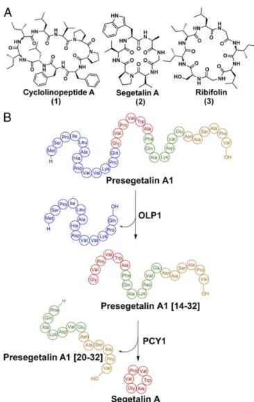
Orbitides encompass a class of plant homodetic macrocyclic natural products, usually 5–12 amino acids in size, which are characterized by the intramolecular condensation between the N and C termini of their linear precursors (10). The chemical structures of orbitides consist solely of α-amide linkages and are distinguished from other macrocyclic plant peptide natural prod-ucts such as cyclotides, which are further interlocked through multiple disulfide bonds (18), and amatoxins, which contain an unusual tryptathionine moiety formed by a crosslink between a Cys and Trp residue (19). The first macrocyclic orbitide, cyclo-linopeptide A, was isolated in 1959 from flaxseed (Linum usita-tissimum) oil and determined to be a cyclic peptide of ILVPPFFLI (Fig. 1A) (20). Since then, over 168 Caryophyllaceae-like non-redundant orbitides have been identified, and encoding sequences have been confirmed in Annonaceae, Rutaceae, Euphorbiaceae, and Linaceae (21). Many orbitides demonstrate biological activi-ties including antimalarial (22), vasodilatory (23), immunomodu-lating (24), and xenoestrogenic activities (25).
Analysis of expressed sequence tag libraries from developing seeds of Saponaria vaccaria (Vaccaria hispanica) identified genes that likely encode for peptide precursors of orbitides. Expres-sion of candidate precursors in the roots of a nonsegetalin A-producing S. vaccaria strain resulted in the production of fully processed cyclic peptide products (26), confirming that orbitides are indeed RiPPs. Subsequent biochemical assays with seed ex-tracts, using synthetic peptide substrates, revealed the order of posttranslational modifications that generate the orbitide sege-talin A (Fig. 1 A and B) (27). Briefly, segesege-talin A is first produced ribosomally as a 32-aa peptide termed presegetalin A1. A serine protease (oligopeptidase 1 or OLP1) cleaves the first 15 amino acids to yield a product containing an N-terminal glycine (termed presegetalin A1 [14–32]). A second enzyme [peptide cyclase 1 (PCY1)] acts on this product to excise the linear C-terminal
Significance
The class of bioactive cyclic plant natural products called orbi-tides was first identified nearly half a century ago. Here we describe how a single enzyme can catalyze the cyclization of a range of ribosomally synthesized linear peptides into the cor-responding cyclic products of varying ring sizes. These studies may provide a means for producing large libraries of cyclic peptides without any sequence bias.
Author contributions: S.K.N. designed research; J.R.C., P.E., and S.K.N. performed re-search; P.S.C. contributed new reagents/analytic tools; S.K.N. analyzed data; and J.R.C. and S.K.N. wrote the paper.
The authors declare no conflict of interest. This article is a PNAS Direct Submission.
Data deposition: Crystallographic coordinates have been deposited in the Protein Data Bank,www.pdb.org[PDB ID codes5UW3(PCY1 with follower peptide),5UW5(PCY1 H695A variant with follower peptide),5UW6(PCY1 with follower peptide and covalent inhibitor ZPP),5UW7(PCY1 Y481F with follower peptide), and5UZW(PCY1 G696 inser-tion variant with follower peptide and covalent inhibitor ZPP)].
1To whom correspondence should be addressed. Email: snair@illinois.edu.
This article contains supporting information online atwww.pnas.org/lookup/suppl/doi:10.
BIOCHE
13 residues (termed presegetalin A1 [20–32]), with the con-comitant macrocyclization of the remaining N-terminal six resi-dues to yield the cyclo[GVPVWA] segetalin A product (Fig. 1B). The biosynthesis of cyclotides (28, 29), amatoxins (30), and cyanobactins (31) uses similar biosynthetic strategies in which a sequence-specific peptidase catalyzes the transamidation of a linear peptide substrate to produce a macrocyclic product.
Primary sequence analysis identifies PCY1 as a member of the S9A protease family that includes several clades of prolyl oligo-peptidases (POP). Proteases of the S9A class catalyze the hydrolytic cleavage of peptide substrates at the amide linkage C-terminal to Pro to yield two linear products. However, PCY1 is distinguished from canonical S9A proteases by its unique product profile, as the enzyme generates a macrocyclic product rather than the linear counterpart. In addition, PCY1 does not require a Pro in the se-quence of the cyclized peptide. Analysis of the predicted primary sequences of dozens of putative orbitide precursor peptides reveals that residues within the core sequence that is cyclized are highly divergent, whereas residues in the preceding leader sequence (re-moved by OLP1) and the trailing follower sequence (excised as a linear peptide by PCY1) are highly conserved. Similar patterns of conservation are observed for the precursors of other cyclic peptides including cyanobactins (31) and amatoxins (19, 32). Alanine scan-ning mutational analysis of presegetalin A1 demonstrates that the necessary elements for substrate recognition by PCY1 reside solely
in the 12-residue follower sequence (hereafter, presegetalin A1 [20– 32]) that is excised during the formation of the macrocyclic product (27). Notably, PCY1 can accept a range of substrates leading to rings of different sizes as this single enzyme is capable of processing almost all of the orbitides that are observed in S. vaccaria. Conse-quently, PCY1 may be used as a biotechnology tool for the pro-duction of macrocycles of diverse sequences. A similar strategy has been used by employing the peptide cyclase butelase 1 to produce cyclic bacteriocins (33).
The structure of the unrelated cyanobactin transamidating pro-tease (PatG) revealed a peptidase S8 (subtilisin-like) fold that is decorated with a “capping” domain, and biochemical studies sug-gest that this domain insertion likely facilitates macrocyclization of the peptide substrate (34, 35). The primary sequence of PCY1 fails to identify any such obvious insertions, relative to canonical S9A protease family members, raising the question of how an enzyme from a class of generic proteases can be adapted to produce a macrocyclic product. To address this issue, we have carried out biochemical and crystallographic characterization of PCY1. Results
Kinetic Analysis of PCY1.Incubation of recombinant PCY1 with a
synthetic peptide corresponding to presegetalin A1 [14–32] resulted in a near-complete conversion into cyclic[GVPVWA] segetalin A (and corresponding linear follower peptide) (Fig. 2). The yield of linear core peptide was minimal, demonstrating that the enzyme favors production of macrocyclic products relative to the linear counterpart. To determine steady-state kinetic parameters, we used a LC-MS–based assay to quantify rates of product formation. Complete Michaelis–Menten kinetics were carried out for the pre-segetalin A1 [14–32] substrate, yielding a kcatvalue of 6.0 × 10⁻²·s−1
with a KM of 0.77 μM (SI Appendix, Fig. S1 and Table S3). In
comparison, the asparaginyl endopeptidases involved in cyclotide macrocyclization (butelase 1 and AEP) have similar kcatvalues, but
with KMvalues that are 5- to 10-fold higher (28, 29). Additionally,
the transamidative protease involved in the biosynthesis of amatoxin (GmPOPB) was initially shown to have a similar KMvalue and a
100-fold faster rate (kcat of 5.7·s−1) (30), whereas more extensive
kinetic analysis indicated a KMabout 75-fold higher and a kcat
ap-proximately 4-fold faster than PCY1 (36).
Fig. 1. (A) Structures of the orbitides cyclolinopeptide A, segetalin A, and ribifolin. (B) Scheme showing the biosynthesis of segetalin A by S. vaccaria.
Fig. 2. Extracted-ion chromatograms of products derived upon incubation of recombinant PCY1 with a synthetic peptide corresponding to presegetalin A1 [14–32]. Chromatograms showing the mass to charge ratio (m/z) values of the segetalin A product (m/z = 610.5, z = 1; green trace), of the linear [GVPVWA] product (m/z = 628.5, z = 1; red trace), of the presegetalin A1 [14–32] substrate (m/z = 993.2, z = 2; blue trace), and the presegetalin A1 [20–32] by-product (m/z = 688.5, z = 2; purple trace) are shown as indicated. The wild-type PCY1 used most of the presegetalin A1 [14–32] substrate to produce almost exclusively cyclic segetalin A product after a 1-h reaction.
Structural Characterization of PCY1.To elucidate how an S9A
pro-tease family can catalyze macrocyclization, we determined the crystal structure of PCY1 that had been incubated with the pre-segetalin A1 [14–32] substrate (Fig. 3 A and B) (37). The 1.9-Å resolution structure of this PCY1 complex revealed an overall ar-chitecture similar to other prolyl oligopeptidase family members, such as the eukaryotic enzyme from porcine muscle (Sus scorfa) (PDB ID code: 1QFS; rmsd of 1.6 Å over 691 aligned Cα atoms) (38, 39) and the bacterial enzyme from Myxococcus xanthus (PDB ID code: 2BKL; rmsd of 1.7 Å over 691 aligned Cα atoms) (40) (Fig. 3 C and E). The structure of PCY1 consists of an α/β hy-drolase domain composed of residues Met1-Val77 and Asp437-Asp724 and a β-propeller domain consisting of residues Cys82-Glu432. A hinge region encompassing residues Asn78-Arg81 and Ser433-Pro436 links the two domains and presumably facilitates movement between an open form competent for binding substrate and a closed form where chemistry is carried out (41, 42). The PCY1–presegetalin A1 complex was crystallized in the closed form with a large internal cavity (10,317 Å3 as calculated by
CASTp), which can accommodate the binding of large peptides (41, 43). A close-up view of the active sites of canonical prolyl oligopeptidases from S. scorfa and M. xanthus reveal the protrusion of various loops that significantly decrease the corresponding in-ternal cavity (Fig. 3 D and F). In PCY1, residues Asp653, His695, and Ser562 make up the active-site catalytic triad, and a hydro-phobic pocket enriched in aromatic residues forms a binding pocket for the target peptide (Fig. 4A). Finally, Tyr481 acts to stabilize the tetrahedral intermediate oxyanion that is formed following the at-tack of the Ser562 alkoxide on the peptide substrate.
Although PCY1 crystallization was carried out using pre-segetalin A1 [14–32], the active site revealed unambiguous electron density only for the terminal six residues (NASAPV) of the follower peptide (Fig. 3B). The residues within the follower sequence bind to PCY1 near the hinge region and engage in hydrogen bonds with amino acids in both the α/β hydrolase and the β-propeller domains. Hydrogen-bonding interactions can be observed between PCY1 residues Arg81, Asn97, Asn104, and backbone carbonyl and amide of Ala28 and Ser29, respectively, of the follower peptide. The only sequence-specific interaction with the follower peptide occurs between its side chain of
Asn27 and the side chain of Asp104 and backbone carbonyl of Leu132 of PCY1. The binding of the carboxyl terminus of the peptide is of particular interest, as it forms hydrogen bonds with PCY1 residues Ser495 and Ser493. This observation explains prior alanine-scanning mutational analysis of presegetalin A1 [14–32], which demonstrates that PCY1 is tolerant of mutation along the entirety of the primary sequence of the substrate but cannot process a substrate with the deletion of the C-terminal Val32, indicating the importance of the follower peptide C-terminal carboxylate (27).
To visualize a structure of PCY1 bound to intact presegetalin A1, we carried out cocrystallization with two different active-site variants, His695→Ala and Tyr481→Phe. In both instances, electron density could be observed only for the six-residue fol-lower sequence, despite the presence of the presegetalin A1 [20– 32] peptide in the crystals (as determined by mass spectrometry), consistent with disorder/flexibility for the remainder of the sub-strate (SI Appendix, Figs. S2 and S3). Hence, only the follower region of presegetalin A1 is engaged by PCY1, allowing for flexibility in the substrate N-terminal to the cleavage site. This flexibility explains how S. vaccaria can produce nine different orbitides of different ring sizes and sequence composition, as only the follower sequences are conserved in the progenitors of the macrocyclic products (SI Appendix, Fig. S4).
Most RiPP biosynthetic enzymes use a PqqD domain (RiPP precursor peptide recognition element) (44) to engage the leader precursor peptide through a limited number of hydrophobic in-teractions. This has been observed in at least some of the en-zymes involved in the biosynthesis of pyrroloquinoline quinone (45), lantibiotics (46), lasso peptides (44), and cyanobactins (47). In contrast, the follower peptide binding observed in PCY1 does not rely on hydrophobic interactions, but rather on hydrogen-bonding interactions. Hydrogen-hydrogen-bonding interactions also me-diate binding of leader peptides by microvirdin biosynthetic
en-Fig. 3. (A) The 1.9-Å PCY1 structure with the α/β hydrolase domain shown in green and β-propeller domain shown in red. (B) The C-terminal six amino acids of the presegetalin A1 [14–32] substrate (orange) is bound near the hinge region of PCY1. A simulated annealing difference Fourier maps (Fo-Fc), calculated with the
coordinates of the peptide omitted, is superimposed and contoured to 2.5σ (blue). (C) Structure of the canonical POP enzyme from S. scorfa (PDB ID code 1QFS) (38, 39) bound to covalent inhibitor ZPP, with the active site demarcated in the rect-angle, and (D) a close-up view of the active site. (E) Structure of the bacterial en-zyme from M. xanthus (PDB ID code 2BKL) (40) bound to covalent inhibitor
Z-Ala-Fig. 4. (A) Active-site alignment of PCY1 (red, pink) and porcine muscle POP (cyan, blue) with covalently bound ZPP. All residues are conserved and align well except for the catalytic histidine. Residues are labeled according to PCY1’s numbering. (B) Sequence alignment of PCY1 and POP enzymes from S. scorfa [domestic pig, National Center for Biotechnology Information (NCBI): NP_001004050.1], Beta vulgaris (beet, NCBI: XP_010667346.1) and Morus nota-bilis (mulberry, NCBI: XP_010101294.1). (C) Changes in structures at regions distal to the active site between PCY1 (red) and S. scorfa POP (blue) indicate significant difference in the orientation of the hairpin loop. The hairpin is stabilized in PCY1 by a stacking interaction between His149 and Tyr696.
BIOCHE
PCY1-Binding Affinity Measurements. To determine the affinity of
PCY1 for various follower peptides, we carried out competition binding measurements (Table 1). A six-residue presegetalin A1 [27– 32] peptide conjugated to fluorescein isothiocyanate bound to PCY1 with a dissociation constant (Kd) of 4.83 μM as determined by
fluorescence polarization (SI Appendix, Fig. S5A). Competitive bind-ing between this labeled peptide and various unlabeled peptides was used to determine the Kivalues for the unlabeled peptides. These
competition experiments demonstrate that unlabeled presegetalin A1 [27–32] bound to PCY1 with a Kiof 31.0 μM (Table 1 andSI Ap-pendix, Fig. S5B). The longer, full-length linear by-product of the cyclization reaction, presegetalin A1 [20–32], binds with a much higher affinity with a Kiof 0.40 μM (Table 1 andSI Appendix, Fig. S5C). The nearly 80-fold greater binding affinity for the longer pre-segetalin A1 [20–32] peptide is curious given that only the last six residues of the peptide are involved in contacts with PCY1 in the cocrystal structure. The additional binding affinity, relative to pre-segetalin A1 [27–32], may be due to solvent exclusion or another nonsequence-dependent mechanism. The amino acids that make up the final cyclic product do not seem to contribute to binding as sub-strate presegetalin A1 [14–32] was measured to bind to the catalyti-cally inhibited PCY1 H695A with a Kiof 1.10 μM, very similar to that
of presegetalin A1 [20–32] (Table 1 andSI Appendix, Fig. S5D). This contrasts with binding studies completed with GmPOPB, in which the cyclized region of the peptide enhances binding affinity (36).
To verify the roles of individual interactions observed in the structure, we measured the affinity of PCY1 for variants of the presegetalin A1 [27–32] peptide. Based on the crystal structure, the side chain of Asn27 of presegetalin A1 [27–32] is engaged in specific hydrogen-bonding interactions. Consequently, the Asn27→Ala mutation in presegetalin A1 [27–32] results in a re-duction in the PCY1-binding affinity (Kiof 131 μM) (Table 1 and SI Appendix, Fig. S5E). The C terminus of the follower peptide forms hydrogen bonds with PCY1 residues Ser495 and Ser493, and the binding affinity of PCY1 for the presegetalin A1 [27–32] variant with a C-terminal amide is diminished and outside the detection range of the assay (Table 1 andSI Appendix, Fig. S5F). Mutations were made to PCY1 residues Ser493 and Asn97, both of which bind the follower peptide. Ser493→Ala had minimal changes on catalytic efficiency relative to the wild type, whereas Asn97→Ala greatly diminished activity (SI Appendix, Table S3 and Fig. S6).
Follower Peptide Induces Conformational Change in PCY1.Based on
the kinetic studies, we hypothesized that the high affinity of PCY1 for the presegetalin A1 [27–32] follower peptide may ensure that the enzyme is maintained in the closed conformation throughout catalysis and is displaced only after turnover. To test this, we fluorescently labeled PCY1 with the thiol-specific probe N-1-pyrene maleimide. Addition of the presegetalin A1 [27–32] to labeled PCY1 resulted in the quenching of fluorescence compared with a buffer control (SI Appendix, Fig. S7). Con-versely, addition of a control peptide that does not bind (pre-segetalin A1 [27–32] amide) showed no change in fluorescence. As there are no cysteines present near the binding site of the
peptide (SI Appendix, Fig. S8), these changes in fluorescence have been attributed to changes in the conformation of PCY1. These findings are consistent with the notion that binding of presegetalin A1 [27–32] alone induces a conformational change in PCY1 to the closed state.
As presegetalin A1 [27–32] binding induces closure of the PCY1 structure, addition of exogenous peptide would be expected to inhibit productive turnover of the presegetalin A1 [14–32] substrate. We tested this hypothesis using a 1-h end-point reaction of PCY1 incubated with 30 μM of substrate in the presence of stoichiometric and 10-fold excess of the presegetalin A1 [20–32] linker+follower peptide product, as well as 100-fold excess of the presegetalin A1 [27–32] follower peptide (SI Appendix, Fig. S9). Although PCY1 consumed nearly all of the presegetalin A1 [14–32] substrate alone (Fig. 2), addition of the follower peptides resulted in a decrease in turnover in a concentration-dependent manner. These data are consistent with the follower peptide maintaining a closed state for PCY1. The role of the leader/follower peptide in stabilizing active conformations of biosynthetic enzymes is an emerging theme in RiPP biosynthesis. For example, during cyanobactin biosynthesis, binding of the PatE′ leader peptide to the LynD cyclodehydratase stabilizes the enzyme in a catalytically competent form (47).
Using the structural data as a guide, we generated and charac-terized mutants of several active-site residues to test their roles in catalysis (SI Appendix, Table S3 and Fig. S10A). As expected, both the Ser562→Ala and His695→Ala mutation of the catalytic triad residues produced inactive enzyme, confirming the necessity of these conserved residues. Finally, Tyr481 is necessary to stabilize the oxyanion of the tetrahedral intermediate as the Try481→Phe variant is catalytically inactive. We next interrogated the extent to which the enzyme could accommodate primary amines other than a peptide α-amino group for transamidation. First, we used a variant pre-segetalin A1 [14–32] substrate peptide that contained both the α-amino terminus and a side-chain amine (Val15→Lys). Kinetic characterization of PCY1 using the Val15→Lys presegetalin A1 [14– 32] as a substrate demonstrates that the KMincreased by 10-fold
relative to the cognate presegetalin A1 [14–32] peptide as a substrate but the kcatalso increased by almost 10-fold, resulting in a catalytic
efficiency similar to that using the cognate peptide. Although PCY1 was able to completely convert this substrate to the cyclic product (SI Appendix, Table S3 and Fig. S10B), tandem mass spectrometric analysis revealed that the α-amino group was used for transamidation (SI Appendix, Fig. S11). We did not observe any products consistent with the use of the side chain of Lys15 as part of the macrocycle. We then tested whether PCY1 could generate a macrocyclic product using a nonproteinogenic amine. To this end, we used a presegetalin A1 [14–32] substrate that contained an N-terminal aminohexanoic acid (Ahx) in place of Gly14. PCY1 largely produced a linear product with this substrate, although a minor amount of cyclic [AhxVPVWA] could also be detected (SI Appendix, Table S3 and Fig. S12). These data suggest that PCY1-catalyzed macrocyclization is most effective via the use of a peptide α-amine.
Dual Function of His695 Allows for Cyclization.Prior studies on other
macrocyclases, such as the PatG protease involved in cyanobactin biosynthesis, suggest a plausible route to cyclic peptide production (34, 35). Briefly, following the formation of an acyl-enzyme in-termediate during the first half protease reaction, hydrolysis of the intermediate is averted by solvent exclusion from the active site. Instead, the α-amino terminus of the bound peptide substrate is oriented back toward the catalytic Ser, and deprotonation of the amine can then facilitate a transpeptidation reaction to yield the cyclic peptide product. A comparison of the structure of PCY1 with other prolyl oligopeptidases reveals a strong conser-vation of overall structure and a near-identical alignment of pu-tative active-site residues (Fig. 4A). While nearly all of these features in the PCY1 active site superimpose with the structures of
Table 1. Binding constants for the follower peptide and mutants Ligand IC50(μM) Ki(μM) Presegetalin A1 [20–32] 1.97 ± 0.34 0.40 ± 0.007 Presegetalin A1 [14–32]* 3.03 ± 0.61 1.10 ± 0.22 Presegetalin A1 [27–32] 47.4 ± 1.0 31.0 ± 0.65 Presegetalin A1 [27–32] N27A 197 ± 8 131 ± 5 Presegetalin A1 [27–32] amide >1,000 >650
*Presegetalin A1 [14–32] binding experiments were completed with PCY1 H695A to prevent turnover.
other S9A proteases, a notable exception is the ∼2.4-Å displace-ment of His695 away from the catalytic Ser562.
Studies of hydrolytic serine proteases have established that the catalytic His moves following formation of a tetrahedral in-termediate, both to tune pKaand to serve as an effective proton
donor to the leaving group (49–51). To determine if the mis-aligned His695 is reflective of a catalytically competent active site, we determined the 3.3 Å cocrystal structure of PCY1 covalently bound to an aldehyde inhibitor that mimics a tetrahedral in-termediate [Z-Proprolinal (ZPP)] (Fig. 4A). Although the struc-ture of ZPP differs from that of the true presegetalin substrate peptide, PCY1 is sufficiently tolerant for the sequence preceding the scissile bond to accommodate this inhibitor. The cocrystal structure shows that ZPP is covalently bound to Ser562 and adopts a conformation similar to that in canonical S9A enzymes, with the Pro oriented within the aromatic hydrophobic pocket. Surpris-ingly, the disposition of His695 relative to the unliganded enzyme remained unchanged (0.9-Å movement of Nγ) and was outside the range of a hydrogen-bonding interaction with Ser562.
The obvious change in the orientation of the catalytic His695 suggested that it may play some role in directing the ability of PCY1 to catalyze macrocyclization. A cursory inspection of the PCY1 active site revealed a single residue deletion at a Gly that is normally adjacent to the catalytic His in canonical S9A proteases (Fig. 4B). Surmising that this deletion may force the displacement of His695 away from Ser562, we generated a Gly insertion mutant in PCY1, which would mimic the canonical S9A protease active site. If the misalignment of His695 indeed plays a role in the macrocyclase activity of PCY1, this insertion mutant should generate linear, hy-drolytic products, similar to that of canonical S9A proteases. Nota-bly, this PCY1 mutant slowly produces a significant amount of the linear, hydrolytic product (ratio of cyclic:linear product of ∼3.7:1), whereas the wild-type enzyme almost exclusively produces the macrocyclic product (Fig. 2 andSI Appendix, Table S3 and Fig. S13). These data support the assertion that the altered position of His695 plays some role in macrocycle formation by PCY1.
Although the insertion mutants showed the expected pheno-type of production of both linear and cyclic products, the cata-lytic competency of the enzyme was severely compromised. To determine the basis for the diminished activity, we determined the 2.8-Å resolution structure of the PCY1 Gly696 insertion variant. The structure reveals significant disorder in the loop harboring His695 and could not be confidently modeled. A more detailed comparison of wild-type PCY1 against canonical S9A proteases reveals additional structural deviations near the active site, relative to canonical S9A proteases, in addition to the single Gly deletion (Fig. 4C). In particular, in canonical S9A enzymes, such as porcine prolyl oligopeptidase (PDB ID code 1QFS), the loop of an adjacent β-hairpin (encompassing Leu143 through Val156) is longer and buttresses the orientation of the catalytic His in the active site. In PCY1, the equivalent loop is one residue shorter and is displaced away from the active site. The side chain of Tyr696 (normally an Ala) occupies the volume created by this loop displacement. The Tyr696→Ala/Gly insertion double mu-tant had a greatly compromised activity, further underscoring that the repositioning of His695 in PCY1 involves a confluence of many local changes (SI Appendix, Fig. S14).
Based on the above data, we hypothesized that the His695 in PCY1 may serve two roles. It could serve to deprotonate the catalytic Ser562 (as in other serine proteases), but also may deprotonate the α-amino terminus of the peptide substrate to facilitate trans-amidation. To further investigate the dual functionality of the cata-lytic His, we analyzed the PCY1 His695→Ala variant under single turnover conditions (Fig. 5A). If His695 does play a role in the deprotonization of Ser562, this variant should still be unable to yield the hydrolytic, linear presegetalin A1 [20–32] by-product that forms after generation of the acyl-enzyme intermediate. As expected,
His695 is necessary for activation of Ser562 despite the long dis-tance between the two residues. Next, we sought to determine if His695 played a role in proton removal from the substrate α-amino group by testing the activity of PCY1 His695→Ala in the presence of imidazole. At the pH of the assay (pH = 8.5), imidazole should be deprotonated and may be able to function as an imperfect, mobile surrogate for His. Surprisingly, the addition of 20 mM im-idazole resulted in a gain of function for the His695→Ala variant, which could generate both linear and cyclic [GVPVWA] segetalin A products from the presegetalin A1 [14–32] substrate (Fig. 5B). Competition between the mobile imidazole surrogate and solvent for attack on the acyl-enzyme intermediate may account for the observance of both linear and cyclic products in the His695→Ala variant. This result suggests that imidazole (and presumably His695 in the wild-type enzyme) can both deprotonate Ser562 to facilitate formation of an acyl-enzyme adduct and deprotonate the α-amino terminus of the substrate to facilitate transamidation. Discussion
Recent studies of various RiPP biosynthetic enzymes have provided several examples of biocatalysts that can generate a macrocyclic product from a linear peptide substrate (14, 28, 30, 31, 52–54). Such enzymes represent a next-generation technological utility, but many of these catalysts are often slow or require sequence constraints on substrates. The biosynthesis of orbitides, as well as other RiPPs such as cyclotides (28, 29), amatoxins (30), and cyanobactins (31), all use convergent strategies based on Ser proteases that can catalyze the formation of a cyclic product via transamidation, rather than a linear product via hydrolysis. Structural and biochemical studies of the cyanobactin macrocyclase PatG reveal that the enzyme has a helix-turn-helix insertion that sits above the active site, which presumably shields the acyl-enzyme intermediate from solvent. Full enclosure of the PatG active site upon binding of substrate is completed by the AYDG follower peptide sequence necessary for substrate recogni-tion. The structural and biochemical data presented here on the orbitide macrocyclase PCY1 reveals a more nuanced strategy of achieving the same result. Rather than containing obvious insertions, transamidation by PCY1 is facilitated by the binding of the follower peptide, which maintains the enzyme in a closed state that precludes solvent from the active site, potentially limiting the competing hy-drolysis reaction. The selectivity for substrates is maintained via recognition of the follower sequence, and the lack of appreciable affinity for the residues to be cyclized (as demonstrated by fluores-cence polarization data) allows for product diversity. Specificity dic-tated by recognition of the follower peptide is not common in RiPP biosynthetic enzymes, and the only other known cases are of enzymes involved in the biosynthesis of cyanobactins and bottromycin (34).
Fig. 5. (A) Formation of presegetalin A1 [20–32] was observed under single turnover conditions by LC-MS. After a 5-min incubation, wild-type PCY1 was able to catalyze formation of the acyl-enzyme intermediate, producing the presegetalin A1 [20–32] by-product. The H695A mutant did not catalyze for-mation of presegetalin A1 [20–32], indicating that it could not form the acyl-enzyme intermediate. (B) An 18-h incubation of H695A with 20 mM imidazole allowed for the production of both linear [GVPVWA] and cyclic segetalin A. However, the H695A variant produces a significantly greater amount of hydrolytic, linear product (ratio of cyclic:linear product of ∼3.7:1), whereas
BIOCHE
The active site of PCY1 is also altered to more efficiently catalyze the cyclization reaction. The placement of His695 facilitates two roles: the canonical role of activating the Ser nucleophile to help form an acyl-enzyme intermediate and an unexpected role in deprotonating the α-amine of the peptide substrate to facilitate transamidation (SI Appendix, Fig. S15). The dual functionality for His695 is aided by its location in a mobile loop, which could ac-commodate the range of movements necessary for each function. Our biochemical studies provide a foundation for understanding the mechanistic basis for peptide macrocyclization in plants, an activity that is observed in growing classes of RiPP natural products.
Materials and Methods
Methods describing the cloning, expression, purification, biochemical anal-ysis, kinetic and biochemical characterization, and crystallization of PCY1 and ligand complexes are described in detail inSI Appendix, SI Materials and
Methods. Chemical data and analytical methods are also provided.
ACKNOWLEDGMENTS. We thank the entire staff at the Life Sciences Collab-orative Access Team at Argonne National Laboratory for their assistance with X-ray data collection. J.R.C. was supported in part by the Lowell P. Hager Fellowship from the Department of Biochemistry. The Bruker UltrafleXtreme mass spectrometer was purchased in part with a grant from the National Center for Research Resources, National Institutes of Health (S10 RR027109 A). 1. Passioura T, Katoh T, Goto Y, Suga H (2014) Selection-based discovery of druglike
macrocyclic peptides. Annu Rev Biochem 83:727–752.
2. Driggers EM, Hale SP, Lee J, Terrett NK (2008) The exploration of macrocycles for drug discovery: An underexploited structural class. Nat Rev Drug Discov 7:608–624. 3. White TR, et al. (2011) On-resin N-methylation of cyclic peptides for discovery of orally
bioavailable scaffolds. Nat Chem Biol 7:810–817.
4. Rezai T, Yu B, Millhauser GL, Jacobson MP, Lokey RS (2006) Testing the conforma-tional hypothesis of passive membrane permeability using synthetic cyclic peptide diastereomers. J Am Chem Soc 128:2510–2511.
5. Scott CP, Abel-Santos E, Wall M, Wahnon DC, Benkovic SJ (1999) Production of cyclic peptides and proteins in vivo. Proc Natl Acad Sci USA 96:13638–13643.
6. Kohli RM, Walsh CT, Burkart MD (2002) Biomimetic synthesis and optimization of cyclic peptide antibiotics. Nature 418:658–661.
7. Zhang C, Dai P, Spokoyny AM, Pentelute BL (2014) Enzyme-catalyzed macrocyclization of long unprotected peptides. Org Lett 16:3652–3655.
8. Hipolito CJ, Suga H (2012) Ribosomal production and in vitro selection of natural product-like peptidomimetics: The FIT and RaPID systems. Curr Opin Chem Biol 16: 196–203.
9. Velásquez JE, van der Donk WA (2011) Genome mining for ribosomally synthesized natural products. Curr Opin Chem Biol 15:11–21.
10. Arnison PG, et al. (2013) Ribosomally synthesized and post-translationally modified peptide natural products: Overview and recommendations for a universal nomen-clature. Nat Prod Rep 30:108–160.
11. Shimamura H, et al. (2009) Structure determination and total synthesis of bot-tromycin A2: A potent antibiotic against MRSA and VRE. Angew Chem Int Ed Engl 48: 914–917.
12. Sivonen K, Leikoski N, Fewer DP, Jokela J (2010) Cyanobactins-ribosomal cyclic pep-tides produced by cyanobacteria. Appl Microbiol Biotechnol 86:1213–1225. 13. Gross E, Morell JL (1971) The structure of nisin. J Am Chem Soc 93:4634–4635. 14. Schramma KR, Bushin LB, Seyedsayamdost MR (2015) Structure and biosynthesis of a
macrocyclic peptide containing an unprecedented lysine-to-tryptophan crosslink. Nat Chem 7:431–437.
15. Hegemann JD, Zimmermann M, Xie X, Marahiel MA (2015) Lasso peptides: An in-triguing class of bacterial natural products. Acc Chem Res 48:1909–1919. 16. Bagley MC, Dale JW, Merritt EA, Xiong X (2005) Thiopeptide antibiotics. Chem Rev
105:685–714.
17. Sardar D, Lin Z, Schmidt EW (2015) Modularity of RiPP enzymes enables designed synthesis of decorated peptides. Chem Biol 22:907–916.
18. Craik DJ, Malik U (2013) Cyclotide biosynthesis. Curr Opin Chem Biol 17:546–554. 19. Hallen HE, Luo H, Scott-Craig JS, Walton JD (2007) Gene family encoding the major
toxins of lethal Amanita mushrooms. Proc Natl Acad Sci USA 104:19097–19101. 20. Kaufmann HP, Tobschirbel A (1959) An oligopeptide from flaxseed. Chem Ber 92:
2805–2809.
21. Gui B, et al. (2012) Identification and quantification of cyclolinopeptides in five flaxseed cultivars. J Agric Food Chem 60:8571–8579.
22. Pinto MEF, et al. (2015) Ribifolin, an orbitide from Jatropha ribifolia, and its potential antimalarial activity. J Nat Prod 78:374–380.
23. Morita H, et al. (2006) Structure of a new cyclic nonapeptide, segetalin F, and vaso-relaxant activity of segetalins from Vaccaria segetalis. Bioorg Med Chem Lett 16: 4458–4461.
24. Gaymes TJ, Cebrat M, Siemion IZ, Kay JE (1997) Cyclolinopeptide A (CLA) mediates its immunosuppressive activity through cyclophilin-dependent calcineurin inactivation. FEBS Lett 418:224–227.
25. Itokawa H, Yun Y, Morita H, Takeya K, Yamada K (1995) Estrogen-like activity of cyclic peptides from Vaccaria segetalis extracts. Planta Med 61:561–562.
26. Condie JA, et al. (2011) The biosynthesis of Caryophyllaceae-like cyclic peptides in Saponaria vaccaria L. from DNA-encoded precursors. Plant J 67:682–690. 27. Barber CJS, et al. (2013) The two-step biosynthesis of cyclic peptides from linear
precursors in a member of the plant family Caryophyllaceae involves cyclization by a serine protease-like enzyme. J Biol Chem 288:12500–12510.
28. Nguyen GKT, et al. (2014) Butelase 1 is an Asx-specific ligase enabling peptide mac-rocyclization and synthesis. Nat Chem Biol 10:732–738.
29. Bernath-Levin K, et al. (2015) Peptide macrocyclization by a bifunctional endopro-tease. Chem Biol 22:571–582.
30. Luo H, et al. (2014) Peptide macrocyclization catalyzed by a prolyl oligopeptidase involved in α-amanitin biosynthesis. Chem Biol 21:1610–1617.
31. Lee J, McIntosh J, Hathaway BJ, Schmidt EW (2009) Using marine natural products to discover a protease that catalyzes peptide macrocyclization of diverse substrates. J Am Chem Soc 131:2122–2124.
32. Li P, Deng W, Li T (2014) The molecular diversity of toxin gene families in lethal Amanita mushrooms. Toxicon 83:59–68.
33. Hemu X, Qiu Y, Nguyen GKT, Tam JP (2016) Total synthesis of circular bacteriocins by butelase 1. J Am Chem Soc 138:6968–6971.
34. Koehnke J, et al. (2012) The mechanism of patellamide macrocyclization revealed by the characterization of the PatG macrocyclase domain. Nat Struct Mol Biol 19: 767–772.
35. Agarwal V, Pierce E, McIntosh J, Schmidt EW, Nair SK (2012) Structures of cyanobactin maturation enzymes define a family of transamidating proteases. Chem Biol 19: 1411–1422.
36. Czekster CM, Naismith JH (2017) Kinetic landscape of a peptide bond-forming prolyl oligopeptidase. Biochemistry 56:2086–2095.
37. Tsilikounas E, Kettner CA, Bachovchin WW (1992) Identification of serine and histi-dine adducts in complexes of trypsin and trypsinogen with peptide and nonpeptide boronic acid inhibitors by 1H NMR spectroscopy. Biochemistry 31:12839–12846. 38. Holm L, Rosenström P (2010) Dali server: Conservation mapping in 3D. Nucleic Acids
Res 38:W545-9.
39. Fülöp V, Böcskei Z, Polgár L (1998) Prolyl oligopeptidase: An unusual beta-propeller domain regulates proteolysis. Cell 94:161–170.
40. Shan L, Mathews II, Khosla C (2005) Structural and mechanistic analysis of two prolyl endopeptidases: Role of interdomain dynamics in catalysis and specificity. Proc Natl Acad Sci USA 102:3599–3604.
41. Li M, Chen C, Davies DR, Chiu TK (2010) Induced-fit mechanism for prolyl endopep-tidase. J Biol Chem 285:21487–21495.
42. Polgár L (2002) The prolyl oligopeptidase family. Cell Mol Life Sci 59:349–362. 43. Dundas J, et al. (2006) CASTp: Computed atlas of surface topography of proteins with
structural and topographical mapping of functionally annotated residues. Nucleic Acids Res 34:W116-8.
44. Burkhart BJ, Hudson GA, Dunbar KL, Mitchell DA (2015) A prevalent peptide-binding domain guides ribosomal natural product biosynthesis. Nat Chem Biol 11:564–570. 45. Tsai TY, Yang CY, Shih HL, Wang AHJ, Chou SH (2009) Xanthomonas campestris PqqD
in the pyrroloquinoline quinone biosynthesis operon adopts a novel saddle-like fold that possibly serves as a PQQ carrier. Proteins 76:1042–1048.
46. Ortega MA, et al. (2015) Structure and mechanism of the tRNA-dependent lantibiotic dehydratase NisB. Nature 517:509–512.
47. Koehnke J, et al. (2015) Structural analysis of leader peptide binding enables leader-free cyanobactin processing. Nat Chem Biol 11:558–563.
48. Li K, Condurso HL, Li G, Ding Y, Bruner SD (2016) Structural basis for precursor protein-directed ribosomal peptide macrocyclization. Nat Chem Biol 12:973–979. 49. Cleland WW (2000) Low-barrier hydrogen bonds and enzymatic catalysis. Arch
Biochem Biophys 382:1–5.
50. Radisky ES, Lee JM, Lu C-JK, Koshland DE, Jr (2006) Insights into the serine protease mechanism from atomic resolution structures of trypsin reaction intermediates. Proc Natl Acad Sci USA 103:6835–6840.
51. Topf M, Richards WG (2004) Theoretical studies on the deacylation step of serine protease catalysis in the gas phase, in solution, and in elastase. J Am Chem Soc 126: 14631–14641.
52. Duquesne S, et al. (2007) Two enzymes catalyze the maturation of a lasso peptide in Escherichia coli. Chem Biol 14:793–803.
53. Yan K-P, et al. (2012) Dissecting the maturation steps of the lasso peptide microcin J25 in vitro. ChemBioChem 13:1046–1052.
54. Li B (2006) et al. Structure and mechanism of the lantibiotic cyclase involved in nisin biosynthesis. Science 311:1464–1467.
Characterization of the Macrocyclase Involved the Biosynthesis of RiPP Cyclic Peptides in
Plants
Jonathan R. Chekan
1, Paola Estrada
1, Patrick S. Covello
2and Satish K. Nair
1,3,4*Materials and Methods………S2
Table S1. Primers used to generate PCY1 variants……….……. S6
Table S2. Data collection and refinement statistics..….………...……… S7
Table S3. Kinetic parameters for wild-type and variant PCY1……….... S8
Figure S1. Michaelis-Menten kinetic curve for PCY1.. ……….………..S9
Figure S2: Difference Fourier maps of PCY1 variant follower binding sites………..……….. S10
Figure S3. Integrity of the substrate peptide in PCY1 crystals.………... .S11
Figure S4: Sequences alignments of PCY1 substrates………... S12
Figure S5: Binding of presegtalin A1 (PSA1) peptides to PCY1.………... S13
Figure S6: Activity of PCY1 follower peptide binding variants….………... S14
Figure S7: Binding of the follower peptide alters the conformation of PCY1…..………. S15
Figure S8: Possible N-(1-pyrene)maleimide labeling locations………... S16
Figure S9: Product inhibition of PCY1 by the follower peptide ………..…… .S17
Figure S10: Biochemical activity of PCY1 variants……….…..……… S18
Figure S11: PCY1 does not cyclize substrates through a Lys side chain amine ………….. ...S19
Figure S12: Macrocyclase activity is dependent on the length of the N-terminal amine ……. .S20
Figure S13: Activity profile of the PCY1 696G insertion variant...……….. .S21
Figure S14: Activity of the Tyr696
!Ala/Gly insertion variants of PCY1………... .S22
Figure S15: Proposed mechanism of PCY1………... S23
Figure S16: Extent of modification of PCY1 with fluorescent dye………... ….S24
Supplementary References………...…... .S25
MATERIALS AND METHODS
Materials. Peptides were purchased from GenScript with a purity of 90% or greater as verified
by HPLC analysis. Chemicals were purchased from Fisher Scientific and Sigma-Aldrich. IPTG
and antibiotics were purchased from Gold Biotechnology.
Enzyme Expression, Purification, and Mutagenesis. The previously described overexpression
plasmid (pCB008) for recombinant production of PCY1 (1) was transformed into a Rosetta
Escherichia coli expression strain (Novagen). PCY1 was expressed in Terrific Broth (BD Difco)
by growing cell cultures to an OD
600of 1 at 37 °C. Cell cultures were cooled in an ice bath for 15
minutes prior to the addition of 0.5 mM IPTG. Cultures were further incubated at 18 °C for
approximately 18 hours before harvesting. Cells were lysed (Avestin) and purified by His
6affinity purification (GE Healthcare Life Sciences). Fractions containing PCY1, as determined
by SDS-PAGE, were collected and further purified using a Superdex 16/60 S200 or S75 size
exclusion column (GE Healthcare Life Sciences) equilibrated with a buffer composed of 100
mM KCl, and 20 mM HEPES free acid pH 7.5. PCY1 mutants were generated using the
QuikChange methodology with the primers listed in Table S1.
Protein Crystallization and Data Collection. Initial crystallization conditions were determined
using sparse matrix screens using a Gryphon robot (Art Robbins). Crystals of wild-type PCY1
were further optimized using hanging drop trays (Hampton) by incubating protein (concentration
of 14 mg/ml) supplemented with 5 mM boric acid and 1 mM presegetalin A1 [14-32] with an
equal volume of precipitant containing 16% PEG 8,000, 0.2 M calcium acetate, and 0.1 M
sodium cacodylate (pH 6.5), prior to equilibration against the same solution. Crystallization trays
were incubated at 9 °C for at least four days. PCY1 His695
!Ala was crystallized using a protein
concentration of 8 mg/mL and precipitant solution of 16 % PEG 8,000, 50 mM calcium acetate,
and 0.1 M sodium cacodylate (pH 6.5). Prior to crystallization, the PCY1 His695
!Ala variant
was incubated with 1 mM presegetalin A1 [14-32]. For crystals of PCY1 with covalently bound
ZPP, the protein was incubated with 1 mM ZPP for 18 hours at 4 °C. Crystals were grown by the
hanging drop method at 9 °C with a presegetalin A1 [27-32] concentration of 1 mM and a
protein concentration of 10 mg/mL using 20% PEG 8,000, 0.1 M calcium acetate, and 0.1 M
sodium cacodylate pH 6.5 as the precipitant. The PCY1 Gly696 insertion variant was crystallized
by pre-incubation with 1 mM ZPP (as before) and 1 mM presegetalin A1 [27-32] at a
concentration of 12 mg/mL. A precipitant solution of 18% PEG 8,000, 0.2 M calcium acetate,
0.1 M sodium cacodylate pH 6.5 was used to form crystals at 9 °C. PCY1 crystals were soaked
in the crystallization buffer supplemented with 20% PEG 400 immediately prior to vitrification
by direct immersion into LN
2. The PCY1 Tyr481
!Phe mutant crystals were produced by first
incubating 1 mM presegetalin A1 [14-32] with protein at a concentration of 7 mg/mL. Crystals
formed at 289 K using a 3:1 ratio of the protein sample mixed with a precipitant containing 23%
PEG 3,350, 0.1 M magnesium chloride, and 0.1 M HEPES free acid (pH 7.5), prior to
equilibration against a solution of the same buffer. The cryo-preservation solution consisted of
the crystallization buffer supplemented to a final concentration of 30% PEG 3,350 was utilized.
Diffraction data was collected using CCD detectors, at LS-CAT Sector 21 of the Advanced
Photon Source (Argonne National Labs, IL).
Crystal Data Processing and Structure Refinement. PCY1 data sets were integrated and
scaled using the autoPROC software package (2). Using PDB code 1QFS as a search model,
phases for wild type PCY1 data set was determined by molecular replacement using PHASER
(3), as implemented in the PHENIX software suite (4). Initial model re-building was carried out
using a combination of phenix.autobuild and Buccaneer (5). All crystallographic model building
was preformed using Coot (6), followed by rounds of refinement using REFMAC5 (7), and
phenix.refine. The refined wild type PCY1 structure was subsequently used to determine the
phases of all mutants and covalently modified PCY1 structures. Data collection, scaling, and
refinement statistics for all data sets can be found in Table S2.
Fluorescence Polarization. PCY1 was gel filtered into assay buffer (50 mM Tris base pH 8.5,
100 mM NaCl, and 5 mM DTT) with a Superdex 16/60 S200 column (GE Healthcare). To
determine the K
dof the FITC(fluorescein isothiocyanate)-presegetalin A1 [27,32] fluorescent
peptide, 100 nM of peptide was incubated with varying concentrations of PCY1 in a 115 µL
reaction. After a 1 hour incubation at 4 °C, 100uL of the solution was transferred to a 96-Well
Low Fluorescence Assay Plate (Corning) and analyzed a Biotek Synergy H1 Hybrid Reader.
Polarization data was fit according to Dunbar et. al. (8) to determine the K
dof FITC-presegetalin
A1 [27-32]. Competitive binding assays were performed using 100 nM FITC-presegetalin A1
[27-32], 2.3 µM PCY1, and varying concentrations of either presegetalin A1 [27-32],
presegetalin A1 [20-32], presegetalin A1 [27-32] amide, or presegetalin A1 [27-32] N27A.
Polarization data was fit to a one-site competitive binding curve to derive an IC
50value for each
peptide. K
ivalues were derived from the IC
50(8).
Activity assays. PCY1 activity assays were completed at 30 °C using 175 nM PCY1 and 30 µM
of peptide in a 100 µL reaction containing 100 mM NaCl, 5 mM DTT, and 20 mM Tris base (pH
8.5). After the designated incubation time, the reactions were quenched with 1 equivalent (100
µL) of methanol. Single turn over assays were completed using 30 µM PCY1 and quenched after
5 min. Samples were centrifuged to remove protein precipitation. All reactions were completed
and analyzed in triplicate.
LC/MS Analysis. LC/MS analysis was completed using an Agilent 1200 series HPLC, an
Agilent G1956B mass spectrometer, and a Grace Davison Denali 250x4.6 mm 5µ C18 HPLC
column. Samples were analyzed using a following method with solvent A (10% ACN, 90% H
2O,
0.1% Formic acid, and 0.1% ammonium formate) and solvent B (90% ACN, 10% H
2O, 0.1%
formic acid, and 0.1% ammonium formate) at 1 mL/min: 1 min at 0% B, linear gradient to 75%
B for 14 min, linear gradient to 100% B for 1 min, 3 min at 100% B, linear gradient to 0% B for
1 min, and 5 min at 0% B. 40 µL of each reaction sample was analyzed using this method.
Extracted ion chromatograms or single ion mode was utilized to observe the following peptides:
segetalin A (m/z=610.5, z=1), linear[GVPVWA] (m/z=628.5 z=1), presegetalin A1 [14-32] (m/z=
993.2, z=2), presegetalin A1 [20-32] (m/z=688.5, z=2), cyclic[GKPVWA] (m/z= 639.5, z=1),
cyclic[Ahx-VPVWA] (m/z= 666.6, z=1), and linear[Ahx-VPVWA] (m/z= 684.6, z=1).
Kinetics. Kinetics assays were completed at 23
oC using 10 nM PCY1 and either 0.5 µM, 1 µM,
5 µM, 15 µM, 30 µM, or 60 µM of the presegetalin A1 [14,32] substrate in a 100 µL reaction
containing 100 mM NaCl, 5 mM DTT, and 20 mM Tris base pH 8.5. After 2, 4, or 6 minutes, the
reaction was quenched with 100 µL of methanol. After centrifugation, 80 µL of sample was
analyzed using the LC/MS method and monitoring the abundance of the linear presegetalin A1
[20-32] byproduct (SIM m/z=688.5, z=2). A standard curve was created using synthetic
presegetalin A1 [20-32] concentrations ranging from 10 nM to 400 nM. Reactions were
completed triplicate. Kinetics of wild-type and variant PCY1 with native and alternative
substrates were conducted in a similar manner.
Fluorescent Monitoring of Conformational Change. To monitor conformational changes,
PCY1 was first labeled with the cysteine specific fluorophore N-(1-pyrene)maleimide. PCY1
was buffer exchanged into 200 mM NaCl, 20 mM HEPES free acid (pH 7.0) and concentrated to
1.25 mL of 50 µM protein. TCEP and N-(1-pyrene)maleimide were added to a concentration of 1
mM with the N-(1-pyrene)maleimide added drop-wise. The reaction was stirred at 23
oC for 2
hours and then quenched with a final concentration of 5 mM DTT. The protein was gel filtered
and buffer exchanged into reaction buffer. Modification of PCY1 was confirmed by MALDI
analysis to reveal a total of three N-(1-pyrene)maleimide added (Figure S16). To monitor the
changes in the environment of the pyrenes, 4 µM of labeled PCY1 was incubated with either
buffer alone, 1 mM presegetalin A1 [27-32], or 1 mM presegetalin [27-32] amide for 10 minutes.
The emission scan was collected using 338 nm excitation wavelength and excitation and
emission slit widths of 5 nm.
Table S1. Primers used for generating PCY1 variants.
Mutagenesis
Primers
Sequence
Y481F For
AAATGCATGGTTTTGGCGGGTTTGG
Y481F Rev
CCAAACCCGCCAAAACCATGCATTT
S562A For
TGAAGGTGGTGCTAATGGTGGCCTTCTCGTTGCTGC
S562A Rev
GGCCACCATTAGCACCACCTTCAATAGCCACTCTTCTAGC
H695A For
CGGATTCAGCGCAAAGCTGCAGCGTACGGACGTGCC
H695A Rev
CGCAAAGCTGCAGCGTACGGACGTGCCACAATGACCCAGATTGC
G696Ins For
CGCAAAGCTGCACATGGCTACGGACGTGCCAC
G696Ins Rev
GTGGCACGTCCGTAGCCATGTGCAGCTTTGCG
G696Ins Y697A
For
CAAAGCTGCACATGGCGCCGGACGTGCCACAATG
G696Ins Y697A
Rev
CATTGTGGCACGTCCGGCGCCATGTGCAGCTTTG
S493A For
GCGTGCCAACAAGTATTTTCACTTCTATGCTTCCGGCCTTCAAGC
S493A Rev
GCTTGAAGGCCGGAAGCATAGAAGTGAAAATACTTGTTGGCACGC
N97A For
GCATAAACATGATGCCAACTTTTGCCGCCAGTCGCA
Table S2: Data collection and refinement statistics
Wild-type H695A ZPP complex 696G –ZPP
complex Y481F
Accession code 5UW3 5UW5 5UW6 5UZW 5UW7
Data collection Space group P1 P1 P1 P1 P21 Cell dimensions a, b, c (Å) 65.5, 85.6, 137.7 65.0, 85.6, 137.9 65.5, 85.5, 138.1 65.0, 85.32, 137.9 87.0, 59.6, 134.1 α, β, γ (°) 87.4, 78.3, 89.3 87.5, 78.5, 89.4 87.6, 78.2, 89.4 87.7, 78.5, 89.6 90.0, 93.1, 90.0 Resolution (Å) 44.9-1.96 (1.967-1.960) 44.90-2.94 (2.950- 2.940) 45.2-3.3 (3.311-3.300) 45.70-2.82 (2.829-2.820) 86.90-2.37 (2.381-2.373) Rsym 12.9 (52.5) 12.7 (50.6) 13.0(27.9) 12.6 (42.6) 9.2 (75.1) I / σI 8.9 (3.3) 11.0 (2.8) 10.2 (5.0) 11.1 (3.3) 14.0 (2.1) Completeness (%) 95.8 (96.8) 98.8 (98.9) 98.9 (99.5) 98.3 (98.1) 99.2 (96.2) Redundancy 3.8 (3.9) 3.9 (3.9) 3.9 (3.9) 4.3 (4.4) 6.2 (6.5) Refinement Resolution (Å) 44.9-1.96 44.90-2.94 45.2-3.3 45.70-2.82 86.90-2.37 No. reflections 191296 58051 41469 65404 52877 Rwork / Rfree 0.224/0.189 0.227/0.186 0.220/0.172 0.270/0.223 0.284/0.236 No. atoms 24731 22798 22866 22661 10789 Protein 22670 22734 22814 22600 10754 Water 2061 64 52 61 35 B-factors Protein 19.68 33.16 33.43 29.66 51.74 Water 24.87 19.19 17.08 13.30 32.58 R.m.s. deviations Bond lengths (Å) 0.0166 0.0102 0.0107 0.0105 0.0104 Bond angles (°) 1.6837 1.4070 1.4751 1.4454 1.3242
![Fig. 5. (A) Formation of presegetalin A1 [20–32] was observed under single turnover conditions by LC-MS](https://thumb-eu.123doks.com/thumbv2/123doknet/14064405.461787/6.877.445.812.837.946/fig-formation-presegetalin-observed-single-turnover-conditions-lc.webp)


![Figure S1. Michaelis-Menten kinetic curve for PCY1. Kinetic measurements were carried out using presegetalin A1 (PSA1) [14-32] as the substrate](https://thumb-eu.123doks.com/thumbv2/123doknet/14064405.461787/16.918.231.687.105.590/figure-michaelis-menten-kinetic-kinetic-measurements-presegetalin-substrate.webp)

![Figure S3 . Integrity of the substrate peptide in PCY1 crystals. MALDI mass spectrometric analysis of the crystallization drop indicated the presence of the PSA1 [20-32] peptide (m/z=](https://thumb-eu.123doks.com/thumbv2/123doknet/14064405.461787/18.918.121.812.110.480/integrity-substrate-crystals-spectrometric-analysis-crystallization-indicated-presence.webp)

![Figure S5: Binding of presegtalin A1 (PSA1) peptides to PCY1. A) PCY1 was titrated into FITC-presegetalin A1 [27-32]](https://thumb-eu.123doks.com/thumbv2/123doknet/14064405.461787/20.918.155.755.100.787/figure-binding-presegtalin-psa-peptides-titrated-fitc-presegetalin.webp)

