arXiv:0905.0320v1 [cond-mat.mes-hall] 4 May 2009
High order vibration modes of glass embedded AgAu
nanoparticles
S. Adichtchev,1 S. Sirotkin,1 G. Bachelier,2 L. Saviot,3 S.
Etienne,4 B. Stephanidis,1 E. Duval,1 and A. Mermet1 1Laboratoire de Physico-Chimie des Mat´eriaux Luminescents,
Universit´e de Lyon, Universit´e Claude Bernard Lyon 1, UMR 5620 CNRS, 69622 Villeurbanne, France
2Laboratoire de Spectroscopie Ionique et Mol´eculaire,
Universit´e de Lyon, Universit´e Claude Bernard Lyon 1, UMR 5579 CNRS, 69622 Villeurbanne, France
3Institut Carnot de Bourgogne, UMR 5209 CNRS-Universit´e de Bourgogne,
9 av. A. Savary, BP 47870, F-21078 DIJON Cedex, France
4Ecole Europ´eenne d’Ing´enieurs en G´enie des Mat´eriaux,
6 rue Bastien Lepage, 54010 Nancy Cedex, Nancy Universit´e, France
(Dated: December 4, 2018)
Abstract
High resolution low frequency Raman scattering measurements from embedded AgAu nanopar-ticles unveil efficient scattering by harmonics of both the quadrupolar and the spherical modes. Comparing the experimental data with theoretical calculations that account for both the embed-ding medium and the resonant Raman process enables a very complete description of the observed multiple components in terms of harmonics of both the quadrupolar and spherical modes, with a dominating Raman response from the former ones. It is found that only selected harmonics of the quadrupolar mode contribute significantly to the Raman spectra in agreement with earlier theoretical predictions.
Over the past twenty years, low frequency vibrational modes from solid nanoparticles have emerged as original features of nanometer sized matter. These modes arise from the recombination of acoustic modes that turn from extended to confined upon reducing the size of the solid. Raman scattering is a dedicated technique to probe nanoparticle oscillation modes provided the used spectrometers enable to reach the low frequency range where these modes typically show up (σ < 10 cm−1). It has proved to be a powerful tool for a quick
and non-destructive evaluation of the sizes of the nanoparticles, especially in the case of metallic ones for which Raman scattering is performed in resonance with the localized sur-face plasmon excitations. Under these conditions, the Raman activity of selected vibration modes of the nanoparticles is strongly enhanced so that intense signals can be obtained from very diluted nanoparticle assemblies1,2. Metallic nanoparticles have allowed significant
progress in the understanding of nanoparticle modes like shape anisotropy effects from Ag nanocolumns and nanolentils3 and elastic anisotropy effects in Au nanocrystals2,4.
Concomi-tantly, the theoretical description of the Raman spectra from nanoparticle vibrations has significantly improved over the years: while the free sphere model of H. Lamb5, combined
with appropriate Raman selection rules6, initially provided satisfactory predictions of the
mode frequencies7
, coupling with the vibrations inside a surrounding matrix in the case of embeddednanoparticles have been well accounted for8,9. On the other hand, quantum
calcu-lations on free metal nanospheres, taking into account the vibration-plasmon coupling, have shown to be able to describe the resonant Raman spectrum in a rather complete way both in terms of frequencies and Raman intensities10. In particular, this latter work predicted that
the Raman intensities from the quadrupolar mode harmonics are not a monotonic function of the mode index. The measurements presented in this work, which show an unusually high number of low-frequency Raman peaks, are an experimental proof of the non monotonic variation of intensity. Thanks to the observation of high order harmonics, we show that the interpretation of low frequency Raman data from metallic nanoparticles requires taking into account the coupling of the surface plasmon with the different vibrations but also the influence of the surrounding matrix as well as the elastic anisotropy inside the nanoparticle. Bimetallic AgAu nanoparticles were produced in a bulk multicomponent glass, starting from the incorporation of silver and gold salts to the initial oxides powder mix. After the colorless glass is made, prolonged annealings slightly above the glass transition tempera-ture generates a characteristic amber shade that arises from the surface plasmon resonance
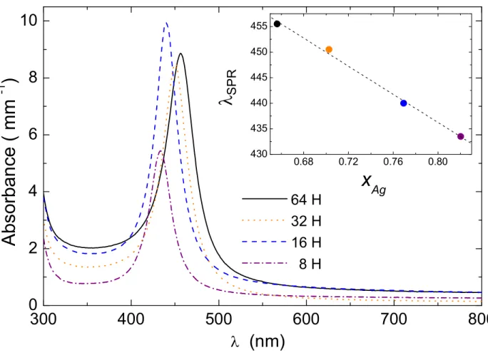
FIG. 1: (color online) Absorption spectra of glasses containing AgxAu1−xnanoparticles for different
annealing times. The inset shows the SPR peak position as a function of the Ag composition, the dashed line is a guide to the eye.
(SPR) of the nucleated metallic nanoparticles. The optical characterization of four samples annealed at 490◦C over 8 H, 16 H, 32 H and 64 H shows a single surface plasmon resonance
peak around 445 nm (Figure 1). This value typically lies between that of pure silver (414 nm) and that of pure gold (529 nm) when embedded as pure nanoparticles with comparable sizes in the same glass, using the same synthesis route. Upon increasing the annealing time, the AgAu SPR is red shifted thus reflecting a gold enrichment of the bimetallic nanopar-ticles. Following a classical interpretation of these data11, one can derive the composition
ratio x of the so-produced AgxAu1−x nanoparticles as a function of annealing time ta using
a linear combination of the SPR peak positions :
λAgxAu1−x(ta) = xλAg(ta) + (1 − x)λAu(ta) (1)
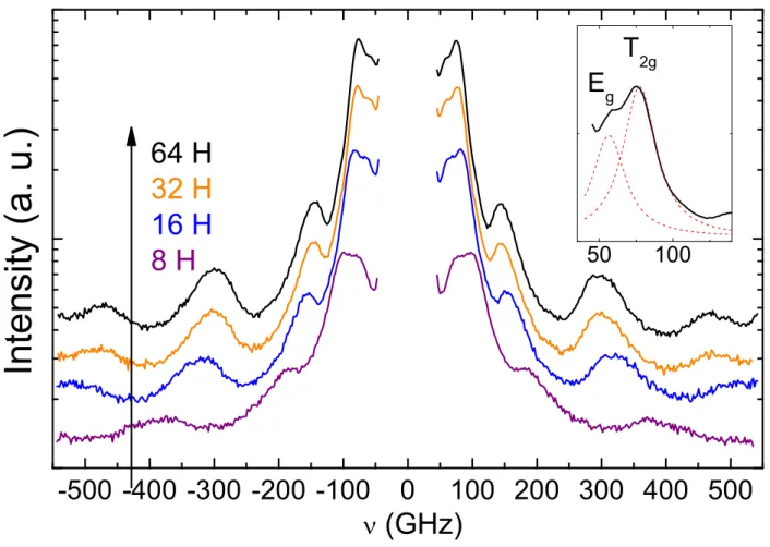
FIG. 2: (color online) Polarized (VV) low frequency Raman spectra (in Log-linear scale) of AgAu nanoparticles embedded in glasses annealed over different times. Inset: enlargement of the lower
(Eg,T2g) doublet for the 64 H sample.
produced in the same glass, at exactly the same annealing times. The so-derived values of x are found to decrease from 0.82 to 0.66 as the annealing times respectively increase from 8 H to 64 H.
The low frequency Raman scattering spectra were recorded with a six pass tandem Fabry-Perot interferometer using the 532 nm line of a continuous doubled Yttrium Aluminum Gar-net laser of 150 mW power. The scattered light was collected in backscattering geometry and the free spectral range set to 550 GHz. The finesse and optical contrast of such inter-ferometer are typically greater than 100 and 1010 respectively. More details on the tandem
Fabry-Perot interferometer can be found elsewhere12,13.
As a general illustration, Figure 2 reports the polarized spectra (with incident and scat-tered light polarized both vertically, VV) of the afore characterized samples. Quite similarly
to recent observations2, these spectra are remarkably richer than those usually reported for
metallic nanoparticles in the sense that they evidence more components than the usually two observed ones, i.e. the fundamental vibrations of both the quadrupolar mode (ℓ = 2, n = 1) and the spherical mode (ℓ = 0, n = 1) (ℓ denotes the angular momentum and n the harmonic rank). Four main bands can already be identified, some of which featuring subcomponents. For purposes of illustration, we will consider in the rest of the paper only the longest annealed sample (Ag0.66Au0.34), provided that the same analysis apply equally well to all samples.
A classical way of identifying the several components of a low frequency Raman scattering spectrum is to compare the polarized (VV) and depolarized (VH) spectra since quadrupolar modes and spherical modes behave differently with regards to polarization. In a first step, we focus on the VH spectrum which is more simple as it only features contributions from the depolarized quadrupolar modes. A multi-Lorentzian decomposition of the VH spectrum reveals 5 components, two of them forming an intense lower frequency doublet (Figure 2 Inset) evidenced thanks to the high resolution Raman setup. Following recent works2,4
, such doublet can be safely identified with the splitting of the fundamental mode of the quadrupolar vibration (ℓ = 2, n = 1) due to the high crystallinity of the nanoparticles (i.e. elastic anisotropy). Indeed, the observed frequency ratio between the lower and the larger components of the doublet, respectively labeled as Eg and T2g according to the irreducible
representation of the corresponding vibrations of a face centered cubic nanocrystal, is close to the value expected for free Au or free Ag nanocrystals4 (νT2g
νEg ≃ 1.4 vs ∼ 1.6 respectively).
As suggested by previous theoretical works10,14, the higher frequency components of the
depolarized Raman spectrum (approximately located at 150, 300 and 460 GHz) may be ascribed to selected high order harmonics of the quadrupolar mode. Similar predictions have been made recently for CdSxSe1−x nanoparticles using different Raman scattering
mechanisms15
. In the following we compare our experimental results to Raman Scatter-ing Intensity (RSI) calculations based on the approach developed by Bachelier et al10. In
this approach, the low frequency Raman spectrum of a metallic nanoparticle is generated from the calculation of the Raman transition probabilities using a three step scattering pro-cess involving the dipolar plasmon state. For the herein investigated case, this calculation method was extended to the case of an embedded nanoparticle, through the determina-tion of the continuum frequency spectrum of the nanoparticle-matrix system. A perfect contact between the particle and the matrix was assumed in order to apply the standard
boundary conditions between the two media. The full derivation of the vibration modes and their normalization is out of the scope of the present paper and will be described in details elsewhere. In addition to the excitation wavelength, the input parameters of these calculations are the matrix acoustic parameters (density ρ = 2.97 g.cm−3; V
L= 5020 m.s−1
and VT = 3010 m.s−1, as derived from Brillouin light scattering measurements on the
sam-ples) together with those of the bimetallic nanoparticles that were assumed to be alloys and thus obeying linear relationships as a function of x.16 Figure 3 compares the experimental
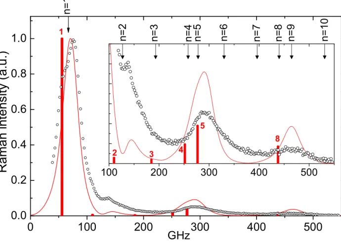
spectrum of the Ag0.66Au0.34 sample to its calculated counterpart. The overall aspect of
the Raman spectrum is rather well reproduced in terms of band positions and relative in-tensities. For further discussion, the top scale of Figure 3 indicates the predictions of the Complex Frequency Method8,9 (CFM), which enables a rapid evaluation of the band
posi-tions for embedded nanoparticles yet without yielding the Raman intensities. In order to identify the filiation of the Raman bands with harmonics of the quadrupolar vibration from a single nanoparticle, Figure 3 shows RSI spectra for the same nanoparticle but considered as free. In the absence of sufficiently reliable Transmission Electron Microscopy data due to the buried nature of the nanoparticles, the mean diameter of the nanoparticles is derived from the best fit of the lower frequency doublet with a single average (ℓ = 2, n = 1) line since at this stage the RSI calculations are unable to account for the elastic anisotropy splitting (nanocrystals are assumed as isotropic nanoparticles). The derived diameter for this system is 23.5 nm, as confirmed from the analysis of the polarized spectrum that shows in addition the spherical mode contributions.
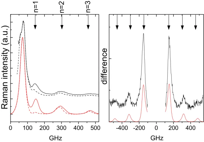
Figure 4 compares the VV and VH experimental spectra of the Ag0.66Au0.34 sample,
together with those derived from the RSI calculations. These comparisons show that the spherical mode contributions are rather small and they significantly overlap with those of the quadrupolar mode. They are better evidenced by the Raman difference spectrum IV V − k.IV H, where k is a scaling factor chosen such that the (ℓ = 2, n = 1) peaks are
normalized (Fig. 4 left). The positions derived from the difference are found to well agree with the CFM predictions for the (ℓ = 0, n = 1, 2, 3) modes, using the same nanoparticle size as that derived from the quadrupolar mode analysis; they are equally well consistent with the RSI predictions of band profile changes induced by the change of polarization.
The present observation of an usually rich low frequency Raman spectrum from embed-ded metallic nanoparticles, and its comparison with RSI and CFM predictions, allows to
FIG. 3: (color online) Experimental (symbols) and calculated (solid line) depolarized (VH) Raman
spectra of the Ag0.66Au0.34 sample with a diameter of 23.5 nm (the spectra were normalized with
respect to the (ℓ = 2, n = 1) peak); high frequency enlargement in the inset. The thick bars are the Raman intensities calculated for the same nanoparticle considered as free, labeled according to their n index (only harmonics with non negligible intensities are shown). Top scale arrows: CFM predictions for the ℓ = 2 mode with the same embedded nanoparticle.
capture several ingredients that are essential for a correct interpretation of similar data. The proper identification of the several Raman bands requires, in a first stage, to com-pare their behaviour as a function of polarization as they may result from the overlap of several contributions (Fig. 4). The detailed analysis of the depolarized spectrum, i.e. of the quadrupolar mode contributions, shows several important points. The first one is that introducing the embedding medium affects the position of the bands as displayed by the frequency mismatch of the observed bands (symbols in Fig. 3) or the CFM positions (top arrows) with the frequencies calculated in the free case (thick bars). This effect, which is
FIG. 4: (color online) Left : Experimental (upper set) and calculated (lower set) of the VV (solid)
and VH (dashed) Raman profiles of the Ag0.66Au0.34 sample (intensities were normalized with
respect to the (ℓ = 2, n = 1) peak). Right: Raman difference spectrum (IV V − IV H), experimental
(thick line) and RSI calculated (thin line). Top scales: CFM predictions for the ℓ = 0 mode.
essentially due to the relatively large mass density of the used glass, is most severe on the (ℓ = 2, n = 1) peak, which is often used for size evaluation due to its prominence. Quantita-tively, using a free case estimation based on the position of the (ℓ = 2, n = 1) band leads to a 19% smaller nanoparticle diameter. The second result is that from the frequency position matching between the experimental curve and the calculated RSI and CFM contributions, it comes out that the high frequency components are affiliated with selected harmonics, or at least harmonic groups, of the single nanoparticle vibration: the bands near 290 GHz and 460 GHz clearly identify with respectively the n = 4, 5 and n = 8 harmonic components of the quadrupolar mode. Their respective Raman cross-sections are relatively consistent with those predicted in the free sphere case (delta functions in Fig. 3)10. The situation
n = 2 harmonic in terms of frequency while its intensity is considerably stronger than that expected from the free sphere evaluation: according to RSI calculations, it is enhanced by a factor 4 going from the free case to the embedded case while the other harmonics are enhanced by a factor 2 (its stronger intensity in the experimental spectrum is certainly due to its merging with the foot of the T2g component that the RSI calculations are unable to
account for at the present stage). This enhancement is rooted in the coupling with the ma-trix that significantly increases the displacement at the surface of the nanoparticle, for this particular harmonic, making it a potential good probe of the coupling interface. Another possible interpretation is that the 150 GHz band results from the redistribution of mode frequencies that arises from the anistropy effect, as is observed from the splitting of the (ℓ = 2, n = 1) component. These two interpretations differ from that given for a similar band observed from similar Au doped glasses2.
Finally, the comparison of the RSI and CFM calculations with the experimental data provides a further interesting aspect related to the specificity of resonant Raman scattering for metallic nanoparticles. Indeed, one observes that while the frequency positions of the spherical mode bands agree well with those predicted by either RSI and CFM calculations (Fig. 4), the CFM predictions for the quadrupolar mode harmonics are systematically slightly red shifted with respect to either the experimental or the RSI calculated bands (Fig. 3). This is due to the dual nature of the quadrupolar modes (longitudinal vs transverse as defined in14) which is also present inside a given CFM resonance. The plasmon-vibration coupling is
therefore significant only for a subset of modes inside the frequency range defined by a CFM peak which can lead to a shift of the observed maxima. This situation does not occur for the spherical mode. As a result, the CFM and RSI peak positions match and these modes can be more reliably used for the size evaluation of the nanoparticles provided they are safely extracted from the dominating quadrupolar mode response.
The first observation of high order quadrupolar and spherical harmonic modes from AgAu nanoparticles embedded in a glass has allowed us to rationalize the interpretation of low frequency Raman spectra from metallic nanoparticles. In accord with theoretical predictions, one observes that Raman scattering by the quadrupolar modes is not a monotonic decreasing function of the harmonic index so that only selected harmonics participate to the Raman spectrum with differing efficiencies. Due to stronger coupling with the plasmon excitations, the Raman spectrum is strongly dominated by high order harmonics of the quadrupolar
modes that overlap with the weaker polarized signatures from the spherical modes. In such case, a detailed polarization analysis of the Raman bands is therefore essential to check the band assignments that are uniquely based on frequency position matchings17,18.
Acknowledgments
The authors acknowledge N. Del Fatti and F. Vall´ee for their relevant comments.
1 A. Courty, I. Lisiecki, and M. P. Pileni, J. Chem. Phys. 116, 8074 (2002), URL
http://link.aip.org/link/?JCP/116/8074/1.
2 B. Stephanidis, S. Adichtchev, S. Etienne, S. Migot, E. Duval, and A. Mermet, Phys. Rev. B
76, 121404 (2007), URL http://link.aps.org/abstract/PRB/v76/e121404.
3 J. Margueritat, J. Gonzalo, C. Afonso, G. Bachelier, A. Mlayah, A. Laarakker, D.
Mur-ray, and L. Saviot, Applied Physics A: Materials Science & Processing 89, 369 (2007), URL http://dx.doi.org/10.1007/s00339-007-4120-8.
4 H. Portal`es, N. Goubet, L. Saviot, S. Adichtchev, D. B. Murray, A.
Mer-met, E. Duval, and M.-P. Pileni, Proceedings of the National Academy of
Sci-ences 105, 14784 (2008), http://www.pnas.org/content/105/39/14784.full.pdf+html, URL http://www.pnas.org/content/105/39/14784.abstract.
5 H. Lamb, Proc. London Math. Soc. s1-13, 189 (1881), URL
http://plms.oxfordjournals.org.
6 E. Duval, Phys. Rev. B 46, 5795 (1992), URL http://link.aps.org/abstract/PRB/v46/p5795.
7 E. Duval, A. Boukenter, and B. Champagnon, Phys. Rev. Lett. 56, 2052 (1986).
8 V. Dubrovskiy and V. Morochnik, Earth. Phys. 17, 494 (1981).
9 P. Verma, W. Cordts, G. Irmer, and J. Monecke, Phys. Rev. B 60, 5778 (1999).
10 G. Bachelier and A. Mlayah, Phys. Rev. B 69, 205408 (2004), URL
http://link.aps.org/abstract/PRB/v69/e205408.
11 S. Link, Z. Wang, and M. El-Sayed, J. Phys. Chem. B 103, 3529 (1999), ISSN 1520-6106, URL
12 J. R. Sandercock, Trends in Brillouin scattering : studies of opaque materials, supported films,
and central modes (Springer-Verlag, 1982), chap. 6, pp. 173–206.
13 R. Mock, B. Hillebrands, and R. Sandercock, Journal of Physics E: Scientific Instruments 20,
656 (1987), ISSN 0022-3735.
14 L. Saviot and D. B. Murray, Phys. Rev. B 72, 205433 (2005), URL
http://link.aps.org/abstract/PRB/v72/e205433.
15 D. Risti´c, M. Ivanda, K. Furi´c, U. V. Desnica, M. Buljan, M. Montagna, M. Ferrari,
A. Chiasera, and Y. Jestin, Journal of Applied Physics 104, 073519 (pages 7) (2008), URL http://link.aip.org/link/?JAP/104/073519/1.
16 Alloy properties Pa (P =ρ, V
L, VT) were evaluated through Pa = x.PAg+ (1 − x).PAu, using
ρAg= 10.5 g.cm−3, ρAu= 19.28 g.cm−3, VAg
L = 3747 m/s, VLAu = 3319 m/s, V
Ag
T = 1740 m/s,
VLAu= 1236 m/s.
17 H. Portal`es, L. Saviot, E. Duval, M. Fujii, S. Hayashi, N. Del Fatti, and F. Vall´ee, J. Chem.
Phys. 115, 3444 (2001), URL http://link.aip.org/link/?JCP/115/3444/1.
18 A. Nelet, A. Crut, A. Arbouet, N. Del Fatti, F. Vall´ee, H. Portal`es, L. Saviot,
and E. Duval, Applied Surface Science 226, 209 (2004), ISSN 0169-4332, URL


