HAL Id: hal-03019238
https://hal.sorbonne-universite.fr/hal-03019238
Submitted on 23 Nov 2020
HAL is a multi-disciplinary open access
archive for the deposit and dissemination of
sci-entific research documents, whether they are
pub-lished or not. The documents may come from
teaching and research institutions in France or
abroad, or from public or private research centers.
L’archive ouverte pluridisciplinaire HAL, est
destinée au dépôt et à la diffusion de documents
scientifiques de niveau recherche, publiés ou non,
émanant des établissements d’enseignement et de
recherche français ou étrangers, des laboratoires
publics ou privés.
The Effect of Selective Laser Trabeculoplasty on
Intraocular Pressure in Patients with Dexamethasone
Intravitreal Implant-Induced Elevated Intraocular
Pressure
Amin Bennedjai, Vincent Theillac, Jad Akesbi, Raphaël Adam, Thibaut
Rodallec, Chafik Keilani, Esther Blumen-Ohana, Antoine Labbé,
Jean-Philippe Nordmann
To cite this version:
Amin Bennedjai, Vincent Theillac, Jad Akesbi, Raphaël Adam, Thibaut Rodallec, et al.. The Effect
of Selective Laser Trabeculoplasty on Intraocular Pressure in Patients with Dexamethasone
Intravit-real Implant-Induced Elevated Intraocular Pressure. Journal of Ophthalmology, Hindawi Publishing
Corporation, 2020, 2020, pp.e3439182. �10.1155/2020/3439182�. �hal-03019238�
Research Article
The Effect of Selective Laser Trabeculoplasty on Intraocular
Pressure in Patients with Dexamethasone Intravitreal
Implant-Induced Elevated Intraocular Pressure
Amin Bennedjai ,
1Vincent Theillac,
1Jad Akesbi,
1Rapha¨el Adam,
1Thibaut Rodallec,
1Chafik Keilani,
1Esther Blumen-Ohana,
1Antoine Labb´e,
2and Jean-Philippe Nordmann
11Department of Ophthalmology 2, Quinze-Vingts National Ophthalmology Hospital, IHU FOReSIGHT,
University Paris Descartes, Paris, France
2Department of Ophthalmology 3, Quinze-Vingts National Ophthalmology Hospital, IHU FOReSIGHT, Paris Saclay University,
Paris, France
Correspondence should be addressed to Amin Bennedjai; aminbennedjai@hotmail.com
Received 8 May 2020; Revised 13 September 2020; Accepted 26 September 2020; Published 13 October 2020 Academic Editor: Ana Raquel Santiago
Copyright © 2020 Amin Bennedjai et al. This is an open access article distributed under the Creative Commons Attribution License, which permits unrestricted use, distribution, and reproduction in any medium, provided the original work is properly cited.
Introduction. To assess the safety and efficacy of selective laser trabeculoplasty (SLT) for ocular hypertension (OHT) induced by a dexamethasone (DEX) intravitreal implant. Materials and Methods. We performed a retrospective study of patients who un-derwent an SLT procedure for ocular hypertension induced by injection of a DEX intravitreal implant. Patients had, at least, one injection of the DEX-implant for symptomatic macular edema. SLT was delivered to 360°of the trabecular meshwork in two
sessions. The primary outcome was a decrease in IOP, evaluated at one, three, and six months after the SLT procedure. Results. Twenty-six eyes of 22 patients were included. The mean intraocular pressure (IOP) measured after DEX-implant injection was 25.4 ± 5.4 mmHg, and the mean increase in IOP was 35.8 ± 14.6%. The mean follow-up after SLT was 18.3 ± 7.7 months. After SLT, the mean IOP dropped by 30.9% at one month (16.9 ± 4.5 mmHg, p � 0.01), 33.6% at three months (16.0 ± 2.7 mmHg, p < 0.01), and 34.9% at six months (15.6 ± 2.1 mmHg, p < 0.01). Each patient had a minimum follow-up of 6 months after SLT. Eight eyes (31%) received a second DEX-implant injection after the SLT procedure without experiencing an increase in the IOP above 21 mmHg or >20%. No glaucoma surgery was required during the follow-up. The mean number of medications (1.65 ± 1.36) was significantly reduced at one (1.19 ± 1.20, p � 0.04), three (0.96 ± 1.03, p < 0.01), and six months (0.77 ± 0.95, p < 0.01) after SLT. Conclusion. SLT is an effective and safe procedure to control OHT following DEX-implant intravitreal injection.
1. Introduction
Dexamethasone intravitreal implant 0.7 mg (Ozurdex
®
, Allergan, Dublin, Ireland) (DEX-implant) is a sustained-release corticosteroid device that is effective in the man-agement of diabetic macular edema [1], age-related mac-ular degeneration [2], retinal vein occlusions [3], noninfectious posterior uveitis [4], and resistant Irvi-ne–Gass syndrome [5]. After DEX-implant injection, an increase in intraocular pressure (IOP) may be observed after 1.5 to 2.5 months [6] in 12.6% [7] to 36.0% [8, 9] of patients. Rajesh et al. reported that antiglaucomamedication or filtering surgery was needed for 91.8% and 3.1% of patients exhibiting a postinjection increase in in-traocular pressure (IOP), respectively [10]. Similarly, 3% of patients treated with a DEX-implant for Irvine–Gass syndrome were eventually injected with anti-VEGF be-cause of ocular hypertension (OHT), despite maximal medical antiglaucoma treatment [5]. After three or more injections, the frequency of mild OHT increased to 53% versus 31% after the first injection [11]. Moreover, the risk of a large increase in IOP is greater for patients with primary open-angle glaucoma (POAG) or suspected glaucoma at the time of DEX implantation [12].
Volume 2020, Article ID 3439182, 6 pages https://doi.org/10.1155/2020/3439182
The physiopathology of glucocorticoid-induced OHT and glaucoma may be related to the impairment of outflow through the trabecular meshwork (TM) via an enzymatic pathway responsible for mucopolysaccharide accumula-tion in the iridocorneal angle [13]. A mouse model has suggested a role for glucocorticoid receptor trans-activation in regulating glucocorticoid-mediated gene expression in the TM [14]. Steroids may also have an effect on the TM extracellular matrix due to altered rates of protein synthesis or degradation or a combination of the two [12].
Selective laser trabeculoplasty (SLT) uses a 532 nm Q-switched, frequency-doubled Nd:YAG laser that delivers a short pulse (3 nanoseconds) [15]. It prevents heat dissi-pation outside of pigmented TM cells and does not cause collateral damage [15]. Selective laser trabeculoplasty is ef-fective in lowering IOP in open-angle glaucoma [16–18] and can be offered as a first-line treatment for POAG or OHT [19]. SLT induces biological changes that modulate increased aqueous outflow through the TM, including changes in cytokine and interleukin-8 (IL-8) secretion and TM remodeling, which are affected by glucocorticoids [20]. Selective laser trabeculoplasty is also effective in treating OHT after intravitreal triamcinolone acetonide injection [21], as well as in preventing an increase in IOP when performed before injection [22].
Various guidelines establishing the management of OHT after intravitreal injection have been published [23]. Topical treatment is the first approach proposed, followed by sur-gical treatment in case of refractory OHT. The exact role of SLT in the algorithm of treatment of OHT after DEX-im-plant injection has not been yet determined. Moreover, no studies have evaluated the effect of SLT in patients with DEX-implant-induced OHT. Thus, the main objective of the present study was to evaluate the effectiveness and safety of the SLT procedure on increased IOP after DEX-implant injection.
2. Patients and Methods
This study was conducted at the Quinze-Vingts National Ophthalmology Hospital, Paris, France, in accordance with the tenets of the Declaration of Helsinki. We retrospectively reviewed the medical records of all patients who underwent an SLT procedure for uncontrolled OHT induced by DEX-implant injection between November 2017 and October 2018.
The study included patients who had, at least, one in-jection of the DEX-implant for symptomatic macular edema. All patients were symptomatic, and diagnosis of macular edema was established based on examination of the fundus and macular optical coherence tomography (OCT) showing a central macular thickness (CMT) of >300 μm (Spectralis HRA + OCT, Heidelberg engineering Inc, Heidelberg
®
, Germany). Patients with uncontrolled IOP (according to their target IOP) despite maximal tolerated topical treatment three months after DEX-implant injection underwent the SLT procedure.2.1. Injection Technique. An implant of 0.7 mg
dexametha-sone (Ozurdex
®
, Allergan, Irvine, CA) was inserted into the vitreous cavity through the pars plana at a distance of 3.5 mm from the limbus in pseudophakic eyes and 4 mm in phakic eyes after topical anesthesia (oxybuprocaine 1.6 mg/ 0.4 ml, Thea®
, Clermont-Ferrand, France) and sterilization of the ocular and periocular surface with 5% povidone io-dine. Patients were seen, at least, one, three, and six months after the injections, allowing measurement of the best corrected visual acuity (BCVA), IOP, CMT, and examina-tion of the fundus.2.2. SLT Procedure. All SLT procedures were performed,
after topical anesthesia (oxybuprocaine), by trained physi-cians of our group with the same machine (TangoTM, EllexInc, Adelaide, Australia) using a Latina SLT lens (Ocular Instruments, Bellevue, Washington, USA) to visu-alize the TM. Selective laser trabeculoplasty was delivered to 360°of the trabecular meshwork in two sessions, one week
apart, with the total number of nonoverlapping impacts varying from 80 to 110. The energy setting was from 0.8 to 1.2 mJ. One drop of 0.1% Indocollyre (Indometacine, Bausch & Lomb, Berlin, Germany) and 0.5% Iopidine (Alcon Laboratories, Inc., Ft. Worth, TX) were immediately ad-ministered after the procedure and for five days, three times a day, for Indocollyre.
2.3. Study Population. Each patient underwent a
stan-dardized examination, performed in our retinal disease department, with measurement of the BCVA in Early Treatment Diabetic Retinopathy Study (ETDRS) letters converted to LogMAR (Minimum Angle of Resolution) [24], IOP measurement using a Goldman applanation tonometer, examination of the fundus, and macular OCT. Every patient had an open angle on the Schaffer scale at gonioscopy ex-amination. Open-angle glaucoma risk factors and the date and number of DEX-implant injections, the date, number, and characteristics of the SLT procedures, number and type of antiglaucoma medications, and surgical history were recorded.
2.4. Statistical Assessment. All statistical analyses were
performed using GraphPad Prism 7
®
for Windows®
(GraphPad Software, La Jolla, CA, USA). The primary outcome was a decrease in IOP evaluated at one, three, and six months after the SLT procedure. Quantitative variables were compared using the Student t-test. Survival curves were generated using the Kaplan–Meier method. Double-sided p values <0.05 were considered statistically significant.3. Results
The study included 26 eyes of 22 patients. Baseline demo-graphics and ocular parameters are presented in Table 1.
Best corrected visual acuity significantly improved fol-lowing DEX-implant injection from 0.58 ± 0.42 to 0.32 ± 0.39 at six months after injection (p � 0.01). The
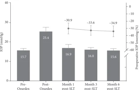
mean time between DEX-implant injection and the diag-nosis of OHT was 55.8 ± 27.9 days. The SLT procedure was performed at a mean of 96.71 ± 14.73 days after DEX-im-plant injection. The mean duration of follow-up after SLT was 18.3 ± 7.7 months. The maximal IOP measured after DEX-implant injection was 25.4 ± 5.4 mmHg, with an in-crease in IOP of 35.8 ± 14.6%. After SLT, the mean IOP dropped by 30.9% at one month (16.9 ± 4.5 mmHg), 33.6% at three months (16.0 ± 2.7 mmHg), and 34.9% at six months (15.6 ± 2.1 mmHg) after SLT, showing a persistent and significant decrease in IOP (p < 0.01 compared to the pre-SLT IOP at each visit) (Figure 1). The pre-SLT procedures are described in Table 2.
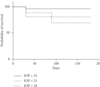
Eight eyes (31%) underwent a second DEX-implant injection after the SLT procedure without experiencing an increase in IOP (>20%) after one week or one, three, or six months of follow-up. Seven eyes did not require additional antiglaucoma drugs. The mean peak IOP after reinjection for patients who had SLT was 18.8 ± 1.5 mmHg, lower than the peak IOP after the first injection for patients requiring reinjection (27.3 ± 1.8 mmHg, p � 0.01). The mean IOP values for the eyes that underwent SLT versus those of the contralateral eyes during the follow-up period are shown in Figure 2. During follow-up, 96.2%, 80.8%, and 69.2% of the treated eyes showed an IOP below 25, 21, and 18 mmHg, respectively (Figure 3).
The mean number of medications (1.65 ± 1.36) was significantly lower at one (1.19 ± 1.20, p � 0.04), three (0.96 ± 1.03, p < 0.01), and six months (0.77 ± 0.95, p < 0.01) after trabeculoplasty. No oral treatment or surgery was required during the follow-up. Six of the 26 patients (23%) required antiglaucoma eye drops after six months, with a mean number of 0.5 ± 0.88 topical treatments, without oral medication or surgery.
Adverse effects after SLT included a moderate transient anterior chamber reaction for one patient (4%) and trau-matic keratitis for another (4%), which healed after one-week of topical treatment (Tobradex
®
(0.3% tobramycin, 0.1% dexamethasone; Alcon Laboratories Inc., Fort Worth, Texas) three times a day and vitamin A ointment associated with artificial tears, respectively).4. Discussion
The SLT procedure appears to be a valuable and safe tool to manage increased IOP after DEX-implant injection. The exact mechanism by which SLT decreases IOP has not been yet fully elucidated. It may involve macrophage activation, resulting from increased chemokine production, allowing TM clearing. The SLT procedure may also allow the TM to release factors that regulate the permeability of Schlemm canal endothelial cells [25]. This effect mirrors the physio-pathology of steroid-induced OHT, which involves in-creased responsiveness of the membranes of goniocytes to steroids, leading to increased production of fibroblasts in the TM and resulting in aqueous retention [12]. The TM ex-tracellular matrix may be remodeled by the expression of stromelysin-1 (MMP-3), resulting in an increase in aqueous outflow [26]. Selective laser trabeculoplasty appears to have some clinical efficacy in secondary glaucoma patients, es-pecially when dysfunction of the TM is involved, such as in DEX-implant-induced OHT. Here, we report a larger de-crease in IOP after SLT than previously reported after SLT performed for pigmentary glaucoma (14.5%) or pseu-doexfoliation glaucoma (16.6%) [27].
This study highlights the potential interest of SLT after steroid-induced OHT. This procedure may be an alternative to the usual treatment, which involves topical ocular anti-glaucoma medications. Two studies have already reported efficacy of the SLT procedure in steroid-induced OHT. Rubin et al. reported the efficacy of SLT, with a significative decrease of IOP (p < 0.007) in five of seven patients [21]. Bozkurt et al., showed that the increase in IOP after intravitreal triamcinolone acetonide injection may be pre-vented by performing SLT if the baseline IOP is >21 mmHg [22]. The SAFODEX study demonstrated that OHT can be observed for 28.5% of DEX-implant injected eyes [28]. A patient who experiences OHT after the first injection has a significant risk of experiencing an increase in IOP after reinjection. The frequency of OHT (>23 mmHg) following one, two, or three reinjections is 31%, 26%, and 53%, re-spectively [11]. In the present study, none of the eight eyes
Table 1: Baseline demographic characteristics of the 22 patients who underwent SLT after DEX-implant injection.
Patient characteristics (n � 22) Male 12 (55) Female 10 (45) Caucasian 18 (82) African 4 (18) Age, mean ± SD 69.6 ± 15.4 Family history of glaucoma 2 (9) Eyes characteristics (n � 26)
Retinal disease
Diabetic macular edema 15 (57) Irvine–Gass syndrome 8 (31) Branch retinal vein occlusion 3 (12) Cup-to-disc ratio ± SD 0.38 ± 0.19 Pseudophakic 19 (73) PPV 5 (19) Retinal laser 8 (31) PPRP 7 (27) Focal laser 1 (4)
The results are presented as n (%) for categorical variables. SD, standard deviation; PPV, pars plana vitrectomy; PPRP, peripherical pan-retical photocoagulation.
Table 2: Characteristics of the SLT procedures. Parameters of the SLT procedures
Total number of SLT procedures 26
Right eyes 10
Left eyes 16
Mean number of spots ± SD 91.6 ± 20.2 Total energy (mJ) ± SD 75.9 ± 24.4 New DEX-implant after SLT 8
that underwent DEX-implant reinjection after the SLT procedure experienced a major peak of IOP after SLT. These results suggest that the SLT procedure may also be useful in preventing new episodes of OHT after reinjection of the DEX-implant in patients who already experienced steroid-induced OHT.
Filtering surgery is more often needed to control cor-ticosteroid-induced OHT in patients with branch or central retinal vein occlusion (RVO). Ocular hypertension in these patients may be multifactorial and the associated retinal
ischemia may be responsible for persistent OHT in RVO [6]. Of note, in our study, the SLT procedure was also effective in controlling corticosteroid-induced OHT for patients with RVO, as the IOP of treated eyes tended to decrease to the same level as that of the adelphic eyes after three to six months.
Our study had several limitations, notably its being a retrospective study with a limited number of patients. Nonetheless, SLT can be considered to lower DEX-implant-induced increases in IOP. Selective laser trabeculoplasty may
0 10 20 30 40 –60 –50 –40 –30 –20 –10 0 15.7 25.4 16.0 15.6 16.9 –30.9 –33.6 –34.9 Pre-Ozurdex Post-Ozurdex Month 1 post-SLT Month 3 post-SLT Month 6 post-SLT IO P (mmH g) P reo p era ti ve I O P lo w er in g (%)
Figure 1: Intraocular pressure lowering after the SLT procedure with a follow-up of 6 months.
10 15 20 25 30 Pre-Ozurdex Post-Ozurdex Month 1 post-SLT Month 3 post-SLT Month 6 post-SLT IO P (mmH g) Fellow eye SLT
Figure 2: Evolution of mean intraocular pressure (IOP) before and after a DEX-implant injection in eyes that underwent selective laser trabeculoplasty versus contralateral eyes at one, three, and six months after treatment.
be a safe and effective alternative to antiglaucoma eye drops as a first-line treatment and probably as a prophylactic procedure to avoid peak IOP in patients presenting corti-costeroid-induced OHT.
Data Availability
The paper data used to support the findings of this study are available from the corresponding author upon request.
Ethical Approval
All procedures performed in these studies involving human participants were in accordance with the ethical standards of the institutional and/or national research committee and with the 1964 Declaration of Helsinki and its later amendments or comparable ethical standards.
Conflicts of Interest
The authors declare no conflicts of interest.
References
[1] M. Iglicki, C. Busch, D. Zur et al., “Dexamethasone implant for diabetic macular edema in naive compared with refractory eyes,” Retina, vol. 39, no. 1, pp. 44–51, 2019.
[2] P. Calvo, A. Ferreras, F. Al Adel, Y. Wang, and M. H. Brent, “Dexamethasone intravitreal implant as adjunct therapy for patients with wet age-related macular degeneration with in-complete response to ranibizumab,” British Journal of Oph-thalmology, vol. 99, no. 6, pp. 723–726, 2015.
[3] A. Ozkok, O. A. Saleh, D. K. Sigford, J. W. Heroman, and S. Schaal, “The omar study,” Retina, vol. 35, no. 7, pp. 1393–1400, 2015.
[4] N. J. S. London, A. Chiang, and J. A. Haller, “The dexa-methasone drug delivery system: indications and evidence,” Advances in Therapy, vol. 28, no. 5, pp. 351–366, 2011. [5] D. Bellocq, V. Pierre-Kahn, F. Matonti et al., “Effectiveness
and safety of dexamethasone implants for postsurgical macular oedema including Irvine-Gass syndrome: the
EPISODIC-2 study,” British Journal of Ophthalmology, vol. 101, no. 3, pp. 333–341, 2017.
[6] E. K. Chin, D. R. P. Almeida, G. Velez et al., “Ocular hy-pertension after intravitreal dexamethasone (OZURDEX) sustained-release implant,” Retina, vol. 37, no. 7, pp. 1345–1351, 2017.
[7] J. A. Haller, F. Bandello, R. Belfort et al., “Dexamethasone intravitreal implant in patients with macular edema related to branch or central retinal vein occlusion,” Ophthalmology, vol. 118, no. 12, pp. 2453–2460, 2011.
[8] D. S. Boyer, Y. H. Yoon, R. Belfort et al., “Three-year, ran-domized, sham-controlled trial of dexamethasone intravitreal implant in patients with diabetic macular edema,” Ophthal-mology, vol. 121, no. 10, pp. 1904–1914, 2014.
[9] R. K. Maturi, A. Pollack, H. S. Uy et al., “Intraocular pressure in patients with diabetic macular edema treated with dexa-methasone intravitreal implant in the 3-year mead study,” Retina, vol. 36, no. 6, pp. 1143–1152, 2016.
[10] B. Rajesh, J. Zarranz-Ventura, A. T. Fung et al., “Safety of 6000 intravitreal dexamethasone implants,” British Journal of Ophthalmology, vol. 104, no. 1, pp. 39–46, 2019.
[11] S. Bahadorani, C. Krambeer, K. Wannamaker et al., “The effects of repeated Ozurdex injections on ocular hyperten-sion,” Clinical Ophthalmology, vol. 12, pp. 639–642, 2018. [12] M. R. Razeghinejad and L. J. Katz, “Steroid-induced iatrogenic
glaucoma,” Ophthalmic Research, vol. 47, no. 2, pp. 66–80, 2012.
[13] J. François, “Corticosteroid glaucoma,” Ophthalmologica, vol. 188, no. 2, pp. 76–81, 1984.
[14] G. C. Patel, J. C. Millar, and A. F. Clark, “Glucocorticoid receptor transactivation is required for glucocorticoid-in-duced ocular hypertension and glaucoma,” Investigative Opthalmology & Visual Science, vol. 60, no. 6, pp. 1967–1978, 2019.
[15] M. A. Latina, S. A. Sibayan, D. H. Shin, R. J. Noecker, and G. Marcellino, “Q-switched 532-nm Nd:YAG laser trabecu-loplasty (selective laser trabecutrabecu-loplasty),” Ophthalmology, vol. 105, no. 11, pp. 2082–2090, 1998.
[16] J. D. Stein and P. Challa, “Mechanisms of action and efficacy of argon laser trabeculoplasty and selective laser trabeculo-plasty,” Current Opinion in Ophthalmology, vol. 18, no. 2, pp. 140–145, 2007. Pro babi lit y of su rv iv al 0 0 50 50 100 100 150 200 Days IOP < 25 IOP < 21 IOP < 18
Figure 3: Kaplan–Meier survival curves plotting the cumulative probabilities that the IOP remains below 25 mmHg, 21 mmHg, and 18 mmHg, respectively, following the SLT procedure.
[17] M. Latina and J. Deleon, “Selective laser trabeculoplasty,” Ophthalmology Clinics of North America, vol. 18, no. 3, pp. 409–419, 2005.
[18] T. S. Acott, J. R. Samples, J. M. Bradley, D. R Bacon, S. S Bylsma, and E. M. van Buskirk, “Trabecular repopulation by anterior trabecular meshwork cells after laser trabeculo-plasty,” American Journal of Ophthalmology, vol. 107, no. 1, pp. 1–6, 1989.
[19] G. Gazzard, E. Konstantakopoulou, D. Garway-Heath et al., “Selective laser trabeculoplasty versus eye drops for first-line treatment of ocular hypertension and glaucoma (LiGHT): a multicentre randomised controlled trial,” The Lancet, vol. 393, no. 10180, pp. 1505–1516, 2019.
[20] D. B. Kagan, N. S. Gorfinkel, and C. M. Hutnik, “Mechanisms of selective laser trabeculoplasty: a review,” Clinical & Ex-perimental Ophthalmology, vol. 42, no. 7, pp. 675–681, 2014. [21] B. Rubin, A. Taglienti, R. F. Rothman, C. H. Marcus, and J. B. Serle, “The effect of selective laser trabeculoplasty on intraocular pressure in patients with intravitreal steroid-in-duced elevated intraocular pressure,” Journal of Glaucoma, vol. 17, no. 4, pp. 287–292, 2008.
[22] E. Bozkurt, N. Kara, A. T. Yazici et al., “Prophylactic selective laser trabeculoplasty in the prevention of intraocular pressure elevation after intravitreal triamcinolone acetonide injection,” American Journal of Ophthalmology, vol. 152, no. 6, pp. 976–981, 2011.
[23] M. Poli, P. Denis, C. Dot, and J.-P. Nordmann, “Conduite `a tenir face au risque d’hypertonie oculaire apr`es injection intra-vitr´eenne,” Journal Français d’Ophtalmologie, vol. 40, no. 3, pp. e77–e82, 2017.
[24] F. L. Ferris, A. Kassoff, G. H. Bresnick, and I. Bailey, “New visual acuity charts for clinical research,” American Journal of Ophthalmology, vol. 94, no. 1, pp. 91–96, 1982.
[25] J. A. Alvarado, R. F. Yeh, L. Franse-Carman, G. Marcellino, and M. J. Brownstein, “Interactions between endothelia of the trabecular meshwork and of Schlemm’s canal: a new insight into the regulation of aqueous outflow in the eye,” Transac-tions of the American Ophthalmological Society, vol. 103, pp. 148–163, 2005.
[26] J. Y. Lee, D. B. Kagan, G. Roumeliotis, H. Liu, and C. M. Hutnik, “Secretion of matrix metalloproteinase-3 by co-cultured pigmented and non-pigmented human trabecular meshwork cells following selective laser trabeculoplasty,” Clinical & Experimental Ophthalmology, vol. 44, no. 1, pp. 33–42, 2016.
[27] B. Koucheki and H. Hashemi, “Selective laser trabeculoplasty in the treatment of open-angle glaucoma,” Journal of Glau-coma, vol. 21, no. 1, pp. 65–70, 2012.
[28] A. Malcl`es, C. Dot, N. Voirin et al., “Safety of intravitreal dexamethasone implant (ozurdex),” Retina, vol. 37, no. 7, pp. 1352–1359, 2017.

![[PDF] Formation PrestaShop : Module TNT Express | Formation à télécharger en PDF](data:image/gif;base64,R0lGODlhAQABAIAAAP///wAAACH5BAEAAAAALAAAAAABAAEAAAICRAEAOw==)