HAL Id: hal-02326078
https://hal.archives-ouvertes.fr/hal-02326078
Submitted on 25 Oct 2019
HAL is a multi-disciplinary open access
archive for the deposit and dissemination of sci-entific research documents, whether they are pub-lished or not. The documents may come from teaching and research institutions in France or abroad, or from public or private research centers.
L’archive ouverte pluridisciplinaire HAL, est destinée au dépôt et à la diffusion de documents scientifiques de niveau recherche, publiés ou non, émanant des établissements d’enseignement et de recherche français ou étrangers, des laboratoires publics ou privés.
Vasculogenesis In Vitro and In Vivo
Hebe Agustina Mena, Paula Romina Zubiry, Blandine Dizier, Virginie
Mignon, Fernanda Parborell, Mirta Schattner, Catherine Boisson-Vidal,
Soledad Negrotto
To cite this version:
Hebe Agustina Mena, Paula Romina Zubiry, Blandine Dizier, Virginie Mignon, Fernanda Parborell, et al.. Ceramide 1-Phosphate Protects Endothelial Colony–Forming Cells From Apoptosis and Increases Vasculogenesis In Vitro and In Vivo. Arteriosclerosis, Thrombosis, and Vascular Biology, American Heart Association, 2019, 39 (10), �10.1161/ATVBAHA.119.312766�. �hal-02326078�
Arterioscler Thromb Vasc Biol is available at www.ahajournals.org/journal/atvb
Correspondence to: Soledad Negrotto, PhD, Experimental Thrombosis Laboratory, Institute of Experimental Medicine, National Academy of Medicine–CONICET, Pacheco de Melo 3081, Buenos Aires, Argentina. Email solenegrotto@hematologia.anm.edu.ar or solenegrotto@hotmail.com
The online-only Data Supplement is available with this article at https://www.ahajournals.org/doi/suppl/10.1161/ATVBAHA.119.312766. For Sources of Funding and Disclosures, see page xxx.
© 2019 American Heart Association, Inc.
ORIGINAL RESEARCH
Ceramide 1-Phosphate Protects Endothelial
Colony–Forming Cells From Apoptosis and
Increases Vasculogenesis In Vitro and In Vivo
Hebe Agustina Mena, Paula Romina Zubiry, Blandine Dizier, Virginie Mignon, Fernanda Parborell, Mirta Schattner, Catherine Boisson-Vidal, Soledad Negrotto
OBJECTIVE: Ceramide 1-phosphate (C1P) is a bioactive sphingolipid highly augmented in damaged tissues. Because of its abilities to stimulate migration of murine bone marrow–derived progenitor cells, it has been suggested that C1P might be involved in tissue regeneration. In the present study, we aimed to investigate whether C1P regulates survival and angiogenic activity of human progenitor cells with great therapeutic potential in regenerative medicine such as endothelial colony– forming cells (ECFCs).
APPROACH AND RESULTS: C1P protected ECFC from TNFα (tumor necrosis factor-α)-induced and monosodium urate crystal– induced death and acted as a potent chemoattractant factor through the activation of ERK1/2 (extracellular signal-regulated kinases 1 and 2) and AKT pathways. C1P treatment enhanced ECFC adhesion to collagen type I, an effect that was prevented by β1 integrin blockade, and to mature endothelial cells, which was mediated by the E-selectin/CD44 axis. ECFC proliferation and cord-like structure formation were also increased by C1P, as well as vascularization of gel plug implants loaded or not with ECFC. In a murine model of hindlimb ischemia, local administration of C1P alone promoted blood perfusion and reduced necrosis in the ischemic muscle. Additionally, the beneficial effects of ECFC infusion after ischemia were amplified by C1P pretreatment, resulting in a further and significant enhancement of leg reperfusion and muscle repair.
CONCLUSIONS: Our findings suggest that C1P may have therapeutic relevance in ischemic disorders, improving tissue repair by itself, or priming ECFC angiogenic responses such as chemotaxis, adhesion, proliferation, and tubule formation, which result in a better outcome of ECFC-based therapy (Visual Overview).
Key Words: cell transplantation ◼ endothelial progenitor cells ◼ humans ◼ ischemia ◼ regeneration
B
uilding new vascular networks to reestablish blood perfusion is one of the therapeutic goals to treat ischemic vascular diseases such as critical limb ischemia, stroke, and myocardial infarction. Restitution of blood flow to the injured site may require angiogenic sprouting of endothelial cells from nearby intact blood vessels, as well as the recruitment of endothelial pro-genitor cells from the bone marrow to initiate vasculo-genesis.1 Several progenitor cell populations with a rolein angiogenesis have been isolated; however, the endo-thelial colony–forming cells (ECFCs) truly represent an
endothelial cell type with potent intrinsic clonal prolifera-tive potential and capacity to contribute to de novo blood vessel formation in vivo.2,3 Moreover, ECFCs are the most
potent vascular reparative cell type among endothelial progenitor cell candidates,4 and its key role on
postisch-emia tissue vascularization has been shown in different models in athymic nude mice.5,6
Sphingolipids are membrane lipids that have been con-sidered for many years as simple structural cell compo-nents. However, in the last decades, increasing evidence has shown that sphingolipids are powerful bioactive
ORIG IN AL R ESE ARCH - V B
molecules, which regulate vital cellular functions such as the cell cycle and survival, as well as the inflammatory and immune responses.7 Ceramide—the fundamental
structural unit of all sphingolipids—can be deacylated to sphingosine, which is then phosphorylated by sphingo-sine kinases (SPHK1 or SPHK2) to yield sphingosphingo-sine 1-phosphate. Also, ceramide 1-phosphate (C1P) can be generated by phosphorylation of ceramide (N-acyl sphin-gosine) by CERK (ceramide kinase).8 Both sphingosine
1-phosphate and C1P production are increased under inflammatory conditions. While sphingosine 1-phos-phate can be secreted by normal activated cells, C1P is released as a consequence of the loss of membrane permeability or cell rupture.9 Thus, C1P is considered
a damage-associated molecular pattern as it rapidly and exponentially increases its levels in sites of tissue injury.10 C1P exerted antiapoptotic effects in several cell
types,11–14 and it is a powerful chemoattractant not only
for macrophages15 but also for different progenitor/stem
cells, including murine bone marrow–derived endothe-lial progenitor cells.9,10 Moreover, C1P stimulates human
umbilical vein endothelial cell (HUVEC)–mediated cap-illary-like tubule formation in vitro and matrigel implant vascularization.10 Interestingly, it has been reported that
dermal microendothelial cells from CERK-deficient mice show profound defects in in vitro angiogenesis assays.16
Based on this background, in the present study, we aimed to investigate whether C1P regulates survival and vasculogenic activity of human progenitor cells with a high potential in regenerative medicine such as ECFC. We found that C1P increases ECFC survival and angio-genic responses including chemotaxis, adhesion, prolif-eration, and tubule formation. Additionally, in an in vivo
model of hindlimb ischemia, C1P improved tissue eration by itself and acted as a primer of ECFC regen-erative properties.
MATERIALS AND METHODS
The authors declare that all supporting data are available within the article and its online-only Data Supplement.
Lipid Preparation and Endothelial Cell Culture
C8-C1P d18:1/8:0 (C8-C1P) and C16-C1P d18:1/16:0 (C16-C1P; C8 cat No. 860532P, C16 cat No. 860533P; Avanti Polar Lipids, Inc, Alabaster, AL) were prepared in ultra-pure water by sonication on ice using a probe sonicator until a clear dispersion was observed. Sonicated ultrapure water served as the appropriate vehicle (untreated control).This study conforms to the principles outlined in the Declaration of Helsinki and received prior approval by the Institutional Ethics Committee (National Academy of Medicine, Argentina). Umbilical cord and cord blood were collected after normal full-term deliveries with the written informed consent of the mother. ECFCs and HUVECs were purified from human umbilical cord blood and umbilical cord, respectively. Both cells were cultured and characterized as described previously.17,18
Only HUVECs from the first 4 passages and ECFCs during the first 40 days of culture were used for experiments.
Experimental Design
Endothelial cells in endothelial basal medium 2 (Lonza, Walkersville, MD) with 2% fetal bovine serum or endothe-lial growth medium 2 (Lonza) were treated with 0.1, 1, or 10 µmol/L C8-C1P, C16-C1P, or vehicle. Pharmacological inhibi-tors of ERK1/2 (extracellular signal-regulated kinases 1 and 2; U-0126, cat No. BML-EI-282; Enzo Life Sciences International, Inc, San Diego, CA) or AKT (LY-294002, cat No. BML-ST420; Enzo Life Sciences International, Inc) were added to ECFC cul-tures 30 minutes before C1P.
Nonstandard Abbreviations and Acronyms
bFGF basic fibroblast growth factor C1P ceramide 1-phosphate CERK ceramide kinase
ECFC endothelial colony–forming cell
ERK1/2 extracellular signal-regulated kinases 1 and 2
HUVEC human umbilical vein endothelial cell ICAM-1 intercellular adhesion molecule 1 IL interleukin
MSU monosodium urate crystals
PI3K phosphatidylinositol 3-kinase pathway SDF-1 stromal cell-derived factor 1
TGFβ1 transforming growth factor-β1 TNFα tumor necrosis factor-α
VEGF vascular endothelial growth factor VEGFR2 vascular endothelial growth factor
recep-tor 2
Highlights
• Ceramide 1-phosphate supported vasculogenesis by increasing endothelial colony forming cell (ECFC) survival and acting as a potent ECFC chemoattrac-tant factor alone or in synergy with SDF-1 (stromal cell-derived factor 1).
• Ceramide 1-phosphate enhanced ECFC angiogenic properties including adhesion, proliferation, and tubule formation in vitro and in vivo.
• In a murine model of hindlimb ischemia, ceramide 1-phosphate induces postischemia revasculariza-tion and tissue repair by itself and further improved ECFC regenerative potential.
• Ceramide 1-phosphate might be considered a new therapeutic tool in ischemic disorders itself or as an improver of the outcome of ECFC-based therapy.
ORIG IN AL R ESE ARCH - V B
Measurement of Cell Viability
ECFCs cultured in endothelial basal medium 2/2% fetal bovine serum were treated with the different stimuli, and cell viability was determined after 24 hours by nuclear morphology analy-sis.18 Briefly, adherent cells were harvested using 0.05% trypsin
and pooled with spontaneously detached cells. Cells were then stained with 100 µg/mL ethidium bromide and acridine orange (Sigma, St. Louis, MA), mounted on slides and immediately ana-lyzed by fluorescence microscopy. At least 300 cells per treat-ment were counted.
A similar procedure was used to measure caspase-3 activa-tion by flow cytometry. Briefly, cells were fixed, permeabilized, and then stained with fluorescein isothiocyanate-conjugated monoclonal antibody against the active fragment of cas-pase-3 (cat No. 51-68654X; BD Biosciences, Bedford, MA) for 30 minutes. Acquisition and analysis were achieved by flow cytometry (FACScalibur; BD Biosciences, San Jose, CA) using FlowJo (Flexera, Itasca, IL).
Measurement of Angiogenic Responses
Chemotaxis driven by C1P or SDF-1 (stromal-derived factor 1) was examined using transwells with 8.0-µm pore polycar-bonate membrane inserts. The number of migrated cells was determined by counting under a high-power microscope.
For cell adhesion assays, ECFCs were stained with carboxy-fluorescein succinimidyl ester and adhesion onto recombinant bovine fibronectin, rat tail collagen type I or HUVEC untreated or activated with TNFα (tumor necrosis factor-α) was deter-mined after 1 hour. Images were obtained with phase-contrast confocal fluorescence microscopy using a FV-1000 micro-scope (Olympus), and the number of adherent cells was deter-mined by cell count in 5 random fields.
ECFC proliferation was determined by cell counting in a Neubauer chamber or by carboxyfluorescein succinimidyl ester staining and flow cytometry.
Tube formation on reduced growth factor basement mem-brane matrix (cat No. A1413202, Geltrex; Gibco, Grand Island, NY) was examined by phase-contrast microscopy, and the total number of branch points was quantified by analyzing images of the entire surface. Image analysis was performed with ImageJ.
Intracellular pathway phosphorylation levels were measured by flow cytometry using specific antibodies against phospho ERK1/2 (Tyr 202/Tyr 204), AKT (Ser 473), P38 (Tyr 182), and P65 (Ser 311) and confirmed by Western blot using primary antibody anti-phospho ERK1/2 (Tyr 204) and anti-phospho AKT (Ser 473) followed by an HRP-conjugated secondary antibody.
Animal Experiments
All experiments involving animals were performed according to protocols approved by the Animal Care and Use Committee of the Paris Descartes University Institutional Committee on Animal Care and Use (APAFIS No. 7443-201 61 020 1540639 v2). Animal care was conformed to French guide-lines (Services Vétérinaires de la Santé et de la Production Animale, Paris, France). Animals were housed in a tempera-ture- and light-controlled (12/12-hour light/dark cycle) facility with free access to water and M20 standard diet (Special Diets Services, Witham, England).
In adherence to the guidelines as described in the ATVB Council Statement (ATVB. 2018;38:292–303), we reported that only 8-week-old nonconsanguine male NMRI-Foxn1nu/ Foxnu were used to exclude any effects of female hormones, such as estrogen, in this study. After treatment, mice were housed in clean cages (Tecniplast 1284 L Eurostandard type II L polysulfone, 5 mice/cage) in a well-ventilated room and allowed unrestricted access to feeds and water.
For Geltrex plug assay, mice were injected subcutaneously with 200 µL of Geltrex alone, or with C8-C1P (1 µmol/L), ECFC alone (1.5×10⁶), or ECFC and C8-C1P. Mice were euthanized
on day 10, and the Geltrex plugs were photographed, and the hemoglobin concentration was measured in the supernatants with Drabkin reagent (Sigma).19
Murine model of hindlimb ischemia, mice underwent sur-gery to induce unilateral hindlimb ischemia by ligation of the right femoral artery as described previously.17 ECFCs (1×10⁵)
untreated or treated with C8-C1P (1 µmol/L) were adminis-tered 5 hours after occlusion in the retro-orbital plexus. Saline solution was used as vehicle. Some animals received intramus-cular injection of C8-C1P (1 µmol/L). The ischemic/normal limb blood flow ratio was determined on day 14 using the laser Doppler perfusion imaging system PeriScan Pim3 (Perimed, Crappone, France). Gastrocnemius muscles from both hindlimbs were fixed in 4% formol and embedded in paraffin and stained with hematoxylin and eosin and then digitized for quantified necrotic, ischemic, or preserved area in each muscle and reported as a percentage of the entire surface area of the section. Vessel density was evaluated in sections stained with primary rat anti-mouse CD31 monoclonal antibody (cat No. 550274; BD Biosciences) and high-definition microangiogra-phy Erase (Tromicroangiogra-phy system) and was expressed as the percent-age of pixels per impercent-age in the quantification area occupied by vessels (CD31+ area).
Statistical Analysis
Results are expressed as means±SEM. P<0.05 was consid-ered statistically significant. The Shapiro-Wilk test was used to define state normality and equal variance. In case of sample normality and equal variances, parametric tests such as 1- and 2-way ANOVA followed by Fisher or Student t test were used. If normality assumption failed, nonparametric tests such as Kruskal-Wallis followed by Dunn or Mann-Whitney U test were performed. All the tests were used according to the experimen-tal design and analyzed using the GraphPad software (PRISM, version 8.0; San Diego, CA).
RESULTS
C1P Promotes ECFC Survival and Migration
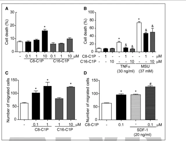
Chemoattraction and cytoprotection are two of the best-known properties of the C1P described in many cell types, except in human endothelial precursors. In the present study, we first aimed to analyze the C1P effect on ECFC survival. ECFCs were treated with 2 different C1P analogs, a synthetic short-chain C8-C1P and the natural long-chain C16-C1P, and cell death was mea-sured after 24 hours by nuclear morphology analysis.
ORIG IN AL R ESE ARCH - V B
We found that while C16-C1P failed to induce ECFC death in all concentrations tested (0.1–10 µmol/L), C8-C1P had a significant cytotoxic effect at 10 µmol/L (Figure 1A). C1P induced ECFC apoptosis since nuclear morphology was compatible with apoptosis (nuclear pyknosis), and caspase-3 was activated by 10 µmol/L C8-C1P (C=2.0±0.5, C1P=18.8±4.8% of positive cells by flow cytometry; P<0.05, n=4).
Numerous studies support the antiapoptotic effect of C1P in different cell types including macrophages, keratinocytes, cochlear hair cells, and retina photorecep-tors.11–14 To mimic the different stress signals present at
the inflammatory and ischemic microenvironment, we induced apoptosis and necrosis with 2 molecules highly elevated in these contexts like TNFα and monosodium urate crystal, respectively.5 C1P analogs were used at
the maximum noncytotoxic concentration. Both C8- and C16-C1P significantly reduced monosodium urate crystal–induced necrosis by 40% approximately and fully suppressed TNFα apoptotic effect (Figure 1B).
Chemoattractant properties of C1P were first reported in macrophages15 and more recently in different murine
bone marrow–residing progenitor cells like mesenchy-mal stem cells, smesenchy-mall embryonic-like cells, and endothelial progenitor cells.10 We here demonstrated that both short
and long versions of this sphingolipid stimulated human ECFC migration. C1P not only increased per se the num-ber of migrated ECFCs in a concentration-dependent manner (Figure 1C) but also potentiated chemotaxis driven by SDF-1—the most powerful chemoattractant factor for undifferentiated cells (Figure 1D).
C1P Enhances Other ECFC Responses Involved
in Vasculogenesis
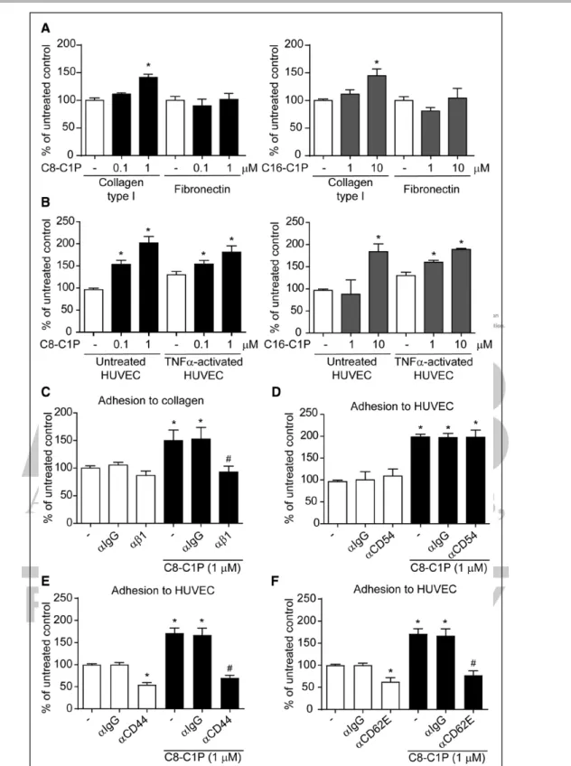
Based on the therapeutic potential of ECFC in regenera-tive medicine, we next studied the effect of C1P in other responses, beyond chemotaxis, involved in vasculogene-sis and tissue repair. To study ECFC adhesion, cells were placed onto a matrix protein-covered surface or cocul-tured with endothelial cells for 1 hour in the presence or absence of C1P. Adhesion to collagen type I, but not fibronectin, significantly was augmented by 1 µmol/L C8- or 10 µmol/L C16-C1P (Figure 2A). Moreover, adhesion
Figure 1. Ceramide 1-phosphate (C1P) promoted endothelial colony–forming cell (ECFC) survival and migration.
A, ECFCs were treated with different concentrations of C8- or C16-C1P, and cell death was analyzed by fluorescence microscopy after 24 h (n=6–8). B, ECFCs were pretreated with C8- or C16-C1P for 30 min and then exposed to TNFα (tumor necrosis factor-α) and monosodium urate crystals (MSUs) at the indicated concentrations. Cell death was analyzed by fluorescence microscopy after 24 h (n=5–10). C-D) Chemotaxis in response to C8- or C16-C1P alone or together with SDF-1 (stromal cell-derived factor 1) was determined using transwell inserts. The number of migrated cells was counted after 6 h (n=4–6). *P<0.05 vs untreated controls; &P<0.05 vs TNFa or MSU alone; #P<0.05 vs SDF-1 alone, 1-way ANOVA.
ORIG IN AL R ESE ARCH - V B
Figure 2. Ceramide 1-phosphate (C1P) enhanced endothelial colony–forming cell (ECFC) adhesion to collagen type I and endothelial cells.
A and B, ECFCs were treated with C8- or C16-C1P and seeded onto collagen type I, fibronectin, untreated or TNFα (tumor necrosis factor-α)-activated human umbilical vein endothelial cell (HUVEC) for 1 h. The number of adherent cells was counted (n=4–7). C–F, ECFCs were treated with C8-C1P in the presence or absence of 2.5 mg/mL blocking antibodies against β1 integrin (αβ1), E-selectin (αCD62E), CD44 (αCD44), ICAM-1 (intercellular adhesion molecule 1; αCD54), or αIgG and seeded onto collagen type I or untreated HUVEC for 1 h. The number of adherent cells was counted (n=3–6). *P<0.05 vs untreated controls; #P<0.05 vs C8-C1P alone, 1-way ANOVA or Kruskal-Wallis.
ORIG IN AL R ESE ARCH - V B
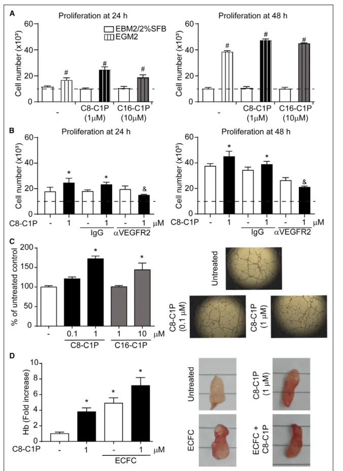
Figure 3. Ceramide 1-phosphate (C1P) increased endothelial colony–forming cell (ECFC) proliferation and tubulogenesis. A, ECFCs were cultured in endothelial basal medium 2 (EBM2)/2% fetal bovine serum (FBS) or endothelial growth medium 2 (EGM2) treated with C8- or C16-C1P at the indicated concentrations, and proliferation was analyzed by cell count in a Neubauer chamber after 24 and 48 h (n=4–5). The dashed line indicates the initial cell number seeded. B, ECFCs were cultured in EGM2 treated with C8-C1P in the presence or absence of 3 mg/mL blocking antibodies against VEGFR2 (vascular endothelial growth factor receptor 2) or αIgG, and (Continued )
ORIG IN AL R ESE ARCH - V B
to mature endothelial cells (HUVECs) was increased by these sphingolipids in a concentration-dependent man-ner, regardless of whether HUVECs were previously acti-vated by TNFα or not (Figure 2B).
To understand the mechanism mediating the strong proadhesive action of C1P, we analyzed the participation of cell adhesion molecules like β1 integrin, coreceptor of collagen type I, as well as the E-selectin (CD62E)/CD44 axis and ICAM-1 (intercellular adhesion molecule 1; CD54), possible mediators of ECFC-HUVEC interaction. Of note, the expression of these adhesion molecules was studied on each cell type before and after C1P treat-ment, and only ICAM-1 was increased, whereas the rest of them remained unchanged (Figure I in the online-only Data Supplement). The enhanced adhesion to collagen type I was completely inhibited after β1 integrin block-ade, whereas IgG control showed no effect (Figure 2C). Moreover, C8-C1P–induced adhesion to HUVEC was fully reduced in the presence of neutralizing antibodies against E-selectin or CD44, but not ICAM-1, indicat-ing that the receptor/counterreceptor E-selectin-CD44 binding is responsible for ECFC-HUVEC adhesion induced by C1P (Figure 2D through 2F). Furthermore, E-selectin mediates the interaction between the 2 endo-thelial cell types after a 1-hour coculture without adding C1P or any activating factor. To investigate the kinetics of E-selectin expression in cocultures, we performed a time course assay in both ECFC and HUVEC grown alone or together. In ECFC, no significant changes were observed in E-selectin expression after 5 and 20 minutes of cocul-ture, whereas a significant increment was detected after 60 and 240 minutes of coculture. Similar data trends were observed in HUVECs, although an earlier signifi-cant alteration tendency was detected in this cell type, after 20 minutes of coculture. In both cases, C1P further increased this process at early stages (20 and 6 minutes of coculture) but failed to potentiate E-selectin expres-sion after 240 minutes (Figure II in the online-only Data Supplement).
Regarding cell proliferation, when ECFCs were cul-tured in endothelial basal medium 2/2% fetal bovine serum, no cell growth was observed in any condition (with or without C1P) because the total cell number at 24 and 48 hours was similar to the amount initially seeded (≈10 000 cells; Figure 3A). As expected, ECFC growth was triggered by endothelial growth medium 2—an effect that was potentiated by C8-C1P and C16-C1P (1 and 10 µmol/L, respectively; Figure 3A). While C8-C1P slightly but significantly increased the total cell number after 24 hours, both C8- and C16-C1P
augmented proliferation after 48 hours (Figure 3A). Flow cytometry analysis of cell division by carboxyfluorescein succinimidyl ester tracking yielded similar results after 24 (untreated: 34±2, C8-C1P 0.1 µM: 29±1, C8-C1P 1 µM: 46±3*, C16-C1P 1 µM: 33±1, C8-C1P 10 µM: 35±1% of positive cells; *P<0.05 versus untreated) and 48 hours (untreated: 49±1, C8-C1P 0.1 µM: 49±3, C8-C1P 1 µM: 71±2*, C16-C1P 1 µM: 58±2, C8-C1P 10 µM: 66±3%* of positive cells; *P<0.05 versus untreated). The mito-genic effect of C1P was completely suppressed in the presence of the neutralizing antibody against VEGFR2 (vascular endothelial growth factor receptor 2), suggest-ing that C1P-induced ECFC proliferation depends on VEGF (vascular endothelial growth factor)/VEGFR2 axis (Figure 3B). On the contrary, we observed that C8-C1P failed to modify the secretion of the proangiogenic fac-tors VEGF and bFGF (basic fibroblast growth factor), as well as TGFβ1 (transforming growth factor-β1) and IL (interleukin)-4, whereas it significantly upregulated the release of the anti-inflammatory cytokine IL-10 and downregulated the release of the proinflammatory IL-8 (Figure III in the online-only Data Supplement).
The formation of cord-like structures in vitro was significantly increased by C1P, C8-C1P being more potent than C16-C1P (Figure 3C). This effect was also observed in vivo using Geltrex plug implants in nude mice loaded (or not) with human ECFC. As shown in Figure 3D, C8-C1P not only has a potent vasculogenic effect by itself but also potentiates plug vascularization mediated by human ECFC.
ERK1/2 and AKT Pathways Are Involved in the
Proangiogenic Effects of C1P
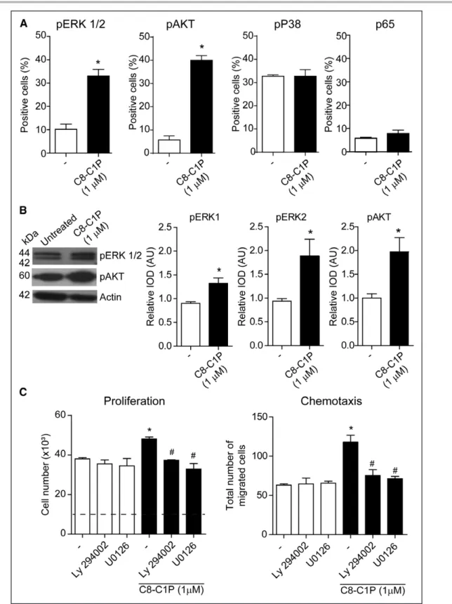
To elucidate the mechanism involved in the proangio-genic effects of C8-C1P, we next studied some of the classical pathways involved in ECFC functions, which have been reported to be regulated by C1P in other cell types (eg, macrophages, cancer cells) such as ERK1/2, PI3K (phosphatidylinositol 3-kinase pathway)/AKT, P38, and NFκB. Flow cytometric analysis showed that C1P increased the percentage of positive cells for pERK1/2 and pAKT but not for P38 and the NFκB subunit p65 (Figure 4A). Activation of these pathways was confirmed by Western blot (Figure 4B). Furthermore, pERK1/2 and pAKT pathways are key mediators of C8-1P proangio-genic effects since the increase in proliferation and che-motaxis induced by C8-C1P was completely inhibited by the pharmacological inhibitors of ERK1/2 (U-0126) and AKT (LY-294002; Figure 4C).
Figure 3 Continued. proliferation was analyzed by cell count in a Neubauer chamber after 24 and 48 h (n=3–4). C, ECFCs were treated with C8- or C16-C1P at the indicated concentrations and then seeded on Geltrex matrix. The number of branch points per field was measured after 18 h. Images are representative of 6 different experiments. D, Geltrex plugs were loaded with C8-C1P, ECFC, or their combination and subcutaneously implanted in nude mice. After 10 d, implants were removed; the concentration of Hb (hemoglobin) was measured using the Drabkin reagent. Images are representative of 5 different experiments. *P<0.05 vs untreated controls; #P<0.05 vs same treatment in EBM2/2%FBS; &P<0.05 vs C8-C1P with IgG, 1-way or 2-way ANOVA, Fisher correction.
ORIG IN AL R ESE ARCH - V B
Figure 4. Intracellular pathways involved in ceramide 1-phosphate (C1P) proangiogenic effects.
A, Endothelial colony–forming cells (ECFCs) treated with C8-C1P for 10 min and the phosphorylation of ERK1/2 (extracellular signal-regulated kinases 1 and 2), AKT, P38, and P65 were measured by flow cytometry (n=3–5). B, ECFCs treated with C8-C1P for 10 min and the phosphorylation of ERK1/2 and AKT was detected by immunoblotting. Each membrane was reprobed with an antibody against actin to calculate the relative IOD (n=4). C, ECFCs were treated with C8-C1P in the presence or absence of pharmacological inhibitors of AKT (LY-294002, 2.5 µmol/L) and ERK1/2 (U-0126, 2.5 µmol/L). Proliferation and C8-C1P–driven chemotaxis were determined after 48 and 6 h, respectively (n=3). *P<0.05 vs untreated controls; #P<0.05 vs C8-C1P alone, Student t test or 1-way ANOVA, Fisher correction.
ORIG IN AL R ESE ARCH - V B
C1P Directly Promotes Tissue Revascularization
Post-Ischemia and Enhances the Therapeutic
Potential of ECFC
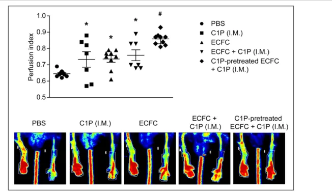
The effect of C1P on the ECFC therapeutic potential for tissue regeneration was studied in a murine model of hindlimb ischemia. As expected, the intravenous injec-tion of untreated ECFC significantly augmented the ischemic-to-nonischemic leg blood flow ratio, relative to vehicle-treated mice (Figure 5). Intramuscular injections of C1P significantly enhanced the perfusion in the ischemic hindlimb in comparison with PBS and slightly increase the revascularization mediated by untreated ECFC alone (Figure 5). Interestingly, the best outcome was observed in animals that received both intravenous administration of C1P-pretreated ECFC together with intramuscular injection of C1P. This latter treatment resulted in further and significant improvement of leg reperfusion compared with each condition alone (Figure 5).
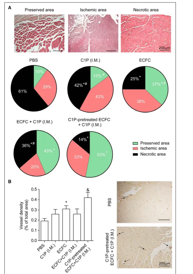
The histomorphometric analysis of the distal gastroc-nemius muscles on day 14 was performed by dividing the digitized muscle sections into 3 areas: (1) preserved area: normal histology including homogeneous fibers and peripheral nuclei; (2) ischemic area: tissue in regeneration with cellular alterations and central nuclei; (3) necrotic area: muscle cell destruction, nonhomogeneous fibers, and mul-ticellular infiltration (Figure 6A). Our results showed that animals receiving intramuscular injections of C1P alone sig-nificantly reduced necrosis in the ischemic muscle in com-parison with PBS-treated mice (Figure 6A), while capillary density was slightly increased (Figure 6B). As expected,
intravenous infusion of untreated ECFC improved tissue repair significantly, although the administration of both untreated ECFC together with intramuscular injection of C1P showed no differences with animals receiving ECFC alone (Figure 6A and 6B). Outstandingly, administration of C1P-pretreated ECFC together with intramuscular C1P reduced necrosis and inflammatory infiltrate, exhibiting a higher percentage of preserved area (Figure 6A). Further-more, this combined treatment resulted in a further and significant enhancement of capillary density compared with all conditions (Figure 6B). Of note, there was no epi-sode of death or defects in ambulation in any group.
Considering that monocytes and macrophages are involved in vasculogenesis and C1P potently induced their migration, we aimed to analyze whether these immune cells play a role in the ECFC/C1P regenera-tive effect. Few immune cells were observed after 7 and 14 days post-surgery, showing no variances between the different experimental conditions (data not shown). An earlier analysis of muscle histology (day 4) showed that PBS-treated animals showed a considerable amount of macrophage infiltrating the ischemic muscles, which was higher when C1P alone was administered intramuscu-larly. However, animals receiving ECFC alone or in com-bination with C1P showed little macrophage infiltration (Figure IV in the online-only Data Supplement).
DISCUSSION
Therapeutic angiogenesis, and in particular, transplan-tation of adult bone marrow cells, including ECFC, has
Figure 5. Ceramide 1-phosphate (C1P) promotes postischemia tissue reperfusion.
PBS, endothelial colony–forming cell (ECFC) alone, and C8-C1P–pretreated ECFC were infused intravenously 5 h after the ischemia-inducing surgery. C8-C1P was administrated intramuscularly (IM) 5 and 29 h after surgery. Perfusion index was calculated by Doppler analysis at day 14. Representative Doppler images are shown (n=6–9). *P<0.05 vs PBS; #P<0.05 vs all other treatments, 1-way ANOVA.
ORIG IN AL R ESE ARCH - V B
Figure 6. Administration of ceramide 1-phosphate (C1P)–pretreated endothelial colony–forming cell (ECFC) together with local C1P reduces necrosis and increased vascular density in the ischemic muscle.
A, Histological analysis of gastrocnemius muscles, stained with hematoxylin and eosin, was performed at day 14. Representative images of preserved, ischemic, and necrotic areas in the gastrocnemius muscle post-ischemia are shown. Original magnification, ×40. Scale bar=200 µm. The surface of each area type is reported as a percentage of the entire histological section surface. B, Vessel density was calculated as the percentage of CD31+ vessels in the total surface. Representative images of immunohistochemistry are shown. Black arrows indicate CD31+ vessels. Original magnification, ×40. Scale bar=200 µm (n=4–9). *P<0.05 vs PBS; #P<0.05 vs C1P-pretreated ECFC+C1P (intramuscularly [IM]); &P<0.05 vs all the treatments, 1-way ANOVA.
ORIG IN AL R ESE ARCH - V B
demonstrated exciting results and constitutes a prom-ising approach for the treatment of ischemia.20 ECFC
recruitment at the site of injury is a key step for tissue revascularization/repair, as well as their engraftment, which largely depends on the survival of the newly arrived cells within the hostile inflammatory microenvironment. In this sense, 2 of the best-known properties of C1P are chemoattraction and cytoprotection. Numerous studies support the antiapoptotic effect of C1P in different cell types. In macrophages and keratinocytes, higher levels of endogenous C1P (after CERK activity upregulation) blocked apoptosis,12,21 whereas the exogenous addition
of C1P to cochlear hair cell and retina photoreceptor cul-tures also increases cell survival.13,14 In line with this
evi-dence, we here demonstrated that C1P protects ECFC not only from apoptosis but also from necrosis triggered by 2 molecules highly elevated in the ischemic tissue like TNFα and monosodium urate crystal, respectively.18
We have used 2 analogs of C1P, a long-chain ana-log (C16-C1P), which is a major type of C1P in cells, considered like the natural C1P,22 and a short chain one
(C8-C1P), because of its higher solubility in water and cellular permeability than the long-chain C1P.23 In fact,
Gangoiti et al24 previously showed that optimal DNA
syn-thesis was attained at lower concentrations when using C8-C1P than when using C16-C1P to stimulate cells. It is worth to mention that C8-C1P was not found in biolog-ical systems; therefore, their effects should be observed, as others suggested,25 as only pharmacological.
Regard-ing cytoprotection, we found that C8-C1P exerted it maximal protective effect at 1 µmol/L and higher con-centrations induced ECFC apoptosis, whereas C16-C1P failed to induce ECFC death in all concentrations tested (0.1–10 µmol/L) and effectively reduced cell death at 10 µmol/L. Both C1P analogs showed similar effects on ECFC functions when used at the maximum noncyto-toxic concentration.
As mentioned above, C1P is a well-known chemoat-tractant factor of monocytes, macrophages,15,26
pancre-atic cancer cells,27 mesenchymal stem cells,28 endothelial
cells,29 and several murine precursors, including bone
marrow–derived endothelial progenitor cells.9,10 Our data
showed that C1P acted as a potent chemoattractant factor for human ECFC and additionally potentiated the chemotactic effect of SDF-1—the major ECFC physi-ological chemotactic factor. Because it has been shown that C1P levels are upregulated in damaged tissues like carotid injury,30 irradiation,9,10 ischemic myocardium,10,31
our findings suggest that C1P, like SDF-1, can create a gradient to attract ECFC to the injury site.
The angiogenic process involves ECFC homing and adhesion to activated endothelium or matrix proteins and their extravasation to target sites. In vivo, endothelial pro-genitor cells usually home to areas of altered shear stress, where the endothelial monolayer is activated and possi-bly denuded, exposing extracellular matrix proteins to the
vascular lumen.32 These areas are believed to influence
the adhesion and extravasation of progenitor cells to isch-emic sites. Our results provide strong evidence that C1P stimulation enhances these events by promoting ECFC adhesion to collagen type I and mature endothelial cells (HUVEC). In this regard, β1 integrin seems to be respon-sible for C1P-induced ECFC adhesion to collagen as the effect was completely suppressed after its blockade. In addition, a complete inhibition of C1P-triggered adhesion to HUVEC was observed in the presence of neutralizing antibodies against E-selectin or CD44, but not ICAM-1, indicating that the E-selectin/CD44 axis is responsible for ECFC-HUVEC adhesion induced by C1P. Moreover, we observed that E-selectin also mediates the interaction between the 2 endothelial cell types after a 1-hour cocul-ture without adding any activating factor, reinforcing the idea that coculturing itself promotes E-selectin expres-sion and further suggesting that C1P-independent endo-thelial cell interaction is also mediated by E-selectin and CD44. These results raised the question: is it possible that E-selectin is significantly expressed on endothelial surface after 1-hour stimulation with C1P? Our results indicated that indeed, E-selectin is rapidly induced on each cell type when cultured together in the presence of C1P—a phenomenon that it is not observed when ECFC and HUVEC are grown separately. To the best of our knowledge, we here report that E-selectin is significantly expressed on ECFC surface as early as 60 minutes after coculture with HUVEC. Similar data were observed in HUVEC, although an earlier significant alteration ten-dency was detected in these cells, after 20 minutes of coculture. A fast expression of E-selectin on HUVEC has been reported before when cultured together with smooth muscle cells,33 after 15-minute stimulation with
nicotine34 or 30-minute post-Flavivirus infection.35
More-over, E-selectin expression was observed as early as 30 minutes after lipopolysaccharide administration in an in vivo model of cutaneous inflammation.36 The mechanisms
of C1P-mediated E-selectin upregulation have not been investigated here. However, considering that the sole interaction between cells triggers E-selectin expression and C1P effect is only observed when cells are cultured together, it is conceivable that C1P acts as an enhancer of the cross talk between them.
It has been previously reported that C1P increased HUVEC adhesion to plastic, although the mechanisms involved were not identified.10 Additionally, C1P increased
the expression of ICAM-1 on ECFC surface, although its blockade has no effect on ECFC-HUVEC interac-tion. Considering that C1P participates and regulates the inflammatory response,37 it is conceivable that ICAM-1
mediates ECFC contact with leukocytes—a fundamen-tal step to allow the migration of these cells to the site of injury. Moreover, our results showing that C1P failed to modify the secretion of proangiogenic factors (VEGF, bFGF, etc) while significantly upregulated the release of
ORIG IN AL R ESE ARCH - V
B the anti-inflammatory cytokine IL-10 and downregulated the release of the proinflammatory IL-8 strengthen the idea that C1P regulates ECFC inflammatory responses. Similarly, it has been previously reported that C1P sup-presses the release of proinflammatory cytokine from macrophages.38–40 How C1P regulates inflammation and
the consequences for tissue repair is an interesting area of future investigation.
Other ECFC responses involved in vasculogenesis were also augmented by C1P. Although C1P had no effect itself on cell proliferation, both C8-C1P and C16-C1P induced ECFC expansion in the presence of a mod-erate amount of growth factors (1:2 dilution endothelial growth medium 2), suggesting a collaborative effect between them and C1P. In addition, this mitogenic action of C1P was completely suppressed by VEGFR2 block-ade. In line with our finding, the involvement of VEGF/ VEGFR2 axis on C1P-induced proliferation has been reported in RAW264.7 macrophages,41 C8-C1P
aug-mented ECFC proliferation over time, whereas C16-C1P only showed significant differences after 48 hours. A sim-ilar C8-C1P–mediated mitogenic effect was previously observed in macrophages42 and aortic vascular smooth
cells30 but not in C16-C1P–stimulated MSCs and mature
endothelial cells (HUVEC, HREC, and HCAEC).9,10,29 The
lack of effect of C16-C1P was probably because pro-liferation was analyzed after 24-hour stimulation—a time point in which we also failed to detect any effects. We also demonstrated that C1P promoted the formation of capillary-like network mediated by ECFC in vitro and the vasculogenesis in vivo in a murine model of subcutaneous implants of ECFC-loaded gel plugs. In addition, a proan-giogenic effect of C1P per se was observed in this model. In agreement with our findings, Kim et al10 showed that a
long-chain analog of C1P, C18-C1P, increased HUVEC-mediated angiogenesis in vitro and in gel plug implants. Moreover, CERK−/− murine microvascular endothelial
cells showed an impaired tubule formation capacity.16
The classical intracellular signaling pathways activated by C1P include PI3K/AKT, MAPK ERK1/2, MAPK P38, and NFκB, which are responsible for the chemotactic effect of this sphingolipid in macrophages and pancre-atic cancer cells.15,26,27 In line with this evidence, we here
showed that C1P triggers phosphorylation of PI3K/AKT and MAPK ERK1/2, although P38 or NFκB were not affected. Likewise, both C1P-enhaced proliferation and chemotaxis were fully abrogated by pharmacological inhibitors of AKT and ERK1/2, suggesting these path-ways as crucial molecular mechanisms mediating C1P proangiogenic action on ECFC.
The relevance of these in vitro findings was con-firmed using an in vivo murine model of hindlimb isch-emia. Animals receiving intramuscular injections of C1P significantly increased recovery of blood perfusion and reduced necrosis in the ischemic muscle in compari-son with PBS-treated mice, while capillary density was
slightly increased. The C1P-mediated tissue protection might be associated with its anti-inflammatory43 and
cytoprotective properties,11–14 although more studies are
required to elucidate this issue. Despite our in vitro data showing that C1P strongly chemoattracts ECFC, intra-venous administration of untreated ECFC together with intramuscular injection of C1P showed no differences with animals receiving ECFC alone. It is conceivable that the elevated endogenous C1P levels at the ischemic tissue,10,31 along with other chemokines, are enough to
achieve the maximum possible chemoattractant effect and the exogenous addition of this sphingolipid failed to produce a greater ECFC attraction. Interestingly, the beneficial effects of ECFC infusion were amplified sig-nificantly by C1P pretreatment of these progenitor cells. Pretreated ECFC together with an intramuscular C1P injection improved their therapeutic outcome, resulting in a further and significant enhancement of leg reperfusion, a tissue necrosis reduction, and a capillary density incre-ment. The angiogenic potency of the C1P-stimulated ECFC is likely to be attributable to both the beneficial effects of C1P on the proangiogenic potential of trans-planted ECFC and the influence of C1P and ECFC on the ischemic hindlimb. In line with our evidence, it has been demonstrated that the priming of MSC with C1P improved the regenerative effect of these cells in a pul-monary artery hypertension rat model.28
Considering that C1P is a well-known chemoattractant factor for monocytes and macrophages, we would expect a higher number of monocytes/macrophages present within the ischemic muscles of animals treated with C1P alone and in combination with ECFC. While this is true for C1P alone, few inflammatory infiltrates were observed in ECFC-treated animals. Although these findings suggest that the number of macrophages/monocytes within the damaged area is not crucial for ECFC/C1P-mediated tissue repair, we cannot rule out that these immune cells still participate in this process at different time points or under other experimental conditions. In addition, the type of monocytes and macrophages migrating from periph-eral blood or their polarization into inflammatory or anti-inflammatory phenotype among the different treatments should be considered as well. These issues are currently under investigation in our laboratory.
In conclusion, our finding suggests that C1P may be considered a new therapeutic tool in ischemic disorders, promoting tissue repair by itself or as a primer of ECFC survival and angiogenic responses, which result in a bet-ter outcome of ECFC-based therapy.
ARTICLE INFORMATION
Received March 18, 2019; accepted August 8, 2019.
Affiliations
From the Experimental Thrombosis Laboratory, Institute of Experimental Medi-cine, National Academy of Medicine–CONICET, Buenos Aires, Argentina (H.A.M.,
ORIG IN AL R ESE ARCH - V B
P.R.Z., M.S., S.N.); Innovative Therapies in Haemostasis, INSERM (B.D., C.B.-V.) and INSERM US025, CNRS UMRS 3612, PTICM (V.M.), Université de Paris, France; and Experimental Medicine and Biology Institute, CONICET, Buenos Ai-res, Argentina (F.P.).
Acknowledgments
We thank I. Dubail, V. Bertrand, Caroline Kharchi, and all the technicians from the animal facilities (UMS 3612 CNRS–US25 Inserm, Paris Descartes University); A. Lokajczyk (Inserm UMRS1140) for help concerning endothelial colony–form-ing cell cultures and experiments; and J. Rogozarski for her help in immunothis-tochemistry assays. We also thank the maternity services of Hospital Rivadavia (Buenos Aires) for providing umbilical cord and cord blood samples.
Sources of Funding
This study was financially supported by ANPCyT grants (PICT 1859/15 and 0612/13). Salary of C. Boisson-Vidal is paid by French CNRS.
Disclosures
None.
REFERENCES
1. Chavakis E, Hain A, Vinci M, Carmona G, Bianchi ME, Vajkoczy P, Zeiher AM, Chavakis T, Dimmeler S. High-mobility group box 1 activates integrin-dependent homing of endothelial progenitor cells. Circ Res. 2007;100:204– 212. doi: 10.1161/01.RES.0000257774.55970.f4
2. Tasev D, Koolwijk P, van Hinsbergh VW. Therapeutic potential of human-derived endothelial colony-forming cells in animal models. Tissue Eng Part B
Rev. 2016;22:371–382. doi: 10.1089/ten.TEB.2016.0050
3. Critser PJ, Yoder MC. Endothelial colony-forming cell role in neoangiogen-esis and tissue repair. Curr Opin Organ Transplant. 2010;15:68–72. doi: 10.1097/MOT.0b013e32833454b5
4. Medina RJ, Barber CL, Sabatier F, Dignat-George F, Melero-Martin JM, Khosrotehrani K, Ohneda O, Randi AM, Chan JKY, Yamaguchi T, et al. Endo-thelial progenitors: a consensus statement on nomenclature. Stem Cells
Transl Med. 2017;6:1316–1320. doi: 10.1002/sctm.16-0360
5. Kalka C, Masuda H, Takahashi T, Kalka-Moll WM, Silver M, Kearney M, Li T, Isner JM, Asahara T. Transplantation of ex vivo expanded endothelial pro-genitor cells for therapeutic neovascularization. Proc Natl Acad Sci USA. 2000;97:3422–3427. doi: 10.1073/pnas.070046397
6. Takahashi T, Kalka C, Masuda H, Chen D, Silver M, Kearney M, Magner M, Isner JM, Asahara T. Ischemia- and cytokine-induced mobilization of bone marrow-derived endothelial progenitor cells for neovascularization. Nat Med. 1999;5:434–438. doi: 10.1038/7434
7. Maceyka M, Spiegel S. Sphingolipid metabolites in inflammatory disease.
Nature. 2014;510:58–67. doi: 10.1038/nature13475
8. Boath A, Graf C, Lidome E, Ullrich T, Nussbaumer P, Bornancin F. Regu-lation and traffic of ceramide 1-phosphate produced by ceramide kinase: comparative analysis to glucosylceramide and sphingomyelin. J Biol Chem. 2008;283:8517–8526. doi: 10.1074/jbc.M707107200
9. Kim CH, Wu W, Wysoczynski M, Abdel-Latif A, Sunkara M, Morris A, Kucia M, Ratajczak J, Ratajczak MZ. Conditioning for hematopoietic transplantation activates the complement cascade and induces a proteolytic environ-ment in bone marrow: a novel role for bioactive lipids and soluble C5b-C9 as homing factors. Leukemia. 2012;26:106–116. doi: 10.1038/leu. 2011.185
10. Kim C, Schneider G, Abdel-Latif A, Mierzejewska K, Sunkara M, Borkowska S, Ratajczak J, Morris AJ, Kucia M, Ratajczak MZ. Ceramide-1-phosphate regulates migration of multipotent stromal cells and endothelial progenitor cells–implications for tissue regeneration. Stem Cells. 2013;31:500–510. doi: 10.1002/stem.1291
11. Gómez-Muñoz A, Kong JY, Salh B, Steinbrecher UP. Ceramide-1-phosphate blocks apoptosis through inhibition of acid sphingomyelinase in macro-phages. J Lipid Res. 2004;45:99–105. doi: 10.1194/jlr.M300158-JLR200 12. Tsuji K, Mitsutake S, Yokose U, Sugiura M, Kohama T, Igarashi Y. Role of
ceramide kinase in peroxisome proliferator-activated receptor beta-induced cell survival of mouse keratinocytes. FEBS J. 2008;275:3815–3826. doi: 10.1111/j.1742-4658.2008.06527.x
13. Le Q, Tabuchi K, Hara A. Ceramide-1-phosphate protection of cochlear hair cells against cisplatin ototoxicity. Toxicol Rep. 2016;3:450–457. doi: 10.1016/j.toxrep.2016.04.003
14. Miranda GE, Abrahan CE, Agnolazza DL, Politi LE, Rotstein NP. Ceramide-1-phosphate, a new mediator of development and survival in
retina photoreceptors. Invest Ophthalmol Vis Sci. 2011;52:6580–6588. doi: 10.1167/iovs.10-7065
15. Granado MH, Gangoiti P, Ouro A, Arana L, González M, Trueba M, Gómez-Muñoz A. Ceramide 1-phosphate (C1P) promotes cell migration Involvement of a specific C1P receptor. Cell Signal. 2009;21:405–412. doi: 10.1016/j.cellsig.2008.11.003
16. Niwa S, Graf C, Bornancin F. Ceramide kinase deficiency impairs microen-dothelial cell angiogenesis in vitro. Microvasc Res. 2009;77:389–393. doi: 10.1016/j.mvr.2009.01.006
17. Mena HA, Lokajczyk A, Dizier B, Strier SE, Voto LS, Boisson-Vidal C, Schattner M, Negrotto S. Acidic preconditioning improves the proangiogenic responses of endothelial colony forming cells. Angiogenesis. 2014;17:867– 879. doi: 10.1007/s10456-014-9434-5
18. Mena HA, Carestia A, Scotti L, Parborell F, Schattner M, Negrotto S. Extracellular histones reduce survival and angiogenic responses of late outgrowth progenitor and mature endothelial cells. J Thromb Haemost. 2016;14:397–410. doi: 10.1111/jth.13223
19. Benslimane-Ahmim Z, Heymann D, Dizier B, Lokajczyk A, Brion R, Laurendeau I, Bièche I, Smadja DM, Galy-Fauroux I, Colliec-Jouault S, et al. Osteoprotegerin, a new actor in vasculogenesis, stimulates endothelial colony-forming cells properties. J Thromb Haemost. 2011;9:834–843. doi: 10.1111/j.1538-7836.2011.04207.x
20. Banno K, Yoder MC. Tissue regeneration using endothelial colony-forming cells: promising cells for vascular repair. Pediatr Res. 2018;83:283–290. doi: 10.1038/pr.2017.231
21. Rivera IG, Ordoñez M, Presa N, Gomez-Larrauri A, Simón J, Trueba M, Gomez-Muñoz A. Sphingomyelinase D/ceramide 1-phosphate in cell survival and inflammation. Toxins (Basel). 2015;7:1457–1466. doi: 10.3390/toxins7051457
22. Arana L, Gangoiti P, Ouro A, Trueba M, Gómez-Muñoz A. Ceramide and ceramide 1-phosphate in health and disease. Lipids Health Dis. 2010;9:15. doi: 10.1186/1476-511X-9-15
23. Gómez-Muñoz A, Gangoiti P, Granado MH, Arana L, Ouro A. Ceramide-1-phosphate in cell survival and inflammatory signaling. Adv Exp Med Biol. 2010;688:118–130.
24. Gangoiti P, Bernacchioni C, Donati C, Cencetti F, Ouro A, Gómez-Muñoz A, Bruni P. Ceramide 1-phosphate stimulates proliferation of C2C12 myo-blasts. Biochimie. 2012;94:597–607. doi: 10.1016/j.biochi.2011.09.009 25. Gómez-Muñoz A. Ceramide 1-phosphate/ceramide, a switch between
life and death. Biochim Biophys Acta. 2006;1758:2049–2056. doi:10.1016/j.bbamem.2006.05.011
26. Arana L, Ordoñez M, Ouro A, Rivera IG, Gangoiti P, Trueba M, Gomez-Muñoz A. Ceramide 1-phosphate induces macrophage chemoat-tractant protein-1 release: involvement in ceramide 1-phosphate-stimulated cell migration. Am J Physiol Endocrinol Metab. 2013;304:E1213–E1226. doi: 10.1152/ajpendo.00480.2012
27. Rivera IG, Ordoñez M, Presa N, Gangoiti P, Gomez-Larrauri A, Trueba M, Fox T, Kester M, Gomez-Muñoz A. Ceramide 1-phosphate regulates cell migration and invasion of human pancreatic cancer cells. Biochem
Pharma-col. 2016;102:107–119. doi: 10.1016/j.bcp.2015.12.009
28. Lim J, Kim Y, Heo J, Kim KH, Lee S, Lee SW, Kim K, Kim IG, Shin DM. Prim-ing with ceramide-1 phosphate promotes the therapeutic effect of mes-enchymal stem/stromal cells on pulmonary artery hypertension. Biochem
Biophys Res Commun. 2016;473:35–41. doi: 10.1016/j.bbrc.2016.03.046
29. Hankins JL, Ward KE, Linton SS, Barth BM, Stahelin RV, Fox TE, Kester M. Ceramide 1-phosphate mediates endothelial cell invasion via the annexin a2-p11 heterotetrameric protein complex. J Biol Chem. 2013;288:19726– 19738. doi: 10.1074/jbc.M113.481622
30. Kim TJ, Kang YJ, Lim Y, Lee HW, Bae K, Lee YS, Yoo JM, Yoo HS, Yun YP. Ceramide 1-phosphate induces neointimal formation via cell proliferation and cell cycle progression upstream of ERK1/2 in vascular smooth muscle cells.
Exp Cell Res. 2011;317:2041–2051. doi: 10.1016/j.yexcr.2011.05.011
31. Klyachkin YM, Nagareddy PR, Ye S, Wysoczynski M, Asfour A, Gao E, Sunkara M, Brandon JA, Annabathula R, Ponnapureddy R, et al. Pharma-cological elevation of circulating bioactive phosphosphingolipids enhances myocardial recovery after acute infarction. Stem Cells Transl Med. 2015;4:1333–1343. doi: 10.5966/sctm.2014-0273
32. Vajkoczy P, Blum S, Lamparter M, Mailhammer R, Erber R, Engelhardt B, Vestweber D, Hatzopoulos AK. Multistep nature of micro-vascular recruitment of ex vivo-expanded embryonic endothelial progenitor cells during tumor angiogenesis. J Exp Med. 2003;197:1755–1765. doi: 10.1084/jem.20021659
33. Chiu JJ, Chen LJ, Lee CI, Lee PL, Lee DY, Tsai MC, Lin CW, Usami S, Chien S. Mechanisms of induction of endothelial cell E-selectin expression by smooth
ORIG IN AL R ESE ARCH - V
B muscle cells and its inhibition by shear stress. Blood. 2007;110:519–528. doi: 10.1182/blood-2006-08-040097
34. Wang Y, Wang Z, Zhou Y, Liu L, Zhao Y, Yao C, Wang L, Qiao Z. Nicotine stimulates adhesion molecular expression via cal-cium influx and mitogen-activated protein kinases in human endothelial cells. Int J Biochem Cell Biol. 2006;38:170–182. doi: 10.1016/j.biocel. 2005.08.004
35. Shen J, T-To SS, Schrieber L, King NJ, Havenga M, Toes R, Melief C, Offringa R. Early E-selectin, VCAM-1, ICAM-1, and late major histocompat-ibility complex antigen induction on human endothelial cells by flavivirus and comodulation of adhesion molecule expression by immune cytokines. J Virol. 1997;71:9323–9332. doi:10.1128/jvi.75.8.3501-3508.2001
36. Silber A, Newman W, Reimann KA, Hendricks E, Walsh D, Ringler DJ. Kinetic expression of endothelial adhesion molecules and relationship to leukocyte recruitment in two cutaneous models of inflammation. Lab Invest. 1994;70:163–175.
37. Gomez-Muñoz A, Presa N, Gomez-Larrauri A, Rivera IG, Trueba M, Ordoñez M. Control of inflammatory responses by ceramide, sphingosine 1-phosphate and ceramide 1-phosphate. Prog Lipid Res. 2016;61:51–62. doi: 10.1016/j.plipres.2015.09.002
38. Hankins JL, Fox TE, Barth BM, Unrath KA, Kester M. Exogenous ceramide-1-phosphate reduces lipopolysaccharide (LPS)-mediated cytokine
expression. J Biol Chem. 2011;286:44357–44366. doi: 10.1074/jbc. M111.264010
39. Lamour NF, Wijesinghe DS, Mietla JA, Ward KE, Stahelin RV, Chalfant CE. Ceramide kinase regulates the production of tumor necrosis factor α (TNFα) via inhibition of TNFα-converting enzyme. J Biol Chem. 2011;286:42808– 42817. doi: 10.1074/jbc.M111.310169
40. Józefowski S, Czerkies M, Łukasik A, Bielawska A, Bielawski J, Kwiatkowska K, Sobota A. Ceramide and ceramide 1-phosphate are nega-tive regulators of TNF-α production induced by lipopolysaccharide. J
Immu-nol. 2010;185:6960–6973. doi: 10.4049/jimmuImmu-nol.0902926
41. Ouro A, Arana L, Riazy M, Zhang P, Gomez-Larrauri A, Steinbrecher U, Duronio V, Gomez-Muñoz A. Vascular endothelial growth factor mediates ceramide 1-phosphate-stimulated macrophage proliferation. Exp Cell Res. 2017;361:277–283. doi: 10.1016/j.yexcr.2017.10.027
42. Gangoiti P, Granado MH, Wang SW, Kong JY, Steinbrecher UP, Gómez-Muñoz A. Ceramide 1-phosphate stimulates macrophage prolifera-tion through activaprolifera-tion of the PI3-kinase/PKB, JNK and ERK1/2 path-ways. Cell Signal. 2008;20:726–736. doi: 10.1016/j.cellsig.2007.12.008 43. Baudiß K, de Paula Vieira R, Cicko S, Ayata K, Hossfeld M, Ehrat N,
Gómez-Muñoz A, Eltzschig HK, Idzko M. C1P attenuates lipopolysaccha-ride-induced acute lung injury by preventing NF-κB activation in neutro-phils. J Immunol. 2016;196:2319–2326. doi: 10.4049/jimmunol.1402681