HAL Id: inserm-00684686
https://www.hal.inserm.fr/inserm-00684686
Submitted on 8 Oct 2013
HAL is a multi-disciplinary open access
archive for the deposit and dissemination of
sci-entific research documents, whether they are
pub-lished or not. The documents may come from
teaching and research institutions in France or
abroad, or from public or private research centers.
L’archive ouverte pluridisciplinaire HAL, est
destinée au dépôt et à la diffusion de documents
scientifiques de niveau recherche, publiés ou non,
émanant des établissements d’enseignement et de
recherche français ou étrangers, des laboratoires
publics ou privés.
Mnk1 kinase activity is required for abscission.
Yoann Rannou, Patrick Salaun, Christelle Benaud, Jabbar Khan, Stéphanie
Dutertre, Régis Giet, Claude Prigent
To cite this version:
Yoann Rannou, Patrick Salaun, Christelle Benaud, Jabbar Khan, Stéphanie Dutertre, et al.. Mnk1
kinase activity is required for abscission.. Journal of Cell Science, Company of Biologists, 2012, 125
(Pt 12), pp.2844-52. �10.1242/jcs.058081�. �inserm-00684686�
Journal
of
Cell
Science
MNK1 kinase activity is required for abscission
Yoann Rannou1,2,*, Patrick Salaun1,2,*, Christelle Benaud1,2, Jabbar Khan1,2, Ste´phanie Dutertre2,3, Re´gis Giet1,2and Claude Prigent1,2,`
1CNRS UMR 6290, IGDR, Rennes, CS34317, 35043 Rennes, France
2Universite´ de Rennes 1, Institut de Ge´ne´tique et De´veloppement de Rennes CS34317, 35043 Rennes, Cedex, France 3CNRS UMS 3480, BIOSIT, CS34317, 35043 Rennes, France
*These authors contributed equally to this work
`Author for correspondence ([email protected])
Accepted 7 February 2012
Journal of Cell Science 125, 2844–2852
ß2012. Published by The Company of Biologists Ltd doi: 10.1242/jcs.058081
Summary
MNK1 is a serine/threonine kinase identified as a target for MAP kinase pathways. Using chemical drug, kinase-dead expression or knockdown by RNA interference, we show that inhibition of MNK1 induces the formation of multinucleated cells, which can be rescued by expressing a form of MNK1 that is resistant to RNA interference. We found that the active human form of MNK1 localises to centrosomes, spindle microtubules and the midbody. Time-lapse recording of MNK1-depleted cells displays cytokinesis defects, as daughter cells fuse back together. When MNK1 activity was inhibited, no microtubule defect at the midbody was detected, however, anchorage of the membrane vesicle at the midbody was impaired as lumenal GFP-positive vesicles did not accumulate at the midbody. At the molecular level, we found that centriolin localisation was impaired at the midbody in MNK1-depleted cells. As a consequence, endobrevin – a v-SNARE protein implicated in the abscission step – was not properly localised to the midbody. Altogether, our data show that MNK1 activity is required for abscission.
Key words: Cytokinesis, Kinase, Mitosis
Introduction
Cytokinesis is a highly conserved mechanism that ends with the cleavage of the mother cell into two daughters. This process is a multi-step mechanism involving the assembly of an equatorial actin and myosin contractile ring, which drives the rapid ingression of a deep cleavage furrow (Field et al., 1999; Glotzer, 2001; Guertin et al., 2002; Zeitlin and Sullivan, 2001). However, although the furrowing is necessary for cytokinesis, it is not sufficient to allow the topological separation of daughter cells. Indeed, furrowing proceeds until the cytoplasm is constricted to a narrow microtubule bridge, known as the midbody. Thus, the terminal event of cytokinesis is the abscission of the intercellular bridge leading to the separation of the two daughter cells. Factors involved in the formation, maintenance and resolution of this bridge are largely unknown (Echard et al., 2004). During mitosis in plant, members of the MAPK family localise to the cell plate as active kinases (Bo¨gre et al., 1999; Calderini et al., 1998) and were found to be involved in cytokinesis (Nishihama and Machida, 2001; Sasabe et al., 2006). In mammalian cells, the MAPK ERK2 directly phosphorylates the centrosome protein CEP55, which induces its relocalisation to the midbody, where it actively participates in cytokinesis (Fabbro et al., 2005). Here, we focused on a kinase downstream of ERK2, MNK1, a serine/threonine kinase identified during screens that are designed to search for ERK substrates (Fukunaga and Hunter, 1997; Waskiewicz et al., 1997). Mammalian MNK1 is also a substrate for the stress-activated p38 MAPK (Tanoue et al., 2000), thereby placing MNK1 as a downstream kinase in the MAPK pathways (Morley, 1997; Wang et al., 1998). MNK1 has been known for a long time to phosphorylate eukaryotic translation initiation factors (eIF4E), which are involved in cap-dependent mRNA translation (Waskiewicz et al., 1999; Wang et al 1998; Morley, 1997). In
Drosophila melanogaster, Lk6 was identified as the Mnk orthologue because both kinases share sequence similarities, conserved residues and domains, and because Lk6 phosphorylates eIF4E in vivo and is a downstream target of ERK2 (Arquier et al., 2005, Parra-Palau et al., 2005). Interestingly, Lk6 was found to interact with microtubules in vitro and to localise to centrosomes (Reiling et al., 2005; Kidd and Raff, 1997). Overexpression of Lk6 also induced defects in the organization of microtubules in eggs and embryos, such as mitotic spindle defects, with centrosomes detaching from the spindle poles (Kidd and Raff, 1997). However, clear mitotic functions of Lk6/MNK1 have never been found.
Here, we provide experimental evidence for a new function of MNK1 kinase during mitosis. MNK1 drug treatment, kinase-dead expression and RNAi downregulation induce the formation of multinucleated cells. The active form of MNK1 is localised to the midbody. Time-lapse recording of live cells shows that microtubules are not affected at the midbody in the absence of MNK1. Inhibition of MNK1 activity eliminates the accumulation of lumenal GFP-positive vesicles at the midbody before abscission. Those vesicles are required to repair damage to the plasma membrane during abscission. At the molecular level, knockdown of MNK1 by RNAi disrupts the localisation of centriolin to the midbody during cytokinesis. In the absence of centriolin, the v-SNARE protein endobrevin, which is required for abscission, is also found mislocalised to the midbody. Thus, MNK1 kinase activity is required for abscission, the terminal step of cytokinesis. Results
Inhibition of MNK1 induces multinucleated cells
We started to investigate MNK1 function by treating proliferating HeLa cells with the Novartis inhibitor CGP57380 at 20 mM for
Journal
of
Cell
Science
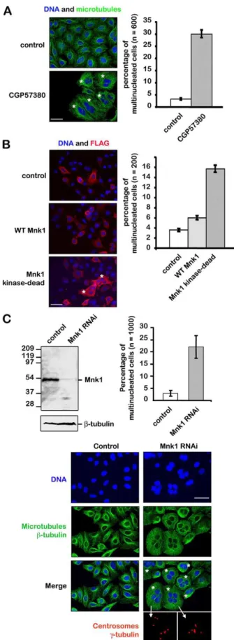
48 hours. Following the drug treatment, 30% (61.5) of the cells were found to be multinucleated (more than two nuclei) whereas ,5% (60.5) of the control cells were multinucleated (Fig. 1A).
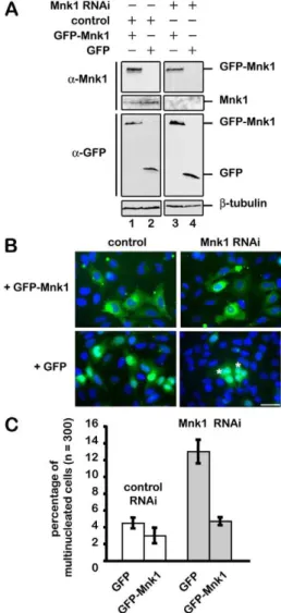
Because CGP57380 inhibits both MNK1 and MNK2, we investigated whether the appearance of multinucleated cells was due to MNK1 inhibition by using a dominant-negative approach, and compared the effect of expressing either active wild-type MNK1 or kinase-dead MNK1 (Pyronnet and Sonenberg, 2001). While overexpression of wild-type MNK1 slightly increased the number of multinucleated cells by 4% (60.5), overexpression of the kinase-dead MNK1 increased this number up to 16% (61) (Fig. 1B). These two experiments using the inhibitor CGP57380 and the kinase-dead MNK1 indicate that the kinase activity of MNK1 is required to avoid the formation of multinucleated cells. We then used an RNA interference approach to more specifically knockdown expression of MNK1. Three different siRNAs were designed against MNK1 mRNA sequences, and 72 hours after siRNA transfection, MNK1 was not detected by western blot (supplementary material Fig. S1). Cells were fixed, stained for microtubules, DNA and centrosomes, and then observed under an epifluorescent microscope. A total of ,3% (61.5) of multinucleated cells were consistently observed in the control, whereas an average of 22% (65) of multinucleated cells were found in cells harbouring a reduced expression of MNK1 (Fig. 1C). Among the population of multinucleated cells, 30% of them had more than two nuclei, indicating that nuclear division was not affected. We did not observe any micronulei, as nuclei were regular in size and shape. However, cell divisions did not occur, strongly suggesting that the lack of MNK1 induced a cytokinesis defect. Multinucleated cells also contained an abnormal number of centrosomes, consistent with a cytokinesis defect. We have eliminated the possibility that the formation of multinucleated cells was due to a lack of eIF4E phosphorylation by overexpressing a mutant form of eIF4E-S209A that cannot be phosphorylated by MNK1 (Ueda et al., 2004; O’Loghlen et al., 2004). Unlike the inhibition of MNK1, overexpression of eIF4E-S209A did not induce the formation of multinucleated cells (supplementary material Fig. S2). Although p38 and ERK1/2 are placed upstream MNK1, their inhibition does not induce the formation of polyploid cells (data not shown). Knockdown of MNK1 expression by RNA interference did not affect the level of ERK1and ERK2 proteins (data not shown). To ensure that the observed phenotype was due to a lack of MNK1, we decided to rescue the treatment of MNK1 siRNA by expressing a GFP-tagged MNK1 resistant to the siRNA. Both MNK1 siRNA and the expression vector were transfected into HeLa cells. Depletion of the endogenous MNK1 and expression of RNAi-resistant GFP-tagged MNK1 protein expression were analyzed by western blot using antibodies against MNK1 and GFP. Control siRNA did not induce any decrease in MNK1 expression (Fig. 2A, lanes 1 and 2),
Fig. 1. Inhibition of MNK1 induces polyploidy. (A) Inhibition of MNK1 with CGP57380 induces multinucleation. Cells were incubated with 20 mM CGP57380 or 1% DMSO (as a control) for 48 hours, then fixed and stained for microtubules (green) and DNA (blue). (B) Overexpression of inactive but not wild-type MNK1 induces multinucleation. Cells were transfected with a vector expressing the FLAG alone or an active FLAG–wt-MNK1 (WT MNK1) or an inactive FLAG-TAA-MNK1 (kinase-dead MNK1). Cells were fixed 72 hours after transfection and stained with an anti-FLAG antibody (red) and DAPI (blue). (C) Depletion of MNK1 using siRNA induces multinucleation. Cells were transfected with MNK1 siRNA or control siRNA. MNK1 protein level was monitored by western blot 72 hours after transfection (b-tubulin was used as a loading control). Cells were fixed and stained for DNA (blue), microtubules (green), and centrosomes (red). Scale bars: 30 mm.
Journal
of
Cell
Science
while siRNA directed against MNK1 eliminated its expression (Fig. 2A, lanes 3 and 4). GFP expression (Fig. 2A, lanes 2 and 4) and MNK1–GFP (Fig. 2A, lanes 1 and 3) were, as expected, insensitive to RNAi. These cells were then fixed and stained for DNA (Fig. 2B). The number of multinucleated cells was quantified by fluorescence microscopy (Fig. 2C). The number of multinucleated cells treated with control siRNA expressing GFP or GFP–MNK1 was equivalent (4% 60.5 versus 3.5% 60.9, n55, .300 cells per experiment, t-test P50.2). By contrast, whereas expression of GFP had no effect on the number of multinucleated cells induced by MNK1 siRNA (13% 61.5), expression of GFP– MNK1 rescued multinucleation (13% 61.5 versus 4.5% 60.6,
n55, .300 cells per experiment, t-test P50.0004). This last result unambiguously demonstrated that the lack of MNK1 is responsible for polyploidy.
The activated phosphorylated form of MNK1 localises to mitotic structures
To understand how MNK1 kinase could be necessary to avoid multinucleation, we examined the localisation of MNK1 and active MNK1 by using antibodies raised against MNK1 phosphorylated at Thr197 and Thr202 (Waskiewicz et al., 1999). MNK1 localised to the cytoplasm and nucleus through out the cell cycle (McKendrick et al., 2001; Parra-Palau et al., 2003). During interphase, the phosphorylated form of MNK1 was hardly detectable, except for a weak signal in the nucleus. In prophase, phosphorylated MNK1 was clearly concentrated at the centrosome (Fig. 3A, white arrow). The activated kinase was then both found at the centrosomes and on the spindle in metaphase, and at the centrosomes and central spindle microtubules in anaphase. In telophase, MNK1 was no longer detectable at the centrosome, but was entirely relocalised to the central spindle and remained on the midbody during cytokinesis. This result indicated that activated MNK1 localised to mitotic structures. During cytokinesis, active MNK1 was detected on both sides of the central ring, and MNK1 RNAi greatly reduced this localisation, demonstrating the specificity of the antibodies used (Fig. 3B). This last localisation is in agreement with a control for the formation of multinucleated cells by MNK1.
Depleted MNK1 cells undergo multinucleation owing to a cytokinesis defect
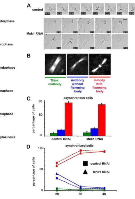
In order to visualise how and when cell division was affected by MNK1 depletion, HeLa cells treated with MNK1 siRNA were recorded using Hoffman contrast video microscopy (Fig. 4A; supplementary material Movies 1, 2). The first stages of mitosis appeared normal in MNK1-depleted cells, as they proceeded to cytokinesis in the same way as the control cells. Midbody abscission occurred ,3–4 hours after the metaphase–anaphase transition. The cells then separated and the flemming body (or central ring) was inherited by one daughter cell (Fig. 4A, control black arrows). By contrast, MNK1-depleted cells did not separate as cytokinesis aborted, and the two daughter cells fused back together to form a single binucleated cell (Fig. 4A, MNK1 RNAi black arrows). The movie clearly shows a cytokinesis defect in absence of MNK1 (supplementary material Movie 2).
Microtubule network at the midbody is not affected in MNK1-depleted cells
To gain a deeper insight into how cytokinesis was affected by the lack of MNK1, we recorded HeLa cells expressing GFP-tagged tubulin, and investigated whether MNK1-depletion affects the microtubule network from metaphase to abscission. MNK1-depleted cells progressed normally through mitosis until cytokinesis just like the control cells. The midbody first formed a thick, stable and straight microtubule intercellular bridge. This bridge became thinner and a flemming body eventually formed. During abscission, the midbody disappeared and the residual flemming body was inherited by one of the daughter cells. In order to follow the evolution of the microtubule network at the midbody during cytokinesis, we used cells expressing tubulin– GFP and defined three types of cell: (1) cells with a thick
Fig. 2. Polyploidy phenotype of MNK1-depleted cells is rescued by expressing a GFP–wt-MNK1 RNA that is resistant to RNAi. Cells were co-transfected with a vector expressing siRNA-resistant GFP–wt-MNK1 or GFP alone, and with MNK1 siRNA or control siRNA. (A) Extracts from cells transfected with GFP (lanes 2 and 4) or GFP–wt-MNK1 (lanes 1 and 3) with MNK1 siRNA (lanes 3 and 4) or with control siRNA (lanes 1 and 2) were probed with antibodies against GFP and MNK1. Note the depletion of endogenous MNK1 (lanes 3 and 4) whereas GFP–wt-MNK1 protein levels are unaffected (lane 3).b-tubulin western blot is used as a loading control. (B) At 72 hours after transfection, cells were fixed and stained for DNA (blue), GFP proteins appear in green. Scale bar: 30 mm. (C) Statistical analysis of the multinucleation rescue by GFP–wt-MNK1 and GFP (5 experiments).
Journal
of
Cell
Science
midbody, (2) cells with a thin mibody and without a flemming body and (3) cells with a thin midbody with a clear flemming body (Fig. 4B). We did not find any differences in the microtubule network at the midbody in asynchronous cells with or without MNK1 (Fig. 4C). In order to compare more precisely cytokinesis in normal and MNK1 depleted cells, we synchronized the cells in the G2-M transition using the CDK1 inhibitor RO-3306. Cells were then released and fixed at different times (2, 3 and 4 hours after release). The number of cells at each stage, which we defined by the shape of the midbody, was again
identical in control and MNK1-depleted cells. Starting from metaphase, numbers evolved identically, indicating that we could not detect any difference with or without MNK1 (Fig. 4D). Together, these results suggest that the kinase does not control the microtubule network at the midbody.
Fig. 4. MNK1 depletion triggers a cytokinesis defect. (A) Selected frames from a time-lapse recording of HeLa cells using Hoffman contrast during mitosis in control cells (top, supplementary material Movie 1) or MNK1 depleted cells (bottom, supplementary material Movie 2). Time is in hours and minutes. (B) Typical midbody morphology during cytokinesis. Three populations of midbody have been considered: thick midbody (left, green), thin midbody without the flemming body (middle, blue) and thin midbody with the flemming body (right, red) (C) Distribution of the three midbody populations in asynchronous HeLa cells expressinga-tubulin–GFP treated with control (left) or MNK1 siRNA (right) for 72 hours; cells were fixed in 3.7% formaldehyde in PHEM. (D) Quantification of three midbody populations in synchronous HeLa cells expressinga-tubulin–GFP treated with control (square) or MNK1 siRNA (triangle) for 72 hours; cells were arrested in metaphase by treating with 10 mM of CDK1 inhibitor IV RO-3306 over the course of 20 hours, then released and fixed 2, 3 or 4 hours after removal of RO-3306.
Fig. 3. Subcellular localisation of active MNK1. (A) Localisation of phosphorylated MNK1 (see Materials and Methods) during mitosis. Cells were fixed and stained for microtubules (green), DNA (blue) and
phosphorylated MNK1 (red). A strong staining of phosphorylated MNK1 on the microtubules spindles (from prophase to telophase) and on the midbody during cytokinesis can be observed. Scale bars: 10 mm. (B) Specificity of phosphorylated-MNK1 localisation during cytokinesis. Cells treated with control (top) or MNK1 (bottom) siRNA were stained for microtubules (green) and phosphorylated MNK1 (red). Scale bar: 2.5 mm.
Journal
of
Cell
Science
MNK1 is required for accumulation of membrane vesicles to the midbody before abscission
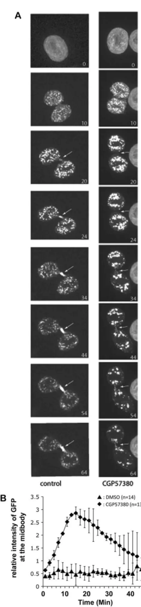
Targeted membrane addition during cleavage furrow formation is a fundamental and widely conserved mechanism of animal cytokinesis (Albertson et al., 2005). When abscission starts, membrane vesicles are recruited at the midbody where they fuse with the plasma membrane to trigger intercellular-bridge abscission. To gain a deeper insight and analyse whether membrane trafficking was affected in the absence of MNK1, we decided to analyse the dynamics of the traffic by using lumenal GFP as a marker (Blum et al., 2000). HeLa cells were transfected with the plasmid encoding lumenal GFP that labelled membrane vesicles. The stable pooled population of transfected cells was incubated for 6 hours either with CGP57380 to inhibit MNK1 or DMSO, and the trafficking of lumenal GFP was filmed live from metaphase to abscission by using spinning confocal microscopy. Analysis of lumenal GFP movement revealed that although recruitment and accumulation of lumenal GFP were clearly observed at the midbody in the control cells before abscission, lumenal GFP was barely detectable at the midbody in the presence of CGP57380 (Fig. 5A; supplementary material Movies 3, 4). GFP-fluorescence intensity at the midbody was quantified and compared in 13 different movies, with or without MNK1. The data obtained clearly show that lumenal GFP was not recruited at the midbody when MNK1 expression was inhibited (Fig. 5B). This strongly suggests that the defect in membrane-vesicle recruitment and accumulation at the midbody induced by the inhibition of MNK1 activity is responsible for the cytokinesis defect observed.
MNK1 is required for endobrevin localisation at the midbody before abscission
As we found a defect in the recruitment of membrane vesicles at the midbody, we checked whether the localisation of the vesicle-fusion SNARE complexes that normally come with the vesicles at the midbody were affected in the absence of MNK1. We used a cell line expressing GFP-tagged a-tubulin to visualise the microtubules and examine the localisation of the vesicle-associated membrane protein 8 (VAMP8 or endobrevin). When abscission starts, endobrevin moves to the central ring where the membrane vesicles fuse with the plasma membrane and triggers intercellular-bridge abscission (Gromley et al., 2005; Low et al., 2003). Typically, during cytokinesis, endobrevin localises to the midbody on both sides of the flemming body (Fig. 6A). We analysed the localisation of endobrevin and recorded three different types of localisation – (1) normal as in Fig. 6A, (2) central but without the flemming body and (3) abnormal – all
Fig. 5. MNK1 is required for membrane vesicle recruitment and accumulation at the midbody. (A) Selected frames from the time-lapse recording of HeLa cells expressing lumenal GFP during cytokinesis in control cells (left, supplementary material Movie 3) or in CGP57380-treated cells (right, supplementary material Movie 4). Cells were observed from metaphase to abscission. Every 2 minutes, a z-series was acquired in fluorescence. Spinning confocal images are the maximal intensity projection of the entire Z-stack. In control cells, lumenal GFP clearly accumulates on the flemming body, whereas it is only weakly detectable on the flemming body of CGP57380-treated cells (white arrows). Time is in minutes. (B) Quantification of the lumenal GFP fluorescence signal at the flemming body during cytokinesis. At the end of telophase, a 10610 pixel square was positioned on the flemming body to measure GFP intensity after maximal-intensity projections were calculated from the original three-dimensional data sets. The background was measured outside the cells and subtracted.
Journal
of
Cell
Science
along the microtubules between the two daughter cells. We observed a normal localisation in 87% (64) of control cells, whereas central staining was only found in 3% (61.5) of control cells and no abnormal localisation was observed. On the contrary, only 22% (61.2) of MNK1-depleted cells showed central staining and 26% (62.5) showed an abnormal localisation of endobrevin with a reduced signal compared with control cells (Fig. 6B). Because the endobrevin protein level was not affected in the absence of MNK1 (Fig. 5C), our interpretation of these data was that MNK1 activity is required for endobrevin localisation to the midbody before abscission.
MNK1 is required for centriolin localisation to the midbody before abscission
Endobrevin localisation to the midbody depends on the presence of centriolin at the midody (Gromley et al., 2005). Centriolin was
first identified as a centrosome protein (Gromley et al., 2003). It localises to the mature centriole throughout cell cycle progression, and upon transition of metaphase to anaphase, a pool of centriolin relocalises to the midbody where it is required for the late stage of cytokinesis (Gromley et al., 2005). Typically, centriolin localises as a ring shape to the midbody (Fig. 7A). We observed that in the absence of MNK1, the number of cells showing centriolin at the midbody decreases from 80% to 50% (Fig. 7B). This was not due to a lack of centriolin synthesis, as we did not observe any decrease in the level of centriolin protein in the absence of the MNK1 (Fig. 7C), indicating that MNK1 is required for localisation of centriolin to the midbody, and that the lack of centriolin at the midbody leads to a defect in the localisation of endobrevin.
Fig. 7. MNK1 is required for centriolin localisation to the midbody. (A) Localisation of centriolin during cytokinesis in control cells. Cells expressing GFP–a-tubulin (green) were fixed, stained for DNA (blue) and centriolin (red). Scale bar: 5 mm. Centriolin appeared as a small ring shape around the flemming body (small panel, middle). (B) Cells were treated with control or MNK1 siRNA for 72 hours, fixed and stained for centriolin and DNA. The number of cells showing centriolin at the midbody was estimated in both cases. (C) Depletion of MNK1 using siRNA had no effect on the level of centriolin as shown by the western blots monitoring MNK1, centriolin and b-tubulin, used as a loading control.
Fig. 6. MNK1 is required for localisation of endobrevin at the midbody. (A) Localisation of endobrevin during cytokinesis in control cells. Cells expressing GFP–a-tubulin (green) were fixed, and stained for DNA (blue) and centriolin (red). Scale bar: 5 mm. Endobrevin appeared as two dots at each side of the flemming body (small panel, right). (B) Cells were treated with control or MNK1 siRNA for 72 hours, and fixed and stained for endobrevin and DNA. The number of cells showing normal endobrevin localisation (top panel), central localisation (middle panel) and abnormal localisation (lower panel) at the midbody was estimated in control and MNK1-depleted cells. (C) Depletion of MNK1 using siRNA had no effect on the level of endobrevin, as shown by the western blots monitoring MNK1, endobrevin andb-tubulin, used as a loading control.
Journal
of
Cell
Science
MNK1 is not required for Mklp1 localisation to the midbody before abscission
In order to get a deeper insight into the mechanism controlled by MNK1, we checked the localisation of the kinesin-like protein MKLP1 (also known as KIF23). MKLP1 is a component of the centralspindlin complex (Mishima et al., 2002), which behaves like a chromosome passenger protein (Kuriyama et al., 2002). It was reported that centriolin localisation was under the control of MKLP1 (Gromley et al., 2005). We monitored the midbody localisation of MKLP1 and did not find any defect in the absence of MNK1 (supplementary material Fig. S3). The data clearly suggest that MNK1 activity can be positioned after MKLP1 and before centriolin in a mechanism that directs cytokinesis through endobrevin localisation. Several other proteins involved in cytokinesis were also controlled. The chromosome passenger
protein Aurora-B localised properly to the midbody
(supplementary material Fig. S3) (Terada et al., 1998). The localisation of the ERK2 substrate CEP55 was not impaired either (supplementary material Fig. S3).
Discussion
During proliferation, at each cell cycle, every cell divides to give two genetically equivalent daughter cells. Every cycle results in the physical separation of the two daughter cells. This is a complex and crucial step in the life of the cell. The position of the cleavage machinery will determine the size of each daughter cell (Oliferenko et al., 2009). The plasma membrane must be broken and repaired without allowing leakage of the cytoplasm (McCollum, 2005; Baluska et al., 2006; Montagnac et al., 2008). Before this final stage, the two cells remain linked by an intercellular bridge made of anti-parallel microtubules associated with many vesicular structures that will be used to repair the plasma membrane (McIntosh et al., 1971). These membrane vesicles are imported from the two daughter cells to fuse with the plasma membrane. In the intercellular bridge, at the middle of the midbody, a ring structure serves as an anchoring point for theses membrane vesicles (Mishima et al., 2002; Zeitlin and Sullivan, 2001).
Here, we present evidence that without MNK1 activity this mechanism is impaired, leading to a defect in cytokinesis that eventually results in the fusion of the two daughter cells. MNK1 is a protein kinase that belongs to the MAPK pathway. It is responsible for the inducible phosphorylation of eIF4E at Ser209 in mammals (Waskiewicz et al., 1999). The exact function of this serine phosphorylation in not known (Scheper and Proud, 2002). It has been reported that suppression of eIF4E phosphorylation by using MNK1 inhibitor CGP57380 or MNK1 knockout in mouse has no effect on the initiation of cap-dependent translation. Although MNK1 is essential for phosphorylation of eIF4E, the function associated with this phosphorylation is not precisely defined. In Drosophila the kinase seems to be required for growth and development (Arquier et al., 2005). When we found that inhibition of MNK1 led to the formation of multinucleated cells, we immediately investigated whether the same phenotype could be obtained by overexpression of unphosphorylatable eIF4E. This was not the case, strongly suggesting that the phenotype observed was not due to a lack of mRNA translation. However, each time we observed a protein localisation defect in the absence of MNK1, we systematically investigated whether the level of the protein was affected. We did not find any change in the level of endobrevin or centriolin proteins in the absence of MNK1.
However, the localisation of both proteins was affected. The number of cells showing the localisation of endobrevin and centriolin to the midbody was strongly decreased in the absence of MNK1. Endobrevin localisation to the midbody requires the presence of centriolin, thus, we suggest that the absence of endobrevin from the midbody is due to the mislocalisation of centriolin. We then checked the localisation of MKLP1 and found that it was not affected by the absence of MNK1. Because the presence of centriolin at the midbody depends on the localisation of MKLP1, we suggest that MNK1 activity is required for centriolin localisation to the midbody independently of MKLP1 (Fig. 8). We are now currently investigating whether centriolin could be a direct target of MNK1. Both proteins move together during mitosis. They both localise to the centrosome at the beginning of mitosis, and they are both present at the midbody before abscission. Our working hypothesis is that the phosphorylation of centriolin would be required for its localisation from the centrosome to the midbody, and MNK1 would be required directly or indirectly in these phosphorylation events. In the absence of MNK1, we observed a defect in the localisation of centriolin that leads to a mislocalisation of endobrevin, resulting in a defect in the anchorage of membrane vesicles at the midbody. The chain of events eventually results in an abortion of abscission and a fusion of the two daughter cells to form a binucleated cell. All these data clearly demonstrate for the first time that MNK1 activity is required for abscission. Materials and Methods
Reagents and antibodies
The following primary antibodies were used: goat polyclonal anti-MNK1 (C-20) (1:1000 for IF and 1:500 for western blot, Santa-Cruz Biotechnology), mouse Fig. 8. MNK1 is required for centriolin localisation to the midbody independently of the presence of Mklp1. In the chain of events leading to cytokinesis defects in the absence of MNK1, we found that even in the presence of MKLP1, centriolin was mislocalised, inducing endobrevin-localisation defects leading to defects in the anchorage of membrane vesicles and eventually abortion of cytokinesis. These data indicate that MNK1 activity is required after MKLP1 localisation and before centriolin localisation.
Journal
of
Cell
Science
anti-phosphorylated-MNK1 (Thr197 and Thr202) (1:100, Cell Signaling), mouse anti-b-tubulin (1:2000, clone 2.1, Sigma Chemicals), mouse anti-c-tubulin (1:2000, clone GTU-88, Sigma Chemicals), mouse anti-GFP (2 mg/ml, clones 7.1 and 13.1, Roche), mouse FLAG (1:1000, Stratagene), rabbit anti-endobrevin (1:1000, gift from Thomas Weimbs), mouse anti-citron kinase (CRIK) (1:1000, BD Transduction Laboratories), rabbit anti-MLKP1 (N-19) (1:1000, Santa-Cruz Biotechnology) and rabbit anti-Rab11 (1:100) antibodies. MNK1 inhibitor CGP57380 was from Novartis (20 mM in DMSO). CDK1 RO-3306 was from Calbiochem.
Plasmid construction and siRNA
siRNA-resistant GFP–MNK1 was obtained by double PCR of MNK1 in pOTB7 (Mammalian Gene Collection: access number: 3629765). Primers used for the first amplification were: 59-CTCGAGGTATCTTCTCAAAAGTTGG-39, 59-GAAGC-AAAAGCACTTTAACGAACGGGAGGCCAGCCGAGTGGTGC-39, 59-CCGCG-GTGGAGCATTTCAGAGTGCTGT-39 and 59-GCACCACTCGGCTGGCCTCC-CGTTCGTTAAAGTGCTTTTGCTTC-39 (mutations are underlined). Primers used for the second amplification were: 59-CTCGAGGTATCTTCTCAAAAGTTGG-39 (XhoI) and 59-CCGCGGTGGAGCATTTCAGAGTGCTGT-39 (SacII) (restriction sites are underlined). The PCR products were cloned into pEGFP-C3 (Clontech). Human MNK1 siRNA oligonucleotide (59-UUCUCGCUCAUUGAAGUGC-39) was purchased from Eurogentec. Random siRNA was used as a control (Petretti et al., 2006). plum–GFP was provided by Irene Schultz (Blum et al., 2000).
Cell culture and transfection
Control HeLa cells or HeLa cells stably expressing GFP-taggeda-tubulin were maintained in Dulbecco’s modified Eagle’s medium supplemented with 10% fetal calf serum. For RNA interference, 150 pmoles of siRNA were mixed with 6 ml of Oligofectamine (Invitrogen, Life Technologies) and applied to cells grown on 12-well culture plates with a confluency of 40%. For rescue assays, cells were transfected with both 150 pmoles of siRNA and 1.6 mg of GFP–MNK1 or pEGFP-C3 vector using Lipofectamine. Cells were cultured for 72 hours before analysis. For lumenal GFP, Hela cells were transfected with plumGFP with JetPrime transfection reagent (Polyplus transfection). The stably transfected pooled population obtained after selection with G418 was used for live-cell imaging.
Indirect immunofluorescence staining
Cells grown on glass coverslips were fixed in –20˚C methanol for 10 minutes or 4% paraformaldehyde. Cells were incubated for 1 hour in PBS plus 0.05% Tween-20 (PBST) containing 5% BSA followed by a 2-hour incubation in PBST-BSA containing primary antibodies. Secondary antibodies were applied for 1 hour at room temperature. Phosphorylated MNK1 was stained with tyramide, as indicated by the manufacturer (TSA Plus Fluorescence Sytems, Perkin Elmer). Coverslips were mounted in Vectashield containing DAPI (Vector Laboratories). A Leica DMIRE2 inverted SP2 confocal microscope using a Plan Apochromat 636 HCX PL APO (NA 1.4) objective was used (Figs 3, 5). For each image, a single confocal plane was presented. For others figures, z-series were acquired using a Leica DMRXA2 fluorescent microscope with a 636 HCX PL APO (NA 1.32) objective and a CoolSnap ES camera (Roper Scientific). The maximum projection of the deconvoluted Z-series was processed by Metamorph software (Universal Imaging). Figures were prepared using Photoshop (Adobe).
Immunoblot analysis
Western blots were performed as described previously (Petretti et al., 2006).
Live-cell imaging
HeLa-lumGFP cells (lumenal-GFP-expressing HeLa cells) were grown in a Lab-Tek I chambered coverglass (Nunc). Cells were treated for 6 hours with either DMSO or with 20 mM CGP57380 (Sigma). Before transferring to the microscope, the medium was changed to CO2-independent medium supplemented with 10%
FBS and 200 mM L-glutamine (Invitrogen). Time-lapse images were captured using a Plan Apo 606 (NA 1.4) objective on an Eclipse Ti-E microscope (Nikon) equipped with a spinning disk (CSU-X1; Yokogawa), a thermostatic chamber (Life Imaging Service), a Piezo Z-stage microscope (Marzhauser), and a charge-coupled device camera (CoolSNAP HQ2; Roper Scientific). Metamorph Software (Universal Imaging) was used to collect the data. GFP frames were recorded every 5 minutes until cells entered anaphase and then every 2 minutes. Images are a maximum projection of 20 Z-planes acquired 0.6 mm apart. Time-lapse data were processed using Metamorph and ImageJ (NIH).
Acknowledgements
We thank Novartis for providing us with CGP57380, Bernard Ducommun for HeLa cells expressing GFP-a-tubulin, Nahum Sonenberg for inactive MNK1 and eIF4ES209A, Thomas Weimbs for anti-endobrevin antibodies, Irene Schultz for lumenal GFP, Stephen Doxsey for anti-centriolin antibody, Georges Baffet for
anti-ERK antibodies, Christian Sardet, Stephen Doxsey and Arnaud Echard for helpful discussions and Rebecca Kay for English proofreading. The microscopy work was performed on the IBiSA platform: Microscopy Rennes Imaging Center. Patrick Salaun and Yoann Rannou were fellows of the Ligue Nationale Contre le Cancer (LNCC).
Funding
Claude Prigent’s lab is supported by the Centre National de la Recherche Scientifique (CNRS), the Agence Nationale de la Recherche (ANR) AURORA, the ‘‘Cance´ropole Grand Ouest’’ [INCa CERIT] and the Ligue Nationale Contre le Cancer (LNCC) [e´quipe labelise´e].
Supplementary material available online at
http://jcs.biologists.org/lookup/suppl/doi:10.1242/jcs.058081/-/DC1
References
Albertson, R., Riggs, B. and Sullivan, W. (2005). Membrane traffic: a driving force in cytokinesis. Trends Cell Biol. 15, 92-101.
Arquier, N., Bourouis, M., Colombani, J. and Le´opold, P. (2005). Drosophila Lk6 kinase controls phosphorylation of eukaryotic translation initiation factor 4E and promotes normal growth and development. Curr. Biol. 15, 19-23.
Baluska, F., Menzel, D. and Barlow, P. W. (2006). Cytokinesis in plant and animal cells: endosomes ‘shut the door’. Dev. Biol. 294, 1-10.
Blum, R., Stephens, D. J. and Schulz, I. (2000). Lumenal targeted GFP, used as a marker of soluble cargo, visualises rapid ERGIC to Golgi traffic by a tubulo-vesicular network. J. Cell Sci. 113, 3151-3159.
Bo¨gre, L., Calderini, O., Binarova, P., Mattauch, M., Till, S., Kiegerl, S., Jonak, C., Pollaschek, C., Barker, P., Huskisson, N. S. et al. (1999). A MAP kinase is activated late in plant mitosis and becomes localized to the plane of cell division. Plant Cell 11, 101-113.
Calderini, O., Bo¨gre, L., Vicente, O., Binarova, P., Heberle-Bors, E. and Wilson, C. (1998). A cell cycle regulated MAP kinase with a possible role in cytokinesis in tobacco cells. J. Cell Sci. 111, 3091-3100.
Echard, A., Hickson, G. R., Foley, E. and O’Farrell, P. H. (2004). Terminal cytokinesis events uncovered after an RNAi screen. Curr. Biol. 14, 1685-1693. Fabbro, M., Zhou, B. B., Takahashi, M., Sarcevic, B., Lal, P., Graham, M. E.,
Gabrielli, B. G., Robinson, P. J., Nigg, E. A., Ono, Y. et al. (2005). Cdk1/Erk2- and Plk1-dependent phosphorylation of a centrosome protein, Cep55, is required for its recruitment to midbody and cytokinesis. Dev. Cell 9, 477-488.
Field, C., Li, R. and Oegema, K. (1999). Cytokinesis in eukaryotes: a mechanistic comparison. Curr. Opin. Cell Biol. 11, 68-80.
Fukunaga, R. and Hunter, T. (1997). MNK1, a new MAP kinase-activated protein kinase, isolated by a novel expression screening method for identifying protein kinase substrates. EMBO J. 16, 1921-1933.
Glotzer, M. (2001). Animal cell cytokinesis. Annu. Rev. Cell Dev. Biol. 17, 351-386. Gromley, A., Jurczyk, A., Sillibourne, J., Halilovic, E., Mogensen, M., Groisman, I.,
Blomberg, M. and Doxsey, S. (2003). A novel human protein of the maternal centriole is required for the final stages of cytokinesis and entry into S phase. J. Cell Biol. 161, 535-545.
Gromley, A., Yeaman, C., Rosa, J., Redick, S., Chen, C. T., Mirabelle, S., Guha, M., Sillibourne, J. and Doxsey, S. J. (2005). Centriolin anchoring of exocyst and SNARE complexes at the midbody is required for secretory-vesicle-mediated abscission. Cell 123, 75-87.
Guertin, D. A., Trautmann, S. and McCollum, D. (2002). Cytokinesis in eukaryotes. Microbiol. Mol. Biol. Rev. 66, 155-178.
Kidd, D. and Raff, J. W. (1997). LK6, a short lived protein kinase in Drosophila that can associate with microtubules and centrosomes. J. Cell Sci. 110, 209-219. Kuriyama, R., Gustus, C., Terada, Y., Uetake, Y. and Matuliene, J.(2002). CHO1, a
mammalian kinesin-like protein, interacts with F-actin and is involved in the terminal phase of cytokinesis. J. Cell Biol. 156, 783-790.
Low, S. H., Li, X., Miura, M., Kudo, N., Quin˜ ones, B. and Weimbs, T. (2003). Syntaxin 2 and endobrevin are required for the terminal step of cytokinesis in mammalian cells. Dev. Cell 4, 753-759.
McCollum, D. (2005). Cytokinesis: breaking the ties that bind. Curr. Biol. 15, R998-R1000.
McIntosh, K., Payne, S. and Russell, W. C. (1971). Studies on lipid metabolism in cells infected with adenovirus. J. Gen. Virol. 10, 251-265.
McKendrick, L., Thompson, E., Ferreira, J., Morley, S. J. and Lewis, J. D. (2001). Interaction of eukaryotic translation initiation factor 4G with the nuclear cap-binding complex provides a link between nuclear and cytoplasmic functions of the m(7) guanosine cap. Mol. Cell. Biol. 21, 3632-3641.
Mishima, M., Kaitna, S. and Glotzer, M. (2002). Central spindle assembly and cytokinesis require a kinesin-like protein/RhoGAP complex with microtubule bundling activity. Dev. Cell 2, 41-54.
Montagnac, G., Echard, A. and Chavrier, P. (2008). Endocytic traffic in animal cell cytokinesis. Curr. Opin. Cell Biol. 20, 454-561.
Journal
of
Cell
Science
Morley, S. J. (1997). Signalling through either the p38 or ERK mitogen-activated protein (MAP) kinase pathway is obligatory for phorbol ester and T cell receptor complex (TCR-CD3)-stimulated phosphorylation of initiation factor (eIF) 4E in Jurkat T cells. FEBS Lett. 418, 327-332.
Nishihama, R. and Machida, Y. (2001). Expansion of the phragmoplast during plant cytokinesis: a MAPK pathway may MAP it out. Curr. Opin. Plant Biol. 4, 507-512. O’Loghlen, A., Gonza´lez, V. M., Salinas, M. and Martı´n, M. E. (2004). Suppression of human Mnk1 by small interfering RNA increases the eukaryotic initiation factor 4F activity in HEK293T cells. FEBS Lett. 578, 31-35.
Oliferenko, S., Chew, T. G. and Balasubramanian, M. K. (2009). Positioning cytokinesis. Genes Dev. 23, 660-674.
Parra-Palau, J. L., Scheper, G. C., Wilson, M. L. and Proud, C. G. (2003). Features in the N and C termini of the MAPK-interacting kinase Mnk1 mediate its nucleocytoplasmic shuttling. J. Biol. Chem. 278, 44197-44204.
Parra-Palau, J. L., Scheper, G. C., Harper, D. E. and Proud, C. G. (2005). The Drosophila protein kinase LK6 is regulated by ERK and phosphorylates the eukaryotic initiation factor eIF4E in vivo. Biochem. J. 385, 695-702.
Petretti, C., Savoian, M., Montembault, E., Glover, D. M., Prigent, C. and Giet, R. (2006). The PITSLRE/CDK11p58 protein kinase promotes centrosome maturation and bipolar spindle formation. EMBO Rep. 7, 418-424.
Pyronnet, S. and Sonenberg, N. (2001). Cell-cycle-dependent translational control. Curr. Opin. Genet. Dev. 11, 13-18.
Reiling, J. H., Doepfner, K. T., Hafen, E. and Stocker, H. (2005). Diet-dependent effects of the Drosophila Mnk1/Mnk2 homolog Lk6 on growth via eIF4E. Curr. Biol. 15, 24-30. Sasabe, M., Soyano, T., Takahashi, Y., Sonobe, S., Igarashi, H., Itoh, T. J., Hidaka, M. and Machida, Y. (2006). Phosphorylation of NtMAP65-1 by a MAP kinase
down-regulates its activity of microtubule bundling and stimulates progression of cytokinesis of tobacco cells. Genes Dev. 20, 1004-1014.
Scheper, G. C. and Proud, C. G. (2002). Does phosphorylation of the cap-binding protein eIF4E play a role in translation initiation? Eur. J. Biochem. 269, 5350-5359. Tanoue, T., Adachi, M., Moriguchi, T. and Nishida, E. (2000). A conserved docking motif in MAP kinases common to substrates, activators and regulators. Nat. Cell Biol. 2, 110-116.
Terada, Y., Tatsuka, M., Suzuki, F., Yasuda, Y., Fujita, S. and Otsu, M. (1998). AIM-1: a mammalian midbody-associated protein required for cytokinesis. EMBO J. 17, 667-676.
Ueda, T., Watanabe-Fukunaga, R., Fukuyama, H., Nagata, S. and Fukunaga, R. (2004). Mnk2 and Mnk1 are essential for constitutive and inducible phosphorylation of eukaryotic initiation factor 4E but not for cell growth or development. Mol. Cell. Biol. 24, 6539-6549.
Wang, X., Flynn, A., Waskiewicz, A. J., Webb, B. L., Vries, R. G., Baines, I. A., Cooper, J. A. and Proud, C. G. (1998). The phosphorylation of eukaryotic initiation factor eIF4E in response to phorbol esters, cell stresses, and cytokines is mediated by distinct MAP kinase pathways. J. Biol. Chem. 273, 9373-9377.
Waskiewicz, A. J., Flynn, A., Proud, C. G. and Cooper, J. A. (1997). Mitogen-activated protein kinases activate the serine/threonine kinases Mnk1 and Mnk2. EMBO J. 16, 1909-1920.
Waskiewicz, A. J., Johnson, J. C., Penn, B., Mahalingam, M., Kimball, S. R. and Cooper, J. A. (1999). Phosphorylation of the cap-binding protein eukaryotic translation initiation factor 4E by protein kinase Mnk1 in vivo. Mol. Cell. Biol. 19, 1871-1880.
Zeitlin, S. G. and Sullivan, K. F. (2001). Animal cytokinesis: breaking up is hard to do. Curr Biol. 11, R514-R516.