HAL Id: hal-02999854
https://hal.archives-ouvertes.fr/hal-02999854
Submitted on 20 Nov 2020HAL is a multi-disciplinary open access archive for the deposit and dissemination of sci-entific research documents, whether they are pub-lished or not. The documents may come from teaching and research institutions in France or abroad, or from public or private research centers.
L’archive ouverte pluridisciplinaire HAL, est destinée au dépôt et à la diffusion de documents scientifiques de niveau recherche, publiés ou non, émanant des établissements d’enseignement et de recherche français ou étrangers, des laboratoires publics ou privés.
STRESS ARE SUFFICIENT TO PRODUCE ORAL
CANDIDIASIS IN BALB/C MICE
Mohammad Halawi, Wissam Yassine, Roudaina Nasser, Hoda Yusef, Jamilah
Borjac, Tareq Al Sagheer, Zaher Zeaiter
To cite this version:
Mohammad Halawi, Wissam Yassine, Roudaina Nasser, Hoda Yusef, Jamilah Borjac, et al.. TWO WEEKS OF CHRONIC UNPREDICTABLE STRESS ARE SUFFICIENT TO PRODUCE ORAL CANDIDIASIS IN BALB/C MICE. Asian Journal of Microbiology, Biotechnology and Environmental Sciences, Global Science Publ., 2020, 22 (2), pp.254-264. �hal-02999854�
ISSN-0972-3005
TWO WEEKS OF CHRONIC UNPREDICTABLE STRESS ARE
SUFFICIENT TO PRODUCE ORAL CANDIDIASIS IN BALB/C MICE
MOHAMMAD HASSAN HALAWI 1*, WISSAM YASSINE2, ROUDAINA NASSER3, HODA YUSEF 1,
JAMILAH BORJAC 1, TAREQ AL SAGHEER3, 4 AND ZAHER ZEAITER3
1Biological Sciences Department, Faculty of Science, Beirut Arab University,
Debbieh, Lebanon
2Department of Chemistry and Biochemistry, Faculty of Sciences, Lebanese University,
Hadath, Beirut, Lebanon
3Biological Sciences Department, Faculty of Sciences, Lebanese University, Hadath, Lebanon 4 Inserm U-1084, Experimental and Clinical Neuroscience Laboratory, University of Poitiers,
Poitiers, France
(Received 15 January, 2020; accepted 11 February, 2020)
Keywords : Candida, Chronic Unpredictable stress, Yeast, Oral Candidiasis
Abstract – The incidence of oral infections caused by Candida species with diverse virulence and
susceptibility profiles has increased in recent years. Due to scarce clinical and experimental data on the ability of stress to induce oral candidiasis, the aim of this study was to assess the impact of stress on oral candidiasis in healthy BALB/c mice and compare its effect to other predisposing factors of oral candidiasis. Immunocompetent and immunocompromised BALB/c female mice were orally infected with C. albicans. A total of four groups of mice each receiving a different treatment were screened. Treatments included antibiotics, corticosteroids and chronic unpredictable stress. Oral tissue colonization and infection was inspected and evaluated comparatively in each group. Tissue burden on day 14 post challenge was measured and mice tongues were inspected for white patches and studied histo-pathologically for evidence of colonization or infection. The induced stress model was able to result in oral colonization and infection without the use of antibiotics or immuno-suppressants. Moreover, the fungal burden was significantly greater in stressed group than that in groups receiving antibiotics treatment or control group. Histopathological examination revealed the abundant presence of C. albicans cells with pseudo-hyphae and in the yeast form, in all tongue tissue samples of treated mice. Tissues were intact in the control group and
Candida cells count was significantly lower in the treated unstressed group. White patches were significantly
more dominant in stressed group than non-stressed and control group. In conclusion, stress maybe a more potent predisposing factor than the use of antibiotics in inducing oral candidiasis, although being a weaker factor than the combined use of antibiotics and corticosteroids together.
INTRODUCTION
Fungi are known to be the leading cause of nosocomial infections (Behzadi et al., 2015). The oral cavity is colonized by C. albicans or other Candida species in 40-60% of healthy individuals (Berberi et
al., 2015). Oropharyngeal candidiasis (OPC) is the
most frequent opportunistic fungal infection known (Carmello et al., 2016). It is a superficial yeast infection caused mainly by C. albicans (Swidergall and Filler, 2017), and is characterized by Candida biofilm formation on the tongue and upon the oral mucosa. OPC results from the adhesion and
subsequent penetration of fungi into the oral tissues (Carmello et al., 2016). It is characterized by formation of creamy white plaques, patches, or papules on an erythematous layer (Mortazavi et al., 2019). It occurs in immune-compromised patients due to Acquired Human Immunodeficiency Syndrome (AIDS), chemotherapy, or the use of immunosuppressant drugs. The use of broad-spectrum antibiotics has also been reported as a predisposing factor for OPC in rather immuno-competent individuals (Salvatori et al., 2016). Similarly, an outstanding increase in incidence of fungal infections was linked to the random use of
antibiotics and corticosteroids in healthy individuals (Vila et al., 2015).
Stress has been a form of our everyday life, and it highly affects the physiological homeostasis. Stress activates the sympathetic nervous system, eventually leading to harmful effects on health. One of the resulting effects of stress is the increase in inflammatory response and thus the promotion of disease progression (Le et al., 2016). Additionally, several studies have shown the suppressive effects of both chronic and acute stressors on the immune system and leukocyte count (Bowers et al., 2008). Therefore, due to the scarce clinical and experimental data on the effect of stress on OPC, the aim of this study was to develop a murine model of oral candidiasis to evaluate the effect of stress in immuno-competent murine model and compare it to that of immunosuppressed mice using a C.
albicans isolate. This study intends to reveal a new
factor that explains some rather idiopathic causes of oral candidiasis among healthy individuals. Such cases, which affect individuals who are thought of as having no predisposing factors, highlights the nature of a yeast that is considered to be opportunistic. The isolate chosen was intentionally selected from a potable water sample instead of a clinical isolate to imitate a realistic scenario, were an immunocompetent stressed consumer may have more chances of being exposed to naturally occurring C. albicans in a water supply than a contagious clinical isolate.
MATERIALS AND METHODS Fungal isolate
One isolate of C. albicans acquired from a Lebanese potable water source (Halawi et al., 2019) was used in this study. The isolate was identified by chromogenic agar and other molecular methods, i.e., HiCrome™ Candida differential agar medium (Himedia, Mumbai, India), Matrix-Assisted Laser Desorption Ionization–Time of Flight Mass Spectrometry MALDI-ToF Vitek-MS (Durham, NC, USA), and sequencing of 26s rRNA (D1/D2 regions) done by Macrogen Inc. (Seoul, South Korea). The sample was stored in YPD broth medium containing 2% glucose, 2% peptone and 15% glycerol at –80°C for further use.
Inoculum preparation
The isolate was cultured twice at 37 °C for 24 hours on Sabouraud dextrose agar (SDA) supplemented
with 0.02% chloramphenicol prior to in vivo use, to ensure viability and purity. For inoculum preparation, isolates were sub-cultured into Sabouraud dextrose broth and allowed to grow overnight in a shaking incubator at 37 °C. After centrifugation, the supernatant was removed and the cells were washed twice in sterile phosphate buffered saline (PBS)(Conti et al., 2014). Yeast cell suspensions were adjusted to an optical density of 0.38 at 520 nm, which corresponds to 1 × 10⁷ cells/
mL using spectrophotometry (Seneviratne et al., 2008). To verify the yeast cell counts, viability of the inoculum was confirmed by placing 10 fold dilutions onto SDA plates at 37 °C for 48 hours.
Animals
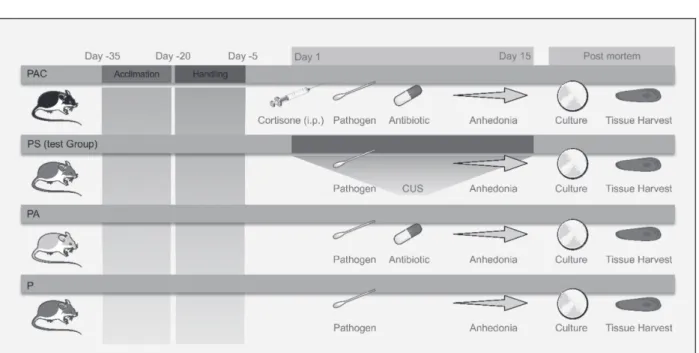
Two weeks old BALB/c female mice (Provided by Beirut Arab University, Debbieh, Lebanon) were used. All mice were given food and water ad libitum and were monitored daily for 30 days prior to challenge. A total of 35 female mice were then used for animal experiments after reaching six weeks of age. Mice were separated into four groups (Fig 1.), each one of the four groups was divided into two shoebox cages, that housed 4-5 mice per cage (Dovigo et al., 2013) as these densities of housing keeps mice less aggressive and healthier (National Research Council (US) Institute for Laboratory Animal Research, 2004). The animal cages were kept in temperature-controlled room (23 ± 2 °C) with 12:12 light/dark cycles (Dovigo et al., 2013). Animals surviving at the end of the experiment or moribund animals (defined according to the CCAC guidelines (Olfert et al., 1998)(Stokes, 2002) were euthanized by an overdose of ketamine-xylazine cocktail administered intraperitoneally, followed by cervical dislocation (Schoell et al., 2009).
Group 1 designated as (PAC) were mice receiving C. albicans inoculum in addition to corticosteroid injections and antibiotics. Group 2 designated as (PS) were the stressed group of mice receiving an inoculum only, with stress treatment. Group 3 designated as (PA) were mice receiving antibiotics along with an inoculum of C. albicans. Group 4 designated as (P) was the control group of mice receiving an inoculum of C. albicans only with no other treatment. Other probabilities of establishing alternative treatment groups was studied carefully and ignored for ethical reasons to prevent unnecessary animal suffering. The presence of a control group receiving corticosteroids
treatment only without an inoculum will subject mice to immunosuppression and probable infections that would be hard to diagnose and link to the current study. The presence of a control group receiving a corticosteroid treatment along with antibiotics, or receiving a sole antibiotic treatment, without an inoculum will lead to probable fatal fungal infections that could be systemic and not caused by Candida spp., which is off the scope of the study.
The animal study protocol was approved by The Institutional Review Board (IRB) of Beirut Arab University-Lebanon (code2019A-0040-S-P-0335), that complies with The Canadian Council on Animal Care’s (CCAC) Guide to the Care and Use of Experimental Animals (Olfert et al., 1993).
Experimental OPC model
In order to induce oral candidiasis in the mice, Carmello et al.(2016)(Carmello et al., 2016) method was used with some modifications. Animals of group 1 were immunosuppressed by subcutaneous injections of methyl prednisolone acetate (Depo-Medrol, Pfizer Manufacturing, Belgium, NV) on days -1, 1, and day 3 and every other day of the infection, at a dose of 100 mg/kg body weight according to the period evaluated (de Oliveira Mima et al., 2010). Tetracycline (0.83 mg/mL) was administered in the drinking water of animals of groups PAC and PA, throughout the test period, beginning at day -1 of the inoculation
(Kamagata-Kiyoura et al., 2004). On day 1, all animals were anesthetized with ketamine hydrochloride 50mg/ mL, (Ketamax™, Rotex- Medica, Trittau, Germany), and xylazine 20 mg/mL, (Xyla™, Interchemie, Holland). Drugs were combined as a cocktail into a single syringe and diluted with sterile 0.9% NaCl to desired concentrations at 0.1 mL per 10 g of body weight. Mice received final concentrations of ketamine (80 mg/kg) and xylazine (8 mg/kg) (Erickson et al., 2016) which induced at least, 60 mins of anesthesia. Mini-sterile swabs soaked in 1 mL of previously calibrated C. albicans blastospores suspension (shaken for resuspension) were inserted sublingually in the mouth and kept for 60 mins. Eye ointment was applied to the eyes of anesthetized mice for lubrication using a sterile cotton applicator (Conti et al., 2014).
Inducing chronic unpredictable stress (CUS)
Mice of group 2 (PS) were the only mice exposed to a procedure of various environmental stressors in random order for two weeks. Stressors included cage tilt (45°, 4–6h), overnight wet cage bedding (14 h), 1–2 min restraint for 2 times in 50mL falcon tube (Wolf et al., 2018), overnight wet cage with no bedding (14h), water deprivation (24h), and forced cold swimming (30 sec. at 10 °C) (Wang et al., 2018). The criterion used to assess anhedonia in susceptible mice was a sucrose preference of less than 65% (Strekalova et al., 2004).
Sucrose Preference test (SPT)
Sucrose preference test was used to evaluate mice chronic depression model. In order to evade isolation stress, the SPT was conducted per cage, reflecting the average consumption of all its residents (mice of the same genotype and treatment) (Wolf et al., 2018). Briefly, two water bottles were placed in the opposite sides of the cages from the start of the habituation period (Fig.3A), and mice were made familiar with their positions for one month. On day 1, water in one of the bottles was replaced with 10% sucrose solution, and mice were permitted to select freely between the two bottles for 24 h. After each 24 h, the weight of each bottle was recorded. The position of the bottles was swiped every 12h to eliminate any side bias. Percentage preference for sucrose was calculated according to the formula: sucrose preference % = (sucrose intake/ total intake) × 100% (Liu et al., 2018).
Fungal burden study
At the end of the experiment period, on day 14, fungal burden was studied, where oral samples were collected by sterile swabs and cultured on SDA supplemented with chloramphenicol plates, and then incubated at 37°C for 48 hours for enumeration. Isolates were then sampled on chromogenic agar for conformation of C. albicans isolation from swabs. After euthanasia the whole tongue tissue was removed and white patches were inspected and measured. Scores were given based on the area of white patches as follows: 0, normal appearing tongue with no white patches; 1, for white patches greater than 0% and less than 20%; 2, white patches between 21 and 90%; 3, white patches greater than 91%; 4, thick white patches that appear pseudomembranous over an area greater than 91% of the tongue (Okada et al., 2013).
Histopathology
After visual inspection and measurements, tongues of mice were immediately and aseptically stored in 10% (wt./vol.) neutral buffered formalin on ice and sent directly for histological examination. The tongues were then dehydrated with 75% ethanol overnight, embedded in paraffin, and 4-m sections were sliced with Leica microtome RM2235 (Leica Microsystems Inc., Buffalo, NY, USA). Samples were then stained with hematoxylin and eosin stain (H&E) and periodic acid–Schiff (PAS)(Zhang et al., 2018) for examination by light microscopy (Olympus optical CO., LTD., Japan).
Statistical analysis
Median survival time (MST) was estimated by the Kaplan–Meier method and compared among groups by the log-rank test. Tissue burden data of tested organs in the different experimental groups were analyzed by using the Kruskal–Wallis test in SPSS (version 17.0 for Windows; Chicago, IL, USA). Mass gain of treatment groups was analyzed by repeated measure two-way ANOVA followed by Sidak multiple comparison test, the percentage of change was analyzed by Kruskal-Wallis test followed by Dunn’s multiple comparison test. SPT data was analyzed by two-way ANOVA, followed by Tukey multiple comparison test. The data of the log10CFU of C. albicans isolated from mice oral cavities were analyzed by ANOVA test. The tongue lesions scoring data were analyzed using Mann-Whitney test. P values of < 0.05 were considered to be significant.
RESULTS
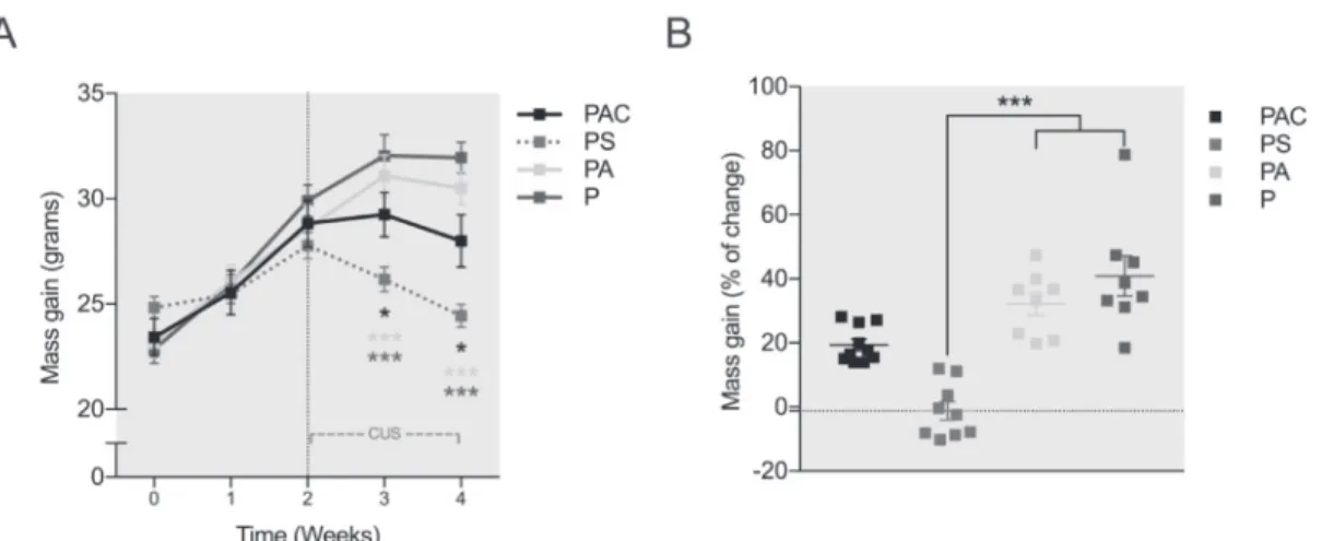
In order to objectively evaluate the impact of the CUS procedure and to confirm the severity of our model, daily mass gain monitoring of the animals was performed on the four groups of mice (PAC, PS, PA, and P) for 4 weeks; among which 2 weeks were during the CUS procedure. Repeated measures two-way ANOVA shows a significant effect of time (F (4, 124) = 128.2; p<0.0001), a trend in the effect of group (F (3, 31) = 2.495; p=0.0782) and the time x group interaction (F (12, 124) = 14.34; p<0.0001). Mass gain was significantly lower in the PS group mice compared to PAC (p<0.05), PA (p<0.001), and P (p<0.0001) as early as one weeks of CUS. Tukey’s Post hoc revealed a similar result in mass gain in PS, at two weeks of CUS, compared to PAC (p<0.05), PA (p<0.0001), and P (p<0.0001). In addition, we found a significant difference between PAC and P groups, where the former displayed a lower mass gain at two weeks of CUS (p<0.01) (Figure 2A).
Furthermore, to examine mass gain change the analysis of the percentage of mass gain was analyzed and the results revealed a significant effect of the group on the percentage attained by the end of the study (p<0.0001; Kurskal-Wallis test). Dunn’s Post hoc analysis revealed a significant difference in PS mice, showing -1.2% gain, as compared to PA that showed +32% (p<0.01), and P littermates with almost +41% of gain (p<0.0001) (Figure 2B). These results show expected decrease in percentage of mass gain change in CUS subjected mice. To an
inferior magnitude, cortisone injection provoked a reduced percentage of mass gain in PAC mice +19% as compared to PA and P littermates, however none of them was enough to reach significance (p=0.2983 and p=0.0855 respectively). Taken together, these results are indicative that the CUS protocol provoked significant decrease in mass gain that were pronounced after 1 week of CUS and lasted till the end of the second week; to a lower extent, the combination of Candida inoculation, antibiotics administration, and cortisone treatment resulted in a plateau effect on mass gain in the PAC group.
In order to determine anhedonia in our mice groups, we compared the sucrose preference for five days using the SPT (Figure 3A). When comparing the four groups, repeated measure two-way ANOVA revealed a significant effect of time (F (4, 52) = 15.43; p<0.0001), group (F (3, 13) = 19.42; p<0.0001) and the time x group interaction (F (12, 52) = 7.051; p<0.0001). Further, statistical analysis showed, at day 4, a significant difference in the sucrose preference in the PS group (60%) as compared to the PA (81%) and P (85%) groups (p<0.01; Tukey’s multiple comparisons test). At day 5, a significant difference in the sucrose preference in the PS group (19%) as compared to the PAC (81%), PA (83%) and P (82%) groups (p<0.0001). It may be mentioned here that our multiple comparisons results revealed a trend at day 3 where the PAC group showed a lower preference (65%) when compared the P mice (83%) (p=0.0871). Similar results were obtained at day 4 with a 68%
sucrose preference for the PAC mice compared to 85% (p=0.0918) for the pathogen only treated mice (Figure 3B).
In the current study, a decrease in sucrose preference below 65% was taken as a criterion for anhedonia (Figure 2B). Our results demonstrate that all the mice in the PS group reached the <65% sucrose preference by the end of the SPT test, and thus were considered anhedonic (Figure 3C). However, animals not exposed to the CUS protocol exhibited a preference for sucrose higher than 65% and where thus considered nonanhedonic. Finally, these differences were even more outstanding when expressed as sucrose preference by day, as PS mice group showed, at day 5, four-fold decrease in sucrose preference (p<0.0001; ordinary one-way ANOVA) when compared to PAC, PA and P mice (Figure 3D). This anhedonic effect was not observed in PAC, PA and P females that showed 80% preference for sucrose, regardless of the group. All together, these results indicate that 2 weeks of CUS protocol provoked significant anhedonic behavior in PS mice.
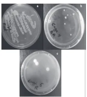
The model used to induce OPC was successful in producing oral candidiasis in all inoculated groups of mice. Cotton swab cultures revealed the successful C. albicans colonization of the oral cavity of all mice, except the negative control group. The culture results on chromogenic agar clearly showed that the isolated yeast from mice buccal cavity was
C. albicans with the characteristic green colonies.
One the other hand, the density of Candida colonies
Fig. 2. Effects of treatment and CUS on general development (mass gain).
PAC in black (n=10), mice under stress protocol (PS) in red (n=9), PA in yellow (n=8), and P in violet (n=8). Data shown as mean +/- SEM, comparison of treatment groups by repeated measure two-way ANOVA followed by Sidak multiple comparison test in (A) and Kruskal-Wallis test followed by Dunn’s multiple comparison test in (B). The P value 0.05 was considered significant, * p0.05; ** p0.01; *** p0.001
in each of the groups varied significantly, where immunosuppressed mice with cortisone acetate in group one had the highest CFU of C. albicans followed by the stressed group two, then by group 3 (Fig. 4).
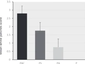
In order to determine the severity of infection on day 14, mice were sacrificed and scores were recorded based on severity and number of white patches on the tongue. The control group P, showed no tongue lesions or white patches whatsoever. The other three groups showed variable lesions. Group PAC showed the most severe lesions with an average score of 2.8±0.44. Group PS showed mild lesions with an average score of 1.75±0.5. Group PA showed minor lesions with a score of 0.66±0.5 (Fig 5). The differences between groups was significant (P<0.05).
Oral swabs and subsequent cultures showed fungal counts of 5.82±0.54 (mean±SD) log10 CFU/ mouse in group PAC, 3.98±0.09 log10 CFU/mouse in group PS, and 3.36±0.29 log10 CFU/mouse in group
Fig. 3. Effects of treatments on the loss of food pleasure in unstressed mice.
Sucrose preference % in PAC in black (n=5), (PS) in red (n=4), PA in yellow (n=4), and P in violet (n=4). A) The Sucrose preference test apparatus. B) The evolution of sucrose preference through 5 days of testing. C) Percentage of anhedonic mice after 2 weeks of CUS calculated by the 65% Anhedonia Criterion. D) Sucrose preference after 2 weeks of CUS. Data presented as mean +/- SEM, two-way ANOVA, followed by Tukey multiple comparison test. P value 0.05 was considered significant, * p0.05; ** p 0.01; *** p0.001.
Fig. 4. Fungal burden showing C. albicans CFU after 48 h
incubation on SDA with 1/1000 dilution; (a) shows group 1 PAC, (b) shows group 2 PS, (c) shows group 3 PA.
PA. Cultures were negative for samples acquired from group P, which is expected for control groups not receiving treatment (Samaranayake and Samaranayake, 2001). Counts were significantly higher in group PAC compared to group PS, and so were counts of groups PS when compared with group PA. The differences between all groups were statistically significant (Fig. 6).
Fig. 5. Average of the tongue lesion scores. The tongue
lesions of PAC group were the highest among groups. Group P had no lesions observable. Statistically, there was significantly more lesions in PS group than that of PA group, but lower than those of PAC group (severity and size of the lesions).
After colonization was confirmed by macroscopically observing white patches on the dorsum of the tongue. Histological studies of mice tongues were undertaken to examine the level of infection and to discriminated between colonization and infection. Colonization was defined as the presence of yeast cells along with pseudo-hypha adhered to tissue lining and severing the papillae (Fig 7a), with infiltration of immune cells in tissue. The deeper invasion of C. albicans of the lamina propria mucosa and into the prickle cell layer was defined as infection (Fig 7b). On the other hand, there were no abnormalities in the control group (Fig 7c.). The overall appearance of tongue tissues confirmed the colonization of mice oral cavities by
C. albicans. Oral cavity inspection revealed white
patches on the dorsum of the tongues of inoculated mice. However, the intensity of white patches varied, with group 1 (PAS) having the highest distribution followed by group 2 (PS) then group 3 (PA), while there was a completely normal appearance of the tongue in mice of group 4 (P),
Fig. 6. Oral log10 CFU/mouse (mean + SD). The highest
colony-forming unit (CFU) counts were obtained in group PAC. Between-group comparisons were statistically significant (P<0.05). CFU counts were the lowest in group PA. Group P recorded nosignificant count.
Fig. 7. PAS of tongue tissue, (a) C. albicans adhering to
papilla of PS(b) C. albicans invading underlying mucosa of PAC group; (c) intact tongue epithelium of group P. (black arrow indicates visible Candida psuedohypha)
which corroborates to spontaneous resolution of macroscopic tongue lesions in control groups stated by Samaranayke and Samaranayke, 2001.
DISCUSSION
Our study is the first to test the effect of CUS on the incidence of oral candidiasis in mice; we used a comparison between groups receiving antibiotics, corticosteroids and stress. It has shown that the effects of Candida inoculation along with CUS or antibiotics or (corticosteroid + antibiotics) are different. The difference was particular with regard to anhedonic behavior, and oral carriage of Candida, which was being pronounced by CUS protocol. The CUS procedure performed here was effective. It was identical for all stressed mice subjected to an unpredictable pattern of chronic stress in order to avoid possible habituation of the mice. The CUS
procedure applied here resulted in less weight gain, and the presence of anhedonia behavior as assessed by reduced sucrose preference.
Nevertheless, our study presents certain limits. First, it may be noted that during the CUS, no monitoring of plasma sugar levels was carried out in the mice given in order to reduce excessive stress in our study groups. For the same reason no mouse was housed alone during the sucrose preference test to reduce isolation stress as mentioned in the materials and methods.
The Candida genus encompasses more than 150 heterogeneous species, nevertheless only a minority of around 20 species is known to cause diseases in animals and humans. Through the last three decades, the frequency of fungal infections caused by Candida spp. has increased dramatically (Szweda
et al., 2015), which set an alarm to figuring out the
real predisposing factors for the infection. Candida
albicans, which is normally a commensal yeast
dwelling as part of the natural oral flora of the buccal cavity of healthy individuals (Hong et al., 2018) is also known to cause superficial and systemic infections. It is a yeast capable of becoming pathogenic and infecting the oral cavity (Richardson
et al., 2018). In fact, multiple studies have listed C. albicans as one of most common and most disease
-causing species of Candida (Marak and Dhanashree, 2018).
Several murine models have been developed for common fungal diseases, which have been useful for evaluating antifungal therapies (Fakhim et al., 2018). The use of BALB/c mice has been reviewed to be acceptable in vivo model of different candidiasis models (Nett, 2019); it also has been accepted as a valid CUS murine model (Mul et al., 2016). However, and despite the challenges faced when using female mice for such studies, females have been chosen due to their reduced aggression compared to males (Vega-Rivera et al., 2016). Moreover, it has been found that females are more resilient to early-life stress, and treatment with glucocorticoid antagonist could not affect their behavioral performance in any behavioral tasks (Loi
et al., 2017). As known, the exposure to stress has
deep and compound effects on motivation and decision-making as stress triggers qualitatively and quantitatively differential behavioral consequences. It is now widely known that chronic stress, especially when being unpredictable, can result in cognitive and emotional deficits characteristic of many neuropsychiatric disorders (Hollon et al.,
2015). Chronic stress induction techniques are usually used as murine models for the initiation of depressive type symptoms such as anhedonia, social interaction and increased immobility in forced swimming test (FST) or tail suspension tests (TST). The mesolimbic dopamine system plays a central role in the control of motivated behavior. A synchronous-choice test as simple as the SPT assesses whether mice prefer a sweet reward rather than a lesser tempting alternative, raw water. This helps to evaluate the motivation in these animals and assess anhedonia, where they are expected to choose sucrose containing water in order to continue to receive the pleasure of reward (Rizvi et
al., 2016). In the current study we hypothesize that
in an immune-competent mouse the presence of chronic stress could trigger oropharyngeal candidiasis. To test this hypothesis, a murine model was exposed to just two weeks of chronic unpredictable stress, and inoculated with C. albicans (Hong et al., 2018), which is part of the normal microbiota of humans, but doesn’t occur naturally in mice (Richardson et al., 2018).
Mice treated to produce CUS lost weight significantly when compared to the rest of the groups, which corroborates with the findings of Keller et al. (2017)(Keller et al., 2017). The other group of mice treated with glucocorticoids also lost weight compared to groups not receiving such treatment, but to a lesser extent than those having CUS induced. This is due to the depressive-like behavioral effects that corticosteroids induce. These findings match those found by Kvarta et al. (2015)(Kvarta et al., 2015), who also found a significant reduction in sucrose preference and linked the reduced weight gain in such groups to an anhedonia-like phenotype. This was true for the first two doses of glucocorticoids, however among continuing the treatment, weight gain increased dramatically in mice receiving glucocorticoids. This phenomenon is similar to that found by Mammi et
al. (2016)(Mammi et al., 2016), who stated that the
excessive exposure to glucocorticoids would lead to obesity in humans and mice, altogether with induction of sugar cravings, which explains the high sucrose preference despite the presence of anhedonia in this case.
Our animal model design using C. albicans showed that oral inoculation using cotton swabs of 1 × 10⁷ CFU/mouse was able to infect and proliferate
in immunocompetent mice under CUS, with high mortality rate. Animals infected with C. albicans had
a high fungal load when given glucocorticoids compared to those receiving antibiotics only. There was severe filiform papilla tissue damage, confirmed by histopathological findings, which are in concordance with previous studies done by Costa
et al. (2013). Both hyphal and blastoconidia of C. albicans were present on infected oral cavities. Thus,
this model authentically reflects key clinical features of human OPC.
The test parameters were hard to maintain and reach solid conclusions when monitoring weight gain and sugar preferences. It is known that the overgrowth of C. albicans leads to several symptoms, some of which may affect tested variables, these symptoms include, sugar cravings, tiredness after feeding, brain fogging and depression (Gustafson, 2016). At all given times, infection is distressing, weight gain may be reduced due to limited intake, and therefore weight may actually be lost (Dickstein, 1964).
Understanding the mechanisms regulating fungal virulence and the interaction with the host is necessary for a better understanding of the infectious process. Moreover, understanding the mechanism governing the depressive state induced by CUS and its effects on the immune system are of high importance. C. albicans being able to infect immune-competent stressed mice highlights the need for further and deeper understanding of the neurological interactions of human immune response and comprehending the exact details and causes of such outcomes. However, the CUS model used, presented to be sufficient in producing an immune-depression in the tested group of mice, the stressed mice showed reduced weights, and high mortality compared to other groups. The SPT test used to assess the level of anhedonia produced in mice revealed very low preference of sucrose.
In conclusion, a fourteen-day CUS treatment of mice was successful in replicating human oral candidiasis model and could explain the increasing prevalence of OPC among rather healthy individuals.
REFERENCES
Behzadi, P., Behzadi, E. and Ranjbar, R. 2015. Urinary tract infections and Candida albicans. Central European
Journal of Urology. 68 (1) : 96.
Berberi, A., Noujeim, Z. and Aoun, G. 2015. Epidemiology of oropharyngeal candidiasis in human immunodeficiency virus/acquired immune deficiency syndrome patients and CD4+ counts.
Journal of International Oral Health: JIOH. 7 (3) : 20.
Bowers, S. L., Bilbo, S. D., Dhabhar, F. S. and Nelson, R. J. 2008. Stressor-specific alterations in corticosterone and immune responses in mice. Brain, Behavior, and
Immunity. 22 (1) : 105–113.
Carmello, J. C., Alves, F., Basso, F. G., de Souza Costa, C. A., Bagnato, V. S., de Oliveira Mima, E. G. and Pavarina, A. C. 2016. Treatment of oral candidiasis using photodithazine®-mediated Photodynamic Therapy in vivo. PloS One. 11(6) : e0156947.
Conti, H. R., Huppler, A. R., Whibley, N. and Gaffen, S. L. 2014. Animal models for candidiasis. Current
Protocols in Immunology. 105(1) : 19–6.
Costa, A. C., Pereira, C. A., Junqueira, J. C. and Jorge, A. O. 2013. Recent mouse and rat methods for the study of experimental oral candidiasis. Virulence. 4(5) : 391– 399.
de Oliveira Mima, E. G., Pavarina, A. C., Dovigo, L. N., Vergani, C. E., de Souza Costa, C. A., Kurachi, C. and Bagnato, V. S. 2010. Susceptibility of Candida albicans to photodynamic therapy in a murine model of oral candidosis. Oral Surgery, Oral Medicine, Oral
Pathology, Oral Radiology, and Endodontology. 109 (3) :
392–401.
Dickstein, B. 1964. Neonatal oral candidiasis: Evaluation of a new chemotherapeutic agent. Clinical Pediatrics. 3(8) : 485–488.
Dovigo, L. N., Carmello, J. C., de Souza Costa, C. A., Vergani, C. E., Brunetti, I. L., Bagnato, V. S. and Pavarina, A. C. 2013. Curcumin-mediated photodynamic inactivation of Candida albicans in a murine model of oral candidiasis. Sabouraudia. 51 (3): 243–251.
Erickson, R. L., Terzi, M. C., Jaber, S. M., Hankenson, F. C., McKinstry-Wu, A., Kelz, M. B. and Marx, J. O. 2016. Intraperitoneal continuous-rate infusion for the maintenance of anesthesia in laboratory mice (Mus musculus). Journal of the American Association for
Laboratory Animal Science.55(5) : 548–557.
Fakhim, H., Vaezi, A., Dannaoui, E., Chowdhary, A., Nasiry, D., Faeli, L., Meis, J. F. and Badali, H. 2018. Comparative virulence of Candida auris with Candida
haemulonii, Candida glabrata and Candida albicans in a
murine model. Mycoses. 61(6) : 377–382.
Gustafson, C. 2016. Ellen Kamhi, PhD, RN: Herbal Support for the HPA Axis. Integrative Medicine: A Clinician’s
Journal.15 (6) : 42.
Halawi, M. H., Borjac, J., Yusef, H. and Zeaiter, Z. 2019. Prevalence of Candida species in Lebanese water.
Asian Journal of Microbiology, Biotechnology and Environmental Sciences. 21 (3) : 92–94.
Hollon, N. G., Burgeno, L. M. and Phillips, P. E. 2015. Stress effects on the neural substrates of motivated behavior. Nature Neuroscience. 18(10) : 1405. Hong, H. J., Son, N. R., Yang, W. Y., Lee, J. M., Kim, J. H.,
Jang, S. M. and Nam, S. H. 2018. Antibacterial and antifungal activities of Lespedeza cuneata extract against Candida albicans. Biomedical Research. 29(20): 3728–3731.
Kamagata-Kiyoura, Y., Abe, S., Yamaguchi, H. and Nitta, T. 2004. Protective effects of human saliva on experimental murine oral candidiasis. Journal of
Infection and Chemotherapy. 10(4) : 253–255.
Keller, S., Lotan, A., Tatarskyy, P., Wolf, G., Avidan, E., Ben-Ari, H., Lifschytz, T., Shbiro, L., Tabachnick, T. and Shaharabany, J. 2017. The Effect of Chronic Stress on Weight and Hypothalamic Insulin and Melanocortin 4 Receptors in Young and old Female Mice. European Neuropsychopharmacology. 27 : 412–413. Kvarta, M. D., Bradbrook, K. E., Dantrassy, H. M., Bailey, A. M. and Thompson, S. M. 2015. Corticosterone mediates the synaptic and behavioral effects of chronic stress at rat hippocampal temporoammonic synapses. Journal of Neurophysiology. 114(3): 1713– 1724.
Le, C. P., Nowell, C. J., Kim-Fuchs, C., Botteri, E., Hiller, J. G., Ismail, H., Pimentel, M. A., Chai, M. G., Karnezis, T. and Rotmensz, N. 2016. Chronic stress in mice remodels lymph vasculature to promote tumour cell dissemination. Nature Communications. 7 : 10634. Liu, M. Y., Yin, C. Y., Zhu, L. J., Zhu, X. H., Xu, C., Luo, C.
X., Chen, H., Zhu, D. Y. and Zhou, Q. G. 2018. Sucrose preference test for measurement of stress-induced anhedonia in mice. Nature Protocols. 13(7) : 1686. Loi, M., Mossink, J., Meerhoff, G., Den Blaauwen, J.,
Lucassen, P. and Joëls, M. 2017. Effects of early-life stress on cognitive function and hippocampal structure in female rodents. Neuroscience. 342: 101– 119.
Mammi, C., Marzolla, V., Armani, A., Feraco, A., Antelmi, A., Maslak, E., Chlopicki, S., Cinti, F., Hunt, H. and Fabbri, A. 2016. A novel combined glucocorticoid-mineralocorticoid receptor selective modulator markedly prevents weight gain and fat mass expansion in mice fed a high-fat diet. International
Journal of Obesity. 40 (6) : 964.
Marak, M. B. and Dhanashree, B. 2018. Antifungal susceptibility and biofilm production of Candida spp. Isolated from clinical samples. International
Journal of Microbiology.
Mul, J. D., Zheng, J. and Goodyear, L. J. 2016. Validity assessment of 5 day repeated forced-swim stress to model human depression in young-adult C57BL/6J and BALB/cJ mice. ENeuro. 3(6).
National Research Council (US) Institute for Laboratory Animal Research. 2004. Effects of Housing Density and
Cage Type on Young Adult C57BL/6J Mice. The Development of Science-based Guidelines for Laboratory Animal Care: Proceedings of the November 2003 International Workshop.
Nett, J. E. 2019. Candida auris: An emerging pathogen “incognito”? PLoS Pathogens.15 (4) : e1007638. Okada, M., Hisajima, T., Ishibashi, H., Miyasaka, T., Abe,
S. and Satoh, T. 2013. Pathological analysis of the
Candida albicans-infected tongue tissues of a murine
oral candidiasis model in the early infection stage.
Archives of Oral Biology. 58 (4) : 444–450.
Olfert, E., Bhasin, J., Latt, R., McCutcheon, K., Rainnie, D.
and Schunk, M. 1998. CCAC Guidelines On: Choosing
an Appropriate Endpoint in Experiments Using Animals for Research, Teaching and Testing.
Olfert, E. D., Cross, B. M. and McWilliam, A. A. 1993. Guide
to the care and use of experimental animals (Vol. 1).
Canadian Council on Animal Care Ottawa.
Oral White Lesions: An Updated Clinical Diagnostic Decision Tree. (n.d.). Retrieved December 19, 2019, from https:/
/www.ncbi.nlm.nih.gov/pmc/articles/PMC6473409/ Richardson, J. P., Mogavero, S., Moyes, D. L., Blagojevic, M., Krüger, T., Verma, A. H., Coleman, B. M., Diaz, J. D. L. C., Schulz, D.and Ponde, N. O. 2018. Processing of Candida albicans Ece1p is critical for Candidalysin maturation and fungal virulence.
MBio.9(1) : e02178-17.
Rizvi, S. J., Pizzagalli, D. A., Sproule, B. A. and Kennedy, S. H. 2016. Assessing anhedonia in depression: Potentials and pitfalls. Neuroscience & Biobehavioral
Reviews. 65 : 21–35.
Salvatori, O., Puri, S., Tati, S. and Edgerton, M. 2016. Innate immunity and saliva in Candida albicans–mediated oral diseases. Journal of Dental Research. 95 (4) : 365– 371.
Samaranayake, Y. H., and Samaranayake, L. P. 2001. Experimental oral candidiasis in animal models.
Clinical Microbiology Reviews. 14(2) : 398–429.
Schoell, A. R., Heyde, B. R., Weir, D. E., Chiang, P.-C., Hu, Y. and Tung, D. K. 2009. Euthanasia method for mice in rapid time-course pulmonary pharmacokinetic studies. Journal of the American Association for
Laboratory Animal Science. 48 (5) : 506–511.
Seneviratne, C., Jin, L., Samaranayake, Y. and Samaranayake, L. 2008. Cell density and cell aging as factors modulating antifungal resistance of
Candida albicans biofilms. Antimicrobial Agents and Chemotherapy. 52(9) : 3259–3266.
Stokes, W. S. 2002. Humane endpoints for laboratory animals used in regulatory testing. ILAR Journal. 43(Suppl_1) : 31–38.
Strekalova, T., Spanagel, R., Bartsch, D., Henn, F. A. and Gass, P. 2004. Stress-induced anhedonia in mice is associated with deficits in forced swimming and exploration. Neuropsychopharmacology. 29 (11) : 2007. Swidergall, M., and Filler, S. G. 2017. Oropharyngeal candidiasis: Fungal invasion and epithelial cell responses. PLoS Pathogens. 13 (1): e1006056. Szweda, P., Gucwa, K., Kurzyk, E., Romanowska, E.,
Dzier¿anowska-Fangrat, K., Jurek, A. Z., Kuœ, P. M. and Milewski, S. 2015. Essential oils, silver nanoparticles and propolis as alternative agents against fluconazole resistant Candida albicans, Candida
glabrata and Candida krusei clinical isolates. Indian Journal of Microbiology. 55 (2) : 175–183.
Vega-Rivera, N. M., Ortiz-López, L., Gómez-Sánchez, A., Oikawa-Sala, J., Estrada-Camarena, E. M. and Ramírez-Rodríguez, G. B. 2016. The neurogenic effects of an enriched environment and its protection against the behavioral consequences of chronic mild stress persistent after enrichment cessation in
six-month-old female Balb/C mice. Behavioural Brain
Research. 301 : 72–83.
Vila, T. V. M., Chaturvedi, A. K., Rozental, S. and Lopez-Ribot, J. L. 2015. Characterization of the in vitro activity of Miltefosine against Candida albicans under planktonic and biofilm growing conditions and in vivo efficacy in the murine model of oral candidiasis.
Antimicrobial Agents and Chemotherapy. AAC-01890.
Wang, B., Chen, X., Zhou, T. and Wang, X. 2018. Antidepressant-like effects of embelin and its possible mechanisms of action in chronic unpredictable stress-induced mice. Neurological
Research. 40 (8) : 666–676.
Wolf, G., Lifschytz, T., Ben-Ari, H., Tatarskyy, P., Merzel, T. K., Lotan, A. and Lerer, B. 2018. Effect of chronic unpredictable stress on mice with developmental under-expression of the Ahi1 gene: Behavioral manifestations and neurobiological correlates.
Translational Psychiatry. 8(1) : 124.
Zhang, X., Liu, H., Hao, Y., Xu, L., Zhang, T., Liu, Y., Guo, L., Zhu, L. and Pei, Z. 2018. Coenzyme Q10 protects against hyperlipidemia-induced cardiac damage in apolipoprotein E-deficient mice. Lipids in Health and