HAL Id: tel-02003499
https://tel.archives-ouvertes.fr/tel-02003499
Submitted on 1 Feb 2019
HAL is a multi-disciplinary open access
archive for the deposit and dissemination of
sci-entific research documents, whether they are
pub-lished or not. The documents may come from
teaching and research institutions in France or
abroad, or from public or private research centers.
L’archive ouverte pluridisciplinaire HAL, est
destinée au dépôt et à la diffusion de documents
scientifiques de niveau recherche, publiés ou non,
émanant des établissements d’enseignement et de
recherche français ou étrangers, des laboratoires
publics ou privés.
A Study of Striatal Markers as Disease Modifiers in
Huntington’s Disease
Laetitia Francelle
To cite this version:
Laetitia Francelle. A Study of Striatal Markers as Disease Modifiers in Huntington’s Disease. Neurons
and Cognition [q-bio.NC]. Université Paris Sud - Paris XI, 2014. English. �NNT : 2014PA11T070�.
�tel-02003499�
UNIVERSITÉ PARIS-SUD
ÉCOLE DOCTORALE 419 :
BIOSIGNE
Laboratoire :
THÈSE DE DOCTORAT SUR TRAVAUX
ASPECTS MOLÉCULAIRES ET CELLULAIRES DE LA BIOLOGIE
par
Laetitia FRANCELLE
A study of striatal markers as disease modifiers
in Huntington's Disease
Date de soutenance : 26/11/2014
Composition du jury :
Directeur de thèse : Dr Emmanuel BROUILLET Laboratoire des maladies neurodégénératives,
URA CEA-CNRS 2210, Service MlRCen, lnstitut d'lmagerie Biomédicale, CEA, Fontenay aux Roses, France.
Président du jury: Pr Dr Hervé DANIEL Pharmacologie et Biochimie de la Synapse,
lnstitut de Biochimie et de Biophysique Moléculaire et Cellulaire, UMR 8619, Centre Universitaire d'Orsay, Orsay, France.
Rapporteurs : Dr Jocelyne CABOCHE SIgnalisation Neuronale et Régulation Génique, Unité Neurosciences Paris Scène,
INSERM/UMR-S1130; CNRS/UMR8246, Paris France.
Dr Karine MERIENNE Interactive Dynamics of Memory Systems,
Laboratoire de Neurosciences Cognitives et Adaptatives (LNCA), UMR7364/ Université de Strasbourg/CNRS, Strasbourg, France.
Examinateurs : Pr Dr Tiago FLEMING OUTEIRO Department of NeuroDegeneration and Restorative Research Center for Nanoscale Microscopy and Molecular Physiology of the Brain,
University Medical Center Göttingen, Göttingen, Germany.
Dr Jean-Vianney BARNIER Centre de Neurosciences Paris-Sud (CNPS),
UMR 8195 CNRS-UPS, Bât 444, Centre Universitaire d'Orsay, Orsay, France.
Page | 3
“Ever tried. Ever failed. No matter. Try Again. Fail again. Fail better.”
Samuel Barclay Beckett
(April 13, 1906 - December 22, 1989)
Irish avant-garde novelist, playwright, theatre director, and poet.
1969 Nobel Prize in Literature.
Page | 4
REMERCIEMENTS
Je remercie mes rapportrices, Jocelyne Caboche et Karine Merienne, pour avoir
accepté de faire la lecture critique de mon manuscrit. Je tiens également à remercier
Jean-Vianney Barnier et Hervé Daniel pour avoir accepté d’être membres de mon jury de thèse. I
thank Tiago Fleming Outeiro to have accepted to be part of my thesis committee, I am
grateful for your time and consideration.
Je remercie Philippe Hantraye d’avoir accepté que je puisse réaliser ma thèse au sein de
MIRCen.
Je ne remercierai jamais assez mon directeur de thèse Emmanuel Brouillet, pour m’avoir fait
confiance en me prenant en thèse, pour avoir fait son maximum, malgré un emploi du temps
surchargé, pour me former et me faire progresser et pour m’avoir permis de parcourir le globe pour ma
formation. J’ai adoré ces années à travailler avec vous.
Merci à Sandrine Humbert, Fréderic Saudou, Diana Zala et leurs équipes à l’Institut Curie
d’Orsay pour avoir toujours accepté de partager leur savoir-faire et leurs compétences scientifiques
avec moi.
Merci à Anselme Perrier pour m’avoir fourni en neurones quelques temps, mais surtout pour
les nombreux échanges que nous avons pu avoir sur le projet DCLK3.
Je remercie Gilles Bonvento et Carole Escartin pour leurs précieux conseils.
Merci à Martine et Diane pour m’avoir formée à l’expérimentation animale, mais aussi à leur
bonne (et mauvaise) humeur partagée en A2/A3.
Je remercie Noëlle pour m’avoir très gentiment fourni toutes ces années en cellules HEK, ainsi
qu’avec l’aide de Charlène et Alexis, pour la production de tous les virus qui m’ont servi à faire de
mon travail ce qu’il est. Du même bureau, merci à Aurélie pour sa bonne humeur, sa gentillesse et son
interprétation des chansons en histo qui donnait plus de vie au montage des coupes….
Merci à Carole, Fanny et Caroline pour m’avoir formée et assistée dans mes diverses
expériences de biochimie ou histologie. Merci à Gwenn et Pauline pour m’avoir faire profiter de leurs
multi-compétences.
Merci à Karine pour son aide et ses précieux conseils en comportement animal, mais
également pour sa bonne humeur.
Merci à Marie Claude pour m’avoir formée et aidée en B.M., et surtout pour m’avoir
encouragée tout au long de ma thèse. Les marqueurs striataux ont commencé dans tes mains, et le
resteront encore un peu j’espère avec DCLK3…
Page | 5
Je tiens à remercier les autres étudiants en thèse à MIRCen, notamment Lucile pour avoir
supporté mon mutisme intempestif, et avec Noémie pour avoir partagé notre bureau, de nombreuses
discussions scientifiques passionnantes, et plus encore … Merci à Chloé et Marie-Anne pour leur
gentillesse et leur capacité à être très rassurante et encourageante par moment. Merci à Juliette,
Micka, Romain, Marie, Charlotte et Brice pour leur bonne humeur et leur entrain à sortir et faire mil
et une activités en-dehors de MIRCen. Gardez ce cap et bon courage pour la suite !
Merci à Sandro pour ses nombreux conseils.
Bon courage aux post-doc Jérôme, Benoit et Pierrick dans cet univers impitoyable qu’est la
Recherche.
Merci aux ex-MIRCenniens, notamment Fabien et Maria-Angeles avec qui j’ai partagé mon
bureau pendant quelques temps, pour leur bonne humeur et amitié. Merci à Emilie et Matthias V.
pour tous nos moments de décompression autour de quelques bières…
Je remercie Marc, Nicolas, Léo et Lina pour m’avoir encouragée à reprendre mes études,
ainsi que Pierre, Rémi C. et Natacha pour leur amitié depuis le Master.
Merci à Kento, Olivier, Marco et Matthias T. pour m’avoir fait découvrir tout un univers
musical électro qui me fait toujours autant vibrer, ainsi que Laurence, Sylvie, Clotilde, Franck,
Gérard, Valérie et Florence pour avoir mis un peu de son des années 70’ dans mes oreilles, cordes
vocales et doigts.
Merci à Rémi N. pour sa patience, sa motivation et sa bonne humeur durant nos sessions
d’escalade, on finira par faire notre via ferrata un jour !
Je remercie ma famille pour avoir toujours cru en moi, et en particulier ma grand-mère qui
m’a toujours poussée et encouragée à aller toujours plus loin, mon grand-père pour sa générosité, ma
mère et mes frères pour être toujours présents dans les bons moments, comme les moments difficiles,
qui ont faits de ces 3 ans et demi une période riche en émotions et moins difficile que ça aurait pu être.
Merci à Gisèle, Laurent et Annie pour leur bienveillance et générosité. Merci à Sarah et Caro pour leur
gentillesse.
Je tiens à remercier Sina pour n’avoir cessé de m’encourager, de me soutenir et de m’aimer
quand je m’essoufflais et en devenais impossible à vivre ! Je n’y serais pas arrivée sans toi, ton
optimisme à toute épreuve, ton humour fracassant et ton amour sans faille !
Page | 6
TABLE OF CONTENTS
REMERCIEMENTS ___________________________________________________________ 4
TABLE OF CONTENTS ________________________________________________________ 6
LIST OF FIGURES ____________________________________________________________ 9
LIST OF TABLES ____________________________________________________________ 10
ABBREVIATIONS ___________________________________________________________ 12
PART1 INTRODUCTION ______________________________________________________ 15
1.1
Neurodegenerative disorders ______________________________________________ 17
1.2
Huntington’s disease _____________________________________________________ 17
1.2.1 First descriptions of Huntington’s disease ____________________________________________ 17 1.2.2. Actual description of HD ________________________________________________________ 18 1.2.2.1. Motor dysfunction __________________________________________________________ 18 1.2.2.2. Cognitive decline ___________________________________________________________ 18 1.2.2.3. Psychiatric disturbance ______________________________________________________ 19 1.2.3. Prevalence ___________________________________________________________________ 19 1.2.4. Neuropathology ______________________________________________________________ 20 1.2.5. Striatal neurotransmission ______________________________________________________ 22 1.2.6. Cause of the disease ___________________________________________________________ 26 1.2.6.1. Normal functions of wt-Htt protein _____________________________________________ 27 1.2.6.2. Loss of function of wt-Htt and toxic gain of function of mHtt ________________________ 29 1.2.6.3. Molecular Mechanisms in HD _________________________________________________ 33
A. Huntingtin cleavages, aggregation and elimination __________________________________ 34
B. Abnormal protein-protein interaction _____________________________________________ 36
C. Dysfunction calcium signaling ___________________________________________________ 38
D. Energy defects________________________________________________________________ 38
E. Transcriptional deregulation ____________________________________________________ 39
F. Abnormal axonal transport _____________________________________________________ 43 1.2.7. Animal models of HD __________________________________________________________ 44 1.2.7.1. Non genetic models _________________________________________________________ 44 1.2.7.2. Genetic models _____________________________________________________________ 44 1.2.7.3. Transgenic models __________________________________________________________ 46 1.2.7.4. Knock-in models ____________________________________________________________ 46 1.2.7.5. Viral-mediated HTT gene transfer models _______________________________________ 47 1.2.7.6. HD models diversity _________________________________________________________ 47 1.2.8. Past and future HD therapies ____________________________________________________ 48 1.2.8.1. Symptomatic treatments _____________________________________________________ 48 1.2.8.2. Preventive or curative treatments______________________________________________ 49 a) Gene silencing therapy ___________________________________________________________ 50 b) Cystamine and cysteamine ________________________________________________________ 52 c) Autophagy enhancers ____________________________________________________________ 53 d) Synaptic plasticity _______________________________________________________________ 53 e) Modulation of inflammation _______________________________________________________ 54 f) HDAC inhibitors _________________________________________________________________ 54 g) Caspase inhibitors _______________________________________________________________ 55
Page | 7
h) Apoptosis ______________________________________________________________________ 55 i) Cell therapy strategies ___________________________________________________________ 55 j) Dietary supplements _____________________________________________________________ 56PART2 THESIS OBJECTIVES ___________________________________________________ 59
2.1.
Working hypothesis to study striatal vulnerability ______________________________ 60
Review article -
Possible involvement of self-defense mechanisms in the preferential
vulnerability of the striatum in Huntington’s disease. ________________________________ 61
2.2.
Thesis objectives _________________________________________________________ 63
PART3 RESULTS ____________________________________________________________ 65
3.
Disease modifiers ______________________________________________________ 67
3.1.
Article #1
-
The long non-coding RNA Abhd11os is neuroprotective against an
N-terminal Fragment of Mutant Huntingtin in vivo. ___________________________________ 67
3.2.
Article #2 -
Loss of the thyroid hormone binding protein Crym renders striatal
neurons more vulnerable to mutant huntingtin in Huntington’s disease. ________________ 69
3.3.
Article #3
-
Role of Doublecortin-like kinase 3 (DCLK3) in the preferential
vulnerability of the striatum in Huntington's disease (draft). __________________________ 71
PART4 GENERAL DISCUSSION _________________________________________________ 73
4.1. Abhd11os _________________________________________________________________ 74
4.2. CRYM ___________________________________________________________________ 77
4.3. DCLK3 __________________________________________________________________ 79
4.3.1. Protein characterization ____________________________________________________________ 79 4.3.2. Disease-modifying mechanisms ______________________________________________________ 79 4.3.3. Troubleshooting __________________________________________________________________ 814.4. First conclusion ____________________________________________________________ 84
4.5. Others models to study neuroprotective effects of striatal markers _________________ 84
4.6. Impact of our study in current spotlights_______________________________________ 85
PART5 CONCLUSION AND PERSPECTIVES _______________________________________ 91
5.1. Therapeutic perspectives ____________________________________________________ 92
5.2. How to improve methods to optimize the testing of modifiers ______________________ 94
5.3. Further perspectives: combinatory therapy ____________________________________ 96
PART6 ANNEXES ___________________________________________________________ 97
6.1. Collaborative work #1
-
High Throughput Screening for Inhibitors of REST in Neural
Derivatives of Human Embryonic Stem Cells Reveals a Chemical Compound that Promotes
Expression of Neuronal Genes. __________________________________________________ 99
6.2. Collaborative work #2
-
Mutant huntingtin alters Tau phosphorylation and subcellular
distribution. _________________________________________________________________ 101
Page | 8
6.3. Clinical trials status report _________________________________________________ 103
PART7 BIBLIOGRAPHY _____________________________________________________ 105
RESUME _________________________________________________________________ 127
ABSTRACT _______________________________________________________________ 129
Page | 9
LIST OF FIGURES
Figure 1 First page of George Huntington paper "On Chorea", 1872 (Bates 2005). ... 17
Figure 2 Representation of human brain coronal section showing basal ganglia (in blue) in relation to surrounding structures (Adapted from Nieuwenhuys et al 1981) (Wichmann & Delong 2007). ... 20
Figure 3 Whole brain and regional atrophy in the controls, premanifest, and early Huntington’s disease patients (Tabrizi et al 2009). ... 21
Figure 4 Schematic representation of the direct (red lines) and indirect (blue lines) pathway classical model in the physiological condition (Calabresi et al 2014). ... 23
Figure 5 Glutamate and DA in basal ganglia function in intact brain and during HD (André et al 2010). ... 24
Figure 6 Number of CAG repeats in htt gene and elongated stretch of glutamine in htt protein, in healthy and HD conditions. ... 27
Figure 7 Schematic illustrating the biological functions of wt-Htt (Schulte & Littleton 2011). ... 28
Figure 8 Schematic diagram of the huntingtin amino acid sequence (Zuccato et al 2010). ... 30
Figure 9 Potential mechanism of cell death in HD (Cattaneo et al 2001). ... 32
Figure 10 Key cellular pathogenic mechanisms in Huntington’s disease HD (Zuccato et al 2010). ... 33
Figure 11 Mechanisms of dysregulation of gene expression in Huntington's disease (Seredenina & Luthi-Carter 2012). ... 40
Figure 12 Transcription factors, DNA target sequences, and chromatin structure in HD (Zuccato 2010). ... 42
Figure 13 Genetic attributes of animal models of HD (Pouladi et al 2013). ... 45
Figure 14 Schematic depicting current priority preclinical therapeutic targets under investigation for HD (Adapted from Ross et al 2014)(Wild & Tabrizi 2014). ... 50
Figure 15 Schematic illustration of the three main approaches to lowering htt (Wild & Tabrizi 2014). ... 51
Figure 16 Diverse Mechanisms Proposed for lincRNA Function (Ulitsky & Bartel 2013). ... 75
Figure 17 Molecular functions of CRYM on thyroid hormone action (Suzuki et al 2007). ... 77
Figure 18 Schematic representation of the striatal markers that have been experimentally studied as potential modifiers of mutant huntingtin toxicity in HD (adapted from Francelle et al 2014). ... 84
Figure 19 Combinatory therapy model of HD ... 96
Page | 10
LIST OF TABLES
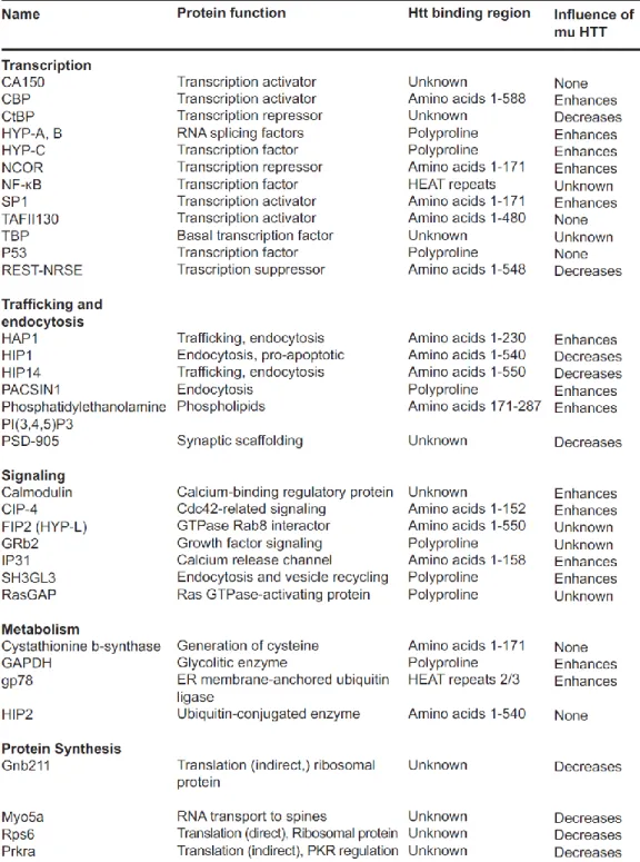
Table 1 Huntingtin interacting proteins and impact of mHtt on interactors protein expression (Clabough 2013). ... 37 Table 2 Pharmacological drugs on symptoms of HD (adapted from Im & Kim 2014). ... 48 Table 3 Similarities (gray) and differences (cyan) between mRNA and lncRNA. ... 74 Table 4 HD-relevant genes located in the four chromosomal regions implicated by both genome-wide scans (Kalathur et al 2012)... 87 Table 5 Current HD clinical trials (source : http://www.clinicaltrials.gov/) ... 103
Page | 12
ABBREVIATIONS
3-NP: 3-nitropropionic acid
AAV: adeno-associated virus
AIF: apoptosis inducing factor
Akt: murine thymoma viral oncogene homolog 1
AMPA: alpha-amino-5-méthyl-3-hydroxy-4-isoxazolepropionoate
ASOs: single-stranded DNA-based antisense oligonucleotides
BDNF: Brain derived neurotrophic factor
cAMP: cyclic adenosine monophosphate
CBP: cAMP response element-binding protein-binding protein
CDK5: cyclin-dependent kinase 5
CNS: central nervous system
COX: cytochrome c oxydase
CRYM: mu-crystallin
CtBP: C-terminal binding protein
DARPP32: dopamine and cyclic AMP-regulated phosphoprotein 32 kDa
DCLK3: doublecortin-like kinase 3
DNA: deoxyribonucleic acid
DTI: Diffusion Tensor Imaging
ENTH: epsin NH2-terminal homology
ENK: enkephalin
GABA: gamma-aminobutyric acid
GAD: glutamic acid decarboxylase
GFAP: glial fibrillary acidic protein
GLUT: glutamate
GPe: external segment of the globus pallidus
GPi: internal segment of the globus pallidus
GTP: Guanosine triphosphate
HAP-1: Huntingtin-associated protein-1,
HEAT: Huntingtin-Elongation-A subunit-TOR
HIP-1: huntingtin-interacting protein 1
HIP-14: huntingtin-interacting protein 14
Hippi: huntingtin-interacting protein 1 interactor
HSP: Heat shock protein
IEG: Immediate Early Gene
InsP3R1: inositol 1,4,5-triphosphate receptor 1
iPSC: induced pluripotent stem cell
kDa: kiloDalton
KI: Knock-In
KO: Knock-Out
miRNA: microRNA
MPTP: 1-Methyl-4-phenyl-1,2,3,6-tetrahydropyridine
MRI: Magnetic Resonance Imaging
mRNA: messenger RNA
MSN: medium spiny neuron
NADP: nicotinamide adenine dinucleotide phosphate
NADPH: reduced nicotinamide adenine dinucleotide phosphate
N-CoR: nuclear receptor corepressor
Page | 13
NOS: synthase de l’oxyde nitrique
NRSE: neuron restrictive silencer element
PACSIN-1: Protein Kinase C and casein kinase Substrate In Neurons 1,
PCR: polymerase
chain reation
PINK-1: PTEN–induced kinase 1
PKA: protein kinase A
PNS: peripheral nervous system
PSD-95/
postsynaptic density protein 95 kDa
RE1/NRSE: repressor element 1 / neuron-restrictive silencer element
REST/NRSF: repressor element-1 transcription factor / neuron restrictive silencer factor
RNA: ribonucleic acid
RNAi: RNA interference
RT-PCR: Reverse transcriptase polymerase chain reaction
RXR: retinoid-x-receptor
SAGE: Serial Analysis of Gene Expression
siRNA: short interfering RNA
SNc: substantia nigra pars compacta
SNr: substantia nigra pars reticulata
SOD: superoxyde dismutase
STN: subthalamic nucleus
T3: triiodothyronine or 3,5,3’-triiodo-L-thyronine
T4: thyroxine or 3,5,3’,5’-tetraiodo-L-thyronine
TAFII 130: RNA polymerase II TATA-binding protein TBP-associated factor de 130kDa
TBP: TATA-binding protein
TR: thyroid hormone nuclear receptor
ZFP : Zinc finger protein
Page | 15
Page | 17
1.1 Neurodegenerative disorders
Neurodegeneration is a term used to describe progressive deterioration of structure and/or
function of neurons that affects different parts of the central nervous system and peripheral
systems, and leads to eventual death.
Important advances have been made in our understanding of the pathways that lead to cell
dysfunction and death in the majority of neurodegenerative disorders, in particular on
Alzheimer’s disease, Parkinson’s disease, Huntington’s disease and Amyotrophic Lateral
Sclerosis. These advances have been informed by both direct analysis of the post-mortem
brain and by study of the biological consequences of the causes of these diseases. Some of the
pathways that have been implicated so far include mitochondrial dysfunction, oxidative stress,
kinase pathways, calcium dysregulation, inflammation and protein handling processes.
Intriguingly, these pathways seem to be important in the pathogenesis of all the diseases and
have led to the identification of molecular targets for candidate interventions designed to slow
or reverse their course.
1.2 Huntington’s disease
1.2.1 First descriptions of Huntington’s disease
Thanks to the precise description by the American physician George Huntington (1850-1916)
in 1872 (Figure 1) of an “hereditary chorea” affecting humans that was widespread in
extreme East of Long Island “limited by few families transmitted as an heritage, appearing in
the adult age and getting worse with time”, it has become possible to give a name to the
illness with jerky dancelike movements that was passing as evil possession at that time in
some parts of the world (
Bates 2005
).
George Huntington noted that in these patients the movement disorder is accompanied by
personality changes and a cognitive decline. He also noticed that families that suffer from this
disease were characterized by an increased incidence of suicide.
Figure 1 First page of George Huntington paper "On Chorea", 1872 (Bates 2005).
Page | 18
This description permits Vessie at that time to identify among the first Americans affected by
Huntington’s disease (HD) three men originated from the East of England that ran away from
witchcraft persecution law to Salem (now called Danvers, New-England, USA).
Unfortunately, the law was voted in this region of USA, which seen executions, including
descendents of the three expatriates, during the famous “Salem witch trials”. The example of
Bures family, one of the three descendant families, helped to better understand the hereditary
hallmark of HD (
Bates 2005
).
1.2.2. Actual description of HD
HD is an autosomal dominant neurodegenerative disease, characterized by a triad of
symptoms: motor dysfunction, cognitive decline, and psychiatric disturbance. It appears in
early adulthood, and progresses to significant functional disability and, ultimately, death
within 15 years after the appearance of the first symptoms.
1.2.2.1.Motor dysfunction
It used to be called Huntington’s chorea because of involuntary jerky dancelike movements as
motor symptoms. These symptoms progressively appear around 35 to 40 years old and are
more and more accentuated and incapacitating with time, even if patients show reduced
awareness of their involuntary movements (
Sitek et al 2014, Snowden et al 1998
).
Before the symptomatic phase, patients are healthy and have no detectable clinical
abnormalities, but in the presymptomatic phase they present subtle changes of motor
functions, as decreased speed of movement and reaction times, and slowed saccadic eye
movements (
Lasker & Zee 1997, Siemers et al 1996
).
Distinct chorea leads to a clinical diagnosis (
HDSG 1996
). Chorea are manifesting in the
same time as incoordination, difficulty to swallow, motor impersistence, difficulty controlling
the speed, decline in fine motor function, gait disturbances, dysarthria, and rigidity (
Gusella &
MacDonald 1995, Shoulson & Chase 1975
). At late stage, patients present severe dystonia
affecting the entire body while chorea disappears. Affected reflexes and oro-lingual muscles
functions lead to “wrong-way” swallowing, broncho-pulmonary complications, pneumonia
and eventually death.
1.2.2.2.Cognitive decline
In the presymptomatic phase, HD patients present also subtle cognitive changes, as
impairment in motor sequence learning (
Feigin et al 2006
), or emotion recognition (
Johnson
et al 2007
).
Cognitive dysfunction in HD, often spares long-term memory, but impairs executive
functions, such as organizing, planning, checking, or adapting alternatives, and affects the
acquisition of new motor skills and recall/recognition memory (
Ho et al 2003, Montoya et al
2006a, Montoya et al 2006b
). These features worsen over time; speech deteriorates faster
than comprehension (
Walker 2007
).
Page | 19
1.2.2.3.Psychiatric disturbance
HD is associated with a wide range of psychiatric disturbances, including affective disorders,
irritability, apathy, and psychosis (
Caine & Shoulson 1983, Craufurd et al 2001
). Some of
these disorders can be measured in asymptomatic patients, as irritability (
Berrios et al 2002
).
Manic and psychotic symptoms can develop (
Cummings 1995
). Depression has been
observed to occur up to 20 years before the onset of motor symptoms (
Folstein
et al 1983
) and
increases as a function of proximity to clinical onset (
Julien et al 2007
).
Despite the denial of symptoms appearance (
Sitek et al 2014
), depression is typical and
suicide is estimated to be about five to ten times that of the general population. Suicidal
ideation is a frequent finding in patients with HD, even if the first cause of death in HD
patients is pneumonia (see as review (
Roos 2010
)).
1.2.3. Prevalence
The prevalence of HD varies greatly geographically as a result of ethnicity, local migration
and past immigration patterns. The Caucasian populations of North America and Western
Europe having 5-10 subjects affected by HD per 100,000 people (
Agostinho et al 2013
). The
rate of occurrence is highest in peoples of Western European descent, averaging around 7 per
100,000 people, and is lower in the rest of the world, e.g. one per million people of Asian and
African descent. One of the highest prevalence is in the isolated populations of the Lake
Maracaibo region of Venezuela, where HD affects up to 700 per 100,000 persons (
Walker
2007
). Other areas of high localization have been found in some regions of Scotland, Wales,
Sweden and Tasmania (
Harper 2002
).
Page | 20
1.2.4. Neuropathology
As many neurodegenerative disorders, HD induces preferential early vulnerability of a
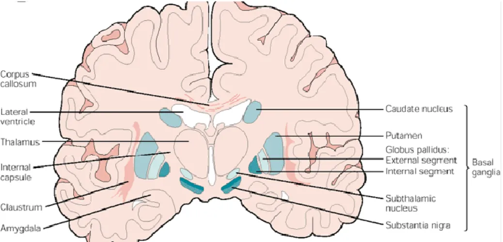
particular region in the brain, which is the striatum, composed in human by the caudate
nucleus and putamen (Figure 2). The striatum play a key role in a group of nuclei called the
basal ganglia.
Basal ganglia consist of four nuclei (Figure 2), portions of which play a major role in normal
voluntary movements. Motor functions of basal ganglia are in large part mediated by motor
areas of frontal cortex (
Wichmann & Delong 2007
).
Figure 2 Representation of human brain coronal section showing basal ganglia (in blue) in relation to surrounding structures (adapted from Nieuwenhuys et al 1981) (Wichmann & Delong 2007).
The striatum is the primary site of HD pathology (
Albin et al 1992, de la Monte et al 1988
),
accompanied by dilatation of the anterior horns of the lateral ventricles, which are obvious on
MRI in advanced cases. Indeed, Hobbs and colleagues measured at onset of HD patient
caudate volume was 2.58 mL smaller than in controls; ventricular volume was 9.27 mL larger.
They showed that differences in caudate and ventricular volumes between patients with HD
and controls were respectively evident 14 years and 5 years, before motor onset (
Hobbs et al
2010, Tabrizi et al 2009
) (Track-HD) (Figure 3A).
The output structures of the striatum, substantia nigra reticulata and globus pallidus (externa
and internal segments) are also altered in symptomatic patients. The nucleus accumbens is
relatively preserved.
Cortical atrophy is also present in advanced cases and detectable thanks to generation of
magnitude map (
Rosas et al 2011, Tabrizi et al 2009
) (Figure 3B).
Page | 21
Figure 3 Whole brain and regional atrophy in the controls, premanifest, and early Huntington’s disease patients (Tabrizi et al 2009).
Figure 3A, 3T volumetric MRI scan in a 50-year-old control, a 55-year-old individual with preHD, and a 49-year-old with early Huntington’s disease. Brain volumes are corrected for intracranial volume. B, Cortical thinning in the Huntington’s disease groups compared with controls. The top panel shows statistical maps corrected with the false discovery rate; magnitude maps are shown below. All results are adjusted for age and sex. ICV=intracranial volume. LH=left hemisphere. RH=right hemisphere.
Interestingly, within the striatal neuronal population, different degrees of degeneration can be
observed (
Ferrante et al 1997, Ferrante et al 1985
). GABAergic medium-sized spiny neurons
(MSN) are found to preferentially degenerate in HD, occurring first in the caudate and then
the putamen (
Rosas et al 2001, Vonsattel & DiFiglia 1998
). Because these neurons represent
95% of the neurons in the striatum, their loss are likely responsible for the atrophy of the
structure.
In contrast, medium-sized aspiny interneurons containing somatostatin, neuropeptide Y, or
NADPH diaphorase, as well as the large cholinergic interneurons which both represent
approximately 5% of striatal neurons are relatively spared. However, the extreme striatal
atrophy and the loss of neurons observed in grade 4 indicate that both spiny and aspiny
neurons are vulnerable at the end stage of the disease (
Reiner et al 1988
).
A
Page | 22
Early neuropathological studies showed that in advanced cases of HD, the cerebral cortex
(particularly layers III, V, and VI), globus pallidus, thalamus, subthalamic nucleus, substantia
nigra, white matter, and the cerebellum could be markedly affected (
Vonsattel & DiFiglia
1998
). Neuropathological characterization of HD brains also showed that the hypothalamus is
probably damaged at very early stages in HD patients (
Kremer et al 1990
). It has been
confirmed using PET scan and MRI imaging [for example
(Douaud et al 2006, Politis et al
2008)
].
In addition, diffusion tensor imaging (DTI) has demonstrated pathology of the white matter in
pre- and early symptomatic patients
(Rosas et al 2006
).
1.2.5. Striatal neurotransmission
Medium-sized spiny neurons constitute 95% of all striatal neurons and utilize
gamma-aminobutyric acid (GABA) as their principal neurotransmitters (
Schaefer et al 2012
). The
dorsal striatum receives parallel sets of diffuse glutamatergic inputs from almost all
neocortical areas and several thalamic nuclei (
Tepper et al 2008
). These inputs primarily
synapse onto spines of MSN (
Leavitt et al 2006
). The striatum also contains a number of
modulatory components including dopamine (DA) projections from the substantia nigra pars
compacta (SNc) (
Raymond et al 2011
) and cholinergic or GABAergic inputs from striatal
interneurons (
Tepper et al 2008
). These elements constitute the basic striatal microcircuit (see
as review (
Raymond et al 2011
)).
Striatal output is largely segregated into two populations of MSN with distinct projections, as
well as DA receptor and neuropeptide expression, constituting the direct and indirect
pathways (
Calabresi et al 2014, Gerfen et al 1990, Hallett 1993, Kawaguchi et al 1995
)
(Figure 4).
In the physiological condition, the direct pathway (red lines, Figure 4) consists of MSN that
predominantly express D1 DA receptors, substance P and dynorphin, and project to the
substantia nigra (SN) and the internal segment of the globus pallidus (GPi).
The indirect pathway (blue lines, Figure 4) is comprised of MSN that express predominantly
D2 DA receptors, enkephalin or neurotensin, and project to the external segment of the globus
pallidus (Gpe).
Page | 23
Figure 4 Schematic representation of the direct (red lines) and indirect (blue lines) pathway classical model in the physiological condition (Calabresi et al 2014).
Abbreviations: DA, dopamine; STN, subthalamic nucleus; SNpc, substantia nigra pars compacta; SNpr, substantia nigra pars reticulata; GPi, globus pallidus pars interna; GPe, globus pallidus pars externa.
DA arising from the SNpc is thought to activate D1-expressing striatal MSN of the direct
pathway and to inhibit D2-expressing striatal neurons of the indirect pathway. The output
nuclei GPi and SNpr project to the thalamus, which in turn send efferents that complete the
cortico-basal ganglia-thalamo-cortical loop.
Two of the most important disorders affecting the basal ganglia are Parkinson’s disease (PD)
and HD. Several studies compared the ravages of this brain structure on the two diseases,
giving an overview on the basal ganglia circuitry and its main pathologies (
Albin et al 1989,
Alexi et al 2000, Fernandez-Ruiz 2009, Fernandez-Ruiz et al 2011
).
PD is also a progressive neurodegenerative disorder whose origin has been associated with
environmental insults, genetic susceptibility or interactions between both causes (
Schapira et
al 2014
). The major clinical symptoms in PD are tremor, bradykinesia, postural instability and
rigidity, symptoms that mainly result from the severe dopaminergic denervation of the
striatum caused by the progressive death of dopaminergic neurons of the SNc (
Clabough &
Zeitlin 2006, Obeso et al 2014
).
HD pathology is associated with abnormal neurotransmitter regulation and synaptic
communication, as well as glutamate-mediated excitotoxicity (
Chen et al 1999, Sepers &
Raymond 2014, Taylor-Robinson et al 1994
).
Page | 24
It has been suggested that one early event in HD is the preferential degeneration of the GABA
(met-enkephalin) neurons of the indirect pathway (producing the arrest of inhibition of the
pallidum), so that an imbalance between the indirect and directly pathways would lead to an
abnormal activation of thalamic motor neurons, in turn abnormally activating cortical motor
neurons, which could explain the hyperkinetic symptoms in HD, in contrast to PD where the
repression of thalamic nuclei leads to loss of activation of cortical neurons and the
hypo/akinetic movements. However, this basal ganglia neurophysiology model of motor
symptoms in HD first suggested by Albin (
Albin et al 1989
) has been debated and did not
help to design efficacious therapy.
Another important part of the imbalanced neurotransmission in HD is the compensatory
mechanism occurring over time that attempts to stabilize this system, well described by
Levine and colleagues (
Andre et al 2010, Raymond et al 2011
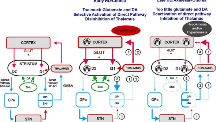
) (Figure 5).
Figure 5 Glutamate and DA in basal ganglia function in intact brain and during HD (André et al 2010).
Left is a simplified schematic showing normal basal ganglia circuitry. Red: glutamate inputs; blue: GABA; green: DA.
Abbreviations: Enk: enkephalin; GLUT: glutamate; GPe: external segment of the globus pallidus; GPi: internal segment of the globus pallidus; SNc: substantia nigra pars compacta; SNr: substantia nigra pars reticulata; STN: subthalamic nucleus.
Under normal conditions (Left panel Figure 5), the thalamus is inhibited by GPi and SNr
GABA projections.
Page | 25
In early HD (Middle panel Figure 5), cortical dysfunction induces excessive glutamate
release in the striatum. Abnormally elevated striatal glutamate and DA levels trigger selective
dysfunction of enkephalin-containing MSN.
Imbalance between the direct and indirect pathways induces hyperkinesia via two pathways:
1) Selective dysfunction/degeneration (dashed lines Figure 5) of
enkephalin-containing MSN leads to decreased release of GABA in the GPe and to its disinhibition. In
turn, overactivation of GABA neurons of the GPe leads to increased release of GABA and to
inhibition of STN which decreases glutamate release and decreases activity of the GPi and
SNr.
2) Overactivation of the direct pathway MSN (by abnormal DA modulation and/or
excessive glutamate) leads to increased release of GABA and to inhibition of GPi and SNr.
Together, alterations in direct and indirect pathways in early HD induce inhibition of GPi and
SNr GABA neurons and to a decreased release of GABA in their output structure, the
thalamus. Disinhibition of the thalamus is responsible for abnormal movements.
In late HD (Right panel Figure 5), corticostriatal and nigrostriatal inputs progressively
degenerate, leading to decreased striatal glutamate and DA release. Low striatal glutamate and
DA levels trigger dysfunction of both direct and indirect pathway MSN.
Imbalance between the direct and indirect pathways induces hyperkinesia and hypokinesia via
two pathways:
1) Alterations in the indirect pathway are similar to early HD and lead to hyperkinetic
movements.
3) Dysfunction/degeneration of direct pathway MSN induces decreased release of
GABA and disinhibition of GPi and SNr. Increased activity in GPi and SNr leads to inhibition
of the thalamus and hypokinesia.
Depending on the stage of dysfunction of direct and indirect pathway MSN, activity in the
basal ganglia could result in hypokinesia or hyperkinesia and would explain why some
symptomatic patients display both chorea and akinesia.
Levine group review evidences that the evolution of neurotransmission alteration in the
striatum and cortex are bi-phasic, with changes in excitation and inhibition in early and late
HD for cortex and striatum compared to healthy situation (Figure 5). In other words, HD is
associated with complex dysfunctions within the cortex and basal ganglia that involve
glutamate and dopamine neurotransmissions. Normally these two systems interact such that
dopamine modulates glutamate-mediated transmission. In HD, this modulation is altered in
complex ways by the initial overactivation of glutamate function which induces concomitant
dysregulation of dopaminergic function. Thus, the normal balance between these systems is
upset.
Page | 26
1.2.6. Cause of the disease
The highest concentration of HD sufferers was found in a tiny and isolated community around
Venezuela’s Lake Maracaibo. Starting from 1979, a group of geneticists and physicians kept
medical records, took blood and skin samples, and charted the transmission of the disease
within families of this community. This allowed to narrow the genetic defect responsible for
HD on the chromosome 4 (
Gusella et al 1983
). Scientists from all over the world joined
together into a team that was collectively named the Huntington Disease Collaborative
Research Group that, after 14 years reported the discovery of the IT15 gene responsible for
HD and of its associated mutation (
HDCRG 1993
). The first exon of the IT15 gene contains a
repetitive DNA element consisting of three nucleotides: C (cytosine), A (adenine), and G
(guanine). When researchers examined this region of IT15 in non-HD controls, they found
that the number of CAG repeats varied from 6 to 35. Analysis of the same region in the IT15
gene in individuals with HD showed that they always had 40 or more CAG repeats (
HDCRG
1993
).
The length of the CAG repeat accounts for 50 to 70% of the variability in age at clinical onset,
whereby individuals with longer repeat lengths commonly have an earlier onset than those
with shorter repeat lengths. When considering hundreds of HD patients, the number of CAG
repeats negatively correlates with the onset of symptoms. Long expansion can lead to juvenile
forms of HD, while short expansion can lead to relatively late onset disease. However, at the
individual level, the number of CAG repeats cannot predict the age of onset.
It was concluded that the CAG repeat expansion in the IT15 gene was responsible for HD.
The IT15 gene is now renamed the huntingtin (HTT) gene because of the name assigned to
the protein. HTT gene is composed by 67 exons and codes for 3144 amino acids (~350kDa).
It is found in metazoans, with the highest degree of conservation among vertebrates
(
Clabough 2013
).
Normal Htt has been found to play a major key role in most if not all cells, especially neurons.
In HD, the mutation results in an elongated stretch of glutamine near the NH2 terminus of the
protein (
Andrew et al 1993, Hannan 1996, Ratovitski et al 2007, Trottier et al 1994
) (Figure
Page | 27
Figure 6 Consequences of the number of CAG repeats in htt gene and elongated stretch of glutamine in Htt protein on neuron survival.
This mutation in Htt protein is thought to confer to the protein a gain of function that is toxic
to neurons (Figure 6). In addition, the disease is likely associated to a loss of function of
normal Htt that would be detrimental to neurons.
1.2.6.1.Normal functions of wt-Htt protein
Htt protein is ubiquitously expressed with a highest expression level in the brain compared to
peripheral nervous system (
Moffitt et al 2009, Vonsattel & DiFiglia 1998
)
.
It is present in neurons, but also in glia cells (
Ross & Tabrizi 2011, Zuccato & Cattaneo 2007
)
and immune cells (
Li & DiFiglia 2012
).
It exists evidences that cell–cell interaction plays an important role in HD pathogenesis (
Ross
& Tabrizi 2011, Zuccato & Cattaneo 2007
). In the brain, glia-neuron interactions are
important for maintaining the normal function of neurons, but in this chapter I will focus on
neurons dysfunction in HD pathology.
At the cellular level, Htt is widely expressed, predominantly in the cytoplasm, but can also be
found in the nucleus (Figure 7), playing roles in transcriptional regulation (
Kegel et al 2002,
Xia et al 2003
), which has been studied in depth by Luthi-Carter and their colleagues over
these past 15 years (more details in paragraph 1.2.6.3.E
Transcriptional deregulation
).
The subcellular localization of Htt is complex and dynamic, and may change conformation
depending on its compartmental localization.
Page | 28
Htt normally localizes along microtubules and participates in the transport of a variety of
cargo, including mRNAs, proteins, vesicles, and organelles, such as mitochondria (
DiFiglia et
al 1995, Kegel et al 2000, Kegel et al 2009, Schulte & Littleton 2011
) (Figure 7).
Notably, Htt is present in excitatory synapses where it associates with synaptic vesicles in the
presynaptic terminal and facilitates neurotransmitter release (
DiFiglia et al 1995, Jeong et al
2009
). In the postsynaptic density, Htt is associated with the postsynaptic scaffolding protein
PSD95 (Figure 7) (
Sun et al 2001
).
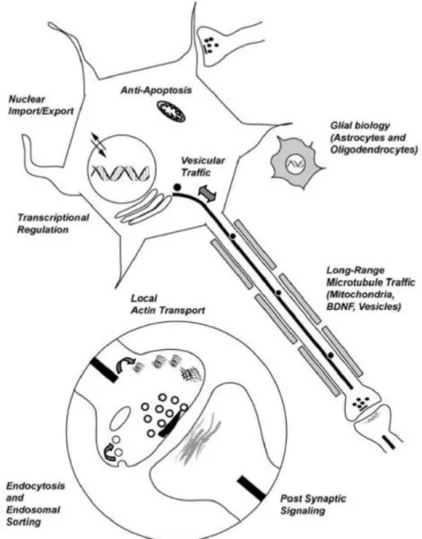
Figure 7 Schematic illustrating the biological functions of wt-Htt (Schulte & Littleton 2011).
The Figure 7 shows a generic neuron with an unsheathed axon (grey boxes represent
oligodendrocyte wrapping) and an astrocyte (grey stellate shape). Enlarged circle is a
magnified view of a synapse. The Htt protein has been suggested to regulate both neuronal
and glial function. Within neurons, Htt has been implicated in nuclear import and
transcriptional regulation. In addition, Htt regulates apoptotic signaling and axonal transport.
At the synapse, Htt has been suggested to have both pre- and post-synaptic roles.
Page | 29
Wt-Htt has multiple effects in development and neuronal physiology, including regulation of
transcription (
Nucifora et al 2001
), membrane dynamics (
Kegel et al 2009
), mitochondrial
efficiency (
Aoyama et al 2006
), brain-derived neurotrophic factor (BDNF) transcription
(
Zuccato et al 2001
), autophagy (
Cortes & La Spada 2014, Jeong et al 2009, Kegel et al 2000
,
Qin et al 2003
), and endosomal recycling (
Li & DiFiglia 2012
) (Figure 7).
Several studies reported multiple physiological functions that are particularly important for
the brain neurons affected in HD.
Htt is required for normal excitatory synapse development in cortical and striatal circuits
(
Margulis & Finkbeiner 2014
). More globally, it has been shown to be essential for
embryonic development, as the knockout of the gene in mice causes embryonic death at the
pre-gastrulation stage (
Muhlau et al 2012
), and in conditional knockout mice (Cre Lox KO
under the CamKII promoter) late disruption of the gene in adult leads to progressive
neurodegeneration (
Dragatsis et al 2000
). The antiapoptotic role of Htt was also supported by
Michael Hayden’s group. They found that primary striatal neurons from YAC18 transgenic
mice (for details on animal models of HD, see paragraph 1.2.7. Animal models of HD)
overexpressing full-length wild-type human huntingtin were protected from apoptosis
compared with cultured striatal neurons from nontransgenic littermates and YAC72 mice
expressing mutant human huntingtin (
Leavitt et al 2006
).
It is known that increased wt-Htt expression improves brain cell survival by modulating
caspase-3 and 9 apoptosis signaling. Another way huntingtin produces antiapoptotic effects is
through regulation of the neurotrophic factor BDNF. Indeed, wt-Htt regulates BDNF gene
transcription, downregulated in HD (
Rigamonti et al 2000, Rigamonti et al 2001, Zuccato et
al 2001, Zuccato et al 2003
). More particularly, Htt levels modulate the sensitivity of neurons
to apoptotic death elicited by N-methyl-D-aspartate (NMDA) receptor-mediated
excitotoxicity (
Leavitt et al 2006
), where wt-Htt can decrease cellular toxicity of mHtt in
vivo.
On the contrary, cells depleted of wt-Htt were more sensitive to apoptotic cell death and
showed increased level of caspase-3 activity, with respect to control cells (
Zhang e
t
al 2008
).
Wild-type Htt has beneficial activities in the development and mature brain. It is therefore
possible that its loss in human HD reduces the ability of neurons to survive and to counteract
the toxic effects of the mutant protein.
1.2.6.2.Loss of function of wt-Htt and toxic gain of function of mHtt
Indeed, this single mutation in Htt is the triggering event that endows the protein with new
toxic functions that are deleterious for brain cells. At the same time, it also impairs the ability
of normal Htt protein to exert molecular activities that are fundamental for the survival and
functioning of the neurons that predominantly degenerate in the disease (
Bjorkqvist et al
2005, Schulte & Littleton 2011
).
Page | 30
Figure 8 Schematic diagram of the huntingtin amino acid sequence (Zuccato et al 2010).
Details:(Q)n is polyglutamine tract, followed by the polyproline sequence (P)n; red emptied rectangles indicate the three main groups of HEAT repeats (HEAT group 1, 2, 3). The small green rectangles indicate the caspase cleavage sites and their amino acid position (513, 552, 586), while the small pink triangles indicate the calpain cleavage sites and their amino acid positions (469, 536). Boxes in yellow: B, regions cleaved preferentially in the cerebral cortex; C, regions of the protein cleaved mainly in the striatum; A, regions cleaved in both. Post-translational modifications: ubiquitination (UBI) and/or sumoylation (SUMO) sites (green); palmitoylation site (orange); phosphorylation at serines 13, 16, 421, and 434 (blue); acetylation at lysine 444 (yellow). NES is the nuclear export signal while NLS is the nuclear localization signal. The nuclear pore protein translocated promoter region (TPR, azure) is necessary for nuclear export. Htt, huntingtin; ER, endoplasmic reticulum.
Thanks to its polar zipper structure (
Perutz 1995
) and its diverse configurations, i.e. alpha
helix, random coil, and extended loop (Ratovitski et al 2007), this polyQ domain may be
important for modulation of wt-Htt function, longevity and energy status of cells (
Clabough &
Zeitlin 2006
), and for stabilization of protein interactions (
Schaefer et al 2012
), but wouldn’t
be essential for normal function of Htt.
Htt is also enriched in consensus sequences called huntingtin elongation factor 3, protein
phosphatase 2A, and TOR 1 (HEAT) repeats that are organized into protein domains
important for protein-protein interactions (Figure 8). Htt protein also goes through
post-translational modifications and presents consensus sites for proteolytic enzymes (Figure 8)
(
Ratovitski et al 2007
).
Page | 31
Functionally active nuclear export signal (NES) sequence and a less active nuclear
localization signal (NLS) are present in Htt (Figure 8), which indicate that the protein is
involved in transporting molecules from the nucleus to the cytoplasm (
Xia et al 2003
).
The first 17 N-terminal amino acids of huntingtin revealed that the sequence forms an
amphipathic-helical membrane-binding domain that is required and is sufficient for Htt
association to mitochondria and for its colocalization with Golgi and endoplasmic reticulum
(ER) (Figure 8) (
Atwal et al 2007, Rockabrand et al 2007
). This sequence also enhances the
formation of visible aggregates.
Post- translational modifications of Htt protein are various: acetylation, phosphorylation,
SUMOylation, ubiquitination, palmitoylation and proteolysis (Figure 8). These processes are
important for modulation of its interactor affinity, subcellular localization, proteolysis or
degradation (
Ehrnhoefer et al 2011
).
These characteristics make Htt indispensable for the proper functioning of cells.
Thus, it appears that Htt has crucial roles in cells, potentially via different domains, and that
loss of the antiapoptotic functions of Htt may contribute to HD (Figures 7, 8 & 9).
Page | 32
Processing of mHtt generates an amino-terminal fragment that translocates into the nucleus
and a C-terminal portion that remains in the cytoplasm. Some of the full-length protein might
also move into the nucleus. The generation of amino-terminal fragments coincides with
increased toxic activity in cells. At the same time, extension of the CAG would cause a loss of
function in the mutant protein (Figure 9a) and/or the mutant protein could act negatively on
the functions of the normal one (Figure 9b). Finally, loss of Htt function might result as a
consequence of decreased protein levels and stability.
Figure 9 Potential mechanism of cell death in HD (Cattaneo et al 2001).
As such HD may be viewed as a double disease (Figure 9), that is caused by both a new toxic
property of mHtt and by a loss of the neuroprotective activity of wt-Htt (see as review
(
Cattaneo et al 2001
)).
Overall, these combined effects alter several cellular processes, such as protein degradation,
mitochondrial respiration and transcription among many others, leading to neuronal
dysfunction and cell loss.
Page | 33
1.2.6.3.Molecular Mechanisms in HD
Figure 10 Key cellular pathogenic mechanisms in Huntington’s disease HD (Zuccato et al 2010).