HAL Id: hal-02945049
https://hal.archives-ouvertes.fr/hal-02945049
Submitted on 22 Sep 2020HAL is a multi-disciplinary open access
archive for the deposit and dissemination of sci-entific research documents, whether they are pub-lished or not. The documents may come from teaching and research institutions in France or abroad, or from public or private research centers.
L’archive ouverte pluridisciplinaire HAL, est destinée au dépôt et à la diffusion de documents scientifiques de niveau recherche, publiés ou non, émanant des établissements d’enseignement et de recherche français ou étrangers, des laboratoires publics ou privés.
Monitoring of XRN4 targets reveals the importance of
co-translational decay during Arabidopsis development
Marie-Christine Carpentier, Jean-Marc Deragon, Viviane Jean, Seng Hour
Vichet Be, Cecile Bousquet-Antonelli, Rémy Merret
To cite this version:
Marie-Christine Carpentier, Jean-Marc Deragon, Viviane Jean, Seng Hour Vichet Be, Cecile Bousquet-Antonelli, et al.. Monitoring of XRN4 targets reveals the importance of co-translational decay during Arabidopsis development. Plant Physiology, American Society of Plant Biologists, 2020, pp.00942.2020. �10.1104/pp.20.00942�. �hal-02945049�
Short title: Co-translational decay dynamics across seedling development 1
2
Corresponding author: Rémy MERRET, email : remy.merret@univ-perp.fr, CNRS-LGDP
3
UMR 5096, 58 av. Paul Alduy 66860 Perpignan, France
4 5
Article title: Monitoring of XRN4 targets reveals the importance of co-translational decay 6
across Arabidopsis development
7 8
Authors: Marie-Christine CARPENTIER1,2, Jean-Marc DERAGON1,2,3 , Viviane JEAN1,2,
9
Seng Hour Vichet BE1,2, Cécile BOUSQUET-ANTONELLI1,2, Rémy MERRET1,2
10 11
1
CNRS-LGDP UMR 5096, 58 avenue Paul Alduy 66860 Perpignan, France
12 2
Université de Perpignan Via Domitia, LGDP-UMR5096, 58 avenue Paul Alduy, 66860
13
Perpignan, France
14 3
IUF, Institut Universitaire de France, 75231 Paris Cedex 05, France
15 16
One sentence summary: 17
Co-translational decay mediated by XRN4 is dynamically regulated across Arabidopsis
18
seedling development and fine-tunes translation efficiency.
19 20
List of authors contributions 21
R.M. designed and supervised the work. R.M conducted all the experiments present in this
22
study. M.-C.C. conducted all the bioinformatic analysis. R.M. performed seedlings sampling
23
with the help of V.J. R.M. performed western blotting with the help of S.H.V.B. R.M. wrote
24
the manuscript, with the help of C.B.A and J.M.D.
25 26
Funding 27
This work was supported by the "Laboratoires d’Excellences (LABEX)" TULIP (ANR-10-28
LABX-41) to RM, by the “IUF Institut Universitaire Français” to JMD, by the ANR
29
3’modRN (ANR-15-CE12-0008) to CBA. 30
31
Email address of author for contact: remy.merret@univ-perp.fr
32 33
ABSTRACT 34
35
RNA turnover is a general process necessary to regulate proper mRNA amount at
post-36
transcriptional level. Although long thought to be antagonistic to translation, the discovery of
37
the 5’-3’ co-translational mRNA decay demonstrated that both pathways can also be 38
intertwined. This pathway globally shapes the transcriptome in different organisms and in
39
response to stress. However, until now, the dynamic of this process in a growing organism
40
was never assessed. In this study, we ran a multi-omic approach to reveal the global landscape
41
of co-translational mRNA decay across Arabidopsis seedling development. We demonstrated
42
that co-translational decay could be regulated by developmental cues. Using xrn4-5 mutant,
43
we demonstrated that XRN4 polyA+ mRNA targets are mainly co-translational decay targets
44
across development. As this pathway is tightly connected with translation, we also assessed
45
its role in the control of translation efficiency. We discovered that clusters of transcripts could
46
be specifically targeted by this pathway in a development-dependant manner to modulate their
47
translation efficiency. Our approach allows the determination of a co-translational decay
48
efficiency that could be an alternative to other methods to assess transcript translation
49
efficiency. Thus, our results demonstrate the prevalence of co-translational mRNA decay in
50
plant development and its role in translational control.
INTRODUCTION 52
53
Over its entire lifetime, any mature cytoplasmic mRNA is in balance between translation,
54
storage and decay. This equilibrium maintains the proper dynamic of gene expression and is
55
crucial to control mRNA homeostasis. Although long thought to be mutually exclusive, there
56
is now a large body of evidence supporting that, in eukaryotes, mRNA translation and decay
57
are interconnected (Heck and Wilusz, 2018). The impact of codon optimality on mRNA
half-58
life is a clear example of this relationship. Codon optimality is defined as the ribosome
59
decoding efficiency according to tRNAs availability. Genome-wide analyses revealed that
60
yeast mRNAs enriched in optimal codons present high ribosome density and are more stable
61
than mRNAs enriched in non-optimal codons (Presnyak et al., 2015). The finding that codon
62
optimality is a key cis determinant of transcript stability places the ribosome as a core
63
component linking translation elongation to mRNA degradation.
64 65
The most relevant interplay between translation and decay is the so called 5’-3’
co-66
translational decay pathway where mRNAs are turned over while still engaged in polysomes
67
and actively translated. This pathway first described in the yeast Saccharomyces cerevisiae,
68
using reporter genes (Hu et al., 2009) was later on found to globally shape the polyadenylated
69
transcriptome in yeast, mammalian cells and Arabidopsis thaliana (Pelechano et al., 2015; Yu
70
et al., 2016; Tuck et al., 2020). For these mRNAs, decapping occurs on polysomes, allowing
71
the 5'-3' exoribonuclease XRN1/4 to chase the last translating ribosome. The genome-wide
72
effect of co-translational decay can be revealed by sequencing of RNA decay intermediates
73
using high-throughput "degradome" approaches. All of them are based on the capture of 5’
74
monophosphate decay intermediates including PARE (Parallel Analysis of RNA Ends,
75
German et al., 2008), 5P-seq (5’P sequencing, Pelechano et al., 2015), Degradome-seq
76
(Addo-Quaye et al., 2008) or GMUCT (Genome-wide Mapping of UnCapped Transcripts,
77
Willmann et al., 2014). These approaches reveal that mRNA decay intermediates follow an
78
XRN1/4-dependant, 3-nucleotide periodicity. This periodicity can be explained by the fact
79
that XRN1/4 follows the last translating ribosome codon per codon, and since it is a
80
processive enzyme, only degradation intermediates protected by ribosomes can be captured.
81
Consequently, all these degradome approaches can not only give a snap-shot of
co-82
translational mRNA degradation, but also reveal ribosome dynamics (Pelechano et al., 2015)
83
and how degradation impacts this dynamic. As an example, in yeast, 5P-seq allowed the
84
identification of general translation termination pauses, and novel codon-specific pausing
sites were detected, such as that at the rare proline codon CCG and at the arginine CGA codon
86
(Pelechano et al., 2015). These additional pausings are explained by the lowest availability of
87
corresponding tRNAs resulting in the slowing-down of the ribosome at these sites.
88
Interestingly, these additional pausings were not detected in Arabidopsis flowers suggesting
89
different ribosome dynamics in plants (Yu et al., 2016). Metagene degradome analyses also
90
revealed 5’P reads accumulation 17 nucleotides upstream of stop codons (UAA, UAG and
91
UGA). This distance corresponds exactly to a ribosome stalled at the A site. As termination
92
step is slower than elongation, general 5’P reads accumulation can be revealed 17 nucleotides 93
upstream of stop codons and can be used as a proxy of the transcriptome-wide co-translational
94
decay activity. In addition, these approaches also unveil small RNA-guided cleavage sites
95
(Franke et al., 2018), RNA-binding protein footprints (Hou et al., 2014), endonucleolytic
96
cleavage sites (Anderson et al., 2018), exon junction complex footprints (Lee et al., 2020) and
97
ribosome stalling sites (Hou et al., 2016). For example, degradome analysis in Arabidopsis
98
revealed that AGO7 can bind to a noncleavable miR390 target site on the TAS3 transcript
99
leading to a ribosome stalling situation that was suggested to control translation of the TAS3
100
gene (Hou et al., 2016). Ribosome stalling at uORFs (upstream Open Reading Frame) is
101
another layer of translational control that can be detected by degradome data. For example,
102
the stalling of 3 ribosomes can be detected on uORFs controlling Arabidopsis BZIP3 main
103
ORF translation (Hou et al., 2016).
104 105
At the physiological level, co-translational decay was shown to play important roles in the
106
response to various stresses. In Arabidopsis, heat stress triggers 5'-ribosome pausing, the over
107
accumulation of XRN4 in polysomes and the 5'-3’ co-translational decay of around 1500
108
transcripts which code for proteins with hydrophobic N-termini (Merret et al., 2013, Merret et
109
al., 2015). Recently, following stress, the yeast Lsm1-7/Pat1 complex was shown to trigger
110
the co-translational decay of stress induced mRNAs limiting their translation and preventing
111
an hyper-response (Garre et al., 2018). Consequently, defect in this pathway triggers the
112
misregulation of translation inhibition under osmotic stress, which correlates with an
113
abnormally high association of stress-induced mRNAs to active polysomes (Garre et al.,
114
2018). Recently, this pathway was also proposed to be important for tubulin autoregulation
115
(Lin et al., 2020). TTC5, a tetratricopeptide protein, recognizes tubulin nascent peptide and
116
triggers the co-translational decay of its own transcripts to maintain proper tubulin
117
homeostasis. TTC5 mutants abolished tubulin autoregulation and display chromosome
118
segregation defects during mitosis. Taken together, these different examples support that
translational decay plays important roles in translation regulation under normal conditions and
120
in response to different stresses. Nonetheless, if and how the co-translational decay process
121
reshapes the transcriptomes and proteomes of cells across organism development remains
122
unknown so far.
123 124
Transcripts can also be turned over through the cytosolic 5’ to 3’ decay pathway which takes
125
place on ribosome free mRNAs. This so-called "general" 5' to 3' cytosolic mRNA turnover
126
process takes place in three steps. Following poly(A) tail shortening (deadenylation), the
127
VCS/DCP1/DCP2 decapping complex hydrolyses the mRNA cap structure. Then, the
5'-128
phosphate end of the decapped mRNA is attacked by the XRN1/4 exoribonuclease, which
129
digests the body of the transcript. In Arabidopsis, XRN4 can target both deadenylated and
130
polyadenylated fractions suggesting that part of the degradation could be deadenylation
131
independent (Nagarajan et al., 2019). For co-translational mRNA decay, the importance of
132
deadenylation is still unclear.
133 134
In Arabidopsis, at the whole organism level, the loss of XRN4 has minimal effects under
135
normal growth conditions. Growth deficiencies were only reported in response to hormones
136
or under stress conditions. Loss-of-function mutants of XRN4 are insensitive to ethylene
137
(Potuschak et al., 2006) and hyper-sensitive to auxin and abscisic acid treatments (Wawer et
138
al., 2018; Windels and Bucher, 2018). Recently, an xrn4 mutant was found to be defective in
139
the dark stress response and during nitrogen supply (Nagarajan et al., 2019). However,
140
whether these deficiencies result from the failure of the general cytosolic or of the
co-141
translational mRNA decay pathways (or a combination of both) is mainly unknown. Only one
142
study distinguished both pathways and reported the exact role of co-translational decay in
143
Arabidopsis heat stress response by analysing pools of mRNAs associated with ribosomes
144
(Merret et al., 2015).
145 146
In the present study, we monitored the impact of co-translational decay across Arabidopsis
147
seedling development. To do so, we assessed the genome-wide impact of XRN4
loss-of-148
function on polyA+ mRNAs at the total, polysome and degradome levels. Through these
149
approaches, we provide evidence that XRN4 mostly catalyzes polyadenylated mRNA
150
degradation in polysomes. We also found that co-translational decay is dynamically
151
modulated across development and can influence transcript translation efficiency, unveiling
152
the importance of co-translational decay across plant development.
RESULTS 154
155
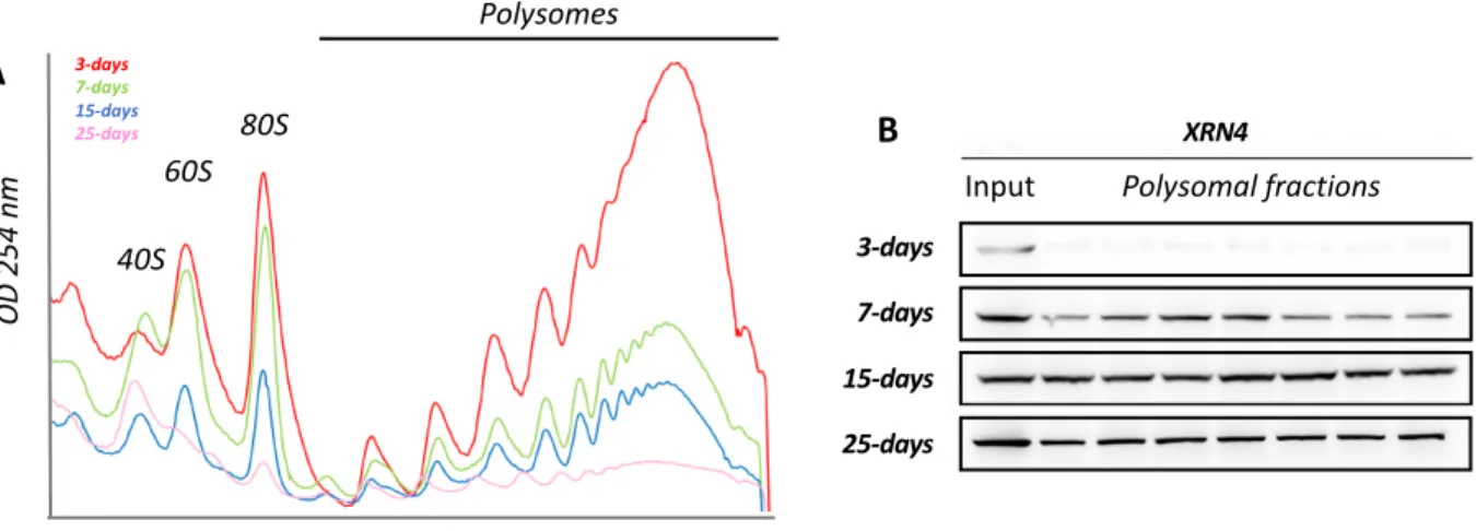
XRN4 differentially accumulates in polysomes across seedling development 156
To explore the hypothesis that translation and co-translational decay are regulated in response
157
to developmental cues, we analyzed two read-outs (Figure 1). Firstly, the global translation
158
activity was assessed by polysome quantification through sucrose density gradient on four
159
developmental stages from 3 to 25-d old seedlings (Figure 1A). Considering that the cellular
160
activity and hence the ribosome load per cell are most likely to be significantly different
161
between developmental stages, we compared polysome contents from identical quantities of
162
biomass rather than identical quantities of total RNA. Indeed, we observed that the polysome
163
content is inversely correlated with the seedling age with the highest levels at 3 days, and a
164
progressive decrease to reach a minimum at 25 days. Secondly, considering that XRN4 is the
165
proposed main catalyzer of 5'-3' co-translational degradation (Merret et al., 2013; Yu et al.,
166
2016), we used as read-out of this turnover activity, its accumulation in polysomes. We hence
167
ran western blotting on input and polysomal fractions using XRN4 specific antibodies (Figure
168
1B). Although, detected at similar levels in all input fractions, XRN4 accumulation in
169
polysomes differs across development. At 3-d-old, XRN4 is mostly absent from polysomes
170
and progressively increases to reach a maximum at the 15-d-old stage a level globally
171
maintained to 25-days (Figure 1B). These results suggest that co-translational decay activity
172
could be regulated across seedling development.
173 174
Co-translational decay efficiency is regulated across seedling development 175
To identify XRN4 targets and get a deeper understanding of the dynamics of co-translational
176
decay, we ran a multi-omic approach (Supplemental Figure 1). For each developmental stage,
177
Col0 (wild type) and xrn4-5 loss-of-function (SAIL_681_E01) seedlings were harvested in
178
two biological replicates. As the loss of XRN4 has minimal effects under normal growth
179
conditions, the observed differences between wild-type and mutant will be a direct
180
consequence of the loss of XRN4 rather than a growth/developmental delay.
181 182
Each sample was used to purify: (i) total RNAs and (ii) RNAs associated to polysomes. Total
183
RNAs were used on the one hand to run a polyA+ RNA sequencing and on the other a
184
degradome analysis through a GMUCT/RNA-seq approach. Briefly, GMUCT (also tagged as
185
5’P-seq) consists through the ligation of a 5’-RNA adapter in the capture and sequencing of 186
polyA+ mRNA molecules that carry a 5’-monophosphate. This also permits to counter select
mRNAs with a 5’-cap structure. Hence GMUCT allows to specifically sequence the 188
population of mRNA molecules (full length and decay intermediates) that are in the course of
189
being exonucleolytically degraded from their 5'-end following a decapping step or an
190
endonucleolytic cleavage. Considering the very high processivity of 5'-exoribonucleases, only
191
mRNAs with features slowing down the progression of the decay enzyme can be captured.
192
Hence, GMUCT mostly to monitor the 5'-co-translational decay process, where the
193
exoribonuclease digests the transcript chasing the elongating ribosomes. Polysomal RNAs
194
were also purified through sucrose density gradient and sequenced following purification of
195
the polyadenylated fraction (Supplemental Figure 1).
196 197
The total polyA+ RNA-seq allows the capture of capped mRNAs free of ribosome, capped
198
translated mRNAs and uncapped co-translational decay intermediates. The polysome polyA+
199
RNA-seq allows the capture of both capped translating mRNAs and uncapped co-translational
200
decay intermediates. The GMUCT approach captures uncapped co-translational decay
201
intermediates (Supplemental Figure 1) hence permitting to monitor the cell 5’-degradome.
202
For sequencing approach, the sequence of the biological repeats of each genotype displays a
203
high reproducibility (R2>0.94, Supplemental Table 1). Only transcripts with at least 1
204
RPKM/1RPM value in at least 1 library wild-type were kept for further analysis, leaving a
205
total of 23,196 transcripts. Fold changes (FC) between xrn4-5 and Col0 were calculated for
206
each transcript at each developmental stage at total and polysome RNA levels (Figure 2,
207
Supplemental Figure 2A-B). Using the DESeq2 pipeline (Love et al., 2014) and cut-off FCs
208
above 2 or below 0.5, we identified mRNAs that differentially accumulate in the absence of
209
XRN4 in the total and/or polysomal fractions (Figure 2). Considering that XRN4 is an RNA
210
decay enzyme, we only focused on up-regulated mRNAs for further analyses. At the total
211
RNA level, from 1 to 13 transcripts were identified as up-regulated in xrn4-5 as compared to
212
Col0 and at the polysome RNA level, from 0 to 23 transcripts were identified as up-regulated
213
(Figure 2). From now on, the pool of mis-regulated transcripts will be referred to as
214
differentially expressed (DEGs). These data are consistent with previous analyses showing
215
that under normal conditions, only a handful of XRN4 targets can be identified at the total and
216
polysomal polyA+ RNA level (Merret et al., 2015).
217 218
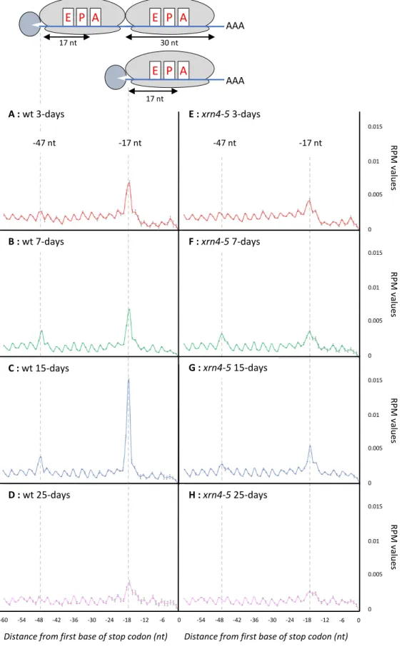
As our main goal in this study is to determine the prevalence of co-translational 5’-3’ mRNA
219
decay across development, we focused on degradome data. The two main features of
co-220
translational decay are the accumulation of the 17 nt long ribosome footprint at the translation
termination site and the 3-nucleotide periodicity of fragments resulting from the degradation
222
of mRNA open reading frames (Pelechano et al., 2015; Yu et al., 2016). To assess the
223
reliability of our analysis, a metagene analysis of the 5’P reads obtained by GMUCT was
224
performed around stop codons at each developmental stage (Figure 3A-D). The relative
225
abundance of reads at each position relative to the stop codon was determined. At all stages, a
226
clear 3-nt periodicity pattern is observed as previously described (Yu et al., 2016).
227
Additionally, a clear over-accumulation of reads 17 nt before stop codons is also detected.
228
This accumulation corresponds precisely to the 5′ boundary of the ribosome with its A site
229
stalled at a stop codon. Interestingly, a differential accumulation at this position is observed
230
across development reaching its maximum at 15-d-old stage (Figure 3C), suggesting that the
231
activity of the co-translational decay pathway could be controlled through development.
232
Moreover, at 7-d and 15-d-old stages, an additional peak is observed 47 nt before stop codons
233
(Figure 3B-C). A similar phenomenon was previously observed in yeast (Pelechano et al.,
234
2015) but was not detected in Arabidopsis flowers (Yu et al., 2016). It corresponds to two
235
ribosomes stalled at stop codons as the distance between the two peaks (30 nt) exactly
236
matches one ribosome footprint. To determine if additional peak(s) could also result from a
237
ribosome stalling at sub-optimal codon(s), we looked for possible enrichment of 5’P read
238
ends in coding regions other than the ones surrounding stop codons (Supplemental Figure 3).
239
We could not identify additional peaks at any codon, other than stop codons, in our four
240
tested developmental conditions. Thus, in our experimental conditions, we find no evidence
241
for ribosome stalling at sub-optimal codons. This also confirms that the -47 nt peak, only
242
observed in developmental stages where the co-translational decay rate is highest, is not
243
associated with a slowing down of ribosome at a specific codon but represents the footprint of
244
two ribosomes stalled at stop codons. Taken together, these observations support that
co-245
translational decay activity is regulated across development.
246 247
Next, we performed the same analysis using xrn4-5 degradome data (Figure 3E-H). For all
248
developmental stages, a decrease in reads accumulation at -17 nt before stop codons is
249
observed, suggesting that the co-translational decay pathway is severely impaired in this
250
mutant and supporting the main role of XRN4 in this pathway. To identify XRN4
co-251
translational decay targets, we used the DESeq2 pipeline on Col0 and xrn4-5 degradome data
252
(Supplemental Figure 2A-B) and compared the FCs across development. For all the
253
differentially accumulated targets, the median fold change is systematically higher than 2.5
254
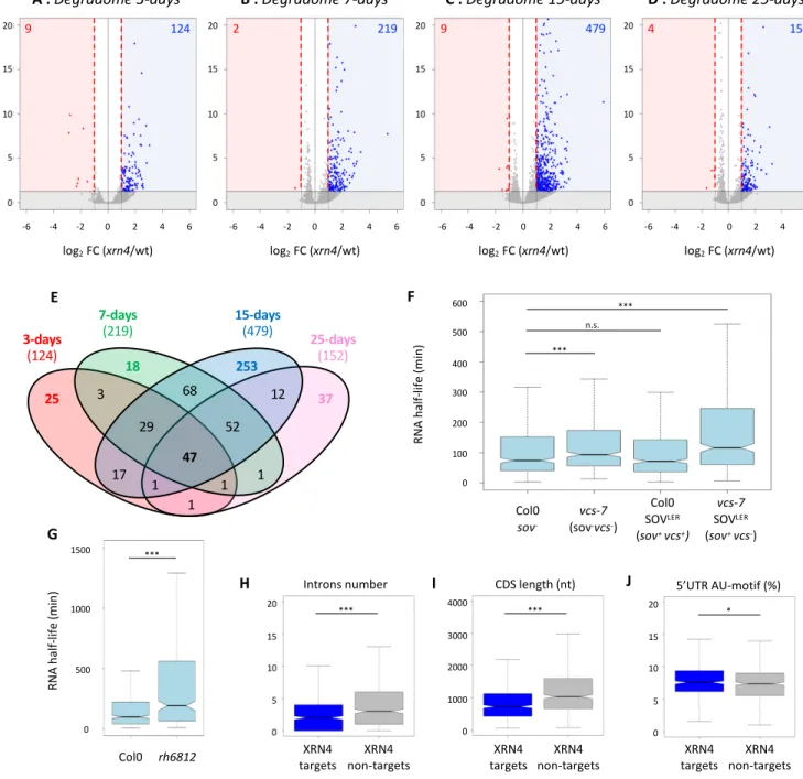
with a maximum at 3-days (Supplemental Figure 4A). To extract the most significant targets,
a cut-off FC above 2 was applied. While in total and polysome RNA data, only a handful of
256
XRN4 targets were identified, the degradome data identifies several hundreds of
mis-257
regulated mRNAs (Figure 4A-D). And consistently with XRN4 function as decay enzyme,
258
98% of differentially expressed genes are up-regulated. The number of co-translational decay
259
targets increases across development reaching it maximum at 15-days (479) before dropping
260
again at 25-days (152). The differential accumulation of 5’P read ends at 17 nt before stop
261
codon suggests that the repertoire of co-translational decay targets and/or their decay rates are
262
developmentally regulated. To test this hypothesis, we performed a Venn diagram on lists of
263
transcripts up-regulated in the xrn4-5 in degradome data (Figure 4E, Supplemental Table 2).
264
A total of 565 unique targets were identified with only 47 targets shared by all stages. Each
265
stage presents specific targets with 25, 18, 253 and 37 mRNAs more sensitive to
co-266
translational decay at 3-, 7-, 15- and 25-d-old stages respectively. Additionally, close
267
developmental stages share more targets than more distant one. As an example, except for the
268
47 common targets, 65 common targets were identified between 25- and 15-d-old stages
269
whereas 3 targets are shared between the 25 and 3-d-old stages (Figure 4E). In order to
270
determine biological processes targeted by co-translational decay, a GO analysis was
271
performed on transcripts identified as up-regulated in xrn4-5 in degradome data at all
272
developmental stages. A clustering approach was performed using DAVID software
273
(Supplemental Table 3). “Redox signalling” processes are mainly affected by XRN4 at
3-d-274
old stage. “Auxin/Growth”, “Response to stress” and “DNA binding” processes are 275
preferentially affected at 7-d-old stage compared to “Ribosome/translation”, “DNA binding”
276
and “RNA binding” processes that are affected at 15-d-old stage (Supplemental Table 3). At 277
25 days, these same GO terms as for the 15-d-old stage are represented but with a lower
278
enrichment score.
279 280
Next, we reasoned that genes we find up-regulated in the absence of XRN4 in our degradome
281
data, should show an augmented half-life when the 5'-3' decay system is impaired.
282
VARICOSE (VCS) is part of the decapping holoenzyme DCP1/DCP2/VCS and the DEAD
283
box RNA helicases RH6, 8 and 12 were recently identified as co-factors of the 5'-3'
284
cytoplasmic mRNA turnover (Sorenson et al., 2018; Chantarachot et al., 2020). We hence
285
used data from these two recent articles that report genome-wide, mRNA half-lives in
wild-286
type and vcs-7 or rh6812 loss-of-function mutants. Of the 565 mRNAs we find up-regulated
287
in xrn4-5, 444 (>78%) are detected in the Sorenson dataset, of which 85% have a half-life
288
below 240 min (Figure 4F, Supplemental Table 2, Col0 data). Consistently, 77 % of the 390
of our co-translational decay targets that are present in the Chantarachot data, also show
half-290
lives below 240 min (Figure 4G, Supplemental Table 2, Col0). This first observation supports
291
that most of the transcripts that are degraded co-translationally from 5', are intrinsically
short-292
lived. Next, we observed that in the absence of an active decapping enzyme (vcs-7 mutant) or
293
in a background without cofactors of the 5'-3' decay (rh6812 mutant), their half-lives
294
significantly increase (Figure 4F, compared the Col0 to vcs-7 boxplot, Figure 4G compare the
295
Col0 boxplot to the rh6812 one). Furthermore, the Sorenson data suggest that these mRNAs
296
are mostly decayed through a 5'-3' process. Indeed, mRNAs can be decayed either from 5',
297
following decapping and/or from 3' either by the exosome complex or by the 3'-5'
298
exoribonuclease, SOV. In addition, mRNAs that are not naturally decayed from 5'- can be
299
turned down by this pathway in the absence of functional exosome or VCS enzymes. The
300
Col0 ecotype was found previously to carry a sov-defective allele (Zhang et al., 2010), hence
301
to ascertain that mRNAs up-regulated in the vcs-7 background are natural targets of the
5'-302
pathway, Sorenson et al. also used Col0 Arabidopsis complemented with a functional allele of
303
SOV. mRNAs we find up-regulated in the absence of XRN4 here again show increased
half-304
lives in the absence of VCS despite the presence of an active SOV (Figure 4F compared Col0
305
to vcs-7 SOVLER). This increase is not seen when VCS is active such as in Col0 SOVLER
306
(Figure 4F compared Col0 to Col0 SOVLER). This further supports that XRN4 co-translational
307
decay targets are actual and specific targets of the 5'-3' degradation pathway.
308 309
A key question is how the 5'-3' decay pathway and more specifically how the
5'-co-310
translational degradation process recognizes its targets amongst the whole cell transcriptome.
311
We hence looked for putative cis elements shared by mRNAs we found as XRN4
co-312
translational decay targets. To do so we retrieved the 5' and 3'-UTRs, the CDS sequences of
313
all targets, and looked for common features as compared to a random set of transcripts
non-314
targeted by XRN4. Although, no differences were found in 5’UTR and 3’UTR lengths, a 315
significant reduction in intron number and CDS length was observed for XRN4
co-316
translational decay targets (Figure 4H-I, Supplemental Figure 4B-C). In addition, AU-motifs
317
were found enriched in their 5’UTR (Figure 4J). This observation is consistent with previous 318
study showing that short-lived mRNAs have less introns and AU-rich motifs in their 5'-UTRs
319
(Narsai et al., 2007; Sorenson et al., 2018). All together, these data suggest that trans and cis
320
elements could regulate co-translational decay activity.
321 322
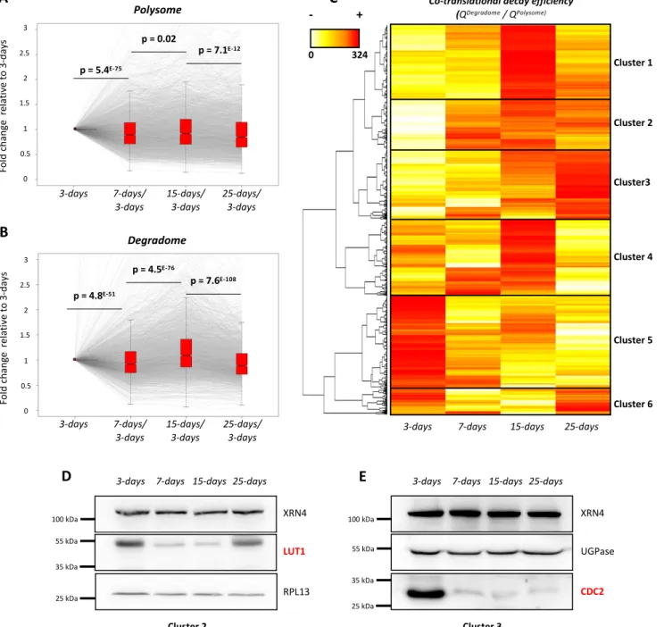
Co-translational decay activity across development can influence translation efficiency 323
Hence our above results suggest that co-translational decay specificity and efficiency is
324
regulated in response to developmental cues. We next wondered about the molecular role of
325
this regulation. Since co-translational decay and translation are interrelated, we asked whether
326
the former could control protein production. We rationalized that, for a given mRNA, a
co-327
translational decay rate higher than the translation rate should result in a decrease in
328
translation efficiency and vice-versa. To explore this, we compared variations across
329
development at polysome and degradome levels in Col0 background (Figure 5A). To limit
330
variations at polysome and degradome levels, we focused our analysis on transcripts detected
331
(RPKM>1 in all libraries) and remaining stable across development at the total RNA level
332
(e.g. fold change between 0.66 and 1.5 between all conditions) resulting in 3,366 mRNAs
333
(Supplemental Figure 2C). For these transcripts, variations at polysome RNA levels were
334
compared to that at the degradome level. Values were normalized to the 3-d-old stage and the
335
dynamic was assessed across development (Figure 5A and 5B). Interestingly, based on
p-336
values, the median variation of the degradome values appears more dynamic across
337
development than that of the polysomal values. To test this, we calculated the ratio between
338
degradome and polysome values for each mRNA (Supplemental Figure 2C). We called this
339
ratio "co-translational decay efficiency" as it reflects the proportion of polysome associated
340
with uncapped mRNA decay intermediates (degradome data) compared to the total amount
341
(capped and uncapped) of polysome-associated mRNAs (polysome data). A high ratio would
342
suggest a high co-translational decay activity resulting in a low translation efficiency
343
compared to a low ratio suggesting a high translation efficiency. A heat map was generated to
344
observe the variation of this efficiency across development (Figure 5C). Interestingly, this
345
efficiency varies between transcripts and is highly modulated across development. Six good
346
resolution clusters were obtained, the behavior of which can be monitored across
347
development. Transcripts from cluster 1 were highly targeted by co-translational decay at
15-348
d-old stage while those from cluster 5 are mostly targeted at the 3-d-old stage. Cluster 3 was
349
also remarkable as it is formed with mRNAs that are progressively targeted by the
co-350
translational decay until the 25-d-old stage. As our hypothesis is that co-translational decay
351
could influence translation efficiency, we set to monitor level variations across development
352
of two proteins LUT1 and CDC2 that belong to cluster 2 and 3 respectively. These mRNAs
353
were selected as case studies because they show important variations of their co-translational
354
efficiency that are mostly due to variations of their degradome values with stable quantities in
355
total and polysomal fractions across development (Supplemental Figure 5). Western blotting
356
shows that LUT1 levels are highest at 3-d-old stage and decrease at 7-d-old and 15-d-old
stages where its transcript’s co-translational decay efficiency is highest. At 25-d-old stage,
358
LUT1 degradome value decreases and consistently its protein levels increase as compared to 7
359
and 15-day (Figure 5D, Supplemental Figure 5). In contrast, CDC2 was mostly detected at
3-360
d-old stage which is consistent with its low co-translational decay efficiency at this stage
361
(Figure 5E, Supplemental Figure 5). Protein level variations are the consequence of both
362
production (translation) and decay rates variations, hence steady-state western blotting is not
363
the most accurate read-out of translation efficiency. Nonetheless, with the LUT1 and CDC2
364
cases we observe a perfect correlation between co-translational efficiency and steady-state
365
protein levels (Figure 5D and E) supporting that indeed co-translational decay could be a
366
mean to fine-tune translation.
367 368
DISCUSSION 369
370
The major objective of this study was to monitor the dynamics of co-translational decay
371
across Arabidopsis seedling development. Using a multifaceted genome-wide approach, we
372
provide evidence that co-translational decay is highly modulated across development with a
373
maximum at 15-d-old stage. Using the xrn4-5 loss-of-function mutant (Souret et al., 2004;
374
Merret et al., 2013), we find that the majority of co-translational mRNA decay in seedling is
375
catalyzed by XRN4, as previously proposed in flower tissue (Yu et al., 2016). However, as
376
the 3-nt periodicity is maintained in xrn4-5, as well as a low level of 17 nt 5’P reads before
377
stop codons, other 5' to 3' exoribonucleases must be involved in this process. In yeast, the
378
nuclear Xrn (Xrn2p) was proposed to relocalize to the cytoplasm of xrn1-null mutant and
379
restore cytosolic mRNA turnover (Johnson, 1997). However, since Arabidopsis has not one
380
but two nuclear 5 to 3' exonucleases (XRN2 and 3) and since the xrn3 loss-of-function mutant
381
is lethal (Gy et al., 2007), the hypothesis of nuclear XRNs complementing a deficient
382
cytosolic enzyme is challenging to assess.
383 384
The polysome fraction was until recently considered to be composed essentially of actively
385
translating mRNAs and was routinely used to assess mRNA translation efficiency after
386
normalization to total RNA levels (see an example in Bai et al., 2017). The discovery of the
387
co-translational decay and its conservation in evolutionary distant eukaryotes (Hu et al., 2009;
388
Hu et al., 2010; Hou et al., 2016; Yu et al., 2016; Simms et al., 2017; Ibrahim et al., 2018),
389
suggests that the proportion of actively translated mRNAs in polysomes might be much lower
390
than initially expected. Translation efficiency is often assessed by ribosome profiling (Ingolia
et al., 2009) or TRAP (Translating Ribosome Affinity Purification, Reynoso et al., 2015). But
392
these approaches do not take into account the fact that part of mRNAs attached to the
393
ribosomes might be undergoing a degradation. Thus, these approaches can give rise to
394
misleading conclusions. In yeast, 5′P decay intermediates represent more than 12% of the
395
polyA+ mRNA fraction (Pelechano et al., 2015). In our study, we found that at least 3,366
396
transcripts present variations in degradome data across development and that their
co-397
translational decay rate could be an efficient way to the cell to control their translation
398
efficiency (Figure 5). Thus, to take into account the co-translational decay in translation
399
efficiency measurements, we calculated the ratio between degradome and polysome values
400
for each transcript as proxy (Figure 5C). This ratio can be determined for each transcript and
401
compared pairwise between conditions or genetic backgrounds. A high ratio would suggest a
402
low translation efficiency and conversely. As a proof of concept, we monitored protein
403
accumulation of the LUT1 and CDC2 genes and found that protein levels vary in an inversely
404
proportional manner to this ratio in both cases (Figure 5, Supplemental Figure 5). We hence
405
propose to use this ratio as an additional way to determine transcript translation efficiency.
406
We also posit that it would be a more accurate read-out than the mere ratio between
407
polysomal and total mRNA levels. The determination of this efficiency will be crucial under
408
conditions where co-translational decay is highly modulated such as development or stress
409
exposure (Merret et al., 2015; Garre et al., 2018).
410 411
In addition to its role in translation efficiency modulation, our analysis allows the
412
identification of specific XRN4 co-translational decay targets (Figure 4). In a landscape
413
analysis of Arabidopsis mRNA half-lives, the authors found that short half-lived mRNAs
414
targeted by decapping and 5’-3’ degradation present less introns that stable ones (Sorenson et 415
al., 2018). Consistently, this feature is shared by transcripts targeted by co-transcriptional
416
decay in our analysis (Figure 4H, Supplemental Table 2). Co-translational decay targets are
417
also enriched in AU-motifs in their 5'-UTRs, a motif also known to be shared by unstable
418
transcripts (Narsai et al., 2007). A low intron complexity and the presence of AU-rich motifs
419
could be some of the cis determinants of co-translational target recognition. Additionally, we
420
found that the mRNA half-life range of XRN4 co-translational decay targets increases in
421
mutants involved in 5’-3’ decapping dependant decay such as vcs-7 and rh6812, key factors 422
of decapping activity (Figure 4F-G). The higher stability of these transcripts in these mutants
423
is consistent as decapping is a necessary and crucial step for XRN4 activity. Interestingly, in
424
yeast DHH1, homologue of RH6, 8 and 12 was proposed to couple mRNA translation to
decay (Radhakrishnan et al., 2016). These findings could support that VCS, RH6, 8 and 12
426
could be trans determinants of co-translational target recognition as recently discussed
427
(Merret and Bousquet-Antonelli, 2020).
428
Analysis of enriched GO terms associated with co-translational decay targets reveals six
429
major GO terms (“Auxin/Growth”, “Response to stress”, “DNA binding”,
430
“Ribosome/translation”, “RNA binding” and “Redox signalling”). Published PARE data 431
identified similar GO terms such as “mRNA processing” or “Ribosome biogenesis” shared by 432
polyadenylated targets of XRN4 (Nagarajan et al., 2019). Recently, it was proposed that
433
XRN4 contributes to root growth under normal conditions and upon salt stress by an
434
unknown mechanism (Kawa et al., 2020). Interestingly, in “auxin/growth” GO term, we
435
identified many genes targeted by the co-translational decay pathway that are associated with
436
growth regulation (such as “response to auxin” GO:0009733, Supplemental Table 3)
437
consistently with the described role of XRN4 in root development. As an example, RVE2
438
(At5g37260), a gene involved in lateral root formation and a VCS substrate (Supplemental
439
Table S16 of Kawa et al., 2020), was identified as a co-translational target of XRN4 at 7 days
440
(Supplemental Table 3). While in these different studies, the respective contributions of both
441
5’-3’ cytosolic and co-translational decay was not addressed, our results suggest that at least 442
part of these XRN4 targets could be decayed co-translationally
443 444
Co-translational decay is an evolutionarily conserved mechanism found in many organisms
445
such as yeast (Pelechano et al., 2015), Arabidopsis (Yu et al., 2016), Soybean (Hou et al.,
446
2016), Barley (Hou et al., 2016), and mammalian cells (Tuck et al., 2020). This pathway was
447
described as being involved in different stress responses (Merret et al., 2015; Pelechano et al.,
448
2015; Garre et al.; 2018). But until now, analyses were only focused on stable lines or specific
449
tissues. Our data demonstrate that the 5'-co-translational decay is dynamically modulated
450
across development and important for proper regulation of protein expression and suggest that
451
this pathway could be important for plant development and physiology.
452 453 454
MATERIAL AND METHODS 455
456
Growth conditions 457
Analyses were carried out with Columbia-0 line as wild type and xrn4-5 mutant
458
(SAIL_681_E01). Plantlets were grown on synthetic Murashige and Skoog medium
459
(Duchefa) containing 1% Sucrose and 0.8% plant agar at 22°C under a 16-h-light/8-h-dark
460
regime. Same growth conditions were applied for soil culture.
461 462
Sampling procedure for RNA sequencing 463
To generate two biological replicates, two distinct batches of seeds (generated from different
464
parent plant) were used for each genotype. All samples were generated at the same time as
465
follows. Seeds were sown in vitro on 20 square plates for each replicate and genotype. After 3
466
days, plantlets from 6 plates were pooled and harvested to generate 3-day samples. The same
467
procedure was performed at 7 days and 15 days to generate 7- and 15-day samples
468
respectively. Plantlets from the 2 remaining plates were transferred to soil for an additional 10
469
days to obtain 25 day-samples. In this case, the rosette and the primary root were collected.
470
For each developmental stage, at least 10 plantlets were pooled to avoid individual-specific
471
bias.
472 473
Polysome profile analysis 474
Polysome profiles were performed as described previously (Merret et al., 2013). In brief, 400
475
mg of tissue powder were homogenized with 1.2 mL of lysis buffer (200 mM Tris-HCl, pH
476
9.0, 200 mm KCl, 25 mM EGTA, 35 mM MgCl2, 1% detergent mix [1% Tween 20, 1% 477
Triton, 1% Brij35, and 1% Igepal], 1% sodium deoxycholate, 0.5% polyoxyethylene tridecyl
478
ether, 5 mM DTT, 50 μg.mL−1 cycloheximide, 50 μg.mL−1 chloramphenicol, and 1% protease
479
inhibitor cocktail [Sigma-Aldrich]). Crude extract was incubated 10 min on ice. After
480
centrifugation, nine hundred microliters of crude extract was loaded on a 15% to 60% Sucrose
481
gradient (9 mL). Ultracentrifugation was performed with an SW41 rotor at 38 000 for 3 h.
482
Polysome profile analyses were performed with an ISCO absorbance detector at 254 nm.
483
Twelve fractions of 650 μL were collected. Proteins were extracted from fractions 6 to 12
484
(corresponding to polysomes). 2 volumes of absolute ethanol were added for each fraction.
485
Proteins were precipitated 6 hours at 4°C before centrifugation. Pellets were washed and
486
resuspended in 10 μL of Laemmli 4X. For polysomal RNA, extraction was performed as in 487
Merret et al., 2015 using Monarch Total RNA Miniprep Kit (New England Biolabs).
489
Total RNA extraction 490
Total RNA was extracted using Monarch Total RNA Miniprep Kit (New England Biolabs).
491
RNA quality was assessed using Agilent RNA 6000 Nano kit (Agilent).
492 493
RNA library preparation 494
RNA library preparation was performed on total or polysomal RNA using NEBNext®
495
Poly(A) mRNA Magnetic Isolation Module and NEBNext Ultra II Directional RNA Library
496
Prep Kit (New England Biolabs) according to manufacturer’s instructions with 1 μg of RNA
497 as starting point. 498 499 GMUCT assay 500
GMUCT library was prepared as described previously with slight modifications (Willmann et
501
al., 2014). Briefly, 50 μg of total RNA were subjected to two polyA+ purification. After
502
5’adapter ligation, excess of adapter was removed by a new round of polyA+
purification.
503
Reverse transcription was performed using Superscript IV system with manufacturer’s
504
instructions. cDNAs were amplified with 11 cycles of PCR. Libraries were purified using
505
SPRIselect beads prior to quality control and normalization.
506 507
RNA sequencing 508
Libraries quality was checked using Agilent High Sensitivity DNA kit (Agilent). Libraries
509
were normalized, multiplexed and sequenced on NextSeq 550 (Illumina) in single-reads 75
510 pb. 511 512 Bioinformatic analysis 513
For total and polysome RNA libraries, after filtering out reads corresponding to chloroplastic,
514
mitochondrial, ribosomal and small RNA sequences using bowtie2, reads were mapped
515
against TAIR10 genome using Hisat2 and the gtf TAIR10 annotation file with standard
516
parameters. Reads count by gene was performed by Cufflinks in RPKM (reads per kilobase
517
by millions mapped reads). For GMUCT analyses, reads were trimmed to 50 pb using
518
Trimmomatic prior to mapping. Reads count was performed using bedcoverage from
519
Bedtools suite and normalized by total of mapped reads (read per millions, RPM). For 5’P
520
reads abundance, the bam file was converted into bed file containing only the first nucleotide
521
of each read. Differential expression analyses were performed using Biocunductor R package
DESeq2, with an FDR of 0.05. P-values were corrected for the multiple tests by
Benjamin-523
Hochberg rule (adjusted p-value). Analysis of codon enrichment was performed as previously
524
described (Yu et al., 2016). For Notched plot analysis, Shapiro test was applied to test
525
normality of the dataset. Then nonparametric Wilcoxon test was performed between each
526
developmental stage. GO analysis was performed using DAVID software (Huang et al., 2009)
527
with default settings, the six major clusters were retained for analysis. For mRNA features
528
analysis, UTRs, introns and CDS sequences were obtained from Araport11 database and are
529
listed on Supplemental Table 2. RNA half-lives data were collected from Dataset_S2 of
530
Sorenson et al., 2018 (Columns “alpha_WT”, “alpha_sov”, “alpha_vcs”, “alpha_vcs sov”) and
531
from Supplementary Table S4 of Chantarachot et al., 2020 (Columns mRNA Half-life
532
“WT_Col-0” and “rh6812”) and are presented on Supplemental Table 2. Only transcripts 533
present in each dataset were kept for statistical analysis. A Wilcoxon test was systematically
534
performed to test significance (presented on Supplemental Table 5).
535 536
Western blot 537
After electrophoretic separation by SDS-PAGE gels, proteins were electrotransferred on
538
polyvinylidene fluoride membranes. Immunoblottings were performed in TBS-5% skimmed
539
milk-1% Tween. Primary antibody was incubated overnight at 4°C under constant agitation.
540
After incubation, membranes were washed 6 times with TBS-1% Tween. A horseradish
541
peroxidase-coupled secondary antibody was incubated in TBS-5% skimmed milk- 1% Tween
542
45 minutes at room temperature. Membranes were again washed 6 times with TBS-1%
543
Tween and revealed with the Immobilon-P kit from Millipore. Image acquisition was
544
performed using Fusion FX imaging system (Vilber). Antibodies against XRN4 (Merret et al.,
545
2013), LUT1 (Agrisera), CDC2 (Agrisera), UGPase (Agrisera) and RPL13 (Merret et al.,
546
2013) were utilized at 1/1,000th, 1/1,000th and 1/3,000th, 1/5,000th and 1/100,000th
547 respectively. 548 549 Accession numbers 550
The accession numbers for the RNA-seq data reported in this article are NCBI Bioprojects
551
PRJNA604882 for total RNA data, PRJNA604883 for polysome RNA data and
552
PRJNA604884 for GMUCT data.
553 554
Acknowledgments 555
We thank Drs. Xiang YU and Brian GREGORY (University of Pennsylvania) for advices on
556
GMUCT bioinformatic analysis. We also thank the sequencing facility of Perpignan
557
University Via Domitia BioEnvironnement platform (Perpignan, France).
FIGURES LEGENDS 559
560
Figure 1 : XRN4 differentially accumulates in polysomes across seedling development.
561
A. Polysomal extracts prepared from 3-, 7-, 15- and 25-d-old seedlings were fractionated on a
562
sucrose gradient and polysome traces obtained through measurement of OD254nm. Polysome 563
profiling were performed starting from identical quantities of N2-pulverized tissues (e.g 300 564
mg of biomass). B. Total proteins extracted from polysomal and input fractions were analyzed 565
by western blotting. The four blots were probed with an antibody specific to XRN4. Inputs 566
correspond to an equivalent of 10 mg of tissue powder for all stages. For polysomal fraction, 567
loaded proteins were precipitated from identical volumes of each fraction. Data are 568
representative of at least three replicates. 569
570
Figure 2 : XRN4 loss-of-function has low impact across seedling development at total 571
(A) and polysome (B) RNA levels. Fold changes between xrn4-5 and Col0 (wt) were 572
calculated for each transcript in each condition. The log2 value of the mean is represented in 573
each graph. The number of transcripts significantly regulated in xrn4-5 is reported (as DEG in
574
red for up-regulated and grey for down-regulated transcripts) and was calculated using
575
DESeq2. Dashed red lines mark the |log2 (2)| values. 576
577
Figure 3 : Metagene analyses displaying the abundance of 5’P reads relative to stop 578
codons. A. wt 3-d-old stage, B. wt 7-d-old stage, C. wt 15-d-old stage, D. wt 25-d-old stage, 579
E. xrn4-5 3-d-old stage, F. xrn4-5 7-d-old stage, G. xrn4-5 15-d-old stage, H. xrn4-5 25-d-old 580
stage. Mean ± SD. The illustrations represent 5’P intermediates accumulation at - 47 nt and -
581
17 nt before stop codons.
582 583
Figure 4 : Identification and features of XRN4 co-translational decay targets across
584
development. A-D. Volcano plot of the change in read abundance in xrn4-5 over wt (Col0).
585
Vertical red dotted lines mark the |log2(2)| values. Log2 fold change and Benjamini-Hochberg 586
adjusted p-values (BH) were calculated through the DESeq2 pipeline (as DEG in blue for up-587
regulated and red for down-regulated transcripts). Horizontal plain black lines demarcate 588
adjusted p-values of 0.05. E. Venn diagram of co-translational decay targets across 589
development. F-G. Majority of XRN4 co-translational decay targets show longer RNA half-590
life in vcs-7, vcs-7 SOVLER mutants (F) and rh6812 mutant (G). RNA half-lives were 591
collected from Sorenson et al., 2019 (F) or from Chantarachot et al., 2020 (G). Only 592
transcripts present in each dataset are represented (N = 444/565 for F or N = 390/565 for G). 593
H-J. Introns number, CDS length, and proportion of AU-motif in 5’UTR respectively of
594
transcripts targeted by XRN4 compared to non-targeted random transcripts. N=565. *** p-595
values < 0.001, * p-values < 0.05, n.s. non significant. 596
597
Figure 5: Co-translational decay is regulated across development and influences protein
598
production. A. Transcript variation at polysome level across development using 3-days as a
599
reference (N=3,366). B. Transcript variation at degradome level across development using 3-600
days as a reference (N=3,366). Gray lines represent individual transcript variation. Transcript 601
distribution is represented by notched boxplots and significance was assessed by p-values 602
(nonparametric Wilcoxon test). C. Heat Map of co-translational decay efficiency (ratio in 603
degradome data over polysome RNAseq data) (N=3,366). Red values correspond to a high 604
decay efficiency and yellow values to a low decay efficiency. D, E. Western blottings using 605
LUT1 and CDC2 antibodies respectively. Both candidates were analyzed on distinct SDS-606
PAGE gels (8% and 10% acrylamide respectively). RPL13 and UGPase antibodies were used 607
as loading controls. Each western blot was performed on two biological replicates. 608
609
SUPPLEMENTAL DATA 610
611
Supplemental Figure 1 : Representation of the experimental procedure. A. To generate 612
two biological replicates, two independent batches of seeds were used for each genotype. A
613
time-course sampling was performed to harvest 3-d-old, 7-d-old, 15-d-old and 25-d-old
614
seedlings in two biological replicates. Col0 (wt) and xrn4-5 (SAIL_681_E01) were used in
615
this study. B. On each sample, total RNA extractions were performed to determine the
616
transcriptome and the degradome through RNA sequencing and GMUCT approaches
617
respectively. From the same biological samples, polysomes were purified through
618
fractionation of sucrose density gradients and their levels determined through measurement of
619
OD254nm in each fraction. Polysomes traces were recorded and polyadenylated RNAs were
620
purified from pooled fractions containing polysomes (translating ribosomes). The total
621
polyA+ RNAseq allows the capture of capped free mRNAs, capped translated mRNAs and
622
uncapped co-translational decay intermediates. The polysome polyA+ RNAseq allows the
623
capture of capped translating mRNAs and uncapped co-translational decay intermediates. The
624
degradome allows the capture of uncapped co-translational decay intermediates.
625 626
Supplemental Figure 2 : Representation of the bioinformatic analysis. A. For each 627
library, reads were mapped against TAIR10 genome using Hisat2. Then, transcripts
628
quantification was performed using Cufflinks for total and polysome libraries (RPKM) and
629
using Bedcoverage for degradome libraries (RPM). B. To identify XRN4 targets, transcripts
630
with a least 1 RPKM/RPM in 1 library were kept (N=23,196). Then, a fold change (FC)
631
analysis was performed between xrn4.5 and Col0. Biological replicates were analyzed
632
separately and differential expression analysis was performed using DESeq2 to determine
633
significant targets. A FC of 2 and a FDR of 0.05 were applied as criteria to identify XRN4
634
targets. C. To determine co-translational decay efficiency, transcripts with a least 1
635
RPKM/RPM in all libraries were kept (N=13,333). Then, transcripts remaining stable across
636
development at the total RNA level (e.g. fold change between 0.66 and 1.5 between all
637
conditions) were kept (N=3,366). Finally, the co-translational decay efficiency (ratio between
638
degradome and polysome values for each transcripts) in Col0 was determined.
639 640
Supplemental Figure 3 : Enrichment of 5′P read ends at the ribosome boundary of 641
mRNA along mRNA coding regions. Red dots denote codons with significant enrichment of 642
5′P read ends at their ribosome boundary sites, while dark dots denote codons that are not 643
significant for this value. The "stop" red dot represents the mean value of the three stop
644
codons. Significance was assessed using a χ2 test. 645
646
Supplemental Figure 4 : A. Boxplots of the range of fold change in xrn4 mutant compared 647
to wt for differentially expressed transcripts in degradome data identified at 3-, 7-, 15- and
25-648
days respectively. Numbers of transcripts and median of fold change are indicated. Red
649
dashed line represents a FC of 2. B-C. Distribution of 5’UTR length or 3’UTR length of
650
XRN4 co-translational targets versus XRN4 non-targets. N=565, n.s. non significant.
651 652
Supplemental Figure 5 : Values obtained for LUT1 and CDC2 transcripts at TOTAL, 653
POLYSOME and DEGRADOME levels in RNAseq data. Data were extracted from
654
Supplemental Table 1. Mean ± SD. Western blot of a second replicate is also presented.
655 656
Supplemental Table 1 : Differential expression analysis of 23,196 genes by DEseq2 of 657
TOTAL, POLYSOME and DEGRADOME mRNA-seq data associated with Figures 2 and 4.
658 659
Supplemental Table 2 : List 565 XRN4 co-transcriptional decay targets identified across 660
development using DEseq2 associated with Figure 4.
661 662
Supplemental Table 3 : Enriched GO terms associated with XRN4 co-translational decay 663
targets across development
664 665
Supplemental Table 4 : Variation of co-translational decay efficiency of 3,366 genes stables 666
at TOTAL RNA level across development associated with Figure 5
667 668
Supplemental Table 5 : Results of statistical analysis used in this manuscript 669
LITERATURE CITED
671 672
Addo-Quaye C, Eshoo TW, Bartel DP, Axtell MJ (2008) Endogenous siRNA and miRNA 673
Targets Identified by Sequencing of the Arabidopsis Degradome. Curr Biol 18: 758–762
674
Anderson SJ, Kramer MC, Gosai SJ, Yu X, Vandivier LE, Nelson ADL, Anderson ZD, 675
Beilstein MA, Fray RG, Lyons E, et al (2018) N6-Methyladenosine Inhibits Local 676
Ribonucleolytic Cleavage to Stabilize mRNAs in Arabidopsis. Cell Rep 25:
1146-677
1157.e3
678
Bai B, Peviani A, van der Horst S, Gamm M, Snel B, Bentsink L, Hanson J (2017) 679
Extensive translational regulation during seed germination revealed by polysomal
680
profiling. New Phytol 214: 233–244
681
Franke KR, Schmidt SA, Park S, Jeong DH, Accerbi M, Green PJ (2018) Analysis of 682
Brachypodium miRNA targets: Evidence for diverse control during stress and
683
conservation in bioenergy crops. BMC Genomics 19: 1–18
684
Garre E, Pelechano V, Sánchez del Pino M, Alepuz P, Sunnerhagen P (2018) The Lsm1-685
7/Pat1 complex binds to stress-activated mRNAs and modulates the response to
686
hyperosmotic shock. PLoS Genet 14: 1–30
687
German MA, Pillay M, Jeong DH, Hetawal A, Luo S, Janardhanan P, Kannan V, 688
Rymarquis LA, Nobuta K, German R, et al (2008) Global identification of 689
microRNA-target RNA pairs by parallel analysis of RNA ends. Nat Biotechnol 26: 941–
690
946
691
Heck AM, Wilusz J (2018) The interplay between the RNA decay and translation machinery 692
in eukaryotes. Cold Spring Harb Perspect Biol. doi: 10.1101/cshperspect.a032839
693
Hou C, Lee W, Chou H, Chen A, Chou S, Chen H (2016) Global Analysis of Truncated 694
RNA Ends Reveals New Insights into Ribosome Stalling in Plants. Plant Cell 28: 2398–
695
2416
696
Hou CY, Wu MT, Lu SH, Hsing YI, Chen HM (2014) Beyond cleaved small RNA targets: 697
Unraveling the complexity of plant RNA degradome data. BMC Genomics. doi:
698
10.1186/1471-2164-15-15
699
Hu W, Petzold C, Coller J, Baker KE (2010) Nonsense-mediated mRNA decapping occurs 700
on polyribosomes in Saccharomyces cerevisiae. Nat Struct Mol Biol 17: 244–247
701
Hu W, Sweet TJ, Chamnongpol S, Baker KE, Coller J (2009) Co-translational mRNA 702
decay in Saccharomyces cerevisiae. Nature 461: 225–229
703
Huang DW, Sherman BT, Lempicki RA (2009) Systematic and integrative analysis of large 704
gene lists using DAVID bioinformatics resources. Nat Protoc 4: 44–57
705
Ibrahim F, Maragkakis M, Alexiou P, Mourelatos Z (2018) Ribothrypsis, a novel process 706
of canonical mRNA decay, mediates ribosome-phased mRNA endonucleolysis. Nat
707
Struct Mol Biol 25: 302–310
708
Ingolia NT, Ghaemmaghami S, Newman JRS, Weissman JS (2009) Genome-wide 709
analysis in vivo of translation with nucleotide resolution using ribosome profiling.
710
Science (80- ) 324: 218–223
711
Johnson AW (1997) Rat1p and Xrn1p are functionally interchangeable exoribonucleases that 712
are restricted to and required in the nucleus and cytoplasm, respectively. Mol Cell Biol
713
17: 6122–6130 714
Kawa D, Meyer AJ, Dekker HL, Abd-El-Haliem AM, Gevaert K, Van De Slijke E, 715
Maszkowska J, Bucholc M, Dobrowolska G, De Jaeger G, et al (2020) SnRK2 716
Protein Kinases and mRNA Decapping Machinery Control Root Development and
717
Response to Salt. Plant Physiol 182: 361–377
718
Lee W, Hou B, Hou C, Tsao S, Kao P, Chen H, Biotechnology A (2020) Widespread Exon 719
Junction Complex Footprints in the RNA Degradome Mark mRNA Degradation Before
720
Steady-state Translation. doi: 10.1105/tpc.19.00666
721
Lin Z, Gasic I, Chandrasekaran V, Peters N, Shao S, Mitchison TJ, Hegde RS (2020) 722
TTC5 mediates autoregulation of tubulin via mRNA degradation. Science 367: 100–104
723
Love MI, Huber W, Anders S (2014) Moderated estimation of fold change and dispersion 724
for RNA-seq data with DESeq2. Genome Biol 15: 1–21
725
Merret R, Bousquet-Antonelli C (2020) Immunity gate-keepers. Nat Plants 6: 608–609 726
Merret R, Descombin J, Juan Y ting, Favory JJ, Carpentier MC, Chaparro C, Charng 727
Y yung, Deragon JM, Bousquet-Antonelli C (2013) XRN4 and LARP1 are required 728
for a heat-triggered mRNA decay pathway involved in plant acclimation and survival
729
during thermal stress. Cell Rep 5: 1279–1293
730
Merret R, Nagarajan VK, Carpentier MC, Park S, Favory JJ, Descombin J, Picart C, 731
Charng YY, Green PJ, Deragon JM, et al (2015) Heat-induced ribosome pausing 732
triggers mRNA co-translational decay in Arabidopsis thaliana. Nucleic Acids Res 43:
733
4121–4132
734
Nagarajan VK, Kukulich PM, von Hagel B, Green PJ (2019) RNA degradomes reveal 735
substrates and importance for dark and nitrogen stress responses of Arabidopsis XRN4.
736
Nucleic Acids Res 47: 9216–9230
737
Narsai R, Howell KA, Millar AH, O’Toole N, Small I, Whelan J (2007) Genome-wide