*Correspondence: Prof. S. Salentinig, E-mail: stefan.salentinig@unifr.ch
Department of Chemistry, University of Fribourg, Chemin du Musée 9, CH-1700 Fribourg, Switzerland
Design and Characterization of
Bio-inspired Antimicrobial Nanomaterials
Mahsa Zabara, Linda Hong, and Stefan Salentinig*Abstract: Colloidal structures are crucial components in biological systems and provide a vivid and seemingly infinite source of inspiration for the design of functional bio-inspired materials. They form multi-dimensional confinements and shape living matter, and transport and protect bioactive molecules in harsh biological envi-ronments such as the stomach. Recently, colloidal nanostructures based on natural antimicrobial peptides have emerged as promising alternatives to conventional antibiotics. This contribution summarizes the recent progress in the understanding and design of these bio-inspired antimicrobial nanomaterials, and discusses their advances in the form of dispersions and as surface coatings. Their potential for applications in future food and healthcare materials is also highlighted. Further, it discusses challenges in the characterization of structure and dynamics in these materials.
Keywords: Antimicrobial peptide · Liquid crystals · Nanocarriers · Nanostructured coatings · Self-assembly
Mahsa Zabara holds a PhD in Materials
Chemistry from the Royal Melbourne Institute of Technology, Australia (2016) on designing photo-tunable antimicro-bial nanomaterials; and a second PhD in Chemical Engineering from TMU University in Tehran, Iran (2012) on de-veloping hybrid nanoparticles as contrast agents. She moved to Switzerland and joined the group of Prof. Salentinig at Empa (2017), and later at the University of Fribourg as postdoctoral fel-low, working on antimicrobial nanomaterials. She has experience in both academic and industrial settings with a constant focus on material science and nanotechnology.
Linda Hong received her PhD from
Monash University (Parkville, Australia) in 2019 studying stimuli-responsive liquid crystalline nanoparticles using microflu-idic approaches and advanced synchrotron-based scattering techniques. She has since moved to Switzerland to undertake a post-doctorate at the University of Fribourg un-der the supervision of Prof. Salentinig. Her research now focuses on investigating the interactions between bacterial membrane components, antimicro-bial peptides, and lipid-based nanocarriers. She is also collaborat-ing with industry to formulate drug delivery systems.
Stefan Salentinig is a professor of
physi-cal chemistry at the University of Fribourg, where he has established the Biocolloids lab, researching bio-inspired materials for health applications. He holds a PhD in phys-ical chemistry focusing on food colloids and scattering methods from the University of Graz, Austria, 2010. He then moved to Australia to take on a scientist position on
separation technology at the CSIRO, and then became lecturer at Monash University in Melbourne working on nanoscale drug delivery systems. In 2015, he moved to Switzerland and joined Empa as a group leader on functional materials, before starting his current role in 2019.
1. Introduction
Bacterial contamination is one of the major challenges for maintaining food quality and safety during processing and stor-age.[1] Further, the attachment and colonization of pathogenic
bacteria on the surface of medical implants or open wounds may result in severe infections.[1a,2]These infections are particularly
concerning if multi-resistant bacteria or difficult to treat microor-ganisms are involved. Hence, the design of suitable and efficient antimicrobial materials is crucial to maintain or even improve the health of our society. The search for alternative materials to con-ventional antibiotics has attracted great interest. Ribosomal syn-thesized, natural, antimicrobial peptides (AMPs) based materials have been emerging as promising alternative candidates.[3]AMPs
are a native part of the human innate immune system, occur as native host-defence molecules in virtually all species of life, and have a broad spectrum of activities against pathogenic bacteria and can modulate the immune response of the host.[4]AMPs were
also discovered in human breast milk and may provide a selec-tive advantage through evolution by protecting both the mother’s mammary gland and her nursing offspring from infection.[5]One
such AMP is lactoferricin B, an AMP obtained from the diges-tion of lactoferrin. It is an iron binding multifuncdiges-tional protein existing in high concentrations in body secretions including co-lostrum, milk, tear, nasal secretion, saliva, bile, urine, and genital secretions.[6]
Selected food-grade AMPs such as Nisin (E 234) are also widely used as food preservatives, for instance in unripen cheese and in heat‐treated meat products.[7]It is interesting to mention that
although these AMPs have been used in food for more than fifty years, no significant bacterial resistance has been reported yet.[8]
However, the major challenge to overcome for the broader clinical application of AMP-based antimicrobials is their lack of
stabil-pH 5 (Fig. 2). The antimicrobially active OA/LL-37 aggregates at approximately pH 5 were highly effective against Escherichia
coli (E. coli) bacteria, whereas the cylindrical micelles at pH 7
were mostly inactive. The geometry of the aggregates together with electrostatic interactions between OA and LL-37 molecules as well as the negatively charged bacteria membrane were dis-cussed to contribute to the underlying mechanism. Such systems may be interesting, for example, in the treatment of infections of the digestive tract, killing bacteria at desired locations such as the stomach, and keeping pro-biotic bacteria in other locations intact.[14]
Lamellar structures (i.e. dispersed in the form of vesicles in solution) are among the most abundant lipid structures in nature. Vesicles can also encapsulate hydrophilic molecules in their hy-drophilic core, hydrophobic molecules within their hydrophobic compartment of the bilayer, and amphiphilic molecules includ-ing AMPs in the lipid–water interface of the bilayer.[10h,20]The
integration of the honeybee-venom AMP, melittin and the AMP, alameticin, from the fungus Trichoderma viride, into the bilayer membrane of 1-palmitoyl-2-oleoyl-sn-glycero-3-phosphocholine (POPC) vesicles has been investigated, observing that these AMPs have a membrane perturbing effect and prefer to interact with (lo-cally) curved lipid surfaces.[21]In addition, it has been demonstrated
that the integration of the 13-residue cathelicidin AMP, indolicidin, into the lipid bilayer membrane of vesicles resulted in the enhance-ment of its antifungal activity.[22]More recently, the encapsulation
of the LL-37 into PEGylated liposomal formulations was shown to significantly reduce its cytotoxicity, and the antiviral activity profile including in the 3D epidermis model was significantly improved while expanding its therapeutic window.[23]
2.2 Non-Lamellar Liquid-crystalline Nanocarriers Certain polar lipids, for instance from the phospholipid family, or selected oil-digestion products such as glycerol-monooleate (GMO) and OA, can self-assemble into highly organized non-lamellar liquid crystalline structures such as inverse (oil-contin-uous) cubic and hexagonal phases.[24]These non-lamellar liquid
crystalline materials contain intricate multi-dimensional networks of hydrophilic nano-channels in their interior. The inverse (oil-continuous) bi-continuous cubic, the inverse hexagonal and the inverse micellar cubic structure are the most famous ones for lip-id-based delivery systems. Depending on composition, the inverse bicontinuous cubic bulk phases are observed to occur in three major geometries: diamond (Pn3m), gyroid (Ia3d) and primitive (Im3m) types. The complex bicontinuous network of confined wa-ter channels with large lipid–wawa-ter inwa-terfacial area, of up to 600 m2 per gram of material, allows them to encapsulate a large capacity of cargo of various polarities.[25]
By incorporating a stimuli-responsive element, controlled and on-demand release of the cargo may be possible through ity in the biological environments.[6,9]Self-assembled structures,
including micelles, microemulsions and inverse lyotropic non-lamellar liquid crystalline phases, have attracted increasing at-tention for their potential applications in peptide/drug delivery.[10]
Encapsulating AMPs in such colloidal structures can preserve them from degradation and improve their antimicrobial prop-erties.[10a,11]A wide range of carrier systems for encapsulating
AMPs has been developed.[10d,12]
The mechanisms underlying the self-assembly of surfactants, and formation of the various liquid-like ‘soft’ structures was re-viewed recently.[13]This work focuses on advances of these
materi-als as antimicrobial nanocarriers and coatings. It sheds light on the functional association of colloids formed by the co-assembly of selected surfactants with AMPs. It discusses advances in (i) low-viscosity, solution-based systems that may be injectable and drink-able; and (ii) high-viscosity films that can be used for the function-alization of surfaces. Related to this, our team recently demonstrat-ed functional lipid-basdemonstrat-ed nanocarriers that can protectAMPs from degradation, and improve their antimicrobial activity.[10a,11a,b,14]
Further, we demonstrated the design of multifunctional nano-bio-interfaces based on the human cathelicidin-derived AMP LL-37 that are antimicrobially active, while simultaneously promoting cell proliferation.[11a]We also pioneered stimuli-responsive
anti-microbial materials that can switch their activity ‘on’ and ‘off’ through external triggers such as pH, humidity or temperature.[14]
Overall, this new class of antimicrobial nanomaterials was in-spired by nature’s own food and nutrient delivery systems. Milk, for instance, contains nanocarriers such as vesicles to protect labile nutrients or metabolic messages from degradation in the harsh environment of the stomach and targets their delivery.[15]
The recently discovered in situ formation of nanostructures during milk digestion may also help maintain the uptake of co-delivered nutrients and drugs even under compromised digestion conditions such as a fat maldigestion, a low bile-salt concentration, or a lim-ited lipase action (Fig. 1).[16]
2. Self-assembled Nanocarriers for AMPs 2.1 Micelles and Vesicles
Micelles have been studied for the encapsulation of a variety of bioactive molecules, including antifungal drugs,[17]and
immu-nosuppressant peptides.[18] Recently, mixed AMP/lipid micelles
in various shapes and forms were discovered to be highly active nanocarriers for AMPs.[11a,14,19]In this context, functional
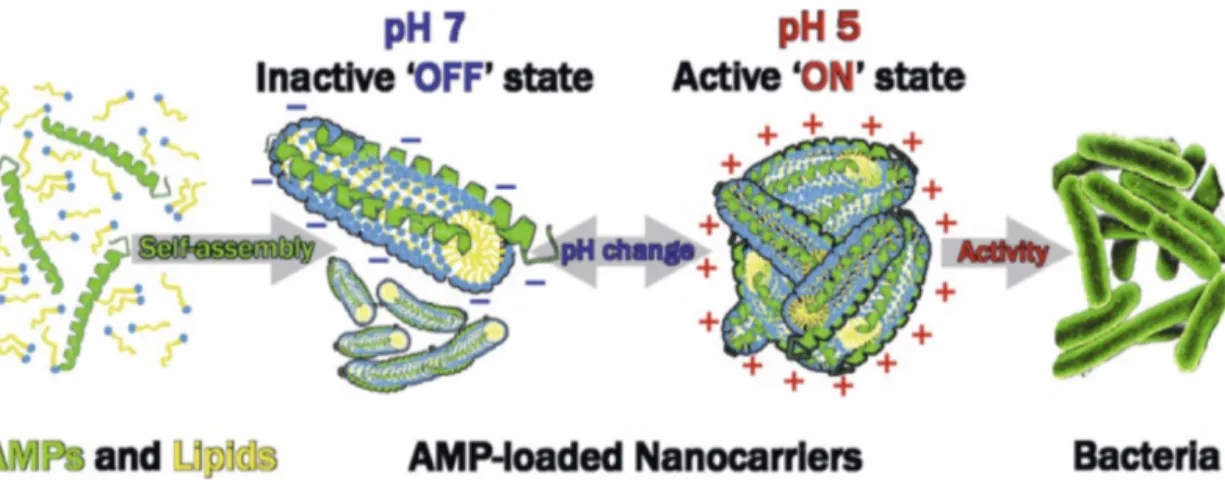
nano-biointerfaces in the form of micelles with pH-triggered biological activity for the site-specific pH-guided delivery of AMPs were de-veloped. The oleic acid (OA)/LL-37 micelles can switch between antimicrobially active and inactive states through pH-triggered structural transformations from cylindrical micelles at approxi-mately pH 7 to aggregates of branched micelles at approxiapproxi-mately
Fig. 1. An integrated online small angle X-ray scattering (SAXS) – in vitro cell co-culture model was developed and applied to study the digestion of a naturally occur-ring emulsion, milk. The impact of the in vitro cell culture on the digestion-triggered formation and evolution of highly ordered nano-structures in milk was demonstrat-ed in this study.[16a]Reprinted from
C. Hempt, M. Gontsarik, T. Buerki-Thurnherr, C. Hirsch, S. Salentinig, J. Colloid Interface Sci. 2020, 574, 430, https://doi.org/10.1016/j. jcis.2020.04.059, under Creative Commons CC-BY license.
ing the digestion of oil-in-water emulsions, including milk and mayonnaise.[16a–e]They possess a range of unique properties such
as biocompatibility, ability to modulate release, and capacity to load different types of cargo, including hydrophobic and amphi-philic molecules in their internal water-channel structure.[20,31]In
addition, based on the selection of the components and environ-mental conditions, these structures can be designed to respond to external stimuli such as pH[10c,11b,14,24d]and temperature.[30,32]
They have been investigated as carriers for the delivery of sev-eral peptides and proteins such as insulin,[33]somatostatin,[34]and
cyclosporine.[35]The nanostructured dispersions have also been
shown to effectively deliver and transport drugs to and through the skin with no observed skin irritation.[36]Non-lamellar liquid
crystalline nanoparticles have been used for the encapsulation of various AMPs, including LL-37.[10a,11,14]Intriguingly, LL-37 was
observed to spontaneously integrate into the internal nanostruc-ture of GMO- and OA-based nanoparticles when added to the aqueous phase of the dispersions (Fig. 3a).[10a,11a,b,14]The GMO/
LL‐37 micelles that resulted from the integration of LL-37 into GMO-based cubosomes were discovered to exhibit significant an-tibacterial activity, eliminating Gram‐negative E. coli faster and more efficiently than free LL‐37 (Fig. 3b).[10a]
phase transformations. For example, transforming from a slow releasing structure (inverse hexagonal phase) to the fast releas-ing cubic structure via a change in temperature,[26]or by
mod-ulating the pH to induce the electrostatic release of cargo.[27]
In particular for the inverse hexagonal phase, the ends of the rod-like water channels in the lipid phase are capped, which impedes the release of polar cargo.[10g]Another unique
charac-teristic of hexagonal phases is their ability to adhere strongly to mucosal linings, thus facilitating sustained absorption despite their slow release profile.[28] Controlling the phase behaviour
of the system can subsequently restrict the release behaviour to specific conditions, which can thus improve the efficiency of the therapeutic effect and reduce the toxicity profile of the encapsulated content.
The highly-viscous bulk phases that are stable in excess sol-vent can be dispersed as low-viscosity particles, even at high con-centrations.[29] This leads to so-called hierarchically organized
systems, where the thermodynamically stable self-assembled phases are dispersed as particles that are kinetically stabilized in the excess solvent.[30]Using recent advances in in situ SAXS
coupled to in vitro digestion models, these hierarchically orga-nized particles were recently discovered to form naturally
dur-Fig. 2. Stimuli-responsive nanocarriers formed through the self-assembly of OA with LL-37. Colloidal transformations from core–shell cylindrical mi-celles at pH 7.0 to aggregates of branched thread-like mimi-celles at pH 5.0 were detected. Antimicrobial in vitro assays using an E. coli bacterial strain showed high antimicrobial activity of the positively charged LL-37/OA aggregates at pH 5.0, which was not caused by the pH conditions them-selves. In contrast, negligible antimicrobial activity was observed at pH 7.0 for the negatively charged cylindrical micelles.[14]Reprinted with
permis-sion from M. Gontsarik, A. Yaghmur, Q. Ren, K. Maniura-Weber, S. Salentinig, ACS Appl. Mater. Interface 2019, 11, 2821, https://doi.org/10.1021/ acsami.8b18618. Copyright (2019) American Chemical Society.
Fig. 3. a) SAXS patterns of the GMO-based samples with varying GMO/LL-37 w/w ratios at 25 °C. The composition-dependent change in lat-tice dimensions and transition from cubic (Im3m) structure to sponge and lamellar structures as well as normal micelles and small vesicles was observed;[10a]b) antibacterial activity against E. coli of different LL-37 nanocarrier formulations. The GMO/LL-37 (50/50) micelles showed significant
antibacterial properties and killed more bacteria after 30 and 60 min compared with free LL-37. LL-37 formulated in cubosomes (at 95/5) showed no significant effect (ANOVA, p < 0.05). Also, unloaded cubosomes did not exhibit any antibacterial properties and were used as negative control.[10a]
Reprinted with permission from M. Gontsarik, M. T. Buhmann, A. Yaghmur, Q. Ren, K. Maniura-Weber, S. Salentinig, J. Phys. Chem. Lett. 2016, 7, 3482, https://doi.org/10.1021/acs.jpclett.6b01622. Copyright (2016) American Chemical Society.
ous cubic structures of Ia3d and Pn3m type upon increasing hy-dration (see Fig. 4).[40b]The fully hydrated Pn3m structure with a
lattice constant of around 9.6 nm could coexist in excess water, in thermodynamic equilibrium with the surrounding aqueous me-dium, which is important for biological applications such as tar-geted drug delivery from implant surfaces or wound pad materi-als. Kang et al. recently prepared GMO/1, 2-dioleoyl-3-trimeth-ylammonium-propane (DOTAP) films on coverslip substrate. These films could adopt three different structures including lamellar, hexagonal and Ia3d structures with proper control of lipid compositions, temperature, and relative air humidity.[38,39]
The films have been used for siRNA delivery and slight structur-al changes were observed compared to lipid films upon adding siRNA molecules into the lipid films via electrostatic interac-tions.[38,39,41]
Nylander et al. investigated the dynamics of the layers in the cubic (Fd3m) phase and inverse hexagonal phase on a silicon substrate surface.[42]The Fd3m layers exhibited more rigidity at
the substrate interface compared to the inverse hexagonal phase layers. The mobility at the interface depended on both the dis-tance from the supporting surface and the type of liquid crystalline phase. The finding has relevance for drug delivery and biomedical formulations, for example, in topical formulations the interfacial behaviour influences the degree of the bio-adhesion and retention of the particles at the intraoral mucosal surfaces.[42,43]
Recently, antimicrobial coatings were designed in our group through the integration of LL-37 into nanostructured GMO-based films (Fig. 5).[40a]Upon LL-37 integration, nanostructural
trans-formations were discovered, similar to the solution-based system discussed above. This conceptual correlation between solution and surface-based self-assembled structures agrees with previous reports: The formation of the various liquid crystalline film struc-tures such as inverse bicontinuous cubic (Pn3m and Ia3d),[40b,44]
inverse hexagonal,[38,42] and inverse micellar cubic (Fd3m)[42]
were reported that correlate well with the structures in the cor-responding bulk or dispersed phase system.
Usually, stabilizers in the form of amphiphilic proteins or tri-block-copolymers such as the Pluronic F127 are added to kineti-cally stabilize the self-assembled nanoparticles in water. However, potential concentration‐dependent cytotoxicity, hemolytic prop-erties, and poor biodegradability of F127 may limit the clinical ap-plication of the stabilized nanoparticles.[37]We recently designed
novel stabilizer‐free antimicrobial nanocarriers based on GMO and LL-37 that are cytocompatible and promote cell prolifera-tion for improved wound healing.[11a]Interestingly, antibacterial
assays showed that these stabilizer-free systems exhibited higher antibacterial activity compared to the GMO/LL‐37/F127 system. Also, the cell viability studies on human dermal fibroblasts (HDF) and THP‐1 cells demonstrated that the stabilizer‐free cubosomes and nanocarriers are nontoxic, contrary to the commonly used F127-stabilized systems that were found cytotoxic. In addition, cell proliferation assays on HDF cells showed that the GMO/ LL‐37 nanocarriers increase the cell proliferation by enhancing the cellular uptake of the LL‐37.[11a]
3. Antimicrobial Surface Functionalization with Lipid-based Coatings
Lipid-based self-assembled films have been studied for sub-strate-mediated gene delivery,[38]as templates for in situ metal
growth into periodic nanostructures,[39] and as host materials
to incorporate nanoparticles with macroscopic alignment.[39a]
Recently, our team demonstrated the design and application of lamellar and non-lamellar liquid crystalline coatings for the an-timicrobial functionalization of surfaces.[40]A facile method was
established to fabricate stable GMO-based coatings on surfaces by depositing GMO/ethanol mixtures using methods such as spin-coating or drop-casting; followed by solvent evaporation.[40]
Synchrotron grazing-incidence small-angle X-ray scattering (GISAXS) and atomic force microscopy (AFM) showed that the resulting GMO-based coatings self-assemble into lamellar struc-tures at low hydration with transformations to a fluid isotropic (sponge) phase followed by a second lamellar and the
bicontinu-Fig. 4. Hydration-induced transformations in the internal nanostructure of the GMO coating on silicon surface studied using in situ GISAXS. Upon hydration of the GMO thin-film, a variety of liquid crystalline nanostructures ranging from fluid isotropic (sponge-) to lamellar and bicontinuous cubic phases were observed between 5 and 100% relative humidity. The identifiable Bragg peaks and further calculated theoretical peak positions are indexed with the corresponding Miller indices for the lamellar structure in black, corresponding to a d-spacing of approximately 3.7 nm; and the Ia3d and Pn3m type cubic structures in blue and red corresponded to lattice constants of 11.5 and 9.6 nm, respectively. The scheme presents an artistic view of the nanostructural transitions from lamellar to inverse bicontinuous cubic structure, in agreement with the GISAXS data. Adapted from S. Salentinig, M. Zabara, P. Parisse, H. Amenitsch, Phys. Chem. Chem. Phys. 2018, 20, 21903, https://doi.org/10.1039/C8CP03205J with permission from the PCCP Owner Societies.
The design of AMP-loaded liquid crystalline surface coat-ings that mimic colloidal structures from natural systems, as discussed for solution systems above, appears to be a conve-nient way for surface modification. Depending on the selec-tion of molecules, the coatings can be food grade and bio-compatible, as well as stimuli-responsive (Fig. 5). Stimuli-induced phase changes in these coatings, similar to the ones discussed in the solution part above, can be associated with distinct differences in the properties of these materials, making them a unique platform for various applications, including bio-sensing, nano-templating, and drug delivery.[39a,41]
For the rational design of such bio-inspired functional coat-ings, a detailed understanding of the interactions of the colloidal structures with the substrate as well as the correlation between structure and (biological) activity is key. However, so far, very little research has been published on these systems that require highly contemporary experimental methods for characterisation, as discussed below.
4. Methods for Nanomaterial Characterization
The characterization of self-assembled nanostructures in so-lution and on surfaces in their ‘native’ state can be a challenging task. As these systems are sensitive to environmental conditions such as temperature and hydration, methods need to be care-fully selected to minimize interference of the experiment with the structure. Usually, these systems are studied using scatter-ing methods (light, X-ray and neutron scatterscatter-ing) combined with imaging techniques such as cryogenic electron microscopy.[45]
To study colloidal structures on surfaces, synchrotron GISAXS and bio-AFM are often applied to study the sample under native (hydrated) conditions or even under water using suitable sample environments.[40b]
Scattering methods are powerful tools for the characterization of nanostructures in solution and on surfaces as they are non-invasive and provide statistical information on the structures in the scattering volume. However, they are indirect methods and the interpretation of the data in real space can be a complex task. In this context, the indirect Fourier transformation method (IFT) provides model-free information on the form factor describing the shape, size and morphology of the particles.[46]The generalized
IFT (GIFT) method simultaneously considers a potential structure factor contribution to the signal, resulting from concentration-de-pendent interparticle interactions.[47]Model-dependent fitting of
the form factor scattering are also commonly applied. The theo-retical scattering curve for a specific geometry is calculated, and parameters such as size, electron densities and morphology can be optimized to obtain the best possible fit to the experimental data.[48]Static light scattering is a technique similar to SAXS and
neutrons, however, it is best suited for the characterization of larger (micron-sized) objects such as emulsions due to the much longer wavelength used.[49]Dynamic light scattering (DLS) has
no such size restriction and can be used throughout the entire colloidal regime. It measures the collective diffusion of the
col-loidal system and is mostly used for particle sizing. The diffusion behaviour is also influenced by particle interactions and gives in-dependent access to these effects.[47a]The study of dynamic
self-assembly processes is only possible through recent advances in experimental techniques and sample manipulations that provide access to the mesoscopic length scale with a high spatio-temporal resolution. Examples of these processes include the response of self-assembled systems to temperature or pressure jumps or the study of enzyme-triggered nanostructure formation such as dur-ing digestion. Additionally, online time-resolved SAXS at high intensity synchrotron sources, as well as combining the technique with microfluidics, can be used for the real-time monitoring of dy-namic structural alterations within fractions of seconds.[50]In situ
GISAXS, potentially combined with custom-made sample envi-ronments such as solvent cells, is a crucial technique to gain in-sights into the nanostructure formation and transformation on sur-faces.[40b,51]However, scattering techniques are statistical methods
providing an average over all structures in the scattering volume. The determination of coexisting structures requires the ap-plication of techniques that can resolve the characteristics of individual nanoparticles in solution. Here, electron microscopy techniques such as cryogenic transmission or scanning electron microscopy (cryo-TEM/cryo-SEM) are integral approaches to gaining further insight into the morphology and can distinguish the possible coexistence different colloidal structures in solu-tion. The application of cryo-TEM to study lipid nanoparticles has been reviewed recently.[52]The coupling of these biophysical
methods with biochemical assays and advanced in vitro models biological assays is crucial to bridge the colloidal structure to the biological activity. In our team , we combined the in vitro intesti-nal digestion model with integrated cell co-culture model and also online SAXS and we could demonstrate the in situ formation of the nanostructures during the digestion of food emulsions.[16a]We
also used the combination of the biophysical evaluation studies using methods such as SAXS, cryo-TEM, ellipsometry and AFM with antimicrobial assays on clinically relevant bacterial strains which led to the design and development of novel antimicrobial nanocarriers and coatings discussed in this contribution.
Acknowledgments
This work was supported by the Swiss National Science Foundation through the National Center of Competence in Research Bio-Inspired Materials.
Received: June 14, 2020
[1] a) T. Wei, Q. Yu, H. Chen, Adv. Healthcare Mater. 2019, 8, 1801381, DOI: 10.1002/adhm.201801381; b) T. Mérian, J. M. Goddard, J. Agri. Food Chem.
2012, 60, 2943, DOI: 10.1021/jf204741p; c) K. Bethke, S. Palantöken, V.
Andrei, M. Roß, V. S. Raghuwanshi, F. Kettemann, K. Greis, T. T. K. Ingber, J. B. Stückrath, S. Valiyaveettil, K. Rademann, Adv. Funct. Mater. 2018, 28, 1800409, DOI: 10.1002/adfm.201800409.
[2] a) Z. K. Zander, M. L. Becker, ACS Macro Lett. 2018, 7, 16, DOI: 10.1021/acsmacrolett.7b00879; b) J. L. Harding, M. M. Reynolds, Trends in Biotechnol. 2014, 32, 140, DOI: 10.1016/j.tibtech.2013.12.004; c) C.
Fig. 5. Antibacterial coatings based on the co-assembly of selected lipids and antimicrobial peptides. These nanostructured coatings can be functionalized with an ‘on/off’ switch for their an-timicrobial activity.[40a]Stimuli such
as temperature, pH, humidity or bacterial metabolites can trigger the activity.
[22] I. Ahmad, W. R. Perkins, D. M. Lupan, M. E. Selsted, A. S. Janoff, Biochim. Biophys. Acta - Biomembr. 1995, 1237, 109, DOI: 10.1016/0005-2736(95)00087-J.
[23] S. Ron-Doitch, B. Sawodny, A. Kühbacher, M. M. N. David, A. Samanta, J. Phopase, A. Burger-Kentischer, M. Griffith, G. Golomb, S. Rupp, J. Control. Release 2016, 229, 163, DOI: 10.1016/j.jconrel.2016.03.025.
[24] a) V. Luzzati, H. Delacroix, A. Gulik, T. Gulik-Krzywicki, P. Mariani, R. Vargas, in ‘Studies in Surface Science and Catalysis’, Vol. 148, Ed. O. Terasaki, Elsevier, 2004, p. 17, DOI: 10.1016/S0167-2991(04)80191-5; b) V. Luzzati, R. Vargas, P. Mariani, A. Gulik, H. Delacroix, J. Mol. Biol. 1993, 229, 540, DOI: 10.1006/jmbi.1993.1053; c) K. Larsson, J. Phys. Chem.
1989, 93, 7304, DOI: 10.1021/j100358a010; d) S. Salentinig, L. Sagalowicz,
O. Glatter, Langmuir 2010, 26, 11670, DOI: 10.1021/la101012a; e) S. Mele, O. Söderman, H. Ljusberg-Wahrén, K. Thuresson, M. Monduzzi, T. Nylander, Chem. Phys. Lipids 2018, 211, 30, DOI: 10.1016/j.chemp-hyslip.2017.11.017; f) H. Qiu, M. Caffrey, Biomater. 2000, 21, 223, DOI: 10.1016/S0142-9612(99)00126-X.
[25] a) S. Engström, T. P. Nordén, H. Nyquist, Eur. J. Pharm. Sci. 1999, 8, 243, DOI: 10.1016/S0928-0987(99)00012-3; b) F. Caboi, G. S. Amico, P. Pitzalis, M. Monduzzi, T. Nylander, K. Larsson, Chem. Phys. Lipids 2001, 109, 47, DOI: 10.1016/S0009-3084(00)00200-0.
[26] W. K. Fong, T. Hanley, B. J. Boyd, J. Control. Release 2009, 135, 218, DOI: 10.1016/j.jconrel.2009.01.009.
[27] T. H. Kim, K. Kwon, D. S. Yoo, S.-J. Lee, C. J. Ma, J. Ahn, J.-C. Kim, J. Disper. Sci. Technol. 2018, 40, 119, DOI: 10.1080/01932691.2018.1467325. [28] J. D. Du, Q. Liu, S. Salentinig, T. H. Nguyen, B. J. Boyd, Int. J. Pharm.
2014, 471, 358, DOI: 10.1016/j.ijpharm.2014.05.044.
[29] S. Salentinig, A. Yaghmur, S. Guillot, O. Glatter, J. Colloid Interface Sci.
2008, 326, 211, DOI: 10.1016/j.jcis.2008.07.021.
[30] L. de Campo, A. Yaghmur, L. Sagalowicz, M. E. Leser, H. Watzke, O. Glatter, Langmuir 2004, 20, 5254, DOI: 10.1021/la0499416.
[31] a) A. C. Groo, N. Matougui, A. Umerska, P. Saulnier, Int. J. Nanomed. 2018, 13, 7565, DOI: 10.2147/IJN.S180040; b) S. Sadiq, M. Imran, H. Habib, S. Shabbir, A. Ihsan, Y. Zafar, F. Y. Hafeez, LWT - Food Sci. Technol. 2016, 71, 227, DOI: 10.1016/j.lwt.2016.03.045.
[32] A. Yaghmur, S. Al-Hosayni, H. Amenitsch, S. Salentinig, Langmuir 2017, 33, 14045, DOI: 10.1021/acs.langmuir.7b03078.
[33] H. Chung, J. Kim, J. Y. Um, I. C. Kwon, S. Y. Jeong, Diabetologia 2002, 45, 448, DOI: 10.1007/s00125-001-0751-z.
[34] C. Cervin, P. Vandoolaeghe, C. Nistor, F. Tiberg, M. Johnsson, Eur. J. Pharm. Sci. 2009, 36, 377, DOI: 10.1016/j.ejps.2008.11.001.
[35] L. B. Lopes, D. A. Ferreira, D. de Paula, M. T. Garcia, J. A. Thomazini, M. C. Fantini, M. V. Bentley, Pharm. Res. 2006, 23, 1332, DOI: 10.1007/ s11095-006-0143-7.
[36] a) L. Boge, K. Hallstensson, L. Ringstad, J. Johansson, T. Andersson, M. Davoudi, P. T. Larsson, M. Mahlapuu, J. Håkansson, M. Andersson, Eur. J. Pharm. Biopharm. 2019, 134, 60, DOI: 10.1016/j.ejpb.2018.11.009; b) M. Sala, R. Diab, A. Elaissari, H. Fessi, Int. J. Pharm. 2018, 535, 1, DOI: 10.1016/j.ijpharm.2017.10.046; c) S. Jain, N. Patel, M. K. Shah, P. Khatri, N. Vora, J. Pharm. Sci. 2017, 106, 423, DOI: 10.1016/j.xphs.2016.10.001. [37] a) I. Hamad, A. C. Hunter, S. M. Moghimi, J. Control. Release 2013, 170,
167, DOI: 10.1016/j.jconrel.2013.05.030; b) I. D. M. Azmi, P. P. Wibroe, L.-P. Wu, A. I. Kazem, H. Amenitsch, S. M. Moghimi, A. Yaghmur, J. Control. Release 2016, 239, 1, DOI: 10.1016/j.jconrel.2016.08.011; c) U. Bazylinska, J. Kulbacka, J. Schmidt, Y. Talmon, S. Murgia, J. Colloid Interface Sci.
2018, 522, 163, DOI: 10.1016/j.jcis.2018.03.063.
[38] M. Kang, C. Leal, Adv. Funct. Mater. 2016, 26, 5610, DOI: 10.1002/ adfm.201600681.
[39] a) D. Steer, M. Kang, C. Leal, Nanotechnol. 2017, 28, 142001, DOI: 10.1088/1361-6528/aa5d77; b) S. J. Richardson, M. R. Burton, P. A. Staniec, I. S. Nandhakumar, N. J. Terrill, J. M. Elliott, A. M. Squires, Nanoscale
2016, 8, 2850, DOI: 10.1039/C5NR06691C.
[40] a) S. Salentinig, M. Zabara, WO 2020/035483 A1, 2020; b) S. Salentinig, M. Zabara, P. Parisse, H. Amenitsch, Phys. Chem. Chem. Phys. 2018, 20, 21903, DOI: 10.1039/C8CP03205J.
[41] M. Kang, M. Tuteja, A. Centrone, D. Topgaard, C. Leal, Adv. Funct. Mater.
2018, 28, 1704356, DOI: 10.1002/adfm.201704356.
[42] T. Nylander, O. Soltwedel, M. Ganeva, C. Hirst, J. Holdaway, M. Y. Arteta, M. Wadsäter, J. Barauskas, H. Frielinghaus, O. Holderer, J. Phys. Chem. B
2017, 121, 2705, DOI: 10.1021/acs.jpcb.6b11038.
[43] J. Barauskas, L. Christerson, M. Wadsäter, F. Lindström, A.-K. Lindqvist, F. Tiberg, Mol. Pharm. 2014, 11, 895, DOI: 10.1021/mp400552u.
[44] a) M. Rittman, H. Amenitsch, M. Rappolt, B. Sartori, B. M. D. O’Driscoll, A. M. Squires, Langmuir 2013, 29, 9874, DOI: 10.1021/la401580y; b) M. Rittman, M. Frischherz, F. Burgmann, P. G. Hartley, A. Squires, Soft Matter
2010, 6, 4058, DOI: 10.1039/C002968H; c) M. Wadsäter, J. Barauskas, T.
Nylander, F. Tiberg, Soft Matter 2013, 9, 8815, DOI: 10.1039/C3SM51385H. [45] Y. Fan, Y. Wang, Curr. Opin. Colloid Interface Sci. 2019, 42, 1, DOI:
10.1016/j.cocis.2019.02.011.
[46] a) O. Glatter, J. Appl. Crystallogr. 1977, 10, 415, DOI: doi:10.1107/ S0021889877013879; b) O. Glatter, in ‘Scattering Methods and their R. Arciola, D. Campoccia, P. Speziale, L. Montanaro, J. W. Costerton,
Biomater. 2012, 33, 5967, DOI: 10.1016/j.biomaterials.2012.05.031; d) D. Campoccia, L. Montanaro, C. R. Arciola, Biomater. 2013, 34, 8533, DOI: 10.1016/j.biomaterials.2013.07.089.
[3] a) A. Lewies, L. H. Du Plessis, J. F. Wentzel, Probiot. Antimicrob. Prot.
2019, 11, 370, DOI: 10.1007/s12602-018-9465-0; b) D. I. Andersson, D.
Hughes, J. Z. Kubicek-Sutherland, Drug Resis. Updates 2016, 26, 43, DOI: 10.1016/j.drup.2016.04.002.
[4] a) H. Jenssen, P. Hamill, R. E. Hancock, Clin. Microbiol. Rev. 2006, 19, 491, DOI: 10.1128/CMR.00056-05; b) R. E. Hancock, D. S. Chapple, Antimicrob. Agents Chemother. 1999, 43, 1317.
[5] D. C. Dallas, A. Guerrero, N. Khaldi, P. A. Castillo, W. F. Martin, J. T. Smilowitz, C. L. Bevins, D. Barile, J. B. German, C. B. Lebrilla, J. Proteome Res. 2013, 12, 2295, DOI: 10.1021/pr400212z.
[6] P. M. Hwang, N. Zhou, X. Shan, C. H. Arrowsmith, H. J. Vogel, Biochem.
1998, 37, 4288, DOI: 10.1021/bi972323m.
[7] J. Delves-Broughton, Int. J. Dairy Technol. 1990, 43, 73, DOI: 10.1111/ j.1471-0307.1990.tb02449.x.
[8] M. Enserink, Science 1999, 286, 2245, DOI: 10.1126/science.286.5448.2245. [9] M.-D. Seo, H.-S. Won, J.-H. Kim, T. Mishig-Ochir, B.-J. Lee, Molecules
2012, 17, 12276, DOI: 10.3390/molecules171012276.
[10] a) M. Gontsarik, M. T. Buhmann, A. Yaghmur, Q. Ren, K. Maniura-Weber, S. Salentinig, J. Phys. Chem. Lett. 2016, 7, 3482, DOI: 10.1021/acs. jpclett.6b01622; b) T. G. Meikle, A. Zabara, L. J. Waddington, F. Separovic, C. J. Drummond, C. E. Conn, Colloid Surf. B: Biointerf. 2017, 152, 143, DOI: 10.1016/j.colsurfb.2017.01.004; c) R. Negrini, R. Mezzenga, Langmuir
2011, 27, 5296, DOI: 10.1021/la200591u; d) R. Nordström, M. Malmsten,
Adv. Colloid Interface Sci. 2017, 242, 17, DOI: 10.1016/j.cis.2017.01.005; e) A. Angelova, V. M. Garamus, B. Angelov, Z. Tian, Y. Li, A. Zou, Adv. Colloid Interface Sci. 2017, 249, 331, DOI: 10.1016/j.cis.2017.04.006; f) D. J. McClements, Adv. Colloid Interface Sci. 2018, 253, 1, DOI: 10.1016/j. cis.2018.02.002; g) S. Milak, A. Chemelli, O. Glatter, A. Zimmer, Eur. J. Pharm. Biopharm. 2019, 139, 279, DOI: 10.1016/j.ejpb.2019.04.009; h) L. M. Were, B. Bruce, P. M. Davidson, J. Weiss, J. Food Prot. 2004, 67, 922, DOI: 10.4315/0362-028x-67.5.922; i) S. Salentinig, Curr. Opin. Colloid Interface Sci. 2019, 39, 190, DOI: 10.1016/j.cocis.2019.02.002.
[11] a) M. Zabara, B. Senturk, M. Gontsarik, Q. Ren, M. Rottmar, K. Maniura-Weber, R. Mezzenga, S. Bolisetty, S. Salentinig, Adv. Funct. Mater.
2019, 29, 1904007, DOI: 10.1002/adfm.201904007; b) M. Gontsarik, M.
Mohammadtaheri, A. Yaghmur, S. Salentinig, Biomater. Sci. 2018, 6, 803, DOI: 10.1039/c7bm00929a; c) L. Boge, H. Bysell, L. Ringstad, D. Wennman, A. Umerska, V. Cassisa, J. Eriksson, M. L. Joly-Guillou, K. Edwards, M. Andersson, Langmuir 2016, 32, 4217, DOI: 10.1021/acs.langmuir.6b00338; d) L. Boge, A. Umerska, N. Matougui, H. Bysell, L. Ringstad, M. Davoudi, J. Eriksson, K. Edwards, M. Andersson, Int. J. Pharm. 2017, 526, 400, DOI: 10.1016/j.ijpharm.2017.04.082.
[12] a) R. Eckert, Fut. Microbiol. 2011, 6, 635, DOI: 10.2217/fmb.11.27; b) M. Mahlapuu, J. Håkansson, L. Ringstad, C. Björn, Front. Cellul. Infect. Microbiol. 2016, 6, DOI: 10.3389/fcimb.2016.00194.
[13] O. Glatter, S. Salentinig, Curr. Opin. Colloid Interface Sci. 2020, DOI: 10.1016/j.cocis.2020.05.003.
[14] M. Gontsarik, A. Yaghmur, Q. Ren, K. Maniura-Weber, S. Salentinig, ACS Appl. Mater. Interface 2019, 11, 2821, DOI: 10.1021/acsami.8b18618. [15] J. Zempleni, A. Aguilar-Lozano, M. Sadri, S. Sukreet, S. Manca, D. Wu, F.
Zhou, E. Mutai, J. Nutr. 2017, 147, 3, DOI: 10.3945/jn.116.238949. [16] a) C. Hempt, M. Gontsarik, T. Buerki-Thurnherr, C. Hirsch, S. Salentinig,
J. Coll. Interf. Sci. 2020, 574, 430, DOI: 10.1016/j.jcis.2020.04.059; b) S. Salentinig, S. Phan, A. Hawley, B. J. Boyd, Angew. Chem. Int. Ed. 2015, 54, 1600, DOI: 10.1002/anie.201408320; c) S. Salentinig, L. Sagalowicz, M. E. Leser, C. Tedeschi, O. Glatter, Soft Matter 2011, 7, 650, DOI: 10.1039/ C0SM00491J; d) S. Salentinig, H. Amenitsch, A. Yaghmur, ACS Omega
2017, 2, 1441, DOI: 10.1021/acsomega.7b00153; e) S. Salentinig, S. Phan,
J. Khan, A. Hawley, B. Boyd, ACS nano 2013, 7, DOI: 10.1021/nn405123j; f) A. Yaghmur, S. Lotfi, S. A. Ariabod, G. Bor, M. Gontsarik, S. Salentinig, Front. Bioeng. Biotechnol. 2019, 7, DOI: 10.3389/fbioe.2019.00384. [17] J. S. Dangi, S. P. Vyas, V. K. Dixit, Drug Dev. Ind. Pharm. 1998, 24, 681,
DOI: 10.3109/03639049809082372.
[18] J. Guo, T. Wu, Q. Ping, Y. Chen, J. Shen, G. Jiang, Drug Deliv. 2005, 12, 35, DOI: 10.1080/10717540590889691.
[19] R. Innocenti Malini, M. Zabara, M. Gontsarik, K. Maniura-Weber, R. M. Rossi, F. Spano, S. Salentinig, RSC Adv. 2020, 10, 8291, DOI: 10.1039/ C9RA10037G.
[20] a) S. Cantor, L. Vargas, A. O. Rojas, C. J. Yarce, C. H. Salamanca, J. Onate-Garzon, Int. J. Mol. Sci. 2019, 20, DOI: 10.3390/ijms20030680; b) P. da Silva Malheiros, D. J. Daroit, N. P. da Silveira, A. Brandelli, Food Microbiol.
2010, 27, 175, DOI: 10.1016/j.fm.2009.09.013; c) V. Juang, H. P. Lee, A. M.
Lin, Y. L. Lo, Int. J. Nanomed. 2016, 11, 6047, DOI: 10.2147/IJN.S117618; d) A. I. Gomaa, C. Martinent, R. Hammami, I. Fliss, M. Subirade, Front. Chem. 2017, 5, 103, DOI: 10.3389/fchem.2017.00103.
[21] P. Wessman, M. Morin, K. Reijmar, K. Edwards, J. Colloid Interface Sci.
License and Terms
This is an Open Access article under the terms of the Creative Commons Attribution License CC BY_NC 4.0. The material may not be used for commercial purposes. The license is subject to the CHIMIA terms and conditions: (http://
chimia.ch/component/sppagebuilder/?view=page&id=12).
The definitive version of this article is the electronic one that can be found at https://doi.org/10.2533/chimia.2020.674
Application in Colloid and Interface Science’, Ed. O. Glatter, Elsevier, 2018, p. 33, DOI: 10.1016/B978-0-12-813580-8.00003-1.
[47] a) O. Glatter, in ‘Scattering Methods and their Application in Colloid and Interface Science’, Ed. O. Glatter, Elsevier, 2018, p. 223; b) B. Weyerich, J. Brunner-Popela, O. Glatter, J. Appl. Crystallogr. 1999, 32, 197, DOI: doi:10.1107/S0021889898011790.
[48] J. S. Pedersen, Adv. Colloid Interface Sci. 1997, 70, 171, DOI: 10.1016/ S0001-8686(97)00312-6.
[49] H. Lindner, G. Fritz, O. Glatter, J. Colloid Interface Sci. 2001, 242, 239. [50] A. Ghazal, M. Gontsarik, J. P. Kutter, J. P. Lafleur, D. Ahmadvand, A.
Labrador, S. Salentinig, A. Yaghmur, J. Phys. Chem. Lett. 2017, 8, 73, DOI: 10.1021/acs.jpclett.6b02468.
[51] a) I. Gunkel, X. Gu, Z. Sun, E. Schaible, A. Hexemer, T. P. Russell, J. Polym. Sci. B: Polym. Phys. 2016, 54, 331, DOI: 10.1002/polb.23933; b) G. Fritz-Popovski, S. C. Bodner, F. Sosada-Ludwikowska, G. A. Maier, R. Morak, L. Chitu, L. Bruegemann, J. Lange, H.-G. Krane, O. Paris, Rev. Sci. Instr. 2018, 89, 035103, DOI: 10.1063/1.5005879; c) S. Jaksch, T. Gutberlet, P. Müller-Buschbaum, Curr. Opin. Colloid Interface Sci. 2019, 42, 73, DOI: 10.1016/j. cocis.2019.04.001.
[52] S. Helvig, I. D. M. Azmi, S. M. Moghimi, A.Yaghmur, AIMS Biophys. 2015, 2, 116, DOI: 10.3934/biophy.2015.2.116.