HAL Id: hal-02105436
https://hal.archives-ouvertes.fr/hal-02105436
Submitted on 6 May 2021
HAL is a multi-disciplinary open access
archive for the deposit and dissemination of
sci-entific research documents, whether they are
pub-lished or not. The documents may come from
teaching and research institutions in France or
abroad, or from public or private research centers.
L’archive ouverte pluridisciplinaire HAL, est
destinée au dépôt et à la diffusion de documents
scientifiques de niveau recherche, publiés ou non,
émanant des établissements d’enseignement et de
recherche français ou étrangers, des laboratoires
publics ou privés.
Distributed under a Creative Commons Attribution| 4.0 International License
ultraviolet B radiation on the Mediterranean plankton
food web
Francesca Vidussi, Behzad Mostajir, Eric Fouilland, Emilie Le Floc’H, Jean
Nouguier, Cécile Roques, Patrice Got, Delphine Thibault, Thierry Bouvier,
Marc Troussellier
To cite this version:
Francesca Vidussi, Behzad Mostajir, Eric Fouilland, Emilie Le Floc’H, Jean Nouguier, et al.. Effects of
experimental warming and increased ultraviolet B radiation on the Mediterranean plankton food web.
Limnology and Oceanography Bulletin, American Society of Limnology and Oceanography, 2011, 56
(1), pp.206-218. �10.4319/lo.2011.56.1.0206�. �hal-02105436�
Effects of experimental warming and increased ultraviolet B radiation on the
Mediterranean plankton food web
Francesca Vidussi,
a,*Behzad Mostajir,
aEric Fouilland,
a,bEmilie Le Floc’h,
a,bJean Nouguier,
aCe´cile Roques,
aPatrice Got,
aDelphine Thibault-Botha,
cThierry Bouvier,
aand Marc Troussellier
aaEcosyste`mes Lagunaires Unite´ Mixte de Recherche (UMR) 5119, Centre National de la Recherche Scientifique (CNRS), Universite´
Montpellier 2, Institut de Recherche pour le De´veloppement (IRD), Institut franc¸ais de recherche pour l’exploitation de la mer (IFREMER), Montpellier, France
bEcosyste`mes Lagunaires UMR 5119 CNRS-Universite´ Montpellier 2- IRD-IFREMER, Station Me´diterrane´enne de l’environnement
Littoral, Se`te, France
cLaboratoire d’Oce´anographie Physique et Bioge´ochimique UMR 6535, Universite´ Aix-Marseille, CNRS, Centre d’Oce´anologie de
Marseille, Marseille, France
Abstract
The responses of the plankton food web to increases in temperature and ultraviolet B radiation (UVBR, 280– 320 nm) were experimentally investigated at a coastal Mediterranean site during spring. Eight moored mesocosms were used to compare natural plankton food web responses (control mesocosms) with three treatments simulating expected future local temperature and UVBR increases, as follows: (1) 3uC increase in water temperature, (2) 20% increase in incident UVBR, and (3) simultaneous 3uC increase in water temperature and 20% increase in incident UVBR. The plankton food web was resistant to elevated UVBR, having only moderate effects on plankton abundances and structure. In contrast, warming induced significant shifts in the plankton food web structure and function. Specifically, the abundance of protozooplankton (ciliates and flagellates) increased and the development time of copepods from nauplii to adults decreased. In the warm mesocosms, the emergence of copepod adult stages midway through the experiment resulted in a decrease in ciliates and consequently in an increase in heterotrophic flagellates. One unexpected result was that warming reduced the abundance of heterotrophic bacteria midway through the experiment. These results indicate a trophic-cascade effect under warming. The increase in adult copepods diminishes ciliates and in turn favors heterotrophic flagellates that consume bacteria. Warming also induced an increase in net oxygen production, indicating an increase in net primary production.
Understanding how organisms respond to multiple environmental stressors induced by global changes requires a better comprehension of the complex ecological mecha-nisms that determine these responses. Responses of organisms to a stressor can occur at the individual level as physiological acclimation (Portner and Farrell 2008) and depend on the tolerance of the species to the stressor (Vinebrooke et al. 2004). Global change can also modify community composition by promoting tolerant species and reducing sensitive ones (Vinebrooke et al. 2004). The simultaneous individual- and community-level responses of organisms to a stressor alter natural interactions among organisms, including predator–prey, competitive, mutual-istic, and parasitic interactions (Davis et al. 1998; Walther et al. 2002), inducing cascading effects along the trophic food web (Weitere et al. 2008).
Previously published studies have mostly focused on the effect of one stressor at the individual level or at a single trophic level. Very few studies have been dedicated to understanding the response mechanisms of the entire aquatic plankton food web to global changes. Aquatic ecosystem functioning is based on the activity of planktonic organisms that are linked by complex trophic, competitive, and mutualistic interactions. Therefore, to evaluate the effect of climate change in aquatic environments, it is critical to understand the response of the entire plankton
food web under global change stressors and the associated ecological mechanisms driving these responses. The re-sponse of the plankton food web to global change is complex and nonlinear. It is difficult to model and predict these complex responses based on results at the individual level and/or on a single stressor. This is because different species and trophic levels display variable sensitivities to a given stressor and because multiple stressors can interact by amplifying or suppressing the effects of other stressors (Vinebrooke et al. 2004; Christensen et al. 2006).
Recent studies investigating the effects of increased temperature on plankton communities have shown that warming modifies the magnitude and composition of the spring phytoplankton bloom (Sommer and Lengfellner 2008). By favoring microbial carbon respiration, warming may ultimately enhance CO2 release from the ocean (Va´zquez-Domı´nguez et al. 2007). The few experiments that have incorporated several trophic levels have shown that, in general, warming adversely affects planktonic consumers while promoting producers (Beisner et al. 1997; Petchey et al. 1999; Strecker et al. 2004). In addition, shifts in the plankton community structure driven by global warming could decouple trophic interactions at higher trophic levels, modifying the temporal matching between predators and their prey (Beaugrand et al. 2003; Edwards and Richardson 2004).
Another stressor linked to global changes that can affect plankton community structure is increased ultraviolet B
* Corresponding author: [email protected]
doi:10.4319/lo.2011.56.1.0206
radiation (UVBR, 280–320 nm). Very few studies on the effect of UVBR have taken into account the entire plankton food web. These studies show complex responses indicating positive feedbacks of UVBR (Sommaruga 2003). Mostajir et al. (1999) have shown that UVBR reduces large planktonic microorganisms (ciliates and diatoms), resulting in accumulation of small microorganisms, such as hetero-trophic flagellates, bacteria, and picophytoplankton.
Recently several studies also focused on another stressor linked to global changes, the increase of atmospheric CO2 and consequent ocean acidification, but very few have been dedicated to elucidating the effects of this stressor on the entire plankton food web (Riebesell et al. 2007) or benthic food web (Hargrave et al. 2009). It has been shown that interactions among multiple stressors due to global change better explain their combined effect on ecosystem func-tioning, compared with the sum of the individual effects of each stressor (Christensen et al. 2006). However, few multifactorial experiments have been dedicated to studying the effect of multiple global change stressors on aquatic ecosystems, and very few have studied the effects at several trophic levels. For example, it has been found that the effect of UVBR may be temperature dependent because temperature enhances photo-enzymatic repair (Williamson et al. 2002; MacFadyen et al. 2004). Another study (Doyle et al. 2005) has shown that the effect of UVBR on phytoplankton growth is not only temperature dependent but also nutrient dependent. Therefore, studying the response of the entire plankton food web to multiple stressors linked to global change with an emphasis on direct (i.e., physiological) and indirect (i.e., positive or negative feedbacks) effects of complex interactions is essential to evaluating how global change will modify the structure and function of aquatic systems.
Water temperature and incident UVBR are expected to increase in coming decades as a result of concomitant global warming and ozone reduction (Weatherhead and Andersen 2006; IPCC 2007). Models predict an increase of 2.2–5.1uC in annual average temperature in the Mediterra-nean region by 2080–2099 (IPCC 2007). UVBR is expected to increase in European regions by about 10% in coming years, particularly during spring (Reuder et al. 2001). However, UVBR recovery to past levels is expected to occur between 2040 and 2070 (WMO 2007). In the context of these environmental changes, shallow coastal waters will be particularly affected by future temperature and UVBR increases. These ecosystems are particularly sensitive to these changes as a result of their relatively shallow depth and lengthy water residence time. In addition, modification of plankton community structure and functioning will have direct socio-economic implications because coastal waters are highly productive areas in terms of primary, secondary, and exploited-resources production (e.g., shellfish farming; Costanza et al. 1997).
The objective of the present study is to investigate the effects of single and combined temperature and UVBR increases on a coastal Mediterranean plankton food web. In particular, changes in the structure of the microbial components were studied during the spring productive season. A factorial mesocosm experiment that applied
increases of 3uC and 20% UVBR (relative to natural conditions) was carried out during spring 2006. These increases correspond to the mean temperature increase in the Mediterranean region expected by 2080–2099 (IPCC 2007) and the high-UVBR scenario for the European region during spring in future years (Reuder et al. 2001).
Methods
Experimental design—A mesocosm experiment was carried out from 29 March to 07 April 2006 at the Mediterranean platform for Marine Ecosystem Experimen-tal Research (MEDIMEER). MEDIMEER is located in Se`te in the South of France (43u249490N, 3u419190E) on the shore of Thau Lagoon, a coastal Mediterranean lagoon. Mesocosms were 3 m high, 1.2 m wide, and were made of 200-mm–thick vinylacetate mixed-polyethylene film (Insin-o¨o¨ritoimisto Haikonen Ky). The polyethylene film trans-mitted 53% and 77% of UVBR and photosynthetically available radiation (400–700 nm), respectively (Nouguier et al. 2007). Mesocosms were held at 1 m above the water surface by floating structures. Mesocosms were covered with the same polyethylene material during the night and during rain events to prevent contamination and water exchange.
Eight moored mesocosms were simultaneously filled on 29 March 2006 (hereafter called day 1) with pooled and screened (, 1000-mm) subsurface (1-m) lagoon water. Mesocosms were filled with water pumped at the subsur-face (1 m) near the MEDIMEER deck (maximum depth , 3 m). Each mesocosm had a maximum water column depth of 2 m and contained 2260 liters of lagoon water. At the end of the experiment, the remaining water volume was approximately 1800 liters. The water column in each mesocosm was constantly homogenized using a pump (Iwaki). The turnover time was set at 1 h and it was adjusted daily using a flowmeter according to the remaining water volume of each mesocosm after sampling. Previous tests showed that the pump did not affect abundances of bacteria, phytoplankton, heterotrophic flagellates, and ciliates, but the effect of pumping and the homogenization system on more fragile species is not excluded.
Four treatments were applied in duplicate mesocosms: (1) Control (C), (2) 20% increase in UVBR (UV) compared to natural incident UVBR received at the water surface in control mesocosms, (3) 3uC increase in water temperature (T) compared to control mesocosms, and (4) combined increase in UVBR (20%) and water temperature (3uC) (UVT). Treatments began on 30 March (day 2) for enhanced temperature and on 31 March (day 3) for increased UVBR.
Increased temperature and UVBR treatments—Water temperature was increased using a submersible heating element (Galvatec) immersed vertically at a depth of , 1 m (Nouguier et al. 2007). Incident UVBR at the water surface was increased using custom-built frames holding two UV fluorescent lamps (Philips TL20RS/01), a double electronic ballast (Bag electronics AD18.22310) controlled by voltage
input, and an ultra-pure polished aluminum reflector. The lamp system was covered with a di-acetate cellulose sheet (which was changed daily) to avoid artificial ultraviolet C radiation. The lamp system was placed on the floating structure at 91 cm above the water surface.
An automatic system to regulate water temperature and UVBR enhancement was developed specifically for this experiment and has been described in Nouguier et al. (2007). We briefly describe the functioning of this automatic system here. The automatic system was based on closed-loop regulation. For temperature, regulation followed several steps: (1) monitoring of water temperature in all mesocosms every 30 s using thermistor probes (Campbell Scientific 107), (2) comparison of water temper-atures in the control (reference temperature) and warmed mesocosms using a Campbell Scientific data logger (CR23X, Campbell Scientific), (3) monitoring of the deviation of water temperature in the warmed mesocosms from the target value (reference temperature plus 3.1uC), and (4) starting the heating elements to reach the target value in the warmed mesocosms.
An automatic device was also designed to perform closed-loop regulation of increased UVBR. Incident UVBR was monitored every 30 s by a UVB radiometer (Skye ultraviolet sensor 430, Skye Instruments) installed on the roof of the control center to avoid shading. To better monitor UVBR increase, a master–slave system was implemented because of the impossibility of obtaining correct UVBR measurements under in situ conditions at the water surface. The master unit was installed in the control data center on a structure mimicking the upper part of the mesocosm. The slave units were placed above the experimental mesocosms. To monitor the UVBR delivered by the fluorescent lamps of the master unit, a UVB radiometer (Skye ultraviolet sensor 430, Skye Instruments) was installed at the base of the master structure. In addition, each fluorescent lamp on the master and slave mesocosms was individually monitored by a custom-built photodiode system (sglux TW30DZ) to control for any eventual lamp failure or discrepancy between the master and the slaves. Regulation of increased UVBR consisted of several steps: (1) rapid sampling (2 Hz) of incident and artificially enhanced UVBR, (2) real-time comparison of incident and enhanced UVBR, (3) establishment of the target value (incident UVBR multiplied by a coefficient), and (4) sending the proportional command to the UV lamps. The UVBR lamps and regulation system were turned on at 09:45 h and switched off at 17:45 h local time (local zenith time was 13:45 h).
Physico-chemical sampling and measurements—Meso-cosm temperature was monitored frequently (every 30 s) at three depths (0.4 m, 0.8 m, and 1.2 m) using thermistor probes (Campbell Scientific 107) from 30 March (day 2) to 07 April (day 10). Incident UVBR and enhanced UVBR were monitored by UVB radiometers from 31 March (day 3) to 07 April (day 10). UVBR (305 nm) was measured daily in the water column of mesocosms and in Thau Lagoon beside the mesocosms using a UV profiling radiometer (PUV-2500, Biospherical Instruments). The
depths at which radiation corresponded to 1% of the UVB surface radiation were compiled for each treatment and for the lagoon throughout the experiment. The quantity of available data varied among treatments because the 1% depth occasionally exceeded the maximum depth reached by the profiling radiometer, depending on the shape of the mesocosm bags (C: n 5 10; UV: n 5 17; T: n 5 5; UVT: n 5 11; lagoon: n 5 9).
All mesocosms were sampled daily from 29 March (day 1) to 07 April (day 10) using 20-liter polycarbonate carboys cleaned with acid. Samples were taken at 09:00 h, before UVB lamps were switched on, except on 29 March (when samples were taken at 21:00 h, after filling the mesocosms). Aliquots were then sampled from the 20-liter polycarbon-ate carboys for chemical and biological analyses, except for metazooplankton and oxygen production analysis (see details below).
Nitrates (NO3+NO2) and phosphates (PO4) were mea-sured in filtered (pre-combusted glass-fiber filters, 47-mm, Whatman GF/F) 80-mL aliquots using an automated colorimeter (Skalar), following standard nutrient analysis methods (Treguer and Le Corre 1975). Ammonium concentrations were measured in triplicate in unfiltered samples (50 mL) using a spectrophotometer (Hitachi U-3000), following the indophenol blue method (Koroleff 1983).
Plankton biomass, abundance, and composition sampling and analysis—For metazooplankton analysis, samples (60 liters) were taken on days 2, 6, and 8 using three 20-liter polycarbonate containers and screened onto a 60-mm sieve to collect and concentrate zooplankton in neutralized formalin (4% final concentration). Metazooplankton was taxonomically enumerated using a Leica MZ6 dissecting microscope, following the technique of Rose (1933).
Heterotrophic flagellates (HF) and ciliates were sampled on days 2, 4, 6, 8, and 10. For HF enumeration, aliquots (30 mL) were preserved with formaldehyde (4% final concentration). Next, 10-mL subsamples were stained with 49,69-diamidino-2-phenyindole hydrochloride and filtered onto 25-mm black nucleopore polycarbonate membranes (0.2-mm pore size). Each filter was placed on a microscope slide, and HF were enumerated using an epifluorescence microscope (Olympus AX-70) with a 1003 objective. For ciliate enumeration and identification, 125-mL aliquots were preserved with acid Lugol’s solution (0.4% final concentration). Samples were then sedimented for 24 h in an Utermo¨hl’s chamber and observed under an inverted microscope (Olympus IX-70).
Phytoplankton samples were collected daily (500 mL) for microscopic identification and preserved with formalde-hyde (8% final concentration) at 4uC until analysis. Samples were then sedimented for 24 h in an Utermo¨hl’s chamber and observed under an inverted microscope (Olympus IX-70).
Chlorophyll a (Chl a) samples (0.5–1 liter) were collected daily, filtered on glass-fiber filters (25 mm, 0.7-mm nominal pore size, Whatman GF/F) at low vacuum (, 200 mm Hg), and stored immediately in liquid nitrogen and then at 280uC until analysis. Chl a was extracted in 3 mL of 95%
methanol for 1 h at 220uC, sonicated with a sonication probe for a few seconds, stored at 4uC for 1 h, and then clarified on glass-fiber filters (25 mm, Whatman GF/F). Chl a was analyzed by high-performance liquid chroma-tography (HPLC), following the method of Zapata et al. (2000), with some adaptations for the HPLC system used. The HPLC system was composed of a pump (600E, Waters) equipped with a 200-mL loop, an automatic refrigerated (4uC) injector (717plus autosampler, Waters), and a photodiode array detector (2996 PDA, Waters). Chl a was identified based on retention time and the absorbance spectra obtained with the photodiode array. The HPLC was calibrated with commercial standards (DHI and Sigma).
Heterotrophic bacteria (hereafter called bacteria) sam-ples (1.6 mL) were collected daily and preserved with formaldehyde (2% final concentration), then frozen in liquid nitrogen and stored at 280uC until analysis. Bacterial samples were thawed at room temperature, and then nucleic acids were stained for 15 min in the dark at 4uC with SYBR Green I (1 : 10,000 vol : vol, Molecular Probes), according to the method described by Marie et al. (1997). Samples were analyzed using a FACSCalibur flow cytometer (Becton Dickinson) equipped with an air-cooled argon laser (488 nm, 15 mW). Bacteria were enumerated using their green fluorescence (FL1) collected at 530 nm. Yellow–green fluorescent cytometry beads of 1-mm and 2-mm diameters (Polysciences) were added to the samples as internal standards for cell size and fluorescence emission, and TruCount control beads (Becton Dickinson) were added to determine the analyzed volumes.
For virus enumeration, subsamples were taken daily, fixed with 0.02-mm of filtered buffered formaldehyde (2% final concentration), and stored at 280uC until further analysis. After thawing, viruses were stained with SYBR Gold nucleic acid stain (Molecular Probes), as described by Chen et al. (2001), and filtrated (15-kPa vacuum) through 0.02-mm pore-size Anodisc membrane filters (Whatman). Virus-like particles were counted under blue light (488 nm) using an Olympus AX-70 epifluorescence microscope.
Oxygen production and dark community respiration rates—Net microbial oxygen production and dark com-munity respiration rates were measured every 2 d (on days 2, 4, 6, 8, and 10) in all mesocosms by following changes in dissolved oxygen concentrations in quartz and in borosil-icate darkened bottles, respectively, both of which were incubated during the light period (from ca. 09:00 h to ca. 18:00 h) in the mesocosms (ca. 0.50-m depth). Mesocosm water for these incubations was sampled using 9-liter carboys cleaned with acid and was kept in darkness. Six 120-mL borosilicate bottles and two 120-mL quartz bottles were carefully filled from each carboy using a silicone tube. Three borosilicate bottles were immediately fixed (time zero) using reagents prepared following the recommenda-tions of Carritt and Carpenter (1966).
The two quartz bottles and three other darkened borosilicate bottles were incubated in their respective mesocosms for about 9 h and were then fixed as described above. Dissolved oxygen concentrations were determined
using an automated Winkler titrator based on potentio-metric detection (Crisson). Net oxygen production rate was calculated as the difference between the dissolved oxygen concentrations measured at time zero and at the end of the incubation period in the quartz bottles. Dark community oxygen respiration rate was calculated as the difference between the dissolved oxygen concentrations measured at time zero and at the end of the incubation period in the darkened bottles. Rates were expressed in mg O2L21h21. Gross oxygen production rates were calculated as the sum of net oxygen production and dark oxygen respiration rates.
Microbial plankton carbon biomass estimates—Microbial carbon biomass was estimated using the following carbon conversion factors: 20 fg C cell21 for bacteria (Sime-Ngando et al. 1995), 0.22 pg C mm23for HF (Booth 1993), and 0.21 pg C mm23for ciliates (Putt and Stoecker 1989); a C : Chl a ratio of 57 was used to estimate the carbon biomass of phytoplankton (Latasa et al. 2005). Total microbial carbon biomass was then computed as the sum of the estimated carbon biomass of bacteria, phytoplankton, heterotrophic flagellates, and ciliates.
Statistical analysis—Repeated-measures analyses of var-iance (RM-ANOVAs) were used to test the effects of UVBR, temperature, and simultaneous increases in UVBR and temperature on plankton abundance and biomass, with days as a repeated factor (SYSTAT version 11). Because the number of subjects (mesocosms) was less than the number of factors (days), univariate tests were computed. All analyses were performed on log10-transformed data to normalize variance, and sphericity was verified before performing RM-ANOVA; p-values less than or equal to 0.05 were considered to indicate statistically significant differences.
Results
Physico-chemical conditions—Average daily temperature naturally increased from day 2 to day 6 from 13.2uC to 15.4uC in control and UV mesocosms (Fig. 1a), followed by a decrease to 14.6uC and then stabilization at 15.1uC. Warmed mesocosms (T and UVT) followed the same trend as controls, with a constant daily difference of 3.1uC (Fig. 1a), except on day 2, when heating began.
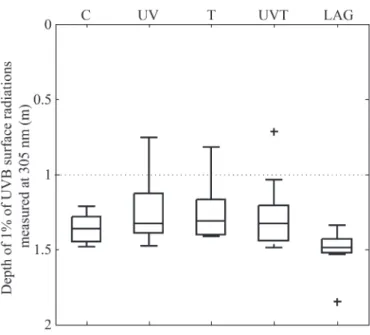
Natural daily UVBR doses received at the water surface of the mesocosms varied from 20 to 47 kJ m22(Fig. 1b), with half the UVBR dose received on cloudy days (day 3). The increased-UVB mesocosms (UV and UVT) received precisely 20% more than the natural daily UVBR, and the maximum daily UVBR dose (natural+added UVBR) never exceeded 57 kJ m22 (Fig. 1b). Less-penetrant UVB radiation (305 nm) reached the lower half of the mesocosm water column on most days of the experiment, as shown in Fig. 2. The 1% depth of UVBR did not differ significantly between treatments and was comparable to the 1% depth measured in the adjacent lagoon water column (Fig. 2).
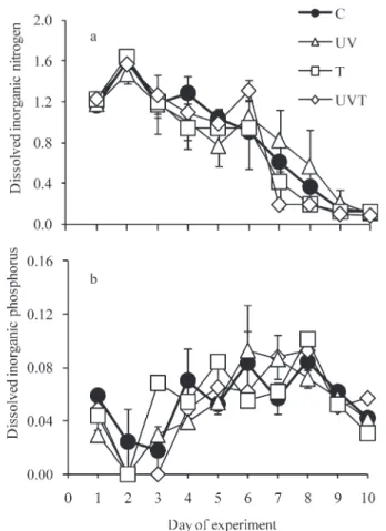
Dissolved inorganic nitrogen (DIN, NO3+NO2+NH4) concentrations ranged from 0.1 to 1.6 mmol L21(treatment
average; Fig. 3a). DIN concentrations increased slightly on the first day of the experiment and then generally decreased in all treatments throughout the experiment (Fig. 3a). Dissolved inorganic phosphorous (DIP, PO4) concentra-tions ranged from undetectable levels to 0.09 mmol L21 (treatment average; Fig. 3b). DIP concentrations decreased to undetectable levels on day 2 or day 3 (according to the treatment), but then tended to increase until day 8 for all treatments, except in UV mesocosms, where DIP tended to increase until day 6 (Fig. 3b). Later, DIP concentrations slightly decreased in all treatments at the end of the experiment. DIN and DIP concentrations did not differ significantly between control mesocosms and treatments.
Meta- and protozooplankton—The metazooplankton community was dominated by copepods, cirripeds,
troco-phore and oyster larvae, a few gastropod mollusks (Limacina sp.), and decapods. Small (ca. 1.2-mm) calanoid copepods (mainly Temora sp. and Centropages sp.) dominated the metazooplankton community in all meso-cosms throughout the experiment. At the beginning of the experiment, only copepodite and nauplii stages were observed. The most interesting result was the significant positive effect of warming on copepod development time, as shown by the earlier observation of adult stages, particularly females, in warmed treatments than in control and increased-UV treatments (on day 6 in T and UVT; Fig. 4; Table 1). This faster development of calanoid copepods was also observed at the nauplii level, as nauplii abundance decreased (through progression into copepodite stages) faster under warmer treatments (T and UVT; Fig. 4c; Table 1). From day 8, copepod adult stages were also observed in C and UV treatments, but in lesser abundance than those observed in T and UVT. Earlier formed females will also have earlier spawning capacities, as observed in the increased number of nauplii between days 6 and 8 in the T and UVT treatments.
The HF community was dominated by small cells (3– 5 mm in diameter). HF abundance responded positively to warming. HF abundances observed on day 6 under T and UVT treatments were 217% and 113% higher, respectively, than those observed in controls (Fig. 5a). This increase under warming was confirmed by inverted-microscope observations of abundant small (3–5-mm) choanoflagellates on day 6.
Ciliates were dominated by small ciliates (10–25 mm). The ciliate community was dominated by oligotrich ciliates
Fig. 1. (a) The daily average water temperature in non-heated mesocosms (white bar, control and increased UVB) and the increased temperature in heated mesocosms (gray bars, increased temperature and simultaneous UVB and temperature increase mesocosms) during the experiment. Symbols represent averages of the daily values (averages of measurements made every 20 s at three different depths) of the two mesocosms receiving the same treatment. (b) The average daily UVBR dose received at the water surface: white bars indicate the natural UVBR dose received by all mesocosms, and gray bars indicate the artificial UVBR dose added in increased UVB and simultaneous UVB and temperature increase treatments.
Fig. 2. Variability of the depth of 1% UVB penetration (305 nm) for each treatment and in the lagoon (LAG) throughout the experiment. Horizontal lines in each box display the lower quartile, median, and upper quartile values. Whiskers show the range of the rest of the data. The maximum whisker length corresponds to 1.5 units of the interquartile range. Crosses represent outliers. The horizontal dotted line at 1 m shows the middle depth of the water column in the mesocosm bags.
(Strombilidium, Strombidium, and Strombidinopsis). Warm-ing significantly affected ciliate abundance (Table 1), but in opposite directions for short-term and long-term effects (Fig. 5b). On day 4, ciliate abundance was greater under warming (by 166% and 86% for T and UVT, respectively), relative to controls. However, on days 8 and 10, ciliate abundance under warming (T and UVT) was as low as 86% of the abundance measured in controls (Fig. 5b).
Phytoplankton, bacteria, and viruses—The phytoplank-ton community was initially composed of cryptophytes, naked dinoflagellates, diatoms, and prasinophytes in all mesocosms. This community shifted to a community dominated by diatoms and prymnesiophytes at the end of the experiment (results not shown). A phytoplankton bloom occurred at days 7 and 9 for warmer and control mesocosms, respectively, as shown by maximum Chl a concentrations (Fig. 6a). Microscopic observations indicat-ed that the phytoplankton bloom observindicat-ed was mainly dominated by the chain-forming diatom Cerataulina pelagica (Cleve) Hendey 1937 (data not shown).
There was no significant treatment effect on bulk phytoplankton as measured by Chl a (Table 1). However, warming (T and UVT) induced an earlier increase in biomass; maximum Chl a biomass was observed 2 d earlier in warmer mesocosms than in control mesocosms (days 7 and 9 for warmer and control mesocosms, respectively;
Fig. 3. (a) DIN and (b) DIP under different treatments: Control (C); 20% increase in ultraviolet B incidence (UV); 3uC increase in temperature (T); simultaneous increases in temperature (3uC) and ultraviolet B incidence (20%) (UVT). Symbols represent means for each treatment (averages of the two mesocosms receiving the same treatment), and error bars indicate the ranges of the observations. Note that DIN on day 1 is the sum of NO3
and NO2only (NH4was not measured on this day).
Fig. 4. Abundances of (a) female copepods, (b) male copepods, and (c) nauplii under different treatments: Control (C); 20% increase in ultraviolet B incidence (UV); 3uC increase in temperature (T); simultaneous increases in temperature (3uC) and ultraviolet B incidence (20%) (UVT). Symbols represent means for each treatment (averages of the two mesocosms receiving the same treatment), and error bars indicate the ranges of the observations.
Fig. 6a). Nevertheless, maximum Chl a concentrations were similar in all treatments (around 1.8 mg L21).
Temporal patterns of bacterial abundance were similar under all treatments, with an initial rapid increase from day 1 to day 3 followed by a decline in abundance until day 7 or day 8, depending on the treatment. Subsequently, constant levels were observed until the end of the experiment (Fig. 6b). Warmer treatments displayed significantly re-duced bacterial abundance from day 5 to day 7 (by 38%
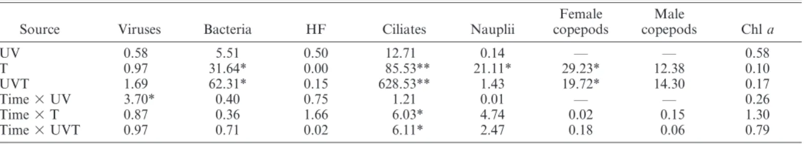
Table 1. RM-ANOVA results for the effects of increased ultraviolet B radiation (UV), increased temperature (T), and simultaneously increased ultraviolet B radiation and temperature (UVT) and time on viruses, heterotrophic bacteria, heterotrophic flagellates (HF), ciliates, nauplii, female copepods, male copepods, and Chl a. Data given are F-values.
Source Viruses Bacteria HF Ciliates Nauplii
Female copepods Male copepods Chl a UV 0.58 5.51 0.50 12.71 0.14 — — 0.58 T 0.97 31.64* 0.00 85.53** 21.11* 29.23* 12.38 0.10 UVT 1.69 62.31* 0.15 628.53** 1.43 19.72* 14.30 0.17 Time 3 UV 3.70* 0.40 0.75 1.21 0.01 — — 0.26 Time 3 T 0.87 0.36 1.66 6.03* 4.74 0.02 0.15 1.30 Time 3 UVT 0.97 0.71 0.02 6.11* 2.47 0.18 0.06 0.79 * p#0.05; ** p#0.01.
Fig. 5. Abundances of (a) HF and (b) ciliates under different treatments: Control (C); 20% increase in ultraviolet B incidence (UV); 3uC increase in temperature (T); simultaneous increases in temperature (3uC) and ultraviolet B incidence (20%) (UVT). Symbols represent means for each treatment (averages of the two mesocosms receiving the same treatment), and error bars indicate the ranges of the observations.
Fig. 6. (a) Chl a concentrations and abundances of (b) bacteria and (c) viruses under different treatments: Control (C); 20% increase in ultraviolet B incidence (UV); 3uC increase in temperature (T); simultaneous increases in temperature (3uC) and ultraviolet B incidence (20%) (UVT). Symbols represent means for each treatment (averages of the two mesocosms receiving the same treatment), and error bars indicate the ranges of the observations.
and 48% for T and UVT, respectively) (Fig. 6b; Table 1), compared to controls. The general pattern of virus abundance showed a decrease from the beginning of the experiment until day 5 (day 6 for UV), followed by an increase, with maximum values reached on day 7 or day 8, depending on the treatment. Warming and increased UVBR had no significant effects on virus abundance (Fig. 6c). However, UVBR delayed by 1 d the decline in virus abundance, which was observed on day 6 in UV and on day 5 under other treatments.
Microbial carbon standing stocks—Estimated microbial carbon biomass was dominated by bacteria, with averaged mesocosm biomass ranging from 71 to 152 mg C L21 (Fig. 7), representing an average of 56% 6 11% (all mesocosms throughout the experiment) of the total microbial carbon biomass. Average phytoplankton carbon concentrations ranged from 43 to 100 mg C L21 (Fig. 7), representing an average of 35% 6 9% of the total microbial carbon biomass. Protozooplankton (HF and ciliates) carbon concentrations were generally low (Fig. 7), contrib-uting averages of 3% 6 3% and 1% 6 1% of total microbial carbon biomass, respectively. Warming (T and UVT) induced a decrease in bacterial carbon biomass and an increase in phytoplankton, especially on days 6 and 8
(Fig. 7). UVBR alone generally had minor effects on microbial carbon biomass.
Oxygen production and respiration—Community net oxygen production ranged from 20.005 to 0.041 mg O2 L21 h21, but most measured values were positive (i.e., mostly oxygen production instead of respiration; Fig. 8a). Community gross oxygen production and dark respiration ranged from 0.001 to 0.048 mg O2L21h21and from 0.002 to 0.010 mg O2L21h21, respectively (Fig. 8b,c). Net oxygen production generally increased throughout the experiment in all mesocosms, except on day 2 in the UVT treatment and on day 4 in the control and UV treatments, when negative values (i.e., mostly oxygen respiration) were measured (Fig. 8a). Increased temperature, alone or combined with increased UVBR, increased net oxygen production by 10% to 106%, respectively, relative to values measured in controls (Fig. 8a). In contrast, increased UVBR alone had no effect, and community net oxygen production levels were similar to those measured in controls (Fig. 8a). Community gross oxygen production followed the same pattern as the net oxygen production (Fig. 8b), with values measured on day 6, under increased temperature alone or combined with increased UVBR, measuring 12 and 15 times higher than those measured in controls (Fig. 8b). In contrast, the
Fig. 7. HF, ciliate, phytoplankton, and bacterial carbon biomasses under (a) control, (b) increased ultraviolet B radiation (UV), (c) increased temperature (T), and (d) simultaneous increased temperature and ultraviolet B radiation (UVT).
different treatments (T, UVT, and UV) had no significant effects on the community dark respiration rates even if a slight increase was observed at the beginning of the experiment under increase of temperature alone and combined with increased UVBR (days 4 and 6; Fig. 8c).
Discussion
It is widely recognized that temperature has a major influence on the physiology of organisms because it controls basic metabolic processes. In particular, maximal growth rates of both autotrophic and heterotrophic organisms increase with temperature (Eppley 1972; Rose and Caron 2007). A major result of our study was the significant response of heterotrophic predators to warming, inducing increases in copepod, ciliate, and HF abundances (until day 4 for ciliates and until day 6 for HF and copepods). Indeed, copepods developed more rapidly under warmer conditions. At the beginning of the experiment, only copepodites were observed, and these copepodites developed into adults 2 d faster (day 6) under warming conditions than in other treatments (day 8). This result is consistent with those of several studies that have reported temperature-dependent development of zooplankton, par-ticularly reduced copepod development time from eggs to adults with increasing temperatures (Hart and McLaren 1978). Spawning will also occur sooner when water temperatures are increased.
Heterotrophic protists have been demonstrated to have high potential growth rates and a rapid response to increasing temperature (Caron et al. 1991), like the ciliates in this experiment. Ciliate abundance increased only a few days after applying the warming treatment. Larger standing stocks of ciliates with increased temperature were correlated to relatively greater abundance of their potential prey (i.e., small flagellates) and scarcity of their potential predators (i.e., adult copepods) at the beginning of the experiment. Weak top-down control of ciliates at the beginning of the experiment is supported by the fact that the standing stock of ciliates declined from day 6 onward under warming, as soon as their potential predators (adult copepods) appeared on day 6 in warmed mesocosms. The decline in ciliates after day 6 under warming explains the concomitant increase of their potential prey (HF). There-fore, our results illustrate a trophic cascade effect (copepods–ciliates–HF) under warming. Heterotrophic protists tightly control the abundance of their prey because they have rapid growth rates similar to those of their prey at intermediate and high temperatures (above 15uC for herbivorous protists; Rose and Caron 2007). The results of the present study also support the capacity of metazoan copepods to develop faster in warmer waters (at temper-atures between 16uC and 18uC). Consequently, copepods can also tightly control the abundance of their prey. Therefore, we did not observe a mismatch between increases in microplankton and zooplankton development, as suggested for Baltic plankton food webs under global warming (Edwards and Richardson 2004). In contrast, we observed rapid growth of copepods, illustrating a typical increase in predator abundance following an increase in abundance of its prey. This indicates an efficient transfer of carbon between microplankton and metazooplankton.
An unexpected result of this experiment was the reduced abundance of heterotrophic bacteria under warming. Viruses did not seem to be affected by temperature, at least in terms of abundance; thus, viral infection is
Fig. 8. (a) Net and (b) gross oxygen production and (c) dark oxygen respiration under different treatments: Control (C); 20% increase in ultraviolet B incidence (UV); 3uC increase in temperature (T); simultaneous increases in temperature (3uC) and ultraviolet B incidence (20%) (UVT). Symbols represent means for each treatment (averages of the two mesocosms receiving the same treatment), and error bars indicate the ranges of the observations.
apparently not the cause of bacterial decrease. Therefore, the decrease in bacterial abundance was probably due to stronger grazing control under warming. This observation is consistent with that reported by Weitere et al. (2008) for natural river plankton; they showed a decrease in bacterial abundance under warming, indicating a food web cascade effect. It has been shown that bacterivorous protists have greater maximal growth rates than herbivorous protists. Thus, they can control their prey more efficiently (Rose and Caron 2007). In addition, bacterivorous grazing rates can be enhanced with increasing temperature (reviewed in Rose and Caron [2007]). Thus, even if temperature can poten-tially enhance bacterial growth rates, increasing their abundance in the absence of bacterivorous protists, bacterial standing stocks may be strongly controlled by bacterivorous protists such as HF. Substrate availability (quality and quantity), which interactively limits bacterial growth with temperature (Pomeroy and Wiebe 2001), is apparently not the cause of the bacterial decrease under warming. Bacterial abundances increased considerably at the beginning of the experiment in all mesocosms, including warmed mesocosms. Conjointly dissolved organic carbon was relatively abundant (F. Vidussi unpubl.). Thus, bacteria were probably not substrate limited, at least at the beginning and at the midway of the experiment.
The increase in microbial predators (principally ciliates) and the positive response of adult copepods (as indicated by their faster appearance) under warming explain the small effect of warming on standing stocks of phytoplank-ton (i.e., Chl a). Two hypotheses can be proposed to explain how zooplankton controlled standing stocks of phytoplankton under warming. The first hypothesis is that herbivorous microzooplankton had growth rates similar to those of phytoplankton at temperatures encountered during the experiment (13–15uC in unheated mesocosms and 16–18uC in warmed mesocosms; Fig. 1a) and con-trolled phytoplankton standing stocks until the midway point of the experiment. Then at the midway point of the experiment, adult copepods appeared and therefore con-trolled phytoplankton standing stocks. The second hypoth-esis is that increased temperature enhanced herbivorous grazing rates. Recently, Rose and Caron (2007) have reviewed the effect of temperature on microzooplankton growth rates and have reported relationships between temperature and maximal growth rates of herbivorous protists, copepods, and their phytoplankton prey. They have shown that maximal growth rates of herbivorous protists exceed maximal growth rates of phototrophic protists (phytoplankton) at temperatures above 15uC, indicating that the mismatch between the growth rates of phytoplankton predators and their prey at temperatures below 15uC can explain phytoplankton blooms. During our study, temperatures in unheated mesocosms were always less than 15uC, except on day 6. In contrast, temperatures in heated mesocosms were always greater than 15uC. Thus, herbivorous protists could have higher growth rates than phytoplankton and thereby control standing stocks of phytoplankton, particularly small phytoplankton, until the midway point of the experiment. Our second hypothesis is that warming enhanced herbivorous grazing rates
(re-viewed in Rose and Caron [2007]; Larsen and Riisgard [2009]). Even if phytoplankton growth was greater under warming, temperature-enhanced grazing rates may have prevented phytoplankton biomass accumulation. Phyto-plankton did not appear to be especially more nutrient limited under warmer conditions; it increased in all mesocosms, and no significant differences in nutrient concentrations were observed between warmer and control mesocosms. However, nutrient limitation may have oc-curred, particularly at the end of the experiment, which could have provoked stronger nutrient competition be-tween different phytoplankton groups and bebe-tween phyto-and bacterioplankton.
Overall, increased UVBR had moderate effects on the structure of the plankton food web. Indeed, only slight decreases in phytoplankton biomass (Chl a; Fig. 6a) and bacterial abundance were observed under increased UVBR (Fig. 6b). The negative effect of increased UVBR on bacteria and phytoplankton, although not statistically significant, might be due to a direct effect and not to an interaction-mediated effect because clear effect of UVBR on predators was not observed. These results are consistent with previous observations, which have indicated that UVBR effects on aquatic ecosystems are subtle rather than drastic (Vincent and Roy 1993). The moderate effects of increased UVBR on the entire plankton food web observed in this study might be explained by the moderate natural daily UVBR dose combined with a relatively UVBR-resistant plankton community in early spring in the northern Mediterranean. Even if increased UVBR during our study exceeded natural doses, it was probably insufficient to induce effective damage in plankton, and repair mechanisms might remain efficient under these conditions. Furthermore, even though potentially damaging UVBR reached most of the mesocosm water column, the water-column mixing applied during the experiment may have contributed to the moderate UVBR effect. Natural mixing is known to facilitate repair processes and reduce the exposure of plankton to UVBR (Neale et al. 1998).
The moderate responses of the Mediterranean microbial food web and copepod communities to increased UVBR observed during this study contrast with the findings of previous studies carried out in coastal waters (Mostajir et al. [1999], in the St. Lawrence Estuary) that have shown significant effects of increased UVBR on large phytoplank-ton and ciliates. Comparison of the present study with previous studies that have reported significant harmful effects of UVBR on planktonic food webs allows us to underline two major concerns. The first concern is the enhanced UVBR dose applied during the experiment. We emphasize the need to apply realistic UVBR doses that mimic future increases in UVBR, taking into account the natural variability of UVBR. The second consideration is that the responses of plankton food webs to increased UVBR depend on local conditions. The effects of UVBR depend not only on the life histories of the organisms (acclimation) but also on the adaptations of local organisms to specific stresses. Therefore, local conditions (e.g., light and temperature levels, resource supply) must be carefully taken into consideration to better predict the effects of
global changes on the functioning and structure of plankton food webs. The results presented here demonstrate that the expected future increase in UVBR has no statistically significant effects at the community level or on the structure of the marine food web during a spring bloom in the Mediterranean Sea. However, our results do not exclude the possibility that natural UVBR levels in the Mediterranean Sea are damaging to sensitive species, as previously reported for summer communities (Sommaruga et al. 2005).
The simultaneous increase in temperature and UVBR generally had no synergistic effects on the plankton food web structure during this study because of the moderate effects of UVBR, as described above. The results showed similar trends in abundance and biomass under warming alone and under warming plus increased UVBR for the majority of planktonic organisms studied here. Therefore, the Mediterranean plankton food web during spring would respond more strongly to future expected warming than to future increases in UVBR.
The experiment described here was of relatively short duration (10 d). Therefore, the validity and robustness of our findings with respect to future global changes may be questioned. This study was designed to elucidate potential mechanisms involved in the functioning of the microbial plankton food web under warming and increased UVBR. A 10-d period corresponds to several generations of micro-organisms, especially in relatively warm waters like those of the Mediterranean. Thus, this short duration is sufficient to highlight potential biologically mediated feedback mecha-nisms, such as trophic cascade effects (Petchey et al. 1999). Further experimental studies in different marine regions and seasons, taking into account local plankton community structures and physico-chemical conditions, will be neces-sary to elucidate how global changes will generally or specifically affect the functioning of the marine plankton food web and whether temperature-mediated trophic cascades are a general rule.
The present experiment clearly shows that increased water temperature alters the functioning of the plankton food web because it increases net oxygen production, essentially through an increase in gross oxygen production. On the contrary, warming has no significant effects on community respiration. This result contrasts with those of previous studies that have shown a significant increase in microbial respiration under warming (Vazquez-Dominguez et al. 2007). The lack of a clear increase in respiration under the increased temperatures observed here is probably caused by the negative indirect effect of warming, via cascading effect, on the heterotrophic bacterial community, which generally accounts for most of the community respiration (Rivkin and Legendre 2001). It has been previously suggested that warming drives the balance in favor of respiration, consequently increasing CO2 production by the microbial community and resulting in oceanic CO2release (Vazquez-Dominguez et al. 2007; Wohlers et al. 2009). However, we do not observe a balance in favor of respiration, indicating that in relatively productive warm waters (such as Mediter-ranean coastal waters during spring), increasing temperature finally can favor net oxygen production (autotrophic O2 production) instead of respiration.
Our study shows that heterotrophs respond rapidly to warming. This result is consistent with the metabolic theory of ecology (Brown et al. 2004), which predicts a strong temperature dependence of heterotrophic metabolism compared to autotrophic metabolism. Although testing our data within the framework of the metabolic theory of ecology is not the purpose of this article, and although we did not measure specific heterotrophic activity, we point out the potential applicability of this theory to understand-ing the functionunderstand-ing of marine and terrestrial ecosystems under global warming, as suggested previously (Lopez-Urrutia and Moran 2007; Lopez-(Lopez-Urrutia 2008). Further studies should test this unifying theory by considering resource limitation and organismal interactions such as predation to better evaluate biological responses to different global change scenarios.
In conclusion, the study presented here shows that the spring Mediterranean plankton community might respond more to future expected warming conditions than to future expected UVBR increases. In particular, warming can affect biological interactions in the plankton food web, inducing trophic cascades. Our results demonstrate the need to consider the responses of the entire food web and interactions therein rather than those of separate compart-ments to describe and predict the complex and sometimes counterintuitive responses of plankton to global changes.
Acknowledgments
We thank numerous students from Montpellier 2 and Aix-Marseille Universities for performing analyses and helping with the experiment. We thank C. Bouvier, M. Bouvy, J.-P. Torreton, and R. De Wit for their support during the mesocosm experiment and B. Charriere, D. Sarno, R. Sempe´re´, and T. Sime-Ngando for help with analyses. We are grateful to Y. Vergne for field assistance. This study is part of the project ‘‘Effects of climate change on marine pelagic food webs: Ultraviolet and temperature effects,’’ supported by the French National Program on Coastal Environments (PNEC-Chantier Lagunes Mediterrae´ennes) and Groupement de Recherche (GDR) 2476 Re´seaux Trophiques Aquatiques. The experimental platform MEDIMEER was founded by Unite´ Mixte de Recherche 5119 Ecosyste`mes Lagunaires, Centre National de la Recherche Scientifique-Institut National Ecologie et Environnement, Institut Fe´de´ratif de Recherche 129 Armand Sabatier, GDR 2476 Re´seaux Trophiques Aquatiques, and Re´gion Languedoc-Roussillon.
References
BEAUGRAND, G., K. BRANDER, J. LINDLEY, S. SOUISSI,ANDP. REID.
2003. Plankton effect on cod recruitment in the North Sea. Nature 426: 661–664, doi:10.1038/nature02164
BEISNER, B., E. MCCAULEY,ANDF. WRONA. 1997. The influence of
temperature and food chain length on plankton predator-prey dynamics. Can. J. Fish. Aquat. Sci. 54: 586–595, doi:10.1139/ cjfas-54-3-586
BOOTH, B. C. 1993. Estimating cell concentration and biomass of
autotrophic plankton using microscopy, p. 199–205. In P. F. Kemp, B. F. Sherr, E. B. Sherr, and J. J. Cole [eds.], Hand-book of methods in aquatic microbial ecology. Lewis. BROWN, J., J. GILLOOLY, A. ALLEN, V. SAVAGE,ANDG. WEST. 2004.
Toward a metabolic theory of ecology. Ecology 85: 1771–1789, doi:10.1890/03-9000
CARON, D., E. LIM, G. MICELI, J. WATERBURY,AND F. VALOIS. 1991. Grazing and utilization of chroococcoid cyanobacteria and heterotrophic bacteria by protozoa in laboratory cultures and a coastal plankton community. Mar. Ecol. Prog. Ser. 76: 205–217, doi:10.3354/meps076205
CARRITT, D.,ANDJ. CARPENTER. 1966. Comparison and evaluation
of currently employed modifications of the Winkler method for determining dissolved oxygen in sea-water; A NASCO report. J. Mar. Res. 24: 286–318.
CHEN, F., J. LU, B. BINDER, Y. LIU, AND R. HODSON. 2001.
Application of digital image analysis and flow cytometry to enumerate marine viruses stained with SYBR gold. Appl. Environ. Microbiol. 67: 539–545, doi:10.1128/AEM.67.2.539-545. 2001
CHRISTENSEN, M., M. GRAHAM, R. VINEBROOKE, D. FINDLAY, M.
PATERSON, AND M. TURNER. 2006. Multiple anthropogenic
stressors cause ecological surprises in boreal lakes. Glob. Change Biol. 12: 2316–2322, doi:10.1111/j.1365-2486. 2006.01257.x
COSTANZA, R., AND oTHERS. 1997. The value of the world’s
ecosystem services and natural capital. Nature 387: 253–260, doi:10.1038/387253a0
DAVIS, A., J. LAWTON, B. SHORROCKS, AND L. JENKINSON. 1998. Individualistic species responses invalidate simple physiolog-ical models of community dynamics under global environ-mental change. J. Anim. Ecol. 67: 600–612, doi:10.1046/ j.1365-2656.1998.00223.x
DOYLE, S., J. SAROS,ANDC. WILLIAMSON. 2005. Interactive effects
of temperature and nutrient limitation on the response of alpine phytoplankton growth to ultraviolet radiation. Limnol. Oceanogr. 50: 1362–1367, doi:10.4319/lo.2005.50.5.1362
EDWARDS, M., AND A. RICHARDSON. 2004. Impact of climate
change on marine pelagic phenology and trophic mismatch. Nature 430: 881–884, doi:10.1038/nature02808
EPPLEY, R. 1972. Temperature and phytoplankton growth in the
sea. Fish. Bull. 70: 1063–1085.
HARGRAVE, C., K. GARY,ANDS. ROSADO. 2009. Potential effects of
elevated atmospheric carbon dioxide on benthic autotrophs and consumers in stream ecosystems: A test using experimen-tal stream mesocosms. Glob. Change Biol. 15: 2779–2790, doi:10.1111/j.1365-2486.2009.01897.x
HART, R.,ANDI. MCLAREN. 1978. Temperature-acclimation and
other influences on embryonic duration in copepod Pseudo-calanus. Mar. Biol. 45: 23–30, doi:10.1007/BF00388974
INTERGOVERNMENTAL PANEL ON CLIMATE CHANGE (IPCC). 2007.
Climate Change 2007: The physical science basis contribution of Working Group I to the fourth assessment report of the Intergovernmental Panel on Climate Change. Cambridge Univ. Press.
KOROLEFF, F. 1983. Determination of ammonia, p. 150–157. In
K. Grasshoff [ed.], Methods of seawater analysis. Verlag Chemie.
LARSEN, P.,ANDH. RIISGARD. 2009. Viscosity and not biological mechanisms often controls the effects of temperature on ciliary activity and swimming velocity of small aquatic organisms. J. Exp. Mar. Biol. Ecol. 381: 67–73, doi:10.1016/ j.jembe.2009.09.021
LATASA, M., X. MORAN, R. SCHAREK, AND M. ESTRADA. 2005.
Estimating the carbon flux through main phytoplankton groups in the northwestern Mediterranean. Limnol. Ocean-ogr. 50: 1447–1458, doi:10.4319/lo.2005.50.5.1447
LOPEZ-URRUTIA, A. 2008. The metabolic theory of ecology and
algal bloom formation. Limnol. Oceanogr. 53: 2046–2047. ———, AND X. MORAN. 2007. Resource limitation of bacterial
production distorts the temperature dependence of oceanic carbon cycling. Ecology 88: 817–822, doi:10.1890/06-1641
MACFADYEN, E., C. WILLIAMSON, G. GRAD, M. LOWERY, W.
JEFFREY, AND D. MITCHELL. 2004. Molecular response to
climate change: Temperature dependence of UV-induced DNA damage and repair in the freshwater crustacean Daphnia pulicaria. Glob. Change Biol. 10: 408–416, doi:10.1111/j.1529-8817.2003.00750.x
MARIE, D., F. PARTENSKY, S. JACQUET, AND D. VAULOT. 1997.
Enumeration and cell cycle analysis of natural populations of marine picoplankton by flow cytometry using the nucleic acid stain SYBR green I. Appl. Environ. Microbiol. 63: 186– 193.
MOSTAJIR, B.,ANDoTHERS. 1999. Experimental test of the effect of ultraviolet-B radiation in a planktonic community. Limnol. Oceanogr. 44: 586–596, doi:10.4319/lo.1999.44.3.0586
NEALE, P., R. DAVIS,ANDJ. CULLEN. 1998. Interactive effects of ozone depletion and vertical mixing on photosynthesis of Antarctic phytoplankton. Nature 392: 585–589, doi:10.1038/ 33374
NOUGUIER, J., B. MOSTAJIR, E. LEFLOC’H,ANDF. VIDUSSI. 2007.
An automatically operated system for simulating global change temperature and ultraviolet B radiation increases: Application to the study of aquatic ecosystem responses in mesocosm experiments. Limnol. Oceanogr.: Methods 5: 269–279.
PETCHEY, O., P. MCPHEARSON, T. CASEY, AND P. MORIN. 1999.
Environmental warming alters food-web structure and ecosystem function. Nature 402: 69–72, doi:10.1038/47023
POMEROY, L.,ANDW. WIEBE. 2001. Temperature and substrates as
interactive limiting factors for marine heterotrophic bacteria. Aquat. Microb. Ecol. 23: 187–204, doi:10.3354/ame023187
PORTNER, H., AND A. FARRELL. 2008. Physiology and climate
change. Science 322: 690–692, doi:10.1126/science.1163156
PUTT, M.,ANDD. STOECKER. 1989. An experimentally determined
carbon-volume ratio for marine oligotrichous ciliates from estuarine and coastal water. Limnol. Oceanogr. 34: 1097–1103, doi:10.4319/lo.1989.34.6.1097
REUDER, J., M. DAMERIS, AND P. KOEPKE. 2001. Future UV
radiation in Central Europe modelled from ozone scenarios. J. Photochem. Photobiol. B Biol. 61: 94–105, doi:10.1016/ S1011-1344(01)00143-9
RIEBESELL, U., AND oTHERS. 2007. Enhanced biological carbon
consumption in a high CO2 ocean. Nature 450: 545–548,
doi:10.1038/nature06267
RIVKIN, R.,ANDL. LEGENDRE. 2001. Biogenic carbon cycling in
the upper ocean: Effects of microbial respiration. Science 291: 2398–2400, doi:10.1126/science.291.5512.2398
ROSE, J.,ANDD. CARON. 2007. Does low temperature constrain
the growth rates of heterotrophic protists? Evidence and implications for algal blooms in cold waters. Limnol. Oceanogr. 52: 886–895, doi:10.4319/lo.2007.52.2.0886
ROSE, M. 1933. Faune de France. Office central de Faunistique,
Paris. [French fauna.]
SIME-NGANDO, T., M. GOSSELIN, S. ROY,ANDJ-P. CHANUT. 1995.
Significance of planktonic ciliated protozoa in the lower St Lawrence Estuary: Comparison with bacterial, phytoplank-ton and particulate organic carbon. Aquat. Microb. Ecol. 9: 243–258, doi:10.3354/ame009243
SOMMARUGA, R. 2003. UVR and its effects on species
interac-tions, p. 487–508. In E. W. Helbling and H. Zagarese [eds.], UV effects in aquatic organisms and ecosystems. The Royal Society of Chemistry.
———, J. HOFER, L. ALONSO-SAEZ, AND J. GASOL. 2005. Differential sunlight sensitivity of picophytoplankton from surface Mediterranean coastal waters. Appl. Environ. Micro-biol. 71: 2154–2157, doi:10.1128/AEM.71.4.2154-2157.2005
SOMMER, U.,ANDK. LENGFELLNER. 2008. Climate change and the
timing, magnitude, and composition of the phytoplankton spring bloom. Glob. Change Biol. 14: 1199–1208, doi:10.1111/ j.1365-2486.2008.01571.x
STRECKER, A., T. COBB, AND R. VINEBROOKE. 2004. Effects of
experimental greenhouse warming on phytoplankton and zooplankton communities in fishless alpine ponds. Limnol. Oceanogr. 49: 1182–1190, doi:10.4319/lo.2004.49.4.1182
TREGUER, P.,ANDP. LE CORRE. 1975. Manuel d’analyse des sels
nutritifs dans l’eau de mer: Utilisation de l’Autoanalyzer II Technicon, 2nd ed. Laboratoire d’Oceanographie chimique Univ. de Bretagne Occidentale. [Textbook of nutrient analysis in marine water: Utilization of the Technicon Autoanalyzer II.]
VAZQUEZ-DOMINGUEZ, E., D. VAQUE,ANDJ. GASOL. 2007. Ocean warming enhances respiration and carbon demand of coastal microbial plankton. Glob. Change Biol. 13: 1327–1334, doi:10.1111/j.1365-2486.2007.01377.x
VINCENT, W.,ANDS. ROY. 1993. Solar ultraviolet-B radiation and
aquatic primary production: Damage protection and recov-ery. Environ. Rev. 1: 1–12.
VINEBROOKE, R., K. COTTINGHAM, J. NORBERG, M. SCHEFFER, S.
DODSON, S. MABERLY, AND U. SOMMER. 2004. Impacts of
multiple stressors on biodiversity and ecosystem functioning: The role of species co-tolerance. Oikos 104: 451–457, doi:10.1111/j.0030-1299.2004.13255.x
WALTHER, G., ANDoTHERS. 2002. Ecological responses to recent
climate change. Nature 416: 389–395, doi:10.1038/416389a
WEATHERHEAD, E.,ANDS. ANDERSEN. 2006. The search for a sign
of recovery of ozone layer. Nature 441: 39–45, doi:10.1038/ nature04746
WEITERE, M., J. DAHLMANN, C. VIERGUTZ,ANDH. ARNDT. 2008.
Differential grazer-mediated effects of high summer temper-atures on pico- and nanoplankton communities. Limnol. Oceanogr. 53: 477–486.
WILLIAMSON, C., G. GRAD, H. DE LANGE, S. GILROY, AND D.
KARAPELOU. 2002. Temperatudependent ultraviolet
re-sponses in zooplankton: Implications of climate change. Limnol. Oceanogr. 47: 1844–1848, doi:10.4319/lo.2002. 47.6.1844
WOHLERS, J.,ANDoTHERS. 2009. Changes in biogenic carbon flow
in response to sea surface warming. Proc. Natl. Acad. Sci. USA 106: 7067–7072, doi:10.1073/pnas.0812743106
WORLDMETEOROLOGICALORGANIZATION(WMO). 2007. Scientific
assessment of ozone depletion: 2006. Global Ozone Research and Monitoring Project-Report No. 50.
ZAPATA, M., F. RODRIGUEZ,ANDJ. GARRIDO. 2000. Separation of
chlorophylls and carotenoids from marine phytoplankton: A new HPLC method using a reversed phase C-8 column and pyridine-containing mobile phases. Mar. Ecol. Prog. Ser. 195: 29–45, doi:10.3354/meps195029
Associate editor: Wade W. Jeffrey Received: 22 February 2010 Accepted: 19 August 2010 Amended: 07 October 2010