HAL Id: hal-02665432
https://hal.inrae.fr/hal-02665432
Submitted on 31 May 2020HAL is a multi-disciplinary open access archive for the deposit and dissemination of sci-entific research documents, whether they are pub-lished or not. The documents may come from teaching and research institutions in France or abroad, or from public or private research centers.
L’archive ouverte pluridisciplinaire HAL, est destinée au dépôt et à la diffusion de documents scientifiques de niveau recherche, publiés ou non, émanant des établissements d’enseignement et de recherche français ou étrangers, des laboratoires publics ou privés.
Virulence strategies in parasitoid Hymenoptera as an
example of adaptive diversity.
Marylene Poirie, Yves Carton, Aurore Dubuffet
To cite this version:
Marylene Poirie, Yves Carton, Aurore Dubuffet. Virulence strategies in parasitoid Hymenoptera as an example of adaptive diversity.. Comptes Rendus Biologies, Elsevier Masson, 2009, 332 (2-3), pp.311-320. �10.1016/j.crvi.2008.09.004�. �hal-02665432�
Les stratégies de virulence des hyménoptères parasitoïdes :
un exemple de diversité adaptative.
Virulence strategies in parasitoid Hymenoptera as
an example of adaptive diversity
Marylène Poirié
1§, Yves Carton
2and Aurore Dubuffet
31
UMR « Interactions Biotiques et Santé Végétale »
Université Nice Sophia Antipolis - CNRS (UMR 6243) - INRA (UMR 1301), 400
route des Chappes,
06903 Sophia-Antipolis, France.
Tel. 33 (0)4 92 38 64 09
Fax. 33 (0)4 92 38 65 87
marylene.poirie@sophia.inra.fr
2
Equipe IRD-UR 072 « Biodiversité et Evolution des complexes plantes - insectes
ravageurs-antagonistes »
Laboratoire Evolution, Génétique et Spéciation,
CNRS 91198 Gif-sur-Yvette Cedex, France.
3
Institute of Integrative and Comparative Biology, Faculty of Biological Sciences
Clarendon Way, University of Leeds, LS2 9JT Leeds UK.
§
To whom correspondence should be addressed
.Résumé: Les insectes parasitoïdes se développent aux dépens d’autres arthropodes qui ne survivront
pas à l’interaction. Leur succès reproducteur dépend donc de leur capacité à réussir le parasitisme (virulence) tandis que celui de l’hôte dépend de sa capacité à l’éviter ou à survivre à l’infestation. Les pressions de sélection intenses exercées sur les populations d’hôte et de parasitoïde ont ainsi conduit à la sélection d’adaptations extrêmement variées dans les stratégies de virulence de ces derniers. Par exemple, des virus spécifiques sont utilisés par certaines familles de parasitoïdes pour bloquer les défenses immunitaires de l’hôte et détourner ses processus physiologiques à leur profit. Cet article synthétise les données physiologiques et moléculaires disponibles sur les stratégies de virulence des parasitoïdes et discute des processus évolutifs mis en jeu.
Mots-clés : Guêpe parasitoïde – immunité – stratégie de virulence – polydnavirus (PDV) – diversité
adaptative – évolution de la virulence
Summary : Parasitoids are mostly insects that develop at the expense of other arthropods, which will
die as a result of the interaction. Their reproductive success thus totally depends on their ability to successfully infest their host whose reproductive success relies on its own ability to avoid or overcome parasitism. Such intense selective pressures have resulted in extremely diverse adaptations in parasitoid strategies that ensure parasitism success. For instance, wasp-specific viruses (polydnaviruses) are injected into the host by parasitoid females to modulate its physiology and immunity. This paper synthesizes available physiological and molecular data on parasitoid virulence strategies and discusses the evolutionary processes at work.
Key-words : Parasitoid wasp – immunity – virulence strategies – polydnavirus (PDV) – adaptive
diversity – evolution of virulence
Besides his work on the mechanisms of artificial selection, Charles Darwin’s reading of the book from Thomas Robert Malthus (1766-1834) "An Essay on the Principle of Population" [1] seemed to have an important impact on the formulation of the principle of natural selection [2]. Among the factors limiting the proliferation of individuals of a given species, Malthus mentioned the lack of “room and nourishment”, but also the fact that “animals are becoming the prey of others”. This led Darwin to consider the “struggle for life” between and within species the biological basis of natural selection. Our current knowledge warrants the idea that interactions between species, described by the famous Darwin’s metaphor of a “tangled bank”, are an essential driving force behind the evolution of life. Darwin’s metaphor hinted that they might be complex to the point of being impossible to understand but data are accumulating at a fast pace, using various approaches ranging from ecology to molecular biology.
Each organism lives continuously in interaction with several other organisms, through relations of predation, competition, mutualism or parasitism. The two last are called “durable interactions”, a concept developed in France by Claude Combes [3]. In these durable interactions, various (and even sometimes spectacular) adaptations can be selected in each species of the interaction as a result of reciprocal selective pressures, under a coevolutionary process. Parasitoids, whose success leads eventually to the death of their host, represent one of the most prevalent lifestyles on earth and particularly original adaptations have been described in parasitoid wasps.
Even if various organisms use the parasitoid life style (as can do some nematodes, ciliates or bacteriophages…) [4,5], it is mainly observed in insects (8 to 25% of them) [6,7]. Parasitoids insects belong to 6 different orders, but more than 80% of the described species are Hymenoptera [7]. Their life style falls between parasitism and predation: they lay their eggs either at the surface (ectoparasitoids) or into (endoparasitoids) an arthropod host, generally another insect, which can be at any stage of its development (but often at the larval or pupal stage), perform their larval development
at its depends [6,8], and end-up killing their host as predators. The host death happens before the host reproduces and, consequently, its fitness directly depends on its ability to escape parasitism or to resist it. In turn, as parasitoids cannot succeed their development without hosts, their fitness directly depends on parasitism success.
Parasitoid populations are thus under strong selection pressures to evolve morphological, behavioural and physiological adaptations to localize the host habitat, localize the host within this habitat, oviposit in a suitable host, and then develop in accordance to the host physiology [9]. The 3 first steps, known as the host-selection process, are a matter of behavioural ecology. Parasitoids are a model of choice in this area since the link between behavioural strategies of females and their fitness is direct [6]. Under natural selection theory, parasitoid behavioural strategies are predicted to evolve towards a preference for the most suitable species (ensuring the maximum number of fertile offspring) and habitats that contain them [10]. This article deals with the fourth step, the host suitability, which depends on adaptations and counter-adaptations of the physiological and/or immunity level.
1. Virulence strategies in parasitoid wasps: a striking example of adaptive diversity.
To ensure the development of its progeny in a parasitized host, an endoparasitoid can stop host development (idiobiont parasitoid) or allow the host to continue developing (koinobiont parasitoid). The host physiology can then be more or less regulated, resulting in changes at the behavioural, endocrinal, nutritional or immunological levels [11]. This review will be devoted to the immunological aspects of the interaction since the great majority of parasitoids, including many ectoparasitoids, have to deal with the immune response of their hosts [12,13,14]. Towards large foreign bodies, like parasitoid eggs, the immune response of the majority of insects is the encapsulation, which consists in the formation of a multicellular, melanized capsule around the foreign body [14] (Figure I). To escape encapsulation, a striking range of virulence strategies has been evolved in parasitoids [8,13] (Table I).
Some parasitoids infest “immuno-incompetent” hosts. Trichogrammas for example, largely used in biological control against Lepidopteran pests, infest the egg stage of their hosts, which does not present any immune defense [15]. Infestation of immunodeficient species, unable to encapsulate a parasitoid egg, can also be an efficient strategy. Such kind of host species has been recently evidenced with the case of Drosophila subobscura. This Drosophila species is deficient for a category of haemocytes (the equivalent of blood cells in insects) that is necessary for the encapsulation process [16].
Against immunocompetent hosts, 2 categories of parasitic strategies have been defined: local immunoevasion and systemic immunosuppression. Parasitoids that use local immunoevasion strategies do not modify the host immune defenses. Some of them perform their development in tissues inaccessible to immune factors. Others, as the braconid Asobara tabida [17], have eggs that stick to host tissues thanks to adhesive properties of the chorion, or that present molecules or surface properties that prevent haemocytes to spread on their surface [18,19]. In all cases, a foreign body newly introduced in the parasitized host will be encapsulated, which shows that the host immune potential is intact.
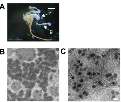
Systemic immunosuppression is on the contrary a general alteration of the host encapsulation ability, generally due to factors injected by parasitoid females during oviposition. These factors are produced by ovaries and/or by the venom gland (also called long gland), a gland associated to the genital tract [20] (Figure IIA). According to the species, they correspond to secreted proteins [21], to viral particles, the polydnaviruses (or PDVs for Polydisperse DNA Viruses) [22] or to “Virus-Like Particules” (VLPs) [23,24] (Figure II, B,C). Note that if the presence of PDVs seems to be restricted to a small number of parasitoid families, the presence or not of VLPs as well as the nature local or systemic of the virulence strategy seem to be relatively independent from phylogenic relations (Table I). PDVs are particularly original virulence factors that can be considered as a tool used by wasps to parasite their host. They represent the first example of symbiosis between a virus and a eukaryote
described up to now [25]. The nature of VLPs is still unknown and their organization extremely variable according to the considered species, their shape varying from a structure amazingly similar to viruses to shapes more or less organized (Figure II B,C).
2. The « viral tools » of parasitoid wasps : a mysterious evolutionary origin.
Polydnaviruses are found only in the superfamilly of Ichneumonoidea that parasitize lepidopteran hosts. Their genome has 2 forms: one segmented, present in virions and composed of multiple double stranded DNA circles [22, 25, 26], and a linear form integrated in the chromosomes of the parasitoid wasp and vertically transmitted to the progeny. The question whether all these viral segments are localised in tandem arrays on one unique chromosome is still discussed [27]. Virions are produced in the calyx fluid, a specialized region of ovaries of parasitoid females, and injected with the egg into the lepidopteran host where they penetrate its cells. Expression of viral genes in host cells is required for the parasitoid success, even if complementation by secretions from the venom gland, the ovaries or the calyx seems to be needed [22, 26] (Table 1). The PDVs cycle has thus two parts: the viral replication which only takes place in parasitoid females, and the gene expression of viral particles, which only occurs in their lepidopteran host.
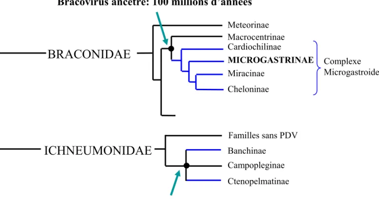
PDVs are associated to more than 30 000 parasitoid species belonging to two families [28] (Figure III). Bracoviruses are associated to species belonging to the braconid family, which constitute a monophyletic group called the microgastroid complex. Only one integration event of a viral genome into the ancestor of this group would have been at the origin of this association, around 100 millions years ago [29]. The symbiosis between species of the ichneumonid family and their associated ichoviruses could similarly come from one “capture” event of a virus, independent from the one described in braconids. However, a recent study suggests that PDVs associated to the subfamily of Banchinae (Ichneumonidae) are different from ichnoviruses isolated into parasitoids of the Campopleginae subfamily. They might thus have a distinct evolutionary origin [30].
The sequencing of the circular genome of polydnaviruses revealed an organisation close to the one of a eucaryote, as well as the presence of several multigenic families [25]. This study could not reveal any gene characteristic of viruses sensu stricto (genes coding for a viral polymerase, for capsid proteins …), which suggests either a non-viral origin of PDVs, with the creation of a pseudo-viral entity by the wasp, either the absence of viral genes in the DNA encapsided into the particles [31]. Indeed, the presence of viral genes into the DNA of particles is not necessary since the viral replication and the production of virions both take place exclusively in the calyx of the wasp and not in the host after injection of the particles. The second hypothesis is thus plausible. The study of the integrated form of PDVs as well as the analysis of the cDNA from the ovarian calyx (where the viral particles are produced) should allow to determine whether these symbiotic particles have a viral origin, and then to discover the type of hypothetical ancestral virus(es) captured by the respective ancestors of Ichneumonidae and of Braconidae. Most of the potential virulence genes, grouped in families into the viral particles DNA, present eucayote characteristics (like the presence of introns) and domains conserved in metazoans. They could thus have been acquired secondarily from the parasitoid genome. Their evolutionary rate seems nevertheless important, since the products of these virulence genes are not closer to hymenopteran proteins than to mammalian ones [32].
Contrary to PDVs, VLPs, which do not contain DNA, have been described in phylogenetically distant species and are produced by various tissues: venom gland in the Figitidae (Cynipoidae)
Leptopilina heterotoma [33] and Leptopilina boulardi [24], ovarian calyx in the Ichneumonidae Venturia canescens [23] and ovaries in the Braconidae Biosteres longicaudatus and Microctonus aethiopoides [34,35]. These VLPs are injected in the host and can be associated to the surface of
parasitoid eggs, as in L. boulardi or V. canescens. The term VLPs is used to describe particles that can have very different aspects, even between species that belong to the same genus or different strains of a species [24]. The questions whether these particles have or not viral origins, if they have a potential
common origin with PDVs and the reason why they are found in distant parasitoid species remain to be elucidated.
3) The nature and diversity of parasitoid virulence molecules.
Even if the antibacterial immune response of insects is nowadays well studied, notably because it presents important homologies with the mammalian innate response, their antiparasitic response is in comparison badly known [14]. For example, the only information available concerning the recognition step of a large foreign body is that it involves circulating haemocytes and corresponds to the perception of the “absence of self molecules” rather than a proper “non-self recognition”. Recognition then rapidly triggers the proliferation and/or the release of haemocytes produced in a specialized organ, the haematopoietic organ [36].
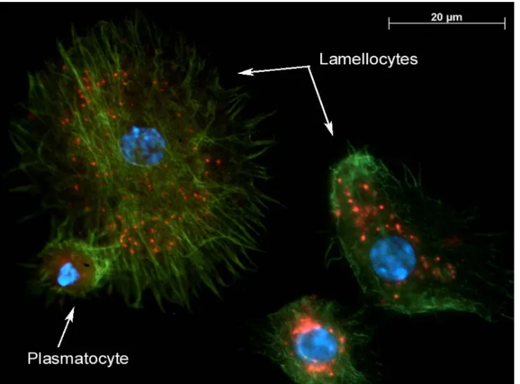
The formation of the capsule itself corresponds to aggregation of multiple layers of haemocytes around the parasitoid egg, the type of cells involved varying according to the host (Diptera, Lepidoptera…). In Drosophila melanogaster, the capsule is constituted by a first layer of plasmatocytes, covered by several layers of lamellocytes (Figure I, C). These lamellocytes are discoidal cells whose number increases from few cells to more than 50 % of circulating cells after a parasitic infection [37]. Another characteristic of the insect immune response is the deposition of melanin at the surface of the foreign body soon after its injection, less than 12 hours in D.
melanogaster [12,14]. Moreover, the parasitoid eggs die generally before the complete formation of
the capsule, confirming that cytotoxic radicals are generated, notably during the melanogenesis. The melanin production results from the activation of a cascade involving several genes and of which the last step is the activation of the pro-phenoloxidase enzyme (PPO) into phenoloxidase (PO) by a serine protease, the pro-phenoloxidase activating enzyme (PPAE) [38,14]. This cascade is regulated by other serine proteases, themselves negatively controlled by serpins. For example, the injection in an
immunocompetent larva of the serpin 27A of D. melanogaster, a molecule known to inhibit the PPAE, strongly decreases the encapsulation rate of L. boulardi eggs [39].
Effects of virulence factors injected by parasitoids on haemocytes and/or the PO cascade have been reported in several host species. These effects can be induced by the expression of PDVs genes, be associated to the presence of VLPs, or due to the injection of ovarian or venom proteins [8]. Alterations of the cellular response can either affect directly the haematopoietic organ [40] and/or affect all or a part of the circulating cells, through apoptosis for example. Modifications of the morphology of haemocytes, often correlated with an alteration of their ability to spread on a foreign surface and thus to form a capsule, have often been reported [41]. Finally, the inhibition of melanisation in the host is also a phenomenon largely described [8, 42].
Virulence factors potentially responsible of these effects, as cystatins, Protein Tyrosine Phosphatases (PTP) or I kappa B-like proteins have been isolated in several parasitoid species, notably thanks to the sequencing of the genome of some PDVs. However, their physiological function in the host has not been clearly demonstrated [8]. Among the very few factors whose effect is known, there are the protein Glc1.8 coded by the PDV of Microplitis demolitor, responsible of the inhibition of the spreading ability of haemocytes [43] and the protein CrV1 coded by the PDV of Cotesia rubecula, which disturbs the actin filaments in the host haemocytes [44]. The presence of a given family of potential virulence genes in PDVs can be more or less restricted to a range of parasitoids: as the case may be, they have been detected in only one species, in different species that belong to the same genus or family, or in species belonging to the two different families that carry PDVs. These differences might reflect the mode and moment of acquisition of the considered genes [8].
As only few virulence genes have been functionally characterized, we have very few data about the targets of these factors in the hosts. Among the recent advances, there is the description of the inhibition of PAP-3 (Prophenoloxidase Activating Protein 3) in the host Manduca sexta by the virulence factor Egf1.0 coded by the PDV of Microplitis demolitor [45]. This factor belongs to the
smapin family and inhibits the host melanisation. Another virulence factor has been largely studied: the protein LbGAP isolated from the venom of the parasitoid L. boulardi, which presents a GAP domain and a RacGAP activity. It has been shown that LbGAP enters the lamellocytes of the host D.
melanogaster and alters their morphology through an interaction with the GTPases Rac1 and Rac2,
both necessary for encapsulation (Figure IV) [46]. The characterization of the function of the virulence factors and their molecular targets opens the doors to study their specificity. For example, some studies are in progress to understand the origins of the specificity of the factor LbGAP, which is not efficient on D. yakuba, a species close though to D. melanogaster. Interestingly, a RhoGAP protein has been also described as a component of VLPs injected by the ichneumonid V. canescens in its hosts. Using comparative studies between virulence molecules from parasitoids that carry or not PDVs or VLPs, or that have different phylogenetic positions, it will be possible to answer the question whether RhoGAP toxins are largely used by parasitoid wasps and whether their presence is correlated with that of VLPs.
One of the major results from the study of Colinet et al. [46] is the evidence of a convergent use of “built” GAP proteins by pathogenic bacteria, which use them to target GTPases involved in the immune defense of mammals, and of “endogenous” GAP proteins by parasitoids, which use them to target the same GTPase family in order to suppress the immune defenses of their insect insect (Figure V). Other proteins, like the PTPs described in the genome of several polydnaviruses, are similarly proteins used as toxins by mammalian pathogenic bacteria. It will be important to determine if these examples are exceptions or if the strong conservation of immune signaling cascades between insects and mammals led to the selection of similar mechanisms in their pathogens and parasites to short-circuit these defenses.
4. The evolution of parasitoid virulence.
Parasitoid wasps can either be highly specialized to one or a few host species or have a quite broad host range. Interestingly, it has often been suggested that virulence strategies may direct the evolution of parasitoid host-specificity. For instance, koinobiont parasitoids should have a narrower
host range than idiobionts since they have to evolve to circumvent host immune defences [47]. The evolution of virulence molecules or the acquisition of new virulence factors in a parasitoid species could thus drive changes in the parasitoid host range and determine the potential for host shift. Several approaches are currently used to address this question: analyzing and comparing virulence factors between closely–related or distant parasitoid species and families (i), characterizing the molecular evolution of virulence genes (ii), and studying intra-specific variability of virulence (iii).
(i) The spectrum of parasitoid virulence molecules described nowadays is probably largely
incomplete, but we already gained indications of the mechanisms at the origin of new virulence factors. Molecules described to date, encoded by PDVs or produced in the venom, belong to “classical” eukaryotic protein families [25]. However, PDV-encoded proteins are directly produced in the cells of the infested lepidopteran host instead of the female wasp itself, and usually in a large amount. In non-PDV parasitoid species, most virulence proteins would also be overproduced in the venom gland [21]. Interestingly, the LbGAP non-PDV virulence factor, which belongs to a classically intra-cellular protein family, was shown to contain a signal peptide that allows secretion in the venom gland [21,46]. Molecular changes driving overexpression and/or secretion of otherwise endogeneous proteins might thus be considered as striking adaptations of these molecules to a new virulence function.
Another important question regarding virulence evolution in parasitoids is the relative importance of phylogenetic constraints and selective pressures due to host species, respectively. It can now be addressed by comparing virulence factors between parasitoid species and families. For instance, occurrence of similar virulence mechanisms in distant parasitoid families parasitizing the same host species – such as Asobara (Braconidae) and Leptopilina (Figitidae) spp. parasitoids of D.
melanogaster - would stress the importance of the selection by the host. At the end, answers to these
questions will prove essential to estimate the potential for evolutionary convergences in parasitoid virulence strategies.
(ii) Most virulence genes in PDV-bearing parasitoids are located in the viral genome which
can be easily purified and sequenced. As a wasp mutualist symbiont, the virus is expected to exhibit a reduction in genome complexity and to evolve under wasp phyletic constraints. However, as a lepidopteran host pathogenic symbiont, the virus is likely undergoing strong selective pressures for the acquisition of new functions by gene acquisition or duplication. Sequencing data have shown that PDV genomes are among the largest and the most complex virus genomes, having multigenic families encoding potential virulence factors [25]. The high divergence of these genes both inside and between wasp species, as well as the demonstration that they would evolve under positive selection, suggest a diversification in relation with changes in the host range [8,48,49]. The studies on virulence factors in species without PDVs have been more heavy going since each factor has to be characterized independently. Such a positive selection phenomenon has thus not been described yet.
(iii) Occurrence of intra-specific variability of virulence is very useful to analyze ongoing
evolutionary pressures driving changes in the efficiency or specificity of virulence molecules. Such a variability has been described in a few parasitoid species [50, 51, 52] where it happens to be due to changes in major genes. However, its molecular bases still remain to be understood. In L. boulardi, the best studied model, two types of females which show opposite virulence properties towards the host species D. melanogaster and D. yakuba have been described. These females differ in their virulence strategies [53] and variation in their virulence levels seem to correlate with variations in their host choice behaviour [54]. Such a correlation between physiological and behavioural traits is one of the required conditions in a model of speciation by host change. More data remain of course to be obtained in order to demonstrate the role of selective pressures by the host on parasitoid diversification.
The concept of « coevolution » cannot be ignored when interactions between parasites and hosts are discussed. However, coevolution could drive changes in life traits involved in the parasitism outcome only if these traits show genetic variability and evolve in response to selection pressures by
the antagonist species. Based on their high number and diversity, host-parasitoids interactions are a good experimental and theoretical model to test ongoing coevolutionary processes. Recent accumulation of data now allows testing different models of coevolution and their predictions, as well as understanding selective pressures at the species-community level.
Aknowledgements : Thanks to JM Drezen for our long-standing PDV-VLP partnership, to D. Colinet
and C. Anselme for fruitful discussions, to C. Combes for its contribution to the world of « durable interactions » and to the members of the GDR 2153 and the resistance group of the Durable Interactions Ecology Network (REID).
References
1. T.R. Malthus, Essai sur le principe de population en tant qu'il influe sur le progrès de la société, Londres, 1798.
2. C. Darwin, On the origin of species by means of natural selection, John Murray, London, 1859. 3. C. Combes, Interactions durables. Écologie et évolution du parasitisme, Éditions Masson,
1995, 525 p.
4. J. Gomez-Gutiérrez, W.T. Peterson, A. De Robertis, R.D. Brodeur, Mass mortality of krill caused by parasitoid ciliates, Science 301 (2003) 339.
5. S.E. Forde, J.N. Thompson, B.J.M. Bohannan, Adaptation varies through space and time in a coevolving host-parasitoid interaction, Nature 431 (2004) 841-844.
6. H.C.J. Godfray, Parasitoids: Behavioral and Evolutionary Ecology, University Press, Princeton, 1994.
7. D.L.J. Quicke, Parasitic wasps, Cambridge University Press edition, Chapman & Hall, London, 1997.
8. F. Pennacchio, M.R. Strand, Evolution of developmental strategies in parasitic hymenoptera, Ann. Rev. Entomol. 51 (2006) 233-258.
9. S.B. Vinson, Biochemical coevolution between parasitoids and their hosts, in: Price P. (Ed), Evolutionary Strategies of Parasitic Insects and Mites, Plenum, New York / London, 1975, pp. 14-48.
10. E.L. Charnov, D.W. Stephens, On the evolution of host selection in solitary parasitoids, Am. Nat. 132(5) (1988) 707-722.
11. N. E. Beckage, Physiological and behavioral host-parasitoid interactions: Future visions, Arch. Insect Biochem. Physiol. 60(4) (2005) 151-152.
12. Y. Carton, A.J. Nappi, Drosophila cellular immunity against parasitoids, Parasitol. Today 13(6) (1997) 218-227.
13. O. Schmidt, U. Theopold, M. Strand, Innate immunity and its evasion and suppression by hymenopteran endoparasitoids, BioEssays 23(4) (2001) 344-351.
14. Y. Carton., M. Poirié, A.J. Nappi, Insect immune resistance to parasitoids, Insect Science 15 (2008) 67-87.
15. S. Salt S, The resistance of insect parasitoids to the defense reactions of their hosts. Biol. Rev. 43 (1968) 200-232.
16. P. Eslin, G. Doury, The fly Drosophila subobscura: A natural case of innate immunity deficiency, Dev. Comp. Immunol. 30(11) (2006) 977-83.
17. P. Eslin, P. Giordanengo, Y. Fourdrain, G. Prévost, Avoidance of encapsulation in the absence of VLP by a braconid parasitoid of Drosophila larvae: an ultrastructural study, Can. J. Zool. 74 (1996) 2193-2198.
18. S. Asgari, U. Theopold, C. Wellby, O. Schmidt, A protein with protective properties against the cellular defense reactions in insects, Proc. Nat. Acad. Sci., U.S.A. 95 (1998) 3690-3695. 19. D.H. Davies, S.B. Vinson, Passive evasion by eggs of the braconid parasitoid Cardiochiles
nigriceps of encapsulation in vitro by haemocytes of host Heliothis virescens. Possible role for fibrous layer in immunity. J. Insect Physiol. 32(12) (1986) 1003-1010.
20. S. J. Moreau, S. Guillot, Advances and prospects on biosynthesis, structures and functions of venom proteins from parasitic wasps, Insect Biochem. Mol. Biol. 35 (2005) 1209-1223. 21. C. Labrosse, K. Stasiak, J. Lesobre, A. Grangeia, E. Huguet, J.M. Drezen, M. Poirié, A
RhoGAP protein as a main immune suppressive factor in the Leptopilina boulardi
(Hymenoptera, Figitidae)–Drosophila melanogaster interaction, Insect Biochem. Mol. Biol. 35 (2005) 93–103.
22. J.M. Drezen, B. Provost, E. Espagne, L. Cattolico, C. Dupuy, M. Poirié, G. Periquet, E. Huguet, Polydnavirus genome: integrated vs. free virus, J. Insect Physiol. 49 (2003) 407-417. 23. I. Feddersen, K. Sanders, O. Schmidt, Virus-like particles with host protein-like antigenic
determinants protect an insect parasitoid from encapsulation, Experientia 42 (1986) 1278-1281.
24. S. Dupas, M. Brehelin, F. Frey, Y. Carton, Immune suppressive virus-like particles in a
Drosophila parasitoid: significance of their intraspecific morphological variations, Parasitology 113 (1996) 207-212.
25. E. Espagne, C. Dupuy, E. Huguet, L. Cattolico, B. Provost, N. Martins, M. Poirié, G. Periquet, J.M. Drezen, Genome sequence of a polydnavirus: insights into symbiotic virus evolution, Science 306 (2004) 286-289.
26. N. E. Beckage, Modulation of immune responses to parasitoids by polydnaviruses, Parasitology 116 (1998) S57-S64.
27. C.A. Desjardins, D.E. Gundersen-Rindal, J.B. Hostetler, L.J. Tallon, R.W. Fuester, M.C. Schatz, M.J. Pedroni, D.W. Fadrosh, B.J. Haas, B.S. Toms, D. Chen, V. Nene, Structure and evolution of a proviral locus of Glyptapanteles indiensis bracovirus, BMC Microbiol. 7 (2007) 61-76.
28. B.A. Webb, Polydnavirus biology, genome structure, and evolution, In: Ball A., Miller L.K. (Eds), The insect viruses, Plenum publishing corporation, New York, 1998, pp. 105–139. 29. N. Murphy, J.C. Banks, J.B. Whitfield, A.D. Austin, Phylogeny of the parasitic microgastroid
subfamilies (Hymenoptera: Braconidae) based on sequence data from seven genes, with an improved time estimate of the origin of the lineage, Mol. Phylogenet. Evol. 47(1) (2008) 378-95.
30. R. Lapointe, K. Tanaka, W.E. Barney, J.B. Whitfield, J.C. Banks, C. Béliveau, D. Stoltz, B.A. Webb, M. Cusson, Genomic and morphological features of a banchine polydnavirus:
comparison with bracoviruses and ichnoviruses, J. Virol. 81(12) (2007) 6491-501.
31. J.M. Drezen, A. Bézier, J. Lesobre, E. Huguet E, L. Cattolico, G. Periquet, C. Dupuy, The few virus-like genes of Cotesia congregata bracovirus, Arch. Insect Biochem. Physiol. 61(3) (2006) 110-22.
32. A. Bézier, J. Herbinière, C. Serbielle, J. Lesobre, P. Wincker, E. Huguet, J.M. Drezen, Bracovirus gene products are highly divergent from insect proteins. Arch. Insect Biochem. Physiol. 67(4) (2008)172-87.
33. R.M. Rizki et T.M. Rizki, Parasitoid virus-like particles destroy Drosophila cellular immunity, Proc. Natl. Acad. Sci. USA 87 (1990) 8388-8392.
34. P.O. Lawrence, D. Akin, Virus-like particles from the poison glands of the parasitic wasp Biosteres longicaudatus (Hymenoptera: Braconidae), Can. J. Zool. 68 (1990) 539-546.
35. B.I.P. Barratt, A.A.Evans, D.B. Stoltz, S.B. Vinson, R. Easingwood, Virus-like particles in the ovaries of Microctonus aethiopoides Loan (Hymenoptera: Braconidae), a parasitoid of adult weevils (Coleoptera: Curculionidae), J. Invert. Pathol. 73(2) (1999) 182-188.
36. R. Lanot, D. Zachary, F. Holder, M. Meister, Postembryonic hematopoiesis in Drosophila. Dev. Biol. 230(2) (2001) 243-257.
37. J. Russo, M. Brehelin, Y. Carton, Haemocyte changes in resistant and susceptible strains of D. melanogaster caused by virulent and avirulent strains of the parasitic wasp Leptopilina
boulardi, J. Insect Physiol. 47 (2001) 167.
38. A.J. Nappi, B.M. Christensen, Melanogenesis and associated cytotoxic reactions: Applications to insect innate immunity, Insect Biochem. Mol. Biol. 35 (2005) 443-459.
39. A.J. Nappi, F. Frey, Y. Carton, Drosophila serpin 27A is a likely target for immune suppression of the blood cell-mediated melanotic encapsulation response. J. Insect Physiol. 51(2) (2005) 197-205.
40. S.J. Moreau, P. Eslin, P. Giordanengo, G. Doury, Comparative study of the strategies evolved by two parasitoids of the genus Asobara to avoid the immune response of the host, Drosophila melanogaster, Dev. Comp. Immunol. 27(4) (2003) 273-82.
41. C. Labrosse, P. Eslin, G. Doury, J.M. Drezen, M. Poirié, Haemocyte changes in D. melanogaster in response to long gland components of the parasitoid wasp Leptopilina boulardi: a Rho-GAP protein as an important factor, J. Insect Physiol. 51(2) (2005) 161-170. 42. L.J. Kohler, Y. Carton, M. Mastore, A.J. Nappi, Parasite suppression of the oxidations of
eumelanin precursors in Drosophila melanogaster. Arch. Insect Biochem. Physiol.66 (2007) 64-75.
43. M. Beck, M.R. Strand (2003) RNA interference silences Microplitis demolitor bracovirus genes and implicates glc1.8 in disruption of adhesion in infected host cells, Virology, 314(2) (2003) 521-535.
44. S. Asgari, O. Schmidt (2002) A coiled-coil region of an insect immune suppressor protein is involved in binding and uptake by haemocytes, Insect Biochem. Mol. Biol., 32(5) (2002) 497-504.
45. M.H. Beck, M.R. Strand, A novel polydnavirus protein inhibits the insect prophenoloxidase activation pathway, Proc. Natl. Acad. Sci. U S A 104 (49) (2007) 19267-72.
46. D. Colinet, A. Schmitz, D. Depoix, D. Crochard, M. Poirié, Convergent use of RhoGAP toxins by eukaryotic parasites and bacterial pathogens, PLoS Pathog 3 (2008) e203.
47. R.R. Askew, M.R Shaw, Parasitoid communities: their size, structure and development, in: Waage, J, Greathead D. (Eds), Insect Parasitoids. Academic Press, London, 1986, pp. 225– 264.
48. S. Dupas, M. W. Turnbull, B.A. Webb, Diversifying selection in a parasitoid's symbiotic virus among genes involved in inhibiting host immunity, Immunogenetics 55 (2003) 351.
49. C. Serbielle, S. Chowdhury, S. Pichon, S. Dupas, J. Lesobre, E. O. Purisima, J.M. Drezen and E. Huguet, Viral cystatin evolution and 3D structure modelling : a case of directional selection acting on a viral protein involved in a host-parasitoid interaction, BMC Biology, in press. 50. Gitau CW, Gundersen-Rindal D, Pedroni M, Mbugi PJ, Dupas S, Differential expression of the
CrV1 haemocyte inactivation-associated polydnavirus gene in the African maize stem borer Busseola fusca (Fuller) parasitized by two biotypes of the endoparasitoid Cotesia sesamiae (Cameron), J. Insect Physiol. 53(7) (2007) 676-84.
51. S. Dupas, Y. Carton, M. Poirié, Genetic dimension of the coevolution of virulence resistance in Drosophila - parasitoid wasps relationships, Heredity 90 (2003) 84.
52. A. Dubuffet, S. Dupas, F. Frey, J.M. Drezen, M. Poirié, Y. Carton, Genetic interactions
between the parasitoid wasp Leptopilina boulardi and its Drosophila hosts, Heredity 98 (2007) 21-27.
53. A. Dubuffet, G. Doury, C. Labrousse, J.M. Drezen, Y. Carton, M. Poirié,Variation of success of Leptopilina boulardi in Drosophila yakuba: The mechanisms explored, Dev. Comp.
Immunol.32 (2008) 597-602.
54. A. Dubuffet, C. Rodriguez Alvarez, J. M. Drezen, J.J.M. van Alphen, M. Poirié, Do parasitoid preferences for different host species match virulence? Physiol. Entomol. 31 (2006) 170-177. 55. A.J. Pruijssers, M.R. Strand, PTP-H2 and PTP-H3 from Microplitis demolitor Bracovirus
localize to focal adhesions and are antiphagocytic in insect immune cells, J. Virol. 81 (2007) 1209.
Figure captions:
Figure I. A. Leptopilina boulardi female searching for Drosophila host larvae (Photo A. Dubuffet). B. Wasp eggs encapsulated within the body cavity of a Drosophila host seen through the larval cuticle (Photo A. Dubuffet). C. Scanning electron micrograph showing haemocytes adhering on the periphery of a fully-formed capsule (Photo A. Nappi).
Figure II. A. Venom gland and reservoir attached to the oviposition apparatus of a Leptopilina
boulardi female wasp (Photo A. Dubuffet) r : reservoir, g : gland. Scale bar : 500 nm. B. Scanning
electron micrograph of VLPs found inside the venom gland and reservoir of a Leptopilina boulardi female parasitoid (Photo C. Labrosse). C. Scanning electron micrograph showing particles of the bracovirus associated with Cotesia congregata (CcBV). Each envelop contains several nucleocapsids. Scale bar : 44 nm.
Figure III. Phylogenetic representation of the Ichnovirus and Bracovirus origin in the Ichneumonoidea super-family. Data have been synthesized from the work of JB Whitfield and AD Austin, especially [29].
Figure IV. Fluorescence micrograph of the LbGAP protein inside Drosophila melanogaster haemocytes (lamellocytes and plasmatocytes). LbGAP is a virulence factor produced in the venom gland and injected by Leptopilina boulardi females. Haemocyte actin cytoskeleton was visualized using phalloidin (green), LbGAP was detected using a specific rabbit polyclonal antibody (red) and nuclei have been visualised using DAPI (blue).
Figure V. Comparison of the effect of bacterial toxins and parasitoid virulence factors on immune cells of their hosts. Pathogenic bacteria such as Yersinia pestis inject Yop toxins in their mammalian hosts that prevent the phagocytic activity of macrophages. Yop E is a RhoGAP protein which has a similar function as the LbGAP factor injected by the parasitoid L. boulardi i.e. modifying the cytoskeleton of Drosophila haemocytes and thus preventing encapsulation. Several Protein Tyrosine Phosphatase (PTPs) are also encoded by the PDV genome. Their function as virulence factors still remains to be demonstrated but one of them has been localized in focal adhesions and shown to affect phagocytosis, such as the Yop H toxin of Yersinia [55].
Table 1. Non exhaustive summary table of the virulence strategies used by parasitoid wasps to avoid immune defenses of their hosts. When possible, known mechanisms or virulence factors are indicated.
Black boxes : parasitoids of the Ichneumonoidae family; Grey boxes: parasitoids of the Braconidae family; Punctuated boxes: parasitoids of the Figitidae family (Cynipoidea superfamily); White boxes : parasitoids of the Chalcidoidea superfamily (1Trichogrammatidae et 2Encyrtidae).
VLPs: Virus Like Particles; PDV: polydnavirus. Polydnaviruses are indicated by initials of the latin
A
B
C
Figure II
A
B
C
g r
Figure III
Bracovirus ancêtre: 100 millions d’années Meteorinae Macrocentrinae
MICROGASTRINAE
Miracinae Cheloninae
BRACONIDAE
Cardiochilinae Complexe MicrogastroideICHNEUMONIDAE
Banchinae Campopleginae Ctenopelmatinae Familles sans PDVBactérie ex: Yersinia sp.