HAL Id: hal-03218266
https://hal.sorbonne-universite.fr/hal-03218266
Submitted on 5 May 2021
HAL is a multi-disciplinary open access
archive for the deposit and dissemination of
sci-entific research documents, whether they are
pub-lished or not. The documents may come from
teaching and research institutions in France or
abroad, or from public or private research centers.
L’archive ouverte pluridisciplinaire HAL, est
destinée au dépôt et à la diffusion de documents
scientifiques de niveau recherche, publiés ou non,
émanant des établissements d’enseignement et de
recherche français ou étrangers, des laboratoires
publics ou privés.
Oncogenetic landscape and clinical impact of IDH1 and
IDH2 mutations in T-ALL
Mathieu Simonin, Aline Schmidt, Christophe Bontoux, Marie-Émilie Dourthe,
Etienne Lengliné, Guillaume Andrieu, Ludovic Lhermitte, Carlos Graux,
Nathalie Grardel, Jean-Michel Cayuela, et al.
To cite this version:
Mathieu Simonin, Aline Schmidt, Christophe Bontoux, Marie-Émilie Dourthe, Etienne Lengliné, et
al.. Oncogenetic landscape and clinical impact of IDH1 and IDH2 mutations in T-ALL. Journal
of Hematology and Oncology, BioMed Central, 2021, 14 (1), �10.1186/s13045-021-01068-4�.
�hal-03218266�
RAPID COMMUNICATION
Oncogenetic landscape and clinical impact
of IDH1 and IDH2 mutations in T-ALL
Mathieu Simonin
1,2,3, Aline Schmidt
4, Christophe Bontoux
1,3, Marie‑Émilie Dourthe
1,3,5, Etienne Lengliné
6,
Guillaume P. Andrieu
1,3, Ludovic Lhermitte
1,3, Carlos Graux
7, Nathalie Grardel
8,9, Jean‑Michel Cayuela
10,
Françoise Huguet
11, Isabelle Arnoux
12, Stéphane Ducassou
13, Elizabeth Macintyre
1,3, Virginie Gandemer
14,
Hervé Dombret
6, Arnaud Petit
2, Norbert Ifrah
4, André Baruchel
5, Nicolas Boissel
6and Vahid Asnafi
1,3*Abstract
IDH1 and IDH2 mutations (IDH1/2
Mut) are recognized as recurrent genetic alterations in acute myeloid leukemia (AML)
and associated with both clinical impact and therapeutic opportunity due to the recent development of specific
IDH1/2
Mutinhibitors. In T‑cell acute lymphoblastic leukemia (T‑ALL), their incidence and prognostic implications
remain poorly reported. Our targeted next‑generation sequencing approach allowed comprehensive assessment of
genotype across the entire IDH1 and IDH2 locus in 1085 consecutive unselected and newly diagnosed patients with
T‑ALL and identified 4% of, virtually exclusive (47 of 49 patients), IDH1/2
Mut. Mutational patterns of IDH1/2
Mutin T‑ALL
present some specific features compared to AML. Whereas IDH2
R140Qmutation was frequent in T‑ALL (25 of 51 muta‑
tions), the IDH2
R172AML hotspot was absent. IDH2 mutations were associated with older age, an immature pheno‑
type, more frequent RAS gain‑of‑function mutations and epigenetic regulator loss‑of‑function alterations (DNMT3A
and TET2). IDH2 mutations, contrary to IDH1 mutations, appeared to be an independent prognostic factor in multivari‑
ate analysis with the NOTCH1/FBXW7/RAS/PTEN classifier. IDH2
Mutwere significantly associated with a high cumulative
incidence of relapse and very dismal outcome, suggesting that IDH2‑mutated T‑ALL cases should be identified at
diagnosis in order to benefit from therapeutic intensification and/or specific IDH2 inhibitors.
Keywords: IDH1, IDH2, T‑ALL
© The Author(s) 2021. Open Access This article is licensed under a Creative Commons Attribution 4.0 International License, which permits use, sharing, adaptation, distribution and reproduction in any medium or format, as long as you give appropriate credit to the original author(s) and the source, provide a link to the Creative Commons licence, and indicate if changes were made. The images or other third party material in this article are included in the article’s Creative Commons licence, unless indicated otherwise in a credit line to the material. If material is not included in the article’s Creative Commons licence and your intended use is not permitted by statutory regulation or exceeds the permitted use, you will need to obtain permission directly from the copyright holder. To view a copy of this licence, visit http:// creat iveco mmons. org/ licen ses/ by/4. 0/. The Creative Commons Public Domain Dedication waiver (http:// creat iveco mmons. org/ publi cdoma in/ zero/1. 0/) applies to the data made available in this article, unless otherwise stated in a credit line to the data.
Introduction
T-cell acute lymphoblastic leukemia (T-ALL) is
aggres-sive neoplasms resulting from the proliferation of
T-lymphoid progenitors blocked at thymic stages of
dif-ferentiation and account for 15% and 25% of pediatric
and adult ALLs, respectively [
1
]. T-ALL is associated
with a wide range of acquired genetic abnormalities that
contribute to developmental arrest and abnormal
pro-liferation [
2
]. Although intensive treatment protocols
have markedly improved the outcomes of children with
T-ALL, cure rates remain below 60% for adults and 85%
for children [
3
–
5
]. The prognosis is particularly poor in
relapsing patients, justifying the development of novel
targeted therapies [
6
,
7
]. For example, alterations
affect-ing epigenetic factors may offer novel targeted
therapeu-tic approaches in high-risk T-ALL [
8
].
Whole-genome sequencing of AML identified acquired
mutations in isocitrate dehydrogenase 1 and 2 (IDH1/2)
[
9
]. These paralogous genes encode two enzymes with
distinct localizations (cytoplasmic for IDH1 and
mito-chondrial for IDH2). Both catabolize the conversion
of isocitrate to α-ketoglutarate (α-KG).
Gain-of-func-tion IDH1/2 mutaGain-of-func-tions (IDH1/2
Mut) confer a
neomor-phic activity on the encoded enzymes, leading to the
Open Access
*Correspondence: vahid.asnafi@aphp.fr
1 Laboratory of Onco‑Hematology, Assistance Publique‑Hôpitaux de Paris (AP‑HP), Hôpital Necker Enfants‑Malades, Université de Paris, 149 rue de Sèvres, 75015 Paris, France
Page 2 of 7 Simonin et al. J Hematol Oncol (2021) 14:74
conversion of α-KG to 2-hydroglutatarate (2-HG) in a
NAD phosphate-dependent manner [
10
]. Accumulation
of the oncometabolite 2-HG induces multiple cellular
alterations, including chromatin methylation and cellular
differentiation, by inhibiting α-KG-dependent enzymes
related to DNA methylation, such as Tet oncogene
fam-ily members (TET2, TET3) [
11
]. IDH1/2
Muthave been
reported in 10 to 20% of AML cases, when they are
predominantly located in the active site of the enzyme
(IDH1
R132, IDH2
R140Qand IDH2
R172). IDH1/2
Mutin AML
are associated with prognostic impact influenced by the
genetic context [
12
,
13
]. Importantly, specific drugs
tar-geting mutant IDH1 or IDH2 have recently shown
prom-ise in IDH1/2
Mutrefractory or relapsed AML patients [
14
,
15
].
In T-ALL, IDH1/2
Muthave been partially explored and
their prognostic impact poorly reported [
16
,
17
]. We now
provide the first comprehensive analysis and
oncoge-netic landscape of IDH1/2
Mutin a cohort of 1085 T-ALL
patients, when the nearly 4% of IDH1/2
Mutare
associ-ated with extremely poor prognosis, specifically in
IDH2-mutated cases.
Methods
Patient’s protocol and clinical trials
Diagnostic peripheral blood or bone marrow samples
from 1085 adults and children with T-ALL were
ana-lyzed after informed consent was obtained at
diagno-sis according to the Declaration of Helsinki. Among
the 1085 T-ALL analyzed, 215 adult patients aged from
16–59 years were included in the GRAALL03/05 trials
(details provide in supplementary) which were registered
at clinicaltrials.gov (GRAALL-2003, #NCT00222027;
GRAALL-2005, #NCT00327678). and 261
pediat-ric patients aged from 1 to 19 years were treated in 10
French pediatric hematology departments, members
of the FRALLE study group, according to the FRALLE
2000 T guidelines (Additional file
2
: Fig. S5 and
Addi-tional file
1
: Table S3).
Gene mutation screening
A custom capture Nextera XT gene panel (Illumina,
San Diego, CA) targeting all coding exons and their
adjacent splice junctions of 80 genes was designed,
based on available evidence in hematological
neo-plasms (Additional file
1
: Table S1). DNA Libraries
were prepared using Nextera Rapid Capture
Enrich-ment protocol and underwent 2 × 150 bp paired-end
sequencing on Illumina MiSeq sequencing system with
MiSeq Reagent Kit v2 (Illumina). Briefly, sequence
reads were filtered and mapped to the human genome
(GRCh37/hg19) using in-house software (Polyweb,
Institut Imagine, Paris). Annotated variants were
selected after filtering out calls according to the
fol-lowing criteria: (1) coverage < 30×, < 10 alternative
reads or variant allelic fraction (VAF) < 7%; (2)
poly-morphisms described in dbSNP, 1000Genomes, EVS,
Gnomad and EXAC with a calculated mean population
frequency > 0.1%. Non-filtered variants were annotated
using somatic database COSMIC (version 78) and
ProteinPaint (St Jude Children’s Research Hospital –
Pediatric Cancer data portal). Lollipop plots were
gen-erated with ProteinPaint (
https:// pecan. stjude. org/#/
prote inpai nt
).
Immunophenotypic and molecular characterization
of T‑ALL samples
Peripheral blood or bone marrow T-ALL samples were
analyzed for immunophenotype, fusion transcripts
(SIL-TAL1, CALM-AF10), oncogenic transcripts (HOXA9,
TLX1 and TLX3) and T-cell receptor (TCR)
recombina-tion and NOTCH1/FBXW7/RAS/PTEN mutarecombina-tions, as
previously described [
4
,
18
,
19
].
Minimal residual disease assessment
Immunoglobulin/T-cell receptor (Ig/TCR) gene
rear-rangement-based Minimal Residual Disease (MRD)
eval-uation was centrally assessed for patients who reached
complete remission after the first induction cycle, on
BM samples after induction (MRD1). MRD was centrally
assessed by real-time quantitative allele-specific
oligo-nucleotide PCR and interpreted according to EuroMRD
group guidelines [
20
–
22
].
Statistical analysis
Comparisons for categorical and continues variables
between IDH1
Mutor IDH2
Mutand IDH
WTsubgroups
were performed with Fisher’s exact test and
Mann–Whit-ney test, respectively. Overall survival (OS) was
calcu-lated from the date of diagnosis to the last follow-up date
censoring patients alive. The cumulative incidence of
relapse (CIR) was calculated from the complete
remis-sion date to the date of relapse censoring patients alive
without relapse at the last follow-up date. Relapse and
death in complete remission were considered as
competi-tive events. Univariate and multivariate analyses
assess-ing the impact of categorical and continuous variables
were performed with a Cox model. Proportional-hazards
assumption was checked before conducting multivariate
analyses. In univariate and multivariate analyses, age and
log10(WBC) were considered as continuous variables.
All analyses were stratified on the trial. Variables with a
p value less than 0.1 in univariate analysis were included
in the multivariable models. Statistical analyses were
per-formed with STATA software (STATA 12.0 Corporation,
College Station, TX). All p-values were two-sided, with
p < 0.05 denoting statistical significance. Circos plots
were generated using R software.
Results and discussion
Incidence of IDH1 and IDH2 mutations in 1085 T‑ALL
A total of 51 (4%) mutations, mainly clonal, in either
IDH1 or IDH2 were apparent in 49 cases (Fig.
1
a and
Additional file
1
: Table S2, Additional file
2
: Figs. S2,
b
a
c
d
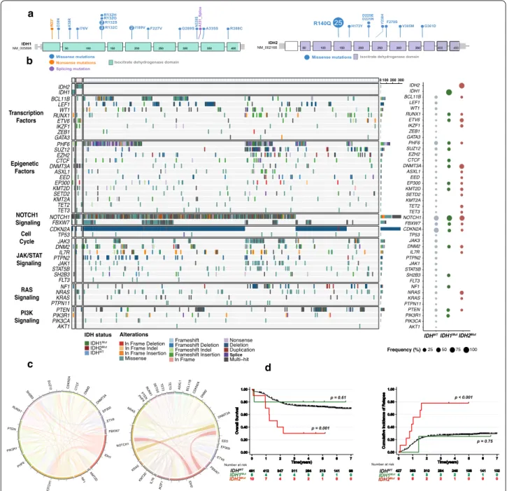
Fig. 1 IDH1 and IDH2 mutations in the GRAALL03/05 and FRALLE2000 studies. a Lollipop plots indicating the observed mutations for each
IDH gene and their consequences. b Oncoplot depicting the genetic anomalies observed in IDH1/2-Mutated or Wild type T‑ALL cases of the GRAALL03/05 and FRALLE2000 studies. Genes are classified by functional groups. The right panel indicates the overall frequency of alterations per gene. c The circos plots depict the co‑occurrences in genetic lesions observed in IDH1 (left panel) and IDH2 mutated T‑ALL (right panel). d Clinical impact of IDH1 and IDH2 mutations in the GRAALL0305 and FRALLE2000 studies. Overall survival (left panel) and cumulative incidence of relapse (right panel). The red curve represents the IDH2‑mutated patients, the green curve the IDH1‑mutated patients and the black curve the IDHWt
Page 4 of 7 Simonin et al. J Hematol Oncol (2021) 14:74
S3). IDH1 mutations were identified in 19 T-ALL cases
(2%) and IDH2 mutations in 32 cases (3%). IDH1/2
Mutwere mutually exclusive except in 2 cases. The IDH2
R140Qmutation was the most prevalent mutation affecting
IDH2 (n = 25, 78%). We identified 7 IDH1 mutations
located in the R132 hotspot (37% of IDH1 mutations),
3 cases with IDH1
R132Cmutation, 2 with IDH1
R132S, 1
with IDH1
R132Hand IDH1
R132Gmutation. The most
com-mon IDH2 mutations in AML occur at R140 followed
by residue IDH2
R172. The latter mutation is virtually the
only IDH mutation found in angio-immunoblastic T cell
lymphoma, reported in about 30% of cases (Additional
file
2
: Fig. S1) [
23
]. IDH2
R172mutation has also been
rarely and inconsistently described in peripheral T-cell
lymphoma not otherwise specified (NOS) with
T-fol-licular helper (T
FH) phenotype [
24
,
25
]. In striking
con-trast, IDH2
R172was not reported in our series of T-ALL.
IDH1
R132, the most frequent IDH1 mutation reported in
our cohort, has recently been recognized to cooperate
with NOTCH1 activation in a T-ALL mouse model [
26
].
These results highlight the specific consequence
associ-ated with IDH1/2
Mutsubtype during immature T-cell
development.
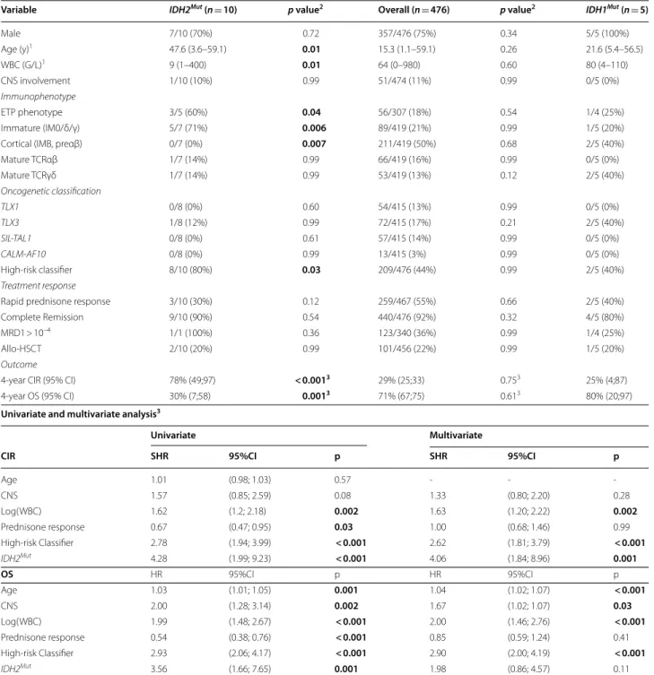
Clinico‑biological characteristics of IDH1/2
Mutin GRAALL
and FRALLE‑treated T‑ALLs
We then investigated the clinical characteristics linked to
IDH1/2
Mutin a subset of 476 patients, including 215 adults
enrolled in the GRAALL-2003/2005 trials and 261
chil-dren enrolled in the FRALLE-2000 trial (Table
1
and
Sup-plemental Methods). The incidence of IDH1/2
Mutin this
cohort was 3% (15/476). IDH1 mutations were detected in
5 patients (4 adult and 1 pediatric case), and IDH2
muta-tions were identified in 10 (6 adult and 4 pediatric cases)
(Additional file
2
: Fig. S2). IDH2R
140Qwas the most
fre-quent mutation (n = 7, 70%) and was most prevalent in
adults’ patients (n = 6/7, 86%). Overall, IDH1/2
Mutwere
observed in 5% of adults and 2% of children (p = 0.1).
IDH1 and IDH2 mutations are associated with both specific
clinical and mutational profiles
Patients with IDH2
Mutwere significantly older than
IDH
WT(median 47.6 years vs 15.0, p = 0.01). IDH2
Mutwere associated with an immature immunophenotype
(5/7, 71% vs 83/407, 20%, p = 0.006) and
ETP-pheno-type (3/5, 60% vs 52/298, 17%, p = 0.04). In line with
this, IDH2
Mutcorrelated positively with abnormalities
known to be associated with an immature phenotype,
including RAS (50% vs 11%, p = 0.02), ETV6 (40% vs 3%,
p < 0.01), DNMT3A (70% vs 3%, p < 0.01), IKZF1 (20% vs
2%, p = 0.02) and TET2 (20% vs 2%, p = 0.04) mutations
(Fig.
1
b, c). IDH2
Mutwere mutually exclusive with
SIL-TAL1 + cases, associated with a mature TCRαβ lineage.
Interestingly, contrary to IDH2-mutated cases, IDH1
Mutdid not statistically differ from IDH
WTpatient regarding
age, immunophenotype or mutational co-occurrence.
IDH2 mutations, but not IDH1, are associated with a poor
prognosis in T‑ALL
To investigate the prognostic value of IDH1/2
Mut,
sur-vival analyses were performed on the 476 patient cohort.
IDH1/2
Mutcases did not differ significantly with regard
to sex, white blood cell count (WBC) or central
nerv-ous system (CNS) involvement (Table
1
). Despite an
ini-tial good treatment response (IDH2
Mutcases achieved
90% complete remission rate and IDH2
Mutdid not
con-fer increased poor prednisone response), patients with
IDH2
Muthad an inferior outcome compared to IDH2
Wt(Table
1
, Fig.
1
d, Additional file
2
: Fig. S4), with an
increased cumulative incidence of relapse (CIR) (4y-CIR:
78% vs 29%; specific hazard ratio (SHR) 4.3, 95%CI (2.0–
9.2); p < 0.001) and a shorter overall survival (OS) (4y-OS:
30% vs 71%; hazard ratio: 3.6, 95%CI (1.7–7.7); p = 0.001).
In multivariate analysis considering variables associated
with CIR and OS in univariate analyses as covariates,
IDH2
Mutpredicted a trend for lower OS (HR: 1.98, 95%CI
(0.86–4.57); p = 0.11) and statistically higher CIR (SHR,
4.06, 95%CI (1.84–8.96), p = 0.001) even after adjustment
on the 4-gene NOTCH1/FBXW7/RAS/PTEN (NFRP)
classifier which identified poor prognosis patients in both
GRAALL and FRALLE trials [
3
,
4
]. Conversely to
IDH-2
Mut, IDH1
Mutwas not associated with poor
prognos-tic impact in T-ALL (4y-CIR: 25% vs 29%, p = 0.75 and
4y-OS: 80% vs 71%, p = 0.61).
We provide the largest comprehensive analysis of IDH1
and IDH2 mutations in T-ALL and highlight for the first
time both their clinical profile and, most importantly, the
extremely poor prognosis impact associated with
IDH-2
Mut. We describe the specific oncogenetic landscape of
IDH1/2
Mutand interestingly report that IDH2
MutT-ALL
conversely to IDH1
Mutwere associated with an immature
phenotype and alterations such as RAS mutations,
tran-scription factors alterations (ETV6, IKZF1) and
epige-netic regulators alterations (TET2, DNMT3A).
Recent studies have shed light on new prognostic
fac-tor in T-ALL allowing sharper prediction of the risk
of relapse (e.g., NFRP classifier, level of MRD1, IKZF1
alterations) [
3
,
4
,
27
]. Despite this, a significant number
of T-ALL relapses remain unpredicted, so new predictive
markers are needed, given the extremely poor prognosis
associated with T-ALL relapse. We therefore consider
that IDH2
MutT-ALL cases should be identified at
diag-nosis to benefit from therapeutic intensification and/or
specific IDH2
Mutinhibitors [
15
].
Table 1 Clinico‑biological and outcome characteristics of adult and pediatric T‑ALL (GRAALL and FRALLE protocols) according to
IDH1/2 status
p-values < 0.05 are indicated in bold
MRD1 correspond to MRD evaluation after induction and was performed by allele-specific oligonucleotides polymerase chain reaction. T-cell receptor status and oncogenic were performed as described in supplemental methods. IDH1Mut and IDH2Mut were statistically compared to IDH1WT and IDH2WT patients, respectively T-ALL: T-cell acute lymphoblastic leukemia; WBC, white blood count; CNS, central nervous system; ETP, early thymic precursor; High Risk classifier, NOTCH1/FBXW7-RAS/
PTEN classifier as previously described [3, 4]; CR, complete remission; MRD, minimal residual disease; Allo-HSCT, allogenic hematopoietic stem cell transplantation; CIR, cumulative incidence of relapse; OS, overall survival; HR: hazard ratio, SHR: specific hazard ratio, CI: confidence interval
1 Statistics presented: Median (Minimum–Maximum)
2 Statistical tests performed: Fisher’s exact test; Wilcoxon rank-sum test 3 Univariate and multivariate Cox analyses stratified on protocol
Variable IDH2Mut (n = 10) p value2 Overall (n = 476) p value2 IDH1Mut (n = 5)
Male 7/10 (70%) 0.72 357/476 (75%) 0.34 5/5 (100%) Age (y)1 47.6 (3.6–59.1) 0.01 15.3 (1.1–59.1) 0.26 21.6 (5.4–56.5) WBC (G/L)1 9 (1–400) 0.01 64 (0–980) 0.60 80 (4–110) CNS involvement 1/10 (10%) 0.99 51/474 (11%) 0.99 0/5 (0%) Immunophenotype ETP phenotype 3/5 (60%) 0.04 56/307 (18%) 0.54 1/4 (25%) Immature (IM0/δ/γ) 5/7 (71%) 0.006 89/419 (21%) 0.99 1/5 (20%) Cortical (IMB, preαβ) 0/7 (0%) 0.007 211/419 (50%) 0.68 2/5 (40%) Mature TCRαβ 1/7 (14%) 0.99 66/419 (16%) 0.99 0/5 (0%) Mature TCRγδ 1/7 (14%) 0.99 53/419 (13%) 0.12 2/5 (40%) Oncogenetic classification TLX1 0/8 (0%) 0.60 54/415 (13%) 0.99 0/5 (0%) TLX3 1/8 (12%) 0.99 72/415 (17%) 0.21 2/5 (40%) SIL-TAL1 0/8 (0%) 0.61 57/415 (14%) 0.99 0/5 (0%) CALM-AF10 0/8 (0%) 0.99 13/415 (3%) 0.99 0/5 (0%) High‑risk classifier 8/10 (80%) 0.03 209/476 (44%) 0.99 2/5 (40%) Treatment response
Rapid prednisone response 3/10 (30%) 0.12 259/467 (55%) 0.66 2/5 (40%) Complete Remission 9/10 (90%) 0.54 440/476 (92%) 0.32 4/5 (80%) MRD1 > 10–4 1/1 (100%) 0.36 123/340 (36%) 0.99 1/4 (25%) Allo‑HSCT 2/10 (20%) 0.99 101/456 (22%) 0.99 1/5 (20%)
Outcome
4‑year CIR (95% CI) 78% (49;97) < 0.0013 29% (25;33) 0.753 25% (4;87) 4‑year OS (95% CI) 30% (7;58) 0.0013 71% (67;75) 0.613 80% (20;97)
Univariate and multivariate analysis3
Univariate Multivariate
CIR SHR 95%CI p SHR 95%CI p
Age 1.01 (0.98; 1.03) 0.57 ‑ ‑ ‑ CNS 1.57 (0.85; 2.59) 0.08 1.33 (0.80; 2.20) 0.28 Log(WBC) 1.62 (1.2; 2.18) 0.002 1.63 (1.20; 2.22) 0.002 Prednisone response 0.67 (0.47; 0.95) 0.03 1.00 (0.68; 1.46) 0.99 High‑risk Classifier 2.78 (1.94; 3.99) < 0.001 2.62 (1.81; 3.79) < 0.001 IDH2Mut 4.28 (1.99; 9.23) < 0.001 4.06 (1.84; 8.96) 0.001 OS HR 95%CI p HR 95%CI p Age 1.03 (1.01; 1.05) 0.001 1.04 (1.02; 1.07) < 0.001 CNS 2.00 (1.28; 3.14) 0.002 1.67 (1.02; 1.07) 0.03 Log(WBC) 1.99 (1.48; 2.67) < 0.001 2.00 (1.46; 2.76) < 0.001 Prednisone response 0.54 (0.38; 0.76) < 0.001 0.85 (0.59; 1.24) 0.41 High‑risk Classifier 2.93 (2.06; 4.17) < 0.001 2.90 (2.00; 4.19) < 0.001 IDH2Mut 3.56 (1.66; 7.65) 0.001 1.98 (0.86; 4.57) 0.11
Page 6 of 7 Simonin et al. J Hematol Oncol (2021) 14:74
Abbreviations
IDH1/2Mut: IDH1‑IDH2 Mutations; AML: Acute myeloid leukemia; T‑ALL: T‑cell
acute lymphoblastic leukemia; NFRP: NOTCH1/FBXW7/RAS/PTEN; CNS: Central nervous system; WBC: White blood cell count; NOS: Not otherwise specified;
TFH: T‑Follicular helper; ETP: Early thymic precursor; MRD: Minimal residual disease.
Supplementary Information
The online version contains supplementary material available at https:// doi. org/ 10. 1186/ s13045‑ 021‑ 01068‑4.
Additional file 1. Supplemental Table 1: Custom capture Nextera XT
gene panel. Supplemental Table 2: IDH1 and IDH2 mutations identified in 1085 patients with T‑ALL. Supplemental Table 3: Chemotherapy in the FRALLE 2000 standard risk group T1 and high risk T2.
Additional file 2. Figure S1: Lollipop plots indicating the observed
mutations for IDH1 and IDH2 in the present series confront with Cosmic‑ reported mutations for AML and AITL. Figure S2: Lollipop plots indicating the observed mutations for IDH1 and IDH2 affecting patients included in FRALLE and GRAALL protocol. Figure S3: Variant Allele Frequency (VAF) of individual IDH1 and IDH2 mutations observed in 1085 T‑ALL. Figure S4: OS and CIR according to the IDH1 or IDH2Mut status in the two subgroups (FRALLE and GRALL 03/05). Figure S5: General design of FRALLE 2000 T guidelines.
Acknowledgements
The authors would like to thank all participants in the GRAALL‑2003 and GRAALL‑2005 study groups, the SFCE and the investigators of the 16 SFCE centers involved in collection and provision of data and patient samples, and V. Lheritier for collection of clinical data.
Authors’ contributions
N.B, V.A and M.S conceived the study and oversaw the project; M.S, A.S, C.B, ME.D, E.L, C.G, N.G, JM.C, I.A, V.G, F.H, S.D, N.I, H.D, A.B, A.P, N.B provided study materials or patients; M.S, C.B, A.S, E.M, G.P.A and V.A performed molecular analyses; M.S, A.S, C.B, V.A. collected and assembled data; N.B and M.S per‑ formed statistical analysis; M.S, A.S, C.B, V.A, N.B, G.P.A analyzed and interpreted data; M.S, N.B, A.S, C.B, E.M, V.A wrote the manuscript. All authors read and approved the final manuscript.
Funding
The GRAALL was supported by grants P0200701 and P030425/AOM03081 from the Programme Hospitalier de Recherche Clinique, Ministère de l’Emploi et de la Solidarité, France and the Swiss Federal Government in Switzerland. Samples were collected and processed by the AP‑HP “Direction de Recherche Clinique” Tumor Bank at Necker‑Enfants Malades. MS was supported by « Action Leucémie» and « Soutien pour la formation à la recherche translation‑ nelle en cancérologie dans le cadre du Plan cancer 2009–2013». This work was supported by grants to Necker laboratory from the “Association Laurette Fugain”, Association pour la Recherche contre le Cancer (Equipe labellisée), Institut National du Cancer PRT‑K 18–071 and the Fédération Leucémie espoir and Horizon Hemato.
Availability of data and materials
The datasets used and/or analyzed during the current study are available from the corresponding author on reasonable request.
Declarations
Ethics approval and consent to participate
Studies were conducted in accordance with the Declaration of Helsinki and approved by local and multicenter research ethical committees.
Consent for publication
Not applicable.
Competing interests
The authors declare no competing financial interests.
Author details
1 Laboratory of Onco‑Hematology, Assistance Publique‑Hôpitaux de Paris (AP‑HP), Hôpital Necker Enfants‑Malades, Université de Paris, 149 rue de Sèvres, 75015 Paris, France. 2 Department of Pediatric Hematology and Oncol‑ ogy, Assistance Publique‑Hôpitaux de Paris (AP‑HP), Armand Trousseau Hospi‑ tal, Sorbonne Université, Paris, France. 3 Institut Necker‑Enfants Malades (INEM), Institut National de la Santé et de la Recherche Médicale (Inserm) U1151, Paris, France. 4 PRES LUNAM, CHU Angers service des Maladies du Sang, INSERM U 892, Angers, France. 5 Department of Pediatric Hematology and Immunology, Assistance Publique‑Hôpitaux de Paris (AP‑HP), Robert Debré Hospital, Univer‑ sity Paris Diderot, Paris, France. 6 Université Paris Diderot, Institut Universitaire d’Hématologie, EA‑3518, Assistance Publique‑Hôpitaux de Paris, University Hospital Saint‑Louis, Paris, France. 7 Department of Hematology, Université Catholique de Louvain, CHU UCL Namur ‑ site Godinne, Yvoir, Belgium. 8 Labo‑ ratory of Hematology, CHRU ‑ Inserm U1172, Lille, France. 9 Inserm U1172, Lille Cedex, France. 10 Laboratory of Hematology, Saint‑Louis Hospital, AP‑HP, Paris, France. 11 Department of Hematology, CHRU ‑ Institut Universitaire de Cancer Toulouse ‑ Oncopole, Toulouse, France. 12 Laboratory of Hematology, Marseille University Hospital Timone, Marseille, France. 13 Pediatric Hematology‑Oncol‑ ogy Department, Centre Hospitalier Universitaire (CHU), Bordeaux, France. 14 Department of Pediatric Hematology and Oncology, University Hospital of Rennes, Rennes, France.
Received: 2 February 2021 Accepted: 25 March 2021
References
1. Hunger SP, Mullighan CG. Acute Lymphoblastic Leukemia in Children. N Engl J Med. 2015;373:1541–52.
2. Girardi T, Vicente C, Cools J, De Keersmaecker K. The genetics and molecular biology of T‑ALL. Blood. 2017;129:1113–23.
3. Petit A, Trinquand A, Chevret S, Ballerini P, Cayuela J‑M, Grardel N, et al. Oncogenetic mutations combined with MRD improve outcome prediction in pediatric T‑cell acute lymphoblastic leukemia. Blood. 2018;131:289–300.
4. Trinquand A, Tanguy‑Schmidt A, Ben Abdelali R, Lambert J, Beldjord K, Lengliné E, et al. Toward a NOTCH1/FBXW7/RAS/PTEN‑based oncoge‑ netic risk classification of adult T‑cell acute lymphoblastic leukemia: a Group for Research in Adult Acute Lymphoblastic Leukemia study. J Clin Oncol. 2013;31:4333–42.
5. Gökbuget N, Kneba M, Raff T, Trautmann H, Bartram C‑R, Arnold R, et al. Adult patients with acute lymphoblastic leukemia and molecular failure display a poor prognosis and are candidates for stem cell transplantation and targeted therapies. Blood. 2012;120:1868–76.
6. Gökbuget N, Stanze D, Beck J, Diedrich H, Horst H‑A, Hüttmann A, et al. Outcome of relapsed adult lymphoblastic leukemia depends on response to salvage chemotherapy, prognostic factors, and performance of stem cell transplantation. Blood. 2012;120:2032–41.
7. Desjonquères A, Chevallier P, Thomas X, Huguet F, Leguay T, Bernard M, et al. Acute lymphoblastic leukemia relapsing after first‑line pediatric‑ inspired therapy: a retrospective GRAALL study. Blood Cancer J. 2016;6:e504.
8. Van der Meulen J, Van Roy N, Van Vlierberghe P, Speleman F. The epige‑ netic landscape of T‑cell acute lymphoblastic leukemia. Int J Biochem Cell Biol. 2014;53:547–57.
9. Mardis ER, Ding L, Dooling DJ, Larson DE, McLellan MD, Chen K, et al. Recurring mutations found by sequencing an acute myeloid leukemia genome. N Engl J Med. 2009;361:1058–66.
10. Ward PS, Patel J, Wise DR, Abdel‑Wahab O, Bennett BD, Coller HA, et al. The common feature of leukemia‑associated IDH1 and IDH2 mutations is a neomorphic enzyme activity converting alpha‑ketoglutarate to 2‑hydroxyglutarate. Cancer Cell. 2010;17:225–34.
•fast, convenient online submission
•
thorough peer review by experienced researchers in your field
• rapid publication on acceptance
• support for research data, including large and complex data types
•
gold Open Access which fosters wider collaboration and increased citations maximum visibility for your research: over 100M website views per year
•
At BMC, research is always in progress. Learn more biomedcentral.com/submissions
Ready to submit your research
Ready to submit your research ? Choose BMC and benefit from: ? Choose BMC and benefit from:
11. Lu C, Ward PS, Kapoor GS, Rohle D, Turcan S, Abdel‑Wahab O, et al. IDH mutation impairs histone demethylation and results in a block to cell differentiation. Nature. 2012;483:474–8.
12. Boissel N, Nibourel O, Renneville A, Gardin C, Reman O, Contentin N, et al. Prognostic impact of isocitrate dehydrogenase enzyme isoforms 1 and 2 mutations in acute myeloid leukemia: a study by the Acute Leukemia French Association group. J Clin Oncol. 2010;28:3717–23.
13. DiNardo CD, Ravandi F, Agresta S, Konopleva M, Takahashi K, Kadia T, et al. Characteristics, clinical outcome, and prognostic significance of IDH mutations in AML. Am J Hematol. 2015;90:732–6.
14. DiNardo CD, Stein EM, de Botton S, Roboz GJ, Altman JK, Mims AS, et al. Durable remissions with ivosidenib in IDH1‑Mutated Relapsed or Refrac‑ tory AML. N Engl J Med. 2018;378:2386–98.
15. Stein EM, DiNardo CD, Pollyea DA, Fathi AT, Roboz GJ, Altman JK, et al. Enasidenib in mutant IDH2 relapsed or refractory acute myeloid leuke‑ mia. Blood. 2017;130:722–31.
16. Van Vlierberghe P, Ambesi‑Impiombato A, Perez‑Garcia A, Haydu JE, Rigo I, Hadler M, et al. ETV6 mutations in early immature human T cell leuke‑ mias. J Exp Med. 2011;208:2571–9.
17. Van Vlierberghe P, Ambesi‑Impiombato A, De Keersmaecker K, Hadler M, Paietta E, Tallman MS, et al. Prognostic relevance of integrated genetic profiling in adult T‑cell acute lymphoblastic leukemia. Blood. 2013;122:74–82.
18. Asnafi V, Beldjord K, Boulanger E, Comba B, Le Tutour P, Estienne M‑H, et al. Analysis of TCR, pT alpha, and RAG‑1 in T‑acute lymphoblastic leukemias improves understanding of early human T‑lymphoid lineage commitment. Blood. 2003;101:2693–703.
19. Bond J, Marchand T, Touzart A, Cieslak A, Trinquand A, Sutton L, et al. An early thymic precursor phenotype predicts outcome exclusively in HOXA‑ overexpressing adult T‑cell acute lymphoblastic leukemia: a Group for Research in Adult Acute Lymphoblastic Leukemia study. Haematologica Haematologica. 2016;101:732–40.
20. van Dongen JJM, Lhermitte L, Böttcher S, Almeida J, van der Velden VHJ, Flores‑Montero J, et al. EuroFlow antibody panels for standardized
n‑dimensional flow cytometric immunophenotyping of normal, reactive and malignant leukocytes. Leukemia. 2012;26:1908–75.
21. Pongers‑Willemse MJ, Verhagen OJ, Tibbe GJ, Wijkhuijs AJ, de Haas V, Roovers E, et al. Real‑time quantitative PCR for the detection of minimal residual disease in acute lymphoblastic leukemia using junctional region specific TaqMan probes. Leukemia. 1998;12:2006–14.
22. van der Velden VHJ, Cazzaniga G, Schrauder A, Hancock J, Bader P, Panzer‑ Grumayer ER, et al. Analysis of minimal residual disease by Ig/TCR gene rearrangements: guidelines for interpretation of real‑time quantitative PCR data. Leukemia. 2007;21:604–11.
23. Lemonnier F, Cairns RA, Inoue S, Li WY, Dupuy A, Broutin S, et al. The IDH2 R172K mutation associated with angioimmunoblastic T‑cell lymphoma produces 2HG in T cells and impacts lymphoid development. Proc Natl Acad Sci U S A. 2016;113:15084–9.
24. Heavican TB, Bouska A, Yu J, Lone W, Amador C, Gong Q, et al. Genetic drivers of oncogenic pathways in molecular subgroups of peripheral T‑cell lymphoma. Blood. 2019;133:1664–76.
25. Watatani Y, Sato Y, Miyoshi H, Sakamoto K, Nishida K, Gion Y, et al. Molecular heterogeneity in peripheral T‑cell lymphoma, not otherwise specified revealed by comprehensive genetic profiling. Leukemia. 2019;33:2867–83.
26. Hao Z, Cairns RA, Inoue S, Li WY, Sheng Y, Lemonnier F, et al. Idh1 muta‑ tions contribute to the development of T‑cell malignancies in genetically engineered mice. Proc Natl Acad Sci U S A. 2016;113:1387–92.
27. Simonin M, Lhermitte L, Dourthe M‑E, Lengline E, Graux C, Grardel N, et al. IKZF1 alterations predict poor prognosis in adult and pediatric T‑ALL. Blood. 2020. https:// doi. org/ 10. 1182/ blood. 20200 07959.
Publisher’s Note
Springer Nature remains neutral with regard to jurisdictional claims in pub‑ lished maps and institutional affiliations.