INTRODUCTION
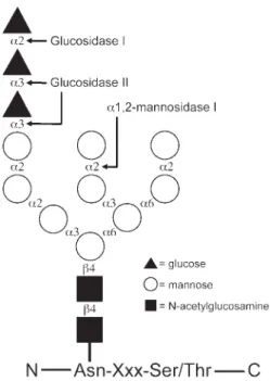
The addition of N-linked glycans to newly synthesized polypeptide chains occurs in the endoplasmic reticulum (ER). Here the 14-sac-charide core glycan (Fig. 1), Glc3Man9GlcNAc2, is added to the growing nascent polypeptide chain from a dolicholpyrophosphate-linked
precursor in a reaction catalyzed by the oligosaccharyl transferase complex (1,2). The modified asparagine side chain occurs within the Asn-Xxx-Ser/Thr consensus sequence, where Xxx denotes any amino acid except pro-line (3). Although only relatively few of the soluble ER-resident proteins seem to be glyco-sylated, the majority of extracellular proteins produced in mammalian cells carry N-linked glycans (4). The large number of different gly-coproteins that traffic through the ER are involved in many fundamental intra- and inter-cellular processes. Consequently, mutations in
Calnexin, Calreticulin, and ERp57
Teammates in Glycoprotein Folding
Lars Ellgaard
*and Eva-Maria Frickel
Institute of Biochemistry, ETH Zurich, CH-8093 Zurich, Switzerland
Abstract
In eukaryotic cells, the endoplasmic reticulum (ER) plays an essential role in the synthesis and maturation of a variety of important secretory and membrane proteins. For glycoproteins, the ER possesses a dedicated maturation system, which assists folding and ensures the quality of final products before ER release. Essential components of this system include the lectin chaperones cal-nexin (CNX) and calreticulin (CRT) and their associated co-chaperone ERp57, a glycoprotein spe-cific thiol-disulfide oxidoreductase. The significance of this system is underscored by the fact that CNX and CRT interact with practically all glycoproteins investigated to date, and by the debilitat-ing phenotypes revealed in knockout mice deficient in either gene. Compared to other important chaperone systems, such as the Hsp70s, Hsp90s and GroEL/GroES, the principles whereby this system works at the molecular level are relatively poorly understood. However, recent structural and biochemical data have provided important new insights into this chaperone system and pre-sent a solid basis for further mechanistic studies.
Index Entries:Calnexin; calreticulin; endoplasmic reticulum; ERp57; lectin; molecular chaper-one; oxidoreductase.
*Author to whom all correspondence and reprint
requests should be addressed. E-mail: lars.ellgaard@ bc.biol.ethz.ch
proteins of the N-glycosylation pathway are known to cause severe disease phenotypes, and a number of congenital disorders of glycosyla-tion are known (5,6).
The sugar itself can directly modulate pro-tein structure and function, e.g., by increasing properties such as stability and solubility (7–11). In addition, N-linked glycans act as tags in intracellular trafficking (12), and accordingly different lectins act as sorting receptors in the secretory pathway (13–15). Despite the many different and widely recog-nized functions of N-linked glycans, the role of the N-linked glycan in protein folding in the ER has only become fully appreciated within the past 10 yr. Specifically, it has been found that modifications of the core glycan to gener-ate a monoglucosylgener-ated (Glc1Man9GlcNAc2) form determine the interaction with two
homologous lectin chaperones, calnexin (CNX) and calreticulin (CRT).
Studies in tissue culture cells and ER-derived microsomes have demonstrated that the interaction with CNX and CRT protects glycoproteins from aggregation and prema-ture degradation (16–20). Repeated cycles of binding to and release from CNX and CRT, controlled by enzymes that modify the core oligosaccharide, ensure the retention of sub-strate glycoproteins in the favorable folding environment of the ER until correctly folded. If permanently misfolded, these substrate proteins are degraded. At least for certain gly-coproteins, the removal of a mannose residue in the middle branch of the glycan is a signal for degradation (21). A recently discovered mannose-binding lectin, EDEM, specifically accelerates the degradation of glycoproteins and could well be directly involved in target-ing Man8GlcNAc2-containing proteins for degradation (22–28). Overall, the oligosaccha-ride and the different enzymes that modify it play important roles in glycoprotein folding and degradation. These aspects have been covered in great detail in other reviews (29–33).
Cell biological studies have provided most of our current knowledge about the CNX/CRT chaperone system. However, more recent work has revealed structural informa-tion at atomic resoluinforma-tion for both CNX and CRT, along with details about their interaction with the co-chaperone ERp57. Moreover, other studies have explored the possibility that protein–protein interactions, in addition to the glycan-mediated interaction, contribute to the function of CNX and CRT as molecular chaperones. Taken together, these investiga-tions have considerably increased our under-standing of the molecular basis for glycoprotein folding and have also raised interesting new questions. This review describes the function of the CNX/CRT chap-erone system. In particular, we emphasize the role of molecular properties of CNX, CRT, and ERp57 in determining the mechanism of chap-erone function in the living cell.
Fig. 1. Schematic representation of the N-linked core oligosaccharide attached to an asparagine side chain in an Asn-Xxx-Ser/Thr amino acid consensus sequence. The linkage for each individual glycosyl residue is indi-cated along with cleavage sites for various ER enzymes that modify the sugar structure.
ER QUALITY CONTROL
The endoplasmic reticulum (ER) plays a fun-damental role in the synthesis, folding, and assembly of numerous important proteins, such as cell-surface receptors, membrane channels, extracellular matrix components, serum pro-teins, and antibodies. The environment in the ER is optimal for the correct folding and maturation of such proteins. For many proteins the matura-tion process involves co- and posttranslamatura-tional modifications, such as signal-peptide cleavage, glycophosphatidylinositol (GPI)-anchor addi-tion and N-linked glycosylaaddi-tion. Furthermore, the oxidizing milieu of the ER supports the for-mation of disulfide bonds. This stabilization of protein conformation is likely to help proteins maintain their structure in the extracellular envi-ronment. Finally, the ER is rich in chaperones and enzymes, which are crucial in assisting the process of correct protein folding (34,35).
Still, incorrectly folded or incompletely assembled proteins are common side products during protein synthesis in the ER. Such prod-ucts, which could be harmful to the cell if allowed to proceed along the secretory pathway to the cell surface or another cellular location, are subject to a stringent quality control (QC) system (36,37). A number of general chaperones, includ-ing BiP, a member of the Hsp70 family of chap-erones, and protein disulfide isomerase (PDI), a thiol-disulfide oxidoreductase, recognize and retain proteins that expose non-native features. This system ensures that misfolded and incor-rectly assembled proteins are retained in the ER and eventually degraded. Many so-called ER storage diseases are known, which arise from the ER retention of mutant alleles of certain proteins. Such diseases include cystic fibrosis and emphy-sema (for reviews, see refs. 38–41).
Defective ER-retained proteins are typically degraded by the proteasome after selective retrotranslocation to the cytosol and ubiquitina-tion (42). This process is referred to as ER-asso-ciated degradation (ERAD). Moreover, the cell responds to increased misfolding of proteins in the ER by the unfolded protein response (UPR) (reviewed in ref. 43). Signaling from the ER to
the nucleus leads to increased transcription of genes encoding ER chaperones to help alleviate the folding problem. In addition, another sig-naling pathway involving phosphorylation of the translation initiation factor eIF2α leads to attenuation of protein translation to further reduce the load on the ER folding machinery. Lastly, yet other genes involved in degradation are upregulated, as recently shown in the case of EDEM (28). Several investigations show that the processes of ERAD and UPR are closely coordinated (44–46).
THE CALNEXIN/CALRETICULIN CYCLE
The QC system described above applies to all proteins that encounter the lumen of the ER or are inserted into the ER membrane. CNX, CRT, and ERp57 are important factors of this general ER QC system (47).CNX and CRT cooperate with a number of enzymes in the process of assisting glycopro-tein folding. The first step of this so-called CNX/CRT cycle (see Fig. 2) involves binding to either chaperone through the monoglucosy-lated glycan, Glc1Man9GlcNAc2, present on nascent chains and on newly synthesized gly-coproteins. This form of the sugar appears either as a trimmed intermediate of the triglu-cosylated core oligosaccharide or by readdition of a glucose residue to the fully deglucosylated glycan (see below). Sequential trimming of the two outermost glucose residues on the core oligosaccharide is executed by glucosidases I and II. The importance of monoglucosylation for CNX and CRT binding has been shown in living cells, in microsomes and in vitro for a number of different proteins (see for instance refs. 48–56). Many such studies have used inhibitors of glucosidase II, such as cas-tanospermine and deoxynojirimycin, to pre-vent the formation of the monoglucosylated, trimmed intermediate of the original core oligosaccharide. Under these experimental con-ditions, access to CNX and CRT is prevented and proteins are retained in the ER where they often form high molecular weight complexes
monoglucosylated glycan, protein–protein interactions of CNX and CRT with their sub-strates could well contribute to the chaperone function of both proteins (see below) (57,58).
assisted by the thiol-disulfide oxidoreductase ERp57, which is found noncovalently associated with both proteins in vivo (47,59,60). The impor-tance of ERp57 in the CNX/CRT cycle has
become apparent from studies showing that the protein interacts with soluble secretory proteins, as well as integral membrane proteins, carrying N-linked glycans (59,61). As observed for sub-strates of CNX and CRT, both the association and release of substrates from ERp57 are modu-lated by glucose trimming (61–63). That the addition of glucosidase inhibitors can result in impaired disulfide bond formation was recently shown in the case of CD1d heavy chain, which interacts with CNX and CRT during folding (64). Furthermore, the direct involvement of ERp57 in the oxidative folding of glycoproteins is evident from the finding that the protein forms transient mixed disulfides with CNX- and CRT-associated glycoproteins during folding in living cells (63). Thus, by interacting with CNX and CRT, ERp57 functions as a specialized thiol-disulfide oxidoreductase for glycoproteins.
Glycoproteins are released from CNX and CRT by the action of glucosidase II, which
removes the terminal glucose of the glycan (49,54). This step most likely occurs irrespective of the folding state of the glycoprotein, and pre-vents its renewed association with CNX and CRT. If not correctly folded at this stage, the glycoprotein is recognized by UDP-glucose:gly-coprotein glucosyltransferase (GT). This enzyme works as a folding sensor and only re-adds a glucose residue to the oligosaccharide on non-native glycoproteins (29). In this way, the action of GT ensures that proteins in a mis-folded conformation can reassociate with CNX and CRT (52,54,65). GT has been shown to detect misfolding on a domain level (66), and even local folding defects in one domain can be distinguished so that only glycans in struc-turally destabilized regions are reglucosylated (C. Ritter, K. Quirin and A. Helenius, personal communication). Therefore, surveillance can take place on a domain-by-domain basis and it can be hypothesized that glycosylation sites in Fig. 2. The calnexin/calreticulin cycle. The folding of newly synthesized glycoproteins in the ER is assisted by calnexin (CNX) and calreticulin (CRT). Both proteins bind to the monoglucosylated form of the glycoprotein generated after the initial removal of two glucoses by glucosidases I and II. Here, we have used the available three-dimensional structures of CNX (the lumenal domain) and CRT (the P-domain) to depict parts of the two molecules (see also Fig. 4). The glycan binds to the lectin domain of both chaperones—in CRT the glycan binding site is represented by the curved black line. Other interactions can occur through protein-protein contacts (not depicted). Disulfide bond formation in glycoprotein substrates of CNX and CRT is catalyzed by the associated thiol-disulfide oxidoreductase ERp57, with which substrates form transient mixed thiol-disulfide intermedi-ates. In both CNX and CRT, the tip region of the P-domain mediates the interaction with ERp57. Removal of the remaining glucose, in a reaction catalyzed by glucosidase II, prevents interaction with CNX and CRT. Upon release, one of three possible fates awaits the glycoprotein. First, if the protein has reached its native conformation it is not longer retained in the ER and is free to travel along the secretory pathway. Second, if the protein is still not correctly folded, it can be recognized by the UDP-glucose:glycoprotein glucosyltransferase (GT). This enzyme uses UDP-glucose as a sugar donor to reglucosylate non-native glycoproteins carrying high-mannose glycans. Consequently, it acts as a folding sensor in CNX/CRT cycle. The readdition of a glucose residue to the N-linked glycan promotes reassociation with CNX and CRT. An important role of CNX and CRT is to retain glycoproteins in the ER, where the conditions for folding are favorable. Finally, pro-longed ER-retention increases the chances of encountering the ER α1,2-mannosidase I. This enzyme removes a mannose residue in the middle branch of the glycan to generate a form with eight man-noses (see also Fig. 1). A novel ER lectin, EDEM, is likely to recognize this form of the glycan and thereby extract the glycoprotein from the CNX/CRT cycle. Moreover, EDEM directs the glycopro-tein for degradation by the ERAD pathway.
domains with a tendency for misfolding have been maintained throughout evolution in order to optimize the folding efficiency of glycopro-teins carrying such domains.
Overall, cycles of binding and release slow down the rate of folding but increase its effi-ciency for many glycoproteins by keeping them exposed to the ER QC system (20). Like other protein QC systems in the cell, the detection of non-native glycoproteins in the ER by GT relies on features of the polypeptide chain that distin-guish it from proteins in a native conformation. The benefit of using sugars as “reporter mole-cules” for the protein folding status likely relates to the fact that the modifications of the core oligosaccharide that control the fate of gly-coproteins in the ER are independent of the spe-cific protein. Thereby the glycan, which is present in a large number of molecules, works as a highly versatile tag that can be modified and recognized by a relatively small number of ER-resident enzymes and lectins.
SUBSTRATE INTERACTIONS
OF CALNEXIN AND CALRETICULIN
A wide variety of important cellular and viral glycoproteins are known substrates of CNX and CRT. These include HIV gp120 and gp160 (67–70), class I major histocompatibility com-plex (MHC) heavy chain (17,71–77), T-cell recep-tor subunits (71,78), α1-antitrypsin (50,79,80), tyrosinase (81–83), the prion protein (84,85), and the cystic fibrosis transmembrane conductance regulator (CFTR) (86,87). Although many glyco-proteins associate with both CNX and CRT, and despite having the same glycan specificity, these two lectins can have distinct roles in glycopro-tein maturation. For instance, they can bind to the same protein at different stages of the fold-ing process as seen for both the MHC class I heavy chain (88,89) and influenza hemagglu-tinin (HA) (18). For the latter, association has been shown to depend on the position of the sugar within the molecule. Whereas CRT associ-ates preferentially with the glycans of the HA top domain, CNX associates more efficientlywith those glycans present in the membrane-proximal stem domain (90).
The set of substrates bound by the two pro-teins is largely, but not completely, overlapping (51,91). In line with the results obtained for HA, the difference in substrate recognition pat-tern has been shown to depend on the presence of the membrane anchor in CNX (92,93). However, the finding that the lumenal domain of CNX cannot complement the function of CRT in MHC class I assembly when expressed in CRT-deficient cells indicates that the two proteins possess protein specific functions despite their many similarities (76).
As mentioned previously, the exact mode of interaction by CNX and CRT with their glyco-protein substrates is not entirely clear. Studies performed in vitro using RNaseB as a model glycoprotein and studies of glycoprotein folding in the protozoan parasite Trypanosoma cruzi sug-gest that the function of CNX and CRT as mole-cular chaperones can be attributed solely to their lectin activity (53,55,94). However, assays of aggregation and refolding using purified pro-teins as substrates show that CNX and CRT could have an additional function as classical chaperones, characterized by protein–protein contacts with hydrophobic regions exposed by non-native polypeptide chains (57,58). As observed for classical chaperones, CNX and CRT can suppress aggregation and preserve proteins in a folding competent state, indepen-dent of their glycosylation status (monogluco-sylated or nonglyco(monogluco-sylated) (57,58). However, the efficiency of suppressing aggregation was enhanced for monoglucosylated proteins, potentially indicating an avidity effect of two binding sites, one for the glycan and one for the polypeptide chain of the substrate (95). Whereas native conformers were shown not to interact with CNX and CRT, complexes were formed with misfolded conformers.
The effects of cofactors, such as Zn2+, adeno-sine triphosphate (ATP) and the monoglucosy-lated glycan on this bonafide chaperone function of CNX and CRT, have also been investigated in vitro. These cofactors are known to modulate structural properties of
CNX and CRT (see below). Although ATP was shown to enhance the ability to suppress pro-tein aggregation, addition of the monoglucosy-lated glycan inhibited this function (57,58). The latter result indicated that occupation of the oligosaccharide binding site is capable of influ-encing the protein–protein interactions pro-posed to mediate the chaperone function.
Overall, these experiments support the notion that protein–protein based contacts of CNX and CRT with their substrates, in addi-tion to an initial glycan based interacaddi-tion, play an important role during folding (57,58). Moreover, ATP binding by CNX and CRT, and potentially Zn2+ binding in the case of CRT, could promote substrate interaction by lead-ing to the exposure of hydrophobic surface. Substrate release would then occur by ATP hydrolysis or dissociation, both processes pos-sibly mediated by a co-chaperone.
MOLECULAR PROPERTIES
OF CALNEXIN AND CALRETICULIN
Besides its well-established role as a molecu-lar chaperone, CRT is a major Ca2+storage pro-tein and plays an important role in Ca2+ homeostasis in the ER (96,97). Thus, the embry-onic lethal phenotype observed in CRT-deficient mice is connected to the function of the protein in Ca2+ signaling during cardiac development (99,100). Although CNX also binds Ca2+ (101,102), the protein does not seem to play a significant role in Ca2+ homeostasis and its major function is that of a molecular chaperone. CNX gene-deficient mice do not show as serious defects as CRT knockout mice. However, 50% die within 2 d of birth, whereas the rest display severe motor disorders, a selective loss of large myelinated fibers and die within 3 mo (103).Biochemical studies of CNX and CRT are abundant, and below the most important bio-chemical properties of the two proteins are summarized. Together with structural data, these molecular characteristics constitute the basis for understanding the mechanism of chaperone function of CNX and CRT in detail.
Primary Structure
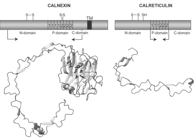
The molecular cloning of CRT (46.5 kDa, 400 residues) (104,105) and CNX (65.4 kDa, 572 residues) (101,102) has revealed that the two proteins are highly similar (Fig. 3). The main differences are found in their carboxyl termi-nal regions. Whereas CRT is a soluble lumitermi-nal protein with a carboxyl terminal KDEL retrieval sequence, CNX is a type I brane protein with a predicted transmem-brane region spanning residues 463–485, followed by a 87-residue carboxyl terminal cytosolic tail. However, the luminal domain of CNX is highly similar to CRT. Based on sequence analysis, both proteins have been suggested to consist of three regions, the N-, P-, and C-domains (Fig. 4) (104–107). The N-domain (CRT residues 1–188, CNX residues 1–253) was originally predicted to comprise a β-sheet rich globular structure (104,105). The unique P-domain (CRT residues 189–283, CNX residues 254–388) constitutes the signature sequence for this family of proteins, and has obtained its name due to the many prolines present in this region. It consists in its entire length of two short sequence repeats, the type 1 and type 2 repeats. Each type of repeat is present in three and four copies in CRT and CNX, respectively. In both proteins, the arrangement of the repeat sequences is such that all type 1 repeats are clustered together, followed by a cluster of type 2 repeats (Figs. 3 and 4). The distinguishing feature of the CRT C-domain (residues 284–400) is the enrich-ment of acidic amino-acid residues in the approx 60 carboxyl terminal residues. This region is known to play a role in low-affinity Ca2+ binding (106,108,109). In contrast, the C-domain of CNX, residues 389–462, does not display noticeable traits. Here, the membrane-proximal region of approx 20 residues is likely to represent a linker sequence between the membrane anchor and a globular lectin domain (see below).
The sequences of both CNX and CRT encode cysteine residues that form intrachain disulfide bonds. In CNX the four cysteines pair to form
Fig. 3. Amino-acid sequence alignment of human CNX and human CRT. Both sequences are shown without amino terminal signal sequences and numbered accordingly. Repeat sequences of the P-domain are indicated above the alignment. Amino-acid residues proposed to coordinate Ca2+ in the crystal structure of the lumenal domain of CNX are labeled with an asterisk, and amino-acid residues proposed to interact with the glycan are marked with a triangle. The black bar denotes the transmembrane region in CNX. Boundaries for the NMR structure of the CRT P-domain (residues 189–288) and for the crystal structure of the lumenal domain of CNX (residues 40–437) are indicated above and below the respective sequences with arrows.
Fig. 4. Overview of the primary amino-acid sequence of CNX and CRT, and the three-dimen-sional structures solved for the two proteins. In the schematic representation, the position of type 1 and type 2 repeats of the P-domain is indicated along with the position of free and disul-fide-bonded cysteine residues. ‘TM’ denotes the transmembrane region in CNX. The arrows indicate the boundaries of the fragments for which the three-dimensional structures are shown below. In the representation of the three-dimensional structure of the lumenal domain of CNX, residues involved in the binding of the glucose are shown as black stick models and the cys-teines forming the two disulfide bonds are shown as CPK models. This figure was prepared using the program MOLMOL.
the Cys140–Cys174 and Cys340–Cys346 disul-fides (110). In CRT a free cysteine is present at position 146, whereas the Cys89–Cys120 fide is equivalent to the Cys140–Cys174 disul-fide in CNX (111). Although certain animal CRT sequences contain a conserved N-glycosy-lation site at residue 326, human placental CRT has been shown not to be glycosylated (111). Animal CNXs do not contain potential sites for N-linked glycosylation. A unique feature of the carboxyl terminal cytosolic tail of CNX is the presence of sites for phosphorylation by casein kinase II at Ser534 and Ser544 and by extracel-lular-signal regulated kinase-1 at Ser563 (112,113). Phosphorylation at these positions has been shown to regulate the association of CNX with ribosomes (114).
Both CNX and CRT have a tissue-specific isoform in testis. The CNX isoform, calmegin, interacts with nascent chains of glycoproteins and is required for male fertility in mice (115,116), whereas the function of the recently discovered CRT isoform, CRT2, has not yet been investigated (117). The sequence similar-ity of both testis isoforms with the respective ubiquitously expressed forms is high (>60%), and it is very likely that the isoforms also func-tion as lectin chaperones (117–119).
Ca
2+Binding
Different cofactors have been found to inter-act with CNX and CRT in vitro and thereby modulate the structural properties of both pro-teins. The effects of metal binding by CRT have been thoroughly characterized by a variety of methods, such as circular dichroism, intrinsic fluorescence, 8-anilino-1-naphtalenesulfonate (ANS) binding and equilibrium dialysis. Of twelve different divalent cations tested for binding to CRT, only Ca2+ and Zn2+ were shown to bind specifically (109). CNX binds Ca2+ (101,102,120,121), whereas there is little evidence that the protein binds other metals.
Owing to its role in Ca2+ homeostasis, the Ca2+-binding properties of CRT have been par-ticularly well investigated (see for instance, refs. 106,108,109,122–125). The protein has two
distinct classes of binding sites: one high-affin-ity binding site that complexes 1 mol Ca2+/mol protein with a Kd of 0.05–11 µM, and several low-affinity sites, located in the acidic C-termi-nal domain, that bind approx 20 mol Ca2+/mol protein with a Kd of 2 mM (106,108,109). As pointed out earlier (107), the rather broad range of Kdvalues determined for high-affinity Ca2+binding by CRT is likely a result of the dif-ferent experimental conditions employed in these studies.
Ca2+binding by CRT seems to have only lit-tle effect on the content of regular secondary structure elements and on the global structure of the protein (109,123,125,126). However, upon occupancy of the high-affinity Ca2+ binding site, local structural effects are observed that indicate a more compact conformation with increased thermal stability (109,125). These results can be rationalized based on the crystal structure of the lumenal domain of CNX, which coordinates one Ca2+ion with a proposed role of structural stabilization (110). In addition, CRT shows increased resistance toward prote-olytic digestion by a variety of proteases at high Ca2+ concentrations (124,125). A similar result has been reported for CNX (121).
Zn
2+Binding
CRT contains one high-affinity binding site for Zn2+(apparent K
dof 0.05 µM) and 14 low-affinity binding sites (apparent Kd of 310 µM) (109). Zn2+ binding by CRT has been mapped to the N-domain (127) and has been shown to induce a considerable conformational change in the protein resulting in the exposure of hydrophobic surface (57,109). In accordance with these results, CRT is found to aggregate at concentrations of Zn2+above 600 µM (109,127), whereas CNX aggregates already in the pres-ence of 100 µM Zn2+(128). In CRT the observed conformational change is accompanied by an increased susceptibility toward proteolytic digestion by trypsin and chymotrypsin and a decreased thermal stability (125).
As proposed by several authors, the expo-sure of hydrophobic surface by CRT observed
in vitro in the presence of concentrations as low as 50 µM ZnCl2could potentially be important for the ability of the protein to suppress aggre-gation through protein–protein interactions with its substrates (57,124,125). Unfortunately, although Zn2+is present in the ER lumen, little is known about its concentration and all the mammalian zinc transporters described to date are localized to cellular compartments other than the ER. However, recent studies show that the ER of Schizosaccharomyces pombe harbors a zinc transporter thought to regulate ER zinc homeostasis in this organism (129,130).
ATP Binding
Another potentially important cofactor of CNX and CRT is ATP. Both proteins bind ATP in vitro (57,58,121,131). Although a weak ATPase activity has been reported for both pro-teins (57,58), others have been unable to detect any such activity (121,124). For both proteins, the binding of ATP is accompanied by a decrease in intrinsic fluorescence emission and enhanced binding of ANS, indicating exposure of hydrophobic surface (57,58). Whereas the presence of ATP protects the carboxyl terminal region of CRT from proteolysis (124), both destabilizing and stabilizing effects of ATP binding by CNX have been reported (121,131). Currently, the physiological significance of ATP binding by CNX and CRT remains unclear. Classical chaperones such as those belonging to the Hsp70 family, including the ER-resident protein BiP, are ATPases. Cycles of ATP binding, hydrolysis and nucleotide exchange control substrate binding and release, and are, in turn, regulated by cofactors that either influence ATPase activity or act as nucleotide exchange factors. The reported ATPase activity of <0.1 pmol/min/µg for CNX and CRT is very low (57,58). In comparison, BiP, which is considered a weak ATPase, has an activity of 5.2 pmol/min/µg (132). Thus, if ATP hydrolysis plays a role for the chaperone function of CNX and CRT, positive regulators of ATPase activity and nucleotide exchange are likely to exist.
Glycan Binding
A number of studies performed in vitro have characterized the direct interaction of CNX and CRT with isolated glycans using biochemical methods. All support the notion that both proteins are lectins that specifically interact with the monoglucosylated form of the core glycan (Fig. 1) (50,131,133–135). These studies also show that CNX and CRT interact with glycans with increasing affinity when com-paring oligosaccharides of different lengths as follows: Glc(G1) << Glcα1-3Man(G1M1) << 3Manα1-2Man(G1M2) < Glcα1-3Manα1-2Manα1-2Man(G1M3). Therefore, the glycan interaction is likely to involve the entire α1-3 branch of the oligosaccharide, which forms a continuous molecular surface in the NMR structure of Glc1Man9GlcNAc2 (136). Direct affinity measurements have shown that the G1M3 tetrasaccharide binds CRT with compara-ble affinity to an IgG molecule carrying the entire monoglucosylated glycan (135). However, gly-can binding by CNX and CRT could potentially also involve contacts to the mannose residues on the α1-6 branch of the glycan (50,133).
Detailed biophysical analysis of the bind-ing of IgG carrybind-ing a sbind-ingle monoglucosy-lated glycan to CRT demonstrated a Kd of approx 2 µM (137). Furthermore, this study showed that—at least under these experi-mental conditions using a native monogluco-sylated protein—no contribution to the binding reaction was observed from pro-tein–protein interactions. As proposed by Surolia and coworkers (137), the relatively low affinity of CRT for the glycan could be advantageous to allow for rounds of glyco-protein association and dissociation. This fea-ture would permit trimming of the remaining glucose of the glycan by glucosidase II to occur on the nonbound substrate. Such a model is supported by the finding that RNaseB bound by the lumenal domain of CNX is protected effectively from glucosidase II and PNGaseF digestion (53).
In vitro, the binding of the oligosaccharide by either CNX or CRT depends critically on the
presence of Ca2+(131,137). Thus, it is possible that the deleterious effect of Ca2+ depletion from the ER, observed for the folding of glyco-proteins known to interact with CNX and CRT, directly reflects structural changes in the lectin domain, which render both proteins incapable of ligand interaction (138).
STRUCTURAL STUDIES OF CALNEXIN
AND CALRETICULIN
The biochemically determined properties of CNX and CRT have been put into new per-spective by recent structural studies on both molecules. For CNX, the crystal structure of a fragment comprising residues 40–437, encom-passing most of the lumenal domain, has been reported to a resolution of 2.9 Å (Fig. 4) (110). This highly unusual structure contains two separate entities: a globular β-sandwich domain homologous to legume lectins and an extended hairpin fold of approx 140 Å in length, corresponding to the P-domain. The globular domain comprises a concave and a convex β-sheet with six and seven β-strands, respectively. In this part of the molecule, the CNX model also shows a putative Ca2+ bind-ing site, with the Ca2+ ion proposed to play a role in structural stabilization rather than lig-and interaction. The only regular secondary structure elements in the P-domain are four short, anti-parallel sheets where each β-strand contains three residues. The disulfide bond connecting the cysteine residues 340 and 346 is found close to the tip of the P-domain.
By soaking the crystal in glucose and deter-mining its location in the electron density, it was found that the concave β-sheet of the glob-ular domain harbors a monovalent glycan binding site. Modeling the binding of the G1M3 tetrasaccharide indicated that steric hindrance is likely to prevent the access of glucosidase II to its sugar substrate. This finding supports the idea that the glycoprotein must dissociate from the lectin for cleavage by glucosidase II to occur. The disulfide bond connecting the cys-teine residues 140 and 174 is present in the
glob-ular domain where it connects two β-strands in the vicinity of the proposed glycan-binding site. This feature can account for the observed sensi-tivity of oligosaccharide binding by CNX toward reduction (121,131).
Interestingly, legume lectins, galectins and neurexin 1β possess a fold closely related to the globular domain of CNX (110). The laminin G-like domain of neurexin 1β contains alternative splice sites in loops connecting the β-strands and the absence or presence of inserted sequences at these sites determines ligand interactions (139). Similarly, the P-domain of CNX is introduced at the same topological position as one of the splice sites in neurexin 1β and also establishes a protein–protein interac-tion (see below).
For CRT, the NMR structure of the P-domain, residues 189–288, has been solved (Fig. 4) (140,141). Like the CNX P-domain it shows an extended hairpin fold, in the case of CRT with a length of approx 110 Å. Three short antiparallel β-sheets and three small hydrophobic clusters stabilize the structure. This threefold repetition of structural elements and the four-fold repeti-tion of similar structural features in the CNX P-domain closely reflects the repetitive nature of the P-domain sequence in the two molecules (Figs. 3 and 4). Roughly, each type 1 repeat sequence pairs up with a type 2 repeat sequence in the structure of both P-domains by forming interactions across the hairpin. In addi-tion, it has recently been shown that a short fragment corresponding to one type 1 repeat and one type 2 repeat, and comprising the β-sheet and the hydrophobic cluster at the tip of CRT P-domain, constitutes an independently folding structure (142). It is tempting to specu-late that the unique sequence of the P-domain has evolved by the sequential insertion of such “12” units into a loop region of the globular lectin domain.
Currently, the structure corresponding to the CNX lectin domain is not known for CRT. However, the crystal structure of the lumenal domain of CNX and the sequence similarity between the two proteins (Figs. 3 and 4) strongly indicate that the CRT N-domain and
residues 284–337 of the C-domain will form a globular domain with structural similarity to the CNX lectin domain. Indeed, this is exactly what the recently modeled structure of the CRT lectin domain also suggests (135). Furthermore, biochemical and biophysical analysis of full-length CRT has shown that the molecule is asymmetric and elongated (126). Based on this information, the known struc-tural data for both proteins and the overall high conservation of sequence and function between the two proteins, it can be assumed that both CNX and CRT show a two domain structure comprising a globular lectin domain and a long protruding P-domain. In addition, the residues of CNX proposed to be involved in the binding of glucose and Ca2+ are largely conserved in CRT. Likewise, residues in CRT proposed to bind the G1M3 tetrasaccharide are well conserved in CNX (Fig. 3).
Whereas the crystal structure of the lumenal domain of CNX revealed the lectin function of the globular domain, the function of the P-domain remained unclear despite detailed structural analysis obtained by both X-ray crys-tallography and NMR spectroscopy. However, it has now been shown that the P-domain of both CNX and CRT binds to ERp57 (128,142,143). Using NMR spectroscopy and deletion mutants of both CNX and CRT, these studies have mapped the site of interaction with ERp57 to the distal end of the P-domain. Furthermore, the Kdof the interaction between ERp57 and the CRT P-domain was determined to approx 9 µM (142,143). Although this affinity is quite weak, it is likely that a more stable ter-tiary complex results in the presence of a cys-teine-containing glycoprotein substrate through the formation of transient mixed disulfide bonds between the glycoprotein and ERp57.
THE ROLE OF ERP57
The growing family of ER thiol-disulfide oxi-doreductases, most of which contain one or more thioredoxin-like domains, promotes the proper oxidation, isomerization and reduction
of disulfide bonds (144). The reactions involving these proteins proceed through intermolecular disulfide-bonded intermediates and both ERp57 and PDI form mixed disulfides with viral glyco-proteins during folding in living cells (63). Therefore, rather than regulating the redox state of other factors in the ER, these proteins directly catalyze the oxidative folding of proteins.
Molecular Studies of ERp57
The best characterized of the redox-active proteins in the ER is PDI. It comprises four thioredoxin-like domains—termed a, b, b’, and a’—followed by an acidic carboxyl terminal c-domain, which also harbors the KDEL ER-retention motif. Thioredoxin is a small 12 kDa protein, which functions as a disulfide reduc-tase in the cytosol. The three-dimensional struc-tures of thioredoxin and related domains show a typical α/β fold with a five-stranded β-sheet surrounded by four α-helices (for a review, see ref. 145). In PDI, the a and a’ domains are cat-alytically active and both contain two cysteine residues in a characteristic ‘CXXC’ sequence motif. Within the thioredoxin superfamily these cysteines are redox-active and switch between the dithiol and disulfide forms.
ERp57 is the closest known homolog of PDI, with which it shares the same domain composi-tion except for the absence of the acidic c-domain. Consequently, the QDEL ER-retention motif in ERp57 is located directly at the car-boxyl terminal end of the a’ domain (146). PDI and ERp57 show an overall amino acid identity of 29%, with the a and a’ domains being most closely related, whereas the b’ domains are the most divergent. In PDI, the b’ domain has been shown to bind peptides, a feature that is likely to be related to its function as a bonafide chap-erone. Since the ERp57 b’ domain does not pos-sess a similar function, it is tempting to speculate that this domain in ERp57 could have evolved to constitute a binding site for CNX and CRT, as proposed recently by Freedman and colleagues (145). Currently, no structural data are available for ERp57. However, NMR structures of the PDI a and b domains show
that they both comprise typical thioredoxin-like folds (149,150). Preliminary NMR data and assignments have been reported for the a’ domain of ERp57 (151).
Using in vitro assays for redox activity, ERp57 has been shown to have disulfide reductase and isomerase activity (152–155). Only one in vitro study has investigated the catalytic activity of ERp57 using an endogenous substrate of the protein, namely partially folded MHC class I heavy chain molecules (156). Toward this sub-strate, ERp57 was shown to exhibit reductase activity, and it was proposed that this activity might be involved in reducing heavy chain mol-ecules prior to retrotranslocation and degrada-tion (156). However, it remains to been seen exactly which type(s) of disulfide exchange reaction(s) ERp57 performs in vivo.
PDI and Glycoprotein Folding
As mentioned previously, ERp57 acts as a glycoprotein specific redox-active enzyme through its cooperative interaction with CNX and CRT. The finding that ERp57-enhanced disulfide bond formation in vitro is dependent on CNX and CRT stresses the functional coop-eration of both lectin chaperones with ERp57 (157). No positive effects on glycoprotein refolding by PDI were observed upon addition of CNX and CRT. Although PDI has been shown to be involved in glycoprotein folding (63), substrate interaction of PDI in vivo is not regulated by glucose trimming (59–61,63,158). These studies all indicate that PDI functions independently of CNX and CRT, which is in agreement with findings that were unable to detect complexes of either lectin with PDI (60,143). However, other data have shown that PDI interacts with CRT (123,159). It is possible that this apparent discrepancy is a result of the different experimental conditions used because the interaction is strongly influenced by changes in Ca2+ concentrations (123). Thus, fluctuations in the free concentration of Ca2+in the ER could affect protein folding, either directly by inducing conformational changes in CNX and CRT resulting in altered substrate
interactions or indirectly by modulating the binding of co-chaperones (97,123).
ERp57 in MHC Class I Assembly
Recently, several investigations have focused on the interaction of ERp57 with glycoproteins during CNX- and CRT-dependent folding (see, for instance, refs. 64,160–162). To date, the best-studied glycoprotein substrate of ERp57 in vivo is the MHC class I complex. This heterodimer is comprised of the glycosylated heavy chain and the associated protein, β2-microglobulin. The MHC class I complex plays a critical role in the immune response by delivering for instance virus- and tumor-derived antigenic peptides to the cell surface for presentation to receptors on cytotoxic T-cells. The process of MHC class I folding, assembly and peptide loading takes place in the ER, and has been shown to involve CNX, CRT and ERp57 together with the more specialized proteins transporter associated with antigen processing (TAP) and tapasin. During the early stages of folding the newly synthe-sized heavy chain interacts with CNX. Upon binding of β2-microglobulin, CRT replaces CNX and together with TAP, tapasin and ERp57 form the so-called peptide loading complex (PLC). In a tapasin-dependent process, the class I mole-cules are then loaded with peptide, the PLC dis-sociates and the mature MHC class I complex can traffic to the cell surface (for recent reviews, see refs. 77,163,164).
ERp57 is involved at various stages of MHC class I assembly. The heavy chain contains two disulfide bonds and folds and oxidizes while associated with CNX, but prior to incorporation into the PLC (165–167). Because a ternary com-plex of heavy chain, CNX and ERp57 has been observed already at this early stage of matura-tion, it is possible that ERp57 is involved in cat-alyzing disulfide bond formation in the heavy chain (167). Moreover, ERp57 is an important component of the PLC (167). The association of ERp57 with the PLC has been shown to be tapasin dependent and correlate with CRT association (169,171). The latter finding sug-gests that a preformed complex of CRT and
ERp57 could enter into the PLC (171). The func-tional cooperation between ERp57 and tapasin is stressed by the finding that the two proteins form an intermolecular disulfide bond in the PLC (172). Analysis of cysteine-lacking mutants of ERp57 and tapasin has led to the conclusion that the two proteins in concert mediate the iso-merization and/or re-oxidation of the disulfide bond in the peptide-binding groove of MHC class I heavy chain (172). Although it is presently not known exactly how this process occurs, it has been shown to be important for the correct loading of the MHC class I β2 -microglobulin heterodimer with high affinity peptides (172).
FROM MOLECULAR PROPERTIES
TO IN VIVO FUNCTION
The insight gained from the many biochem-ical and cell biologbiochem-ical studies of CNX, CRT and ERp57 is providing a more and more detailed picture of the processes involving these three chaperones. Our understanding is now so advanced that many functional fea-tures observed in vivo can be rationalized based on known biochemical and structural properties. As described previously, the pre-sent data strongly indicate that CNX and CRT show a similar overall structural organization. Functionally, the lectin domain is responsible for binding the glycan attached to substrate glycoproteins, whereas the P-domain binds ERp57 at its tip. These features, in combination with the properties of both chaperones deter-mined in vivo, have led us to present a basic model for the cooperative interaction of either chaperone and ERp57 with the nascent chain or non-native conformers of newly synthesized glycoproteins in the ER (143,173).
We propose that substrate binding occurs through the interaction of the monoglucosy-lated glycan with the lectin domain of either CNX or CRT. This feature allows the polypep-tide chain a certain degree of conformational freedom in the process of folding. The struc-tural arrangement of the lectin domain and the
elongated P-domain with ERp57 bound at the tip sequesters the polypeptide chain of the sub-strate in a partially protected space during folding. This partial shielding of the bound glycoprotein substrate should help to suppress intermolecular aggregation that could result from interactions with other ER folding inter-mediates. The role of the P-domain is likely to involve positioning of ERp57 for the formation of intermolecular disulfide bonds with a vari-ety of bound substrate proteins. The conforma-tional plasticity observed in the NMR structure of the CRT P-domain could allow both chaper-ones to adapt to substrates of varying size and shape (140). This complements the idea that the overall molecular flexibility of CRT character-ized biophysically by Bouvier and colleagues is advantageous for the protein in its function as a molecular chaperone by allowing transient and dynamic interactions with a number of substrates (109,123,125,126). An additional function of the P-domain could be to constrain diffusion of the substrate after dissociation from CNX and CRT. This would allow for a more rapid reassociation, provided that the remaining glucose on the glycoprotein sub-strate has meanwhile not been trimmed by glucosidase II. Taken together, the function of CNX and CRT is likely to be dictated by their distinctive structural features and the close cooperation with ERp57, which leads to the productive oxidative folding of glycoprotein substrates.
This basic model supports the view that CNX and CRT function as molecular chaper-ones by keeping their substrates “out of trou-ble” rather than by actively promoting folding. However, it leaves room for the incorporation of additional features of both proteins relating to the binding of cofactors and protein–protein contacts with substrate glycoproteins. The pre-diction of protein–protein interactions during CNX- and CRT-assisted glycoprotein folding is appealing because it provides explanations for results otherwise not easy to rationalize. For instance, when performing immunoprecipita-tion experiments to identify glycoprotein sub-strates of CNX in vivo, it has been observed
that certain complexes persist even after removal of the glycan by glycosidase treatment with Endo H or PNGase F (50,78,174,175). Therefore, it could be that the initial binding of CNX and CRT to the glycoprotein substrate occurs as a result of the lectin activity, but that subsequent protein-protein contacts stabilize the complexes. Polypeptide-based contacts could also help explain the rather high effi-ciency observed for coimmunoprecipitation of substrate proteins with CNX and CRT in spite of the relatively low affinity measured for the interaction between CRT and monoglucosy-lated IgG (137).
Currently, however, the data relating to the function of CNX and CRT as bona fide molecu-lar chaperones are not easy to assess defini-tively. For instance, the physiologic role of potentially important cofactors implicated in this function, such as Zn2+ and ATP, is presently not evident. It has also been argued that immunoprecipitation is not the method of choice to study interactions that involve non-native conformers or nascent chains owing to their hydrophobic nature (29). For example, overexpression of protein or addition of tuni-camycin, which inhibits glycosylation and turns on the UPR, often results in protein mis-folding and aggregation. Potentially, com-plexes of CNX and nonglycosylated proteins immunoisolated under such conditions that perturb the folding environment in the ER lumen are nonphysiologic in nature. Besides, interactions of membrane proteins with CNX could possibly be preserved if caught in the same detergent micelle (20).
The fact that the experimental conditions employed in immunoprecipitation experiments can influence results considerably contributes an additional difficulty. When performing immunoprecipitations under mild detergent conditions (176), interaction of nonglycosy-lated proteins or proteins carrying di- or triglu-cosylated glycans with CNX and CRT can be observed. In contrast, when performing similar experiments under more stringent detergent conditions, no binding to CNX was observed (177). No matter which experimental
condi-tions are employed, any substrate interaction observed with CNX and CRT should be criti-cally assessed to ensure that it is transient and that it results in the formation of a fully mature and correctly folded substrate protein. Only such interactions are expected to occur as a result of the chaperone function of CNX and CRT. In contrast, long-lasting interactions with substrate proteins are likely to be nonproduc-tive and of nonspecific nature.
PERSPECTIVES
Since 1989 when Suh and colleagues observed that a misfolded variant of the vesic-ular stomatitis virus G-protein was retained in the ER in a monoglucosylated form (178), our understanding of the processes involved in glycoprotein folding in the ER has increased tremendously. In particular, the roles of the dif-ferent proteins in the CNX/CRT cycle have become much more evident. The publication of three-dimensional structures of CNX and CRT has provided substantial new insight into the molecular mechanism of the chaperone func-tion of these proteins and has significantly changed the way we think about these two lectin chaperones.
The next goals for the field should involve the determination of a high-resolution struc-ture of the CRT lectin domain, if possible in complex with the G1M3 glycan. More detailed information about the interaction of CNX and CRT with ERp57, as well as structure determi-nation of ERp57, or individual domains thereof, will be important. Additional experi-ments will be needed to clarify the exact role of the P-domain in glycoprotein folding. Despite being structurally similar, the P-domains in CNX and CRT differ with a “12” repeat unit in size. Whether this difference also conveys func-tional distinctions is presently unclear. Addressing such questions relating to the P-domain should also promote a better under-standing of how the cooperation of CNX and CRT with ERp57 assists disulfide bond forma-tion in substrate glycoproteins.
The direct mapping of specific residues in CNX and CRT that decide the interaction with ATP, Zn2+and polypeptide regions of substrate proteins will also be essential. Such information could for instance be obtained by structure determination in the presence of these co-fac-tors, which might also provide direct informa-tion on the local and global conformainforma-tional changes that occur upon cofactor binding. These results should in turn allow the design of muta-tions that specifically remove the co-factor inter-actions individually. Provided that such mutants can be shown to retain structural and functional properties of the wild-type protein, they will become valuable tools for the charac-terization of functions related to cofactor bind-ing both in vivo and in vitro. Application of this experimental strategy will be particularly important to investigate functions such as ATP binding, which can otherwise not easily be tar-geted in vivo without perturbing the cellular environment. To fully appreciate the role of ATP binding by CNX and CRT the identification of potential co-chaperones for ATP hydrolysis, nucleotide exchange or dissociation will be crit-ical. Taken together, the results of such experi-ments should provide us with a more detailed understanding of how the CNX/CRT chaper-one system functions at the molecular level.
ACKNOWLEDGMENTS
We would like to thank M. Molinari and R. Mancini for critical reading of the manuscript and helpful comments. The continued support by Ari Helenius is much appreciated. Financial support by the Swiss National Science Foundation (to L. E.) is gratefully acknowledged.
REFERENCES
1. Kornfeld, R. and Kornfeld, S. (1985) Assembly of asparagine-linked oligosaccharides. Annu.
Rev. Biochem. 54, 631–664.
2. Burda, P. and Aebi, M. (1999) The dolichol pathway of N-linked glycosylation. Biochim.
Biophys. Acta 1426, 239–257.
3. Gavel, Y. and von Heijne, G. (1990) Sequence differences between glycosylated and non-gly-cosylated Asn-X- Thr/Ser acceptor sites: impli-cations for protein engineering. Protein Eng. 3, 433–442.
4. Gahmberg, C. G. and Tolvanen, M. (1996) Why mammalian cell surface proteins are glycopro-teins. Trends Biochem. Sci. 21, 308–311.
5. Stanley, P. and Ioffe, E. (1995) Glyco-syltransferase mutants: key to new insights in glycobiology. FASEB J. 9, 1436–1444.
6. Aebi, M. and Hennet, T. (2001) Congenital dis-orders of glycosylation: genetic model systems lead the way. Trends Cell. Biol. 11, 136–141. 7. O’Connor, S. E. and Imperiali, B. (1996)
Modulation of protein structure and function by asparagine-linked glycosylation. Chem. Biol.
3,803–812.
8. Dwek, R. A. (1996) Glycobiology: Toward understanding the function of sugars. Chem.
Rev. 96, 683–720.
9. Drickamer, K. and Taylor, M. E. (1998) Evolving views of protein glycosylation.
Trends Biochem. Sci. 23, 321–324.
10. Wormald, M. R. and Dwek, R. A. (1999) Glycoproteins: glycan presentation and protein-fold stability. Structure Fold. Des. 7, R155–160. 11. Wormald, M. R., Petrescu, A. J., Pao, Y. L.,
Glithero, A., Elliott, T., and Dwek, R. A. (2002) Conformational studies of oligosaccharides and glycopeptides: complementarity of NMR, X-ray crystallography, and molecular model-ling. Chem. Rev. 102, 371–386.
12. Helenius, A., and Aebi, M. (2001) Intracellular functions of N-linked glycans. Science 291, 2364–2369.
13. Yamashita, K., Hara-Kuge, S., and Ohkura, T. (1999) Intracellular lectins associated with N-linked glycoprotein traffic. Biochim. Biophys.
Acta. 1473, 147–160.
14. Hauri, H., Appenzeller, C., Kuhn, F., and Nufer, O. (2000) Lectins and traffic in the secre-tory pathway. FEBS Lett. 476, 32–37.
15. Schrag, J. D., Procopio, D. O., Cygler, M., Thomas, D. Y., and Bergeron, J. J. (2003) Lectin control of protein folding and sorting in the secretory pathway. Trends Biochem. Sci. 28, 49–57.
16. Ou, W. J., Cameron, P. H., Thomas, D. Y., and Bergeron, J. J. (1993) Association of folding intermediates of glycoproteins with calnexin during protein maturation. Nature 364,
17. Jackson, M. R., Cohen-Doyle, M. F., Peterson, P. A., and Williams, D. B. (1994) Regulation of MHC class I transport by the molecular chap-erone, calnexin (p88, IP90). Science 263, 384–387.
18. Hebert, D. N., Foellmer, B., and Helenius, A. (1996) Calnexin and calreticulin promote fold-ing, delay oligomerization and suppress degradation of influenza hemagglutinin in microsomes. EMBO J. 15, 2961–2968.
19. Labriola, C., Cazzulo, J. J., and Parodi, A. J. (1995) Retention of glucose units added by the UDP-GLC:glycoprotein glucosyltransferase delays exit of glycoproteins from the endo-plasmic reticulum. J. Cell Biol. 130, 771–779. 20. Helenius, A., Trombetta, E. S., Hebert, D. N.,
and Simons, J. F. (1997) Calnexin, calreticulin and the folding of glycoproteins. Trends Cell
Biol. 7, 193–200.
21. Liu, Y., Choudhury, P., Cabral, C. M., and Sifers, R. N. (1999) Oligosaccharide modifica-tion in the early secretory pathway directs the selection of a misfolded glycoprotein for degradation by the proteasome. J. Biol. Chem.
274,5861–5867.
22. Nakatsukasa, K., Nishikawa, S., Hosokawa, N., Nagata, K., and Endo, T. (2001) Mnl1p, an alpha -mannosidase-like protein in yeast Saccharomyces cerevisiae, is required for endoplasmic reticulum-associated degrada-tion of glycoproteins. J. Biol. Chem. 276, 8635–8638.
23. Hosokawa, N., Wada, I., Hasegawa, K., Yorihuzi, T., Tremblay, L. O., Herscovics, A., et al. (2001) A novel ER alpha-mannosidase-like protein accelerates ER-associated degradation.
EMBO Rep. 2, 415–422.
24. Jakob, C. A., Bodmer, D., Spirig, U., Battig, P., Marcil, A., Dignard, D., et al. (2001) Htm1p, a mannosidase-like protein, is involved in glyco-protein degradation in yeast. EMBO Rep. 2, 423–430.
25. Braakman, I. (2001) A novel lectin in the secre-tory pathway. An elegant mechanism for gly-coprotein elimination. EMBO Rep. 2, 666–668. 26. Molinari, M., Calanca, V., Galli, C., Lucca, P.,
and Paganetti, P. (2003) Role of EDEM in the release of misfolded glycoproteins from the calnexin cycle. Science 299, 1397–1400.
27. Oda, Y., Hosokawa, N., Wada, I., and Nagata, K. (2003) EDEM as an acceptor of terminally misfolded glycoproteins released from cal-nexin. Science 299, 1394–1397.
28. Yoshida, H., Matsui, T., Hosokawa, N., Kaufman, R. J., Nagata, K., and Mori, K. (2003) A time-dependent phase shift in the mam-malian unfolded protein response. Dev. Cell 4, 265–271.
29. Parodi, A. J. (2000) Protein glucosylation and its role in protein folding. Annu. Rev. Biochem.
69,69–93.
30. Parodi, A. J. (2000) Role of N-oligosaccharide endoplasmic reticulum processing reactions in glycoprotein folding and degradation.
Biochem. J. 348, 1–13.
31. Trombetta, E. S., and Parodi, A. J. (2001) N-gly-can processing and glycoprotein folding. Adv.
Protein. Chem. 59, 303–344.
32. Roth, J. (2002) Protein N-glycosylation along the secretory pathway: relationship to organelle topography and function, protein quality control, and cell interactions. Chem.
Rev. 102, 285–303.
33. Roth, J., Zuber, C., Guhl, B., Fan, J. Y., and Ziak, M. (2002) The importance of trimming reac-tions on asparagine-linked oligosaccharides for protein quality control. Histochem. Cell
Biol. 117, 159–169.
34. Benham, A. M., and Braakman, I. (2000) Glycoprotein folding in the endoplasmic reticulum. Crit. Rev. Biochem. Mol. Biol. 35, 433–473.
35. Fewell, S. W., Travers, K. J., Weissman, J. S., and Brodsky, J. L. (2001) The action of molecu-lar chaperones in the early secretory pathway.
Annu. Rev. Genet. 35, 149–191.
36. Hurtley, S. M. and Helenius, A. (1989) Protein oligomerization in the endoplasmic reticulum.
Annu. Rev. Cell Biol. 5, 277–307.
37. Ellgaard, L., Molinari, M., and Helenius, A. (1999) Setting the standards: quality control in the secretory pathway. Science 286, 1882–1888. 38. Thomas, P. J., Qu, B. H., and Pedersen, P. L.
(1995) Defective protein folding as a basis of human disease. Trends Biochem. Sci. 20, 456–459.
39. Aridor, M. and Balch, W. E. (1999) Integration of endoplasmic reticulum signaling in health and disease. Nat. Med. 5, 745–751.
40. Kopito, R. R. and Ron, D. (2000) Con-formational disease. Nat. Cell Biol. 2, E207–209. 41. Rutishauser, J. and Spiess, M. (2002) Endoplasmic reticulum storage diseases. Swiss
Med. Wkly. 132, 211–222.
42. Tsai, B., Ye, Y., and Rapoport, T. A. (2002) Retro-translocation of proteins from the
endo-plasmic reticulum into the cytosol. Nat. Rev.
Mol. Cell Biol. 3, 246–255.
43. Ma, Y. and Hendershot, L. M. (2001) The unfolding tale of the unfolded protein response. Cell 107, 827–830.
44. Casagrande, R., Stern, P., Diehn, M., Shamu, C., Osario, M., Zuniga, M., et al. (2000) Degradation of proteins from the ER of S. cere-visiae requires an intact unfolded protein response pathway. Mol. Cell 5, 729–735. 45. Travers, K. J., Patil, C. K., Wodicka, L.,
Lockhart, D. J., Weissman, J. S., and Walter, P. (2000) Functional and genomic analyses reveal an essential coordination between the unfolded protein response and ER-associated degradation. Cell 101, 249–258.
46. Friedlander, R., Jarosch, E., Urban, J., Volkwein, C., and Sommer, T. (2000) A regula-tory link between ER-associated protein degradation and the unfolded-protein response. Nat. Cell Biol. 2, 379–384.
47. High, S., Lecomte, F. J., Russell, S. J., Abell, B. M., and Oliver, J. D. (2000) Glycoprotein fold-ing in the endoplasmic reticulum: a tale of three chaperones? FEBS Lett. 476, 38–41. 48. Hammond, C. and Helenius, A. (1994) Quality
control in the secretory pathway: retention of a misfolded viral membrane glycoprotein involves cycling between the ER, intermediate compartment, and Golgi apparatus. J. Cell Biol.
126,41–52.
49. Hebert, D. N., Foellmer, B., and Helenius, A. (1995) Glucose trimming and reglucosylation determine glycoprotein association with cal-nexin in the endoplasmic reticulum. Cell 81, 425–433.
50. Ware, F. E., Vassilakos, A., Peterson, P. A., Jackson, M. R., Lehrman, M. A., and Williams, D. B. (1995) The molecular chaperone calnexin binds Glc1Man9GlcNAc2 oligosaccharide as an initial step in recognizing unfolded glyco-proteins. J. Biol. Chem. 270, 4697–4704.
51. Peterson, J. R., Ora, A., Van, P. N., and Helenius, A. (1995) Transient, lectin-like asso-ciation of calreticulin with folding intermedi-ates of cellular and viral glycoproteins. Mol.
Biol. Cell 6, 1173–1184.
52. Wada, I., Kai, M., Imai, S., Sakane, F., and Kanoh, H. (1997) Promotion of transferrin folding by cyclic interactions with calnexin and calreticulin. EMBO J. 16, 5420–5432. 53. Zapun, A., Petrescu, S. M., Rudd, P. M., Dwek, R.
A., Thomas, D. Y., and Bergeron, J. J. (1997)
Conformation-independent binding of monoglu-cosylated ribonuclease B to calnexin. Cell 88, 29–38.
54. Cannon, K. S. and Helenius, A. (1999) Trimming and readdition of glucose to N-linked oligosaccharides determines calnexin association of a substrate glycoprotein in liv-ing cells. J. Biol. Chem. 274, 7537–7544.
55. Labriola, C., Cazzulo, J. J., and Parodi, A. J. (1999) Trypanosoma cruzi calreticulin is a lectin that binds monoglucosylated oligosac-charides but not protein moieties of glycopro-teins. Mol. Biol. Cell 10, 1381–1394.
56. Radcliffe, C. M., Diedrich, G., Harvey, D. J., Dwek, R. A., Cresswell, P., and Rudd, P. M. (2002) Identification of specific glycoforms of major histocompatibility complex class I heavy chains suggests that class I peptide loading is an adaptation of the quality control pathway involving calreticulin and ERp57. J. Biol. Chem.
277,46415–46423.
57. Saito, Y., Ihara, Y., Leach, M. R., Cohen-Doyle, M. F., and Williams, D. B. (1999) Calreticulin functions in vitro as a molecular chaperone for both glycosylated and non-glycosylated pro-teins. EMBO J. 18, 6718–6729.
58. Ihara, Y., Cohen-Doyle, M. F., Saito, Y., and Williams, D. B. (1999) Calnexin discriminates between protein conformational states and functions as a molecular chaperone in vitro.
Mol. Cell 4, 331–341.
59. Oliver, J. D., van der Wal, F. J., Bulleid, N. J., and High, S. (1997) Interaction of the thiol-dependent reductase ERp57 with nascent gly-coproteins. Science 275, 86–88.
60. Oliver, J. D., Roderick, H. L., Llewellyn, D. H., and High, S. (1999) ERp57 functions as a sub-unit of specific complexes formed with the ER lectins calreticulin and calnexin. Mol. Biol. Cell
10,2573–2582.
61. Elliott, J. G., Oliver, J. D., and High, S. (1997) The thiol-dependent reductase ERp57 interacts specifically with N- glycosylated integral membrane proteins. J. Biol. Chem. 272, 13849–13855.
62. Van der Wal, F. J., Oliver, J. D., and High, S. (1998) The transient association of ERp57 with N-glycosylated proteins is regulated by glu-cose trimming. Eur. J. Biochem. 256, 51–59. 63. Molinari, M. and Helenius, A. (1999)
Glycoproteins form mixed disulphides with oxidoreductases during folding in living cells.
64. Kang, S. J. and Cresswell, P. (2002) Calnexin, calreticulin and ERp57 cooperate in disulfide bond formation in human CD1d heavy chain.
J. Biol. Chem. 277, 44838-44844.
65. Van Leeuwen, J. E., and Kearse, K. P. (1997) Reglucosylation of N-linked glycans is critical for calnexin assembly with T cell receptor (TCR) alpha proteins but not TCRbeta pro-teins. J. Biol. Chem. 272, 4179–4186.
66. Ritter, C. and Helenius, A. (2000) Recognition of local glycoprotein misfolding by the ER folding sensor UDP-glucose:glycoprotein glu-cosyltransferase. Nature Struct. Biol. 7, 278–280. 67. Otteken, A. and Moss, B. (1996) Calreticulin interacts with newly synthesized human immunodeficiency virus type 1 envelope gly-coprotein, suggesting a chaperone function similar to that of calnexin. J. Biol. Chem. 271, 97–103.
68. Li, Y., Bergeron, J. J., Luo, L., Ou, W. J., Thomas, D. Y., and Kang, C. Y. (1996) Effects of ineffi-cient cleavage of the signal sequence of HIV-1 gp 120 on its association with calnexin, fold-ing, and intracellular transport. Proc. Natl.
Acad. Sci. USA 93, 9606–9611.
69. Li, Y., Luo, L., Thomas, D. Y., and Kang, C. Y. (2000) The HIV-1 Env protein signal sequence retards its cleavage and down-regulates the glycoprotein folding. Virology 272, 417–428. 70. Land, A., and Braakman, I. (2001) Folding of
the human immunodeficiency virus type 1 envelope glycoprotein in the endoplasmic reticulum. Biochimie 83, 783–790.
71. Hochstenbach, F., David, V., Watkins, S., and Brenner, M. B. (1992) Endoplasmic reticulum resident protein of 90 kilodaltons associates with the T- and B-cell antigen receptors and major histocompatibility complex antigens during their assembly. Proc. Natl. Acad. Sci.
USA 89, 4734–4738.
72. Ortmann, B., Androlewicz, M. J., and Cresswell, P. (1994) MHC class I/beta 2-microglobulin complexes associate with TAP transporters before peptide binding. Nature
368,864–867.
73. Sadasivan, B., Lehner, P. J., Ortmann, B., Spies, T., and Cresswell, P. (1996) Roles for calretic-ulin and a novel glycoprotein, tapasin, in the interaction of MHC class I molecules with TAP.
Immunity 5, 103–114.
74. van Leeuwen, J. E. and Kearse, K. P. (1996) Deglucosylation of N-linked glycans is an important step in the dissociation of
calretic-ulin-class I-TAP complexes. Proc. Natl. Acad.
Sci. USA 93, 13997–14001.
75. Vassilakos, A., Cohen-Doyle, M. F., Peterson, P. A., Jackson, M. R., and Williams, D. B. (1996) The molecular chaperone calnexin facilitates folding and assembly of class I histocompati-bility molecules. EMBO J. 15, 1495–1506. 76. Gao, B., Adhikari, R., Howarth, M.,
Nakamura, K., Gold, M. C., Hill, A. B., et al. (2002) Assembly and antigen-presenting func-tion of MHC class I molecules in cells lacking the ER chaperone calreticulin. Immunity 16, 99–109.
77. Williams, A., Peh, C. A., and Elliott, T. (2002) The cell biology of MHC class I antigen pre-sentation. Tissue Antigens 59, 3–17.
78. van Leeuwen, J. E. and Kearse, K. P. (1996) Calnexin associates exclusively with individ-ual CD3 delta and T cell antigen receptor (TCR) alpha proteins containing incompletely trimmed glycans that are not assembled into multisubunit TCR complexes. J. Biol. Chem.
271,9660–9665.
79. Le, A., Steiner, J. L., Ferrell, G. A., Shaker, J. C., and Sifers, R. N. (1994) Association between calnexin and a secretion-incompetent variant of human alpha 1-antitrypsin. J. Biol. Chem.
269,7514–7519.
80. Liu, Y., Choudhury, P., Cabral, C. M., and Sifers, R. N. (1997) Intracellular disposal of incompletely folded human alpha1-antit-rypsin involves release from calnexin and post-translational trimming of asparagine-linked oligosaccharides. J. Biol. Chem. 272, 7946–7951.
81. Halaban, R., Cheng, E., Zhang, Y., Moellmann, G., Hanlon, D., Michalak, M., et al. (1997) Aberrant retention of tyrosinase in the endo-plasmic reticulum mediates accelerated degra-dation of the enzyme and contributes to the dedifferentiated phenotype of amelanotic melanoma cells. Proc. Natl. Acad. Sci. USA 94, 6210–6215.
82. Halaban, R., Svedine, S., Cheng, E., Smicun, Y., Aron, R., and Hebert, D. N. (2000) Endoplasmic reticulum retention is a common defect associated with tyrosinase-negative albinism. Proc. Natl. Acad. Sci. USA 97, 5889–5894.
83. Branza-Nichita, N., Negroiu, G., Petrescu, A. J., Garman, E. F., Platt, F. M., Wormald, M. R., et al. (2000) Mutations at critical N-glycosylation sites reduce tyrosinase activity by altering