Andreas Gass Josef Flammer Lilly Linder Silvana C. Romerio Paul Gasser Walter E. Haefeli
Inverse correlation between endothelin-1-
induced peripheral microvascular
vasoconstriction and blood pressure
in glaucoma patients
Received: 19 August 1996
Revised version received: 4 March 1997 Accepted: 2 April 1997
A. Gass • J. Flammer • S.C. Romerio R Gasser
Department of Ophthalmology, University of Basel, Switzerland A. Gass • L. Linder • S.C. Romerio W.E. Haefeli ( ~ )
Division of Clinical Pharmacology, Department of Medicine,
University Hospital, CH-4031 Basel, Switzerland, Tel. +41-61-265 25 25; Fax +41-61-265 45 60; e-mail haefeli @ ubaclu.unibas.ch L. Linder • W.E. Haefeli Department of Pharmacy, University of Basel, Switzerland R Gasser
Medical Outpatient Department, University Hospital, CH-4031 Basel, Switzerland
Abstract • Background: The po- tent vasoconstrictor peptide endo- thelin-I has been shown to partici- pate in the control of peripheral vascular tone and in the regulation of ocular perfusion. In g l a u c o m a patients v a s o s p a s m s and arterial hy- potension have been identified as risk factors for the progression of glaucomatous damage, and the reg- ulation of endothelin- 1 release is disturbed in some of these patients. The aim of this study was to assess the relationship between resting blood pressure and cutaneous vas- cular responsiveness to endothelin- 1 and phenylephrine in patients with g l a u c o m a and in m a t c h e d controls. • Methods: In 9 patients with p r i m a r y open-angle g l a u c o m a (POAG), 7 patients with normal tension g l a u c o m a (NTG), and 16 age- and sex-matched controls, endothelin- 1 and p h e n y l e p h r i n e responses were assessed in the hu- m a n forearm microcirculation using laser D o p p l e r f l o w m e t r y during
intra-arterial drug administration. Blood pressure was m e a s u r e d intra- arterially. • Results: In contrast to a~-adrenergic effects, endothe- lin-1 responses were inversely cor- related to both systolic (r e = 0.27, P = 0.05) and diastolic (re =- 0.54, P = 0.001) blood pressure in glau- c o m a patients, whereas there was no such correlation in controls. Pa- tients with lower blood pressure val- ues were m o r e sensitive to the vaso- constrictor effects of endothelin- 1. Cutaneous responsiveness to endo- thelin-1 and phenylephrine was similar in g l a u c o m a patients and in controls. • Conclusion: These re- sults reveal that g l a u c o m a patients appear to have peripheral microvas- cular abnormalities which are ex- hibited as altered responsiveness to endothelin-1. Thus, this study sup- ports the hypothesis that endothe- l i n - l - r e l a t e d microvascular dys- function may be involved in the pathogenesis of glaucomatous dam- age.
Introduction
G l a u c o m a t o u s d a m a g e of the eye is characterized by progressive nerve fiber loss c o m b i n e d with excavation of the optic nerve head and visual field damage. Besides increased intraocular pressure and systemic arterial hy- potension [15, 17], vasospastic diathesis has been identi- fied as a further risk factor for the occurrence and pro- gression of such a d a m a g e [8, 12]. Hence, in the patho- genesis of g l a u c o m a additional factors affecting the vas-
culature in general must be involved [9]. Also circulatory defects of the optic disk have been described in patients with g l a u c o m a [32], although it is still unclear whether these disturbances are cause or consequence of the visual field defects. One of the m a j o r endogenous factors regu- lating eye perfusion is endothelin- 1 [23], the p l a s m a con- centration of which is increased in those g l a u c o m a pa- tients who have normal intraocular pressure [18, 24]. In the present study we evaluated the sensitivity of the microcirculation to two p r o t o t y p e vasoconstrictors,
phenylephrine and endothelin-1, and assessed the rela- tionship between individual vascular responsiveness and resting intra-ar terial blood pressure in glaucoma patients and in healthy controls.
tive values. Group comparisons were done with the Mann-Whitney U-test, and correlations were done by linear regression analysis. Pairs of averaged dose-response curves were evaluated with analy- sis of variance (ANOVA) for repeated measurements. A P value of <0.05 was considered significant.
Patients and methods
After giving written informed consent, 16 age- and sex-matched controls (mean age 63 years, range 28-73; 7 women, 9 men) and 16 otherwise healthy glaucoma patients (mean age: 61 years, range 26-74; 6 women, 10 men) with open anterior chamber angle and characteristic glaucomatous visual field defects and/or optic nerve damage were included. The study was approved by the ethics com- mittee of the Department of Medicine, University Hospital Basel, Switzerland. Nine patients had primary open-angle glaucoma (POAG; intraocular pressure consistently >21 mmHg; mean age _+ SEM 62 + 4 years, range 3 4 - 7 l years), and seven had normal-tension glaucoma (NTG; untreated intraocular pressure --<21 mmHg; age 59 _+ 6 years, range 26-75 years). Patients and controls underwent complete blood analysis for exclusion of severe hematologic, renal, or hepatic dysfunction. In addition, serum lipids, including HDL and LDL fractions, were detelwnined and were found to be similar in both groups. Patients with known vas- cular disease, concurrent drug use, or smoking were excluded. In glaucoma patients, systemically administered drugs were discon- tinued at least 14 days, topically administered drugs at least 7 days before the study. Controls were not taking any medication, were not suffering from any concurrent disease, and their medical histo- ry was not remarkable for cardiovascular, liver or kidney disorders. All studies were done in the morning with the patient semire- cumbent in bed. Under local anesthesia an 18-gauge catheter was inserted into the left brachial artery for blood pressure recording (Statham P23 Pb pressure transducer) and regional drug adminis- tration. Drugs were infused at a constant flow rate (1 ml/min), and doses were adjusted according to forearm volume. After reaching a stable baseline during infusion of solvent (Physiogel, 4% gelatin solution, Swiss Red Cross, Bern, Switzerland), increasing d o s e s
of phenylephrine (Neo-Synephrine, Winthrop, Mtinchenstein, Switzerland; 0 . 3 - t 0 btg/min/100 ml tissue) and endothelin-1 (Clinalfa AG, L~ufelfingen, Switzerland; 0.005-25 n g / m i n / 100 ml tissue) were administered into the brachial artery for 5 min to construct cumulative dose-response curves. The two drug ad- ministrations were separated by a washout phase of sufficient length to allow forearm blood flow to return to baseline values (approximately 60 rain). The short-acting phenylephrine was al- ways administered first, since the effect of endothelin-1 may last for several hours and may, therefore, confound the effects of subse- quently administered vasoactive compounds [37]. Changes in cu- taneous blood cell velocity were measured with a laser Doppler flowmeter [2-roW helium-neon laser, light wavelength 632.8 nm, wide band mode (20-12 000 Hz); Periflux PF3, Perimed, Sweden] [3]. The laser probe (PF 308, Periflux) was fixed on the lateral ventral forearm with a thermostat probe holder (PF 206, Periflux) heated to 34°C. The baseline immediately before drug application
w a s set to 100%, and flow was expressed as percentage change from this baseline. The last 24 s of each drug infusion was used for evaluation.
Individual phenylephrine dose-response curves, expressed as percentage change from baseline, were fitted to a sigmoid model by use of a nonlinear least-squares regression program (Allfit) providing estimates of the maximum effect (Emax) and the dose- rate producing 50% of Emax (EDso) [6]. Endothelin-1 dose-re- sponse curves were biphasic and, therefore, not evaluated with a sigmoid model: For these semilogarithmic curves, area under the curve (AUC) values were calculated using the trapezoidal rule, with vasodilation expressed as positive and constriction as nega-
Results
Intra-arterial systolic and diastolic blood pressure values measured at baseline before administration of drugs were 117 ~_~ 4/56 _+ 1 mmHg in glaucoma patients and 122 +
3/57
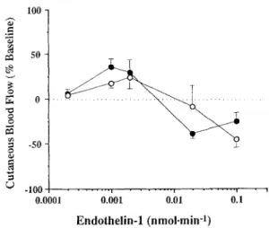
_+ 2 mmHg in the control group (P >0.3 for diastolic and systolic values). The effect of intra-arterial endothelin-1 was biphasic in all controls and in all but one patient with POAG (Fig. 1), and the dose-response100 Fig. 1 50 0 -so -100 ... i ... 1 ... i 0.0001 0.001 0.01 0.1 E n d o t h e l i n - 1 ( n m o l - m i n -1)
Mean changes in cutaneous blood flow recorded in 16 glau-
conqa patients (O) and in 16 age- and sex-matched controls (©) during intra-arterial administration of increasing dosages of en- dothelin- 1 "7. o ¢ - .< 350 250 150 50 -50 - 150 4O O 45 50 55 60 65 70 75 Diastolic Blood Pressure (mmHg)
Fig. 2 Linear correlation between cutaneous vasoconstriction af- ter infusion of endothelin-1 (expressed as AUC values) and intra- arterial diastolic blood pressure (mmHg) at baseline in 16 patients with glaucoma (O) ; t 2 = 0.52, P = 0.002; AUC = - 6 3 4 + 11.3 ( 0 ) (diastolic blood pressure)] and 16 age- and sex-matched con- trols (O; j2 _ 0.068, P = 0.33)
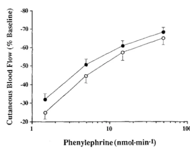
- 8 0 - . m "~ -70- ee~ - 6 0 - ¢> -so- -40- -30- e - %) -20 . . . i . . . . IO Phenylephrine (nmoi.min-l) Ioo
Fig. 3 Mean changes in cutaneous blood flow recorded in 16 pa- tients with glaucoma (Q) and 16 age- and sex-matched controls (©) during intra-arterial administration of increasing dosages of phenylephrine. Emax and EDso values of the two fitted dose- response curves were not significantly different
curves of the two groups were similar (P = 0.8). At lower dose-rate the peptide induced vasodilation, whereas at higher dose-rate pronounced vasoconstriction occurred. While there was no significant relationship between blood pressure values and endothelin-1 responsiveness in control subjects, vasoconstriction was inversely correlat- ed with intra-arterial blood pressure in glaucoma pa- tients. Evaluation by linear regression revealed r 2 values of 0.3 (P = 0.03) for systolic, 0.39 (P = 0.007) for mean, and 0.47 (P = 0.003) for diastolic blood pressure. Also when expressed as AUC values, dose-response relation- ships of endothelin-1 were correlated with systolic ( P = 0.27, P = 0.05), mean ( F = 0.5, P = 0.003), and diastolic blood pressure (r z = 0.54, P = 0.001) (Fig. 2). The mean phenylephrine dose-response relationships of the two groups were similar (P = 0.3) and are shown in Fig. 3. Fitted mean log EDso (log n g / m i n , P = 0.37) and Emax (vasoconstriction expressed as percentage of baseline, P = 0.49) values for both groups were sim- ilar ( c o n t r o l s : - 0 . 3 5 +_ 0.1, 65% _+ 3.6; g l a u c o m a : - 0.47 +_ 0.1, 70% _+ 2.9). There was no correlation be- tween cq-adrenergic sensitivity (expressed as log EDso) or efficacy (expressed as Emax) and intra-arterial sys- tolic or diastolic blood pressure, either in controls or in glaucoma patients.
Discussion
The aim of this investigation was to test the main vaso- constrictor pathways involved in the regulation of vascu- lar tone in the human peripheral microcirculation. Al- though the regulation of the ocular circulation might be different from peripheral vascular beds [ 15], there is evi- dence for a relationship between peripheral forearm cir- culation and the ocular circulation in patients with va-
sospastic syndromes [13]. The forearm vasculature was also chosen because this arterial bed only rarely devel- ops arteriosclerosis, and changes in vascular responsive- ness therefore reflect functional rather than anatomic al- terations of the vessels. Moreover, this vascular bed ap- peared particularly suitable for these experiments since it has repeatedly been used to characterize functional abnormalities of vessels in patients with diseases affect- ing the vasculature [5, 16, 21, 27] and to study respon- siveness of the skin microcirculation [3, 35]. Finally, since only small doses are required to construct complete dose-response curves, systemic counter-regulatory re- flex activation is unlikely to occur or to confound the results.
Whereas eq-adrenoceptor-mediated vasoconstriction was independent of blood pressure, endothelin-1 re- sponses were inversely correlated with systolic and dias- tolic values in glaucoma patients, indicating that patients with lower blood pressure were more sensitive to en- dothelin-1-induced vasoconstriction. This is of interest, since low blood pressure may promote glaucomatous damage although it is unclear by which mechanism. Ap- proximately 50% of the correlation between endothelin- 1 responses and diastolic blood pressure in glaucoma patients was explained by this relationship. This is a rather remarkable finding, since vascular tone of forearm resistance arteries is also modulated by other effector pathways such as spontaneous nitric oxide release [34]. To our knowledge, no data about the sensitivity of human vessels to endothelin-1 with respect to blood pressure are available. However, in accordance with our findings, in hypertensive rats, the sensitivity of vascular smooth muscle to endothelin-1 is reduced [7]. In the same study the response to norepinephrine did not differ between normo- and hypertensive animals, indicating that the re- duced sensitivity is endothelin-l-specific. Similar re- sults have also been reported in small subcutaneous ar- teries from patients with essential hypertension [33].
In almost all patients studied, endothelin-1 evoked a biphasic response in cutaneous blood flow with vasodila- tion at lower and pronounced vasoconstriction at higher dose-rates. Biphasic effects of endothelin-1 have been reported earlier [20] and have been explained by the exis- tence of at least two receptors (ET A and ETB). Whereas the endothelium primarily expresses vasodilator ET B re- ceptors, vasoconstrictive ET A and ET B receptors are lo- cated on vascular smooth muscle cells [23]. Evidence has also been presented for the presence of both endothe- lin receptor subtypes in the microcirculation of porcine eyes and in isolated human choroidal and retinal vessels [22], indicating that endothelin- 1 can evoke dose-depen- dent vasodilator and vasoconstrictor effects also in the ocular microcirculation.
In accordance with findings in patients with other va- sospastic disorders (Raynaud's disease [1]), (z~-adrener- gic responsiveness in glaucoma patients was similar to
controls, suggesting that differences in peripheral %- adrenoceptor sensitivity are absent and do not promote generalized vasospasms which are known to occur in certain g l a u c o m a patients.
The cause for the relationship between blood pressure and endothelin-1 responsiveness in the microcirculation of g l a u c o m a patients is unknown, and the design of this study does not allow us to conclude whether these para- meters are only associated with each other or whether any of them is causally involved in the pathogenesis of glaucoma. However, since endothelin-I responses are functionally linked to the activity of nitric oxide synthas- es, and since chronically administered NO may increase both the expression of ET A receptors and the affinity o f the peptide to its receptor [30], it may be speculated that the blunted constrictor responses to endothelin-1 in hy- pertensive patients [33] and animals [7] are caused by decreases in NO production, which has been shown in some [21] but not all studies addressing this issue [27]. Collectively, these data may suggest that g l a u c o m a pa- tients tend to have higher NO production, which could subsequently cause increased vasoconstrictor responses to endothelin-1. Hence, future studies should focus on the evaluation o f the effects of endothelin- 1, its antago- nists, and on the investigation of NO synthase activities in these patients.
Obviously the results o f this study were obtained in the peripheral microcirculation o f the human forearm and not in the ocular circulation; therefore, they can only
be extrapolated to the eye circulation. The local regula- tion of the ocular circulation is complex, has not yet been studied in all details [10, 36], and may differ in critical aspects from the peripheral microcirculation. Whereas choroidal vessels are amply innervated [11, 25] and flow is largely independent of autoregulation [ 19], retinal ves- sels are not innervated and local flow is controlled by efficient autoregulatory mechanisms similar to those regulating cerebral blood flow [2, 29]. The vascular area of the optic nerve head which is critical in the pathogen- esis o f g l a u c o m a also lacks autonomic innervation and is to some extent autoregulated [26, 28, 31]. However, blood supply of the optic nerve head mainly depends on the perfusion of the posterior ciliary arteries, which are regulated in a way similar to c o r o n a r y and peripheral arteries [14]. In particular, all these ocular and extraocu- lar vessels have in c o m m o n that they are rather sensitive to the vasoconstrictor effects of endothelin- 1 [4, 22, 23]. Thus, since systemic blood pressure is linked to periph- eral vascular sensitivity it appears possible that there is also a correlation with ocular vascular responsiveness in these patients. This, however, remains to be shown in future studies including the ocular circulation.
Acknowledgements We would like to thank Andreas J. Bircher, M.D. for technical support. These studies were supported by grant 32-36575.92 and 32-49825.96 from the Swiss National Research Foundation and by a grant from Sandoz Foundation, Basel, Switzerland.
References
1. Bedarida GV, Kim D, Blaschke TF, Hoffman BB (1993) Venodilation in Raynaud's disease. Lancet 342: 1451- 1454
2. Bill A, Nilsson SFE (1985) Control of ocular blood flow. J Cardiovasc Phar- macol 7 [suppl 3]:$96-S 102 3. Bircher A, De Boer EM, Agner T,
Wahlberg JE, Serup J (1994) Guideli- nes for measurement of cutaneous blood flow by laser Doppler flowme- try. Contact Dermatitis 30:65-72 4. Bursell SE, Clermont AC, Oren B,
King GL (1995) The in vivo effect of endothelins on retinal circulation in nondiabetic and diabetic rats. Invest Ophthalmol Vis Sci 36:596-607 5. Chowienczyk PJ, Watts GF, Cockcroft
JR, Ritter JM (1992) Impaired en- dothelium-dependent vasodilation of forearm resistance vessels in hyperc- holesterolemia. Lancet 340: 1430-
1432
6. De Lean A, Munson PJ, Rodbard D (1978) Simultaneous analysis of families of sigmoidal curves: applica- tion to bioassay, radioligand assay, and physiological dose-response curves. Am J Physiol 235:E97-E102 7. Dohi Y, Lfischer TF (1991) Endothe-
lin in hypertensive resistance arteries. Intraluminal and extraluminal dys- function. Hypertension 18:543-549 8. Drance SM, Douglas GR, Wijsman K,
Schulzer M, Britton RJ (1988) Re- sponse of blood flow to warm and cold in normal and low-tension glaucoma patients. Am J Ophthalmol 105:35-39 9. Ftammer J (1995) To what extent are
vascular factors involved in the patho- genesis of glaucoma? In: Kaiser HJ, Flammer J, Hendrickson P (eds) Ocu- lar blood flow. Glaucoma Meeting. Karger, Basel, pp 12-39
10. Flammer J, Orgtil S (1997) Optic nerve blood flow abnormalities in glaucoma. Prog Retin Res (in press) 11. Fltigel-Koch C, Kaufman P, Ltitjen- Drecoll E (1994) Association of a choroidal ganglion cell plexus with the fovea centralis. Invest Ophthalmol Vis Sci 35:4268-4272
12. Gasser P, Flammer J (1991) Blood- cell velocity in the nailfold capillaries of patients with normal-tension and high-tension glaucoma. Am J Oph- thalmol 111:585-588
13. Guthauser U, Flammer J, Mahler F (1988) The relationship between digi- tal and ocular vasospasm. Graefe's Arch Clin Exp Ophthalmol 226: 224- 226
14. Haefliger IO, Meyer R Flammer J, Ltischer TF (1994) The vascular en- dothelium as a regulator of the ocular circulation: a new concept in ophthal- mology? Surv Ophthalmol 39: 123- 132
15. Hayreh SS (1995) The optic nerve head circulation in health and disease. Exp Eye Res 61:259-272
16. Indolfi C, Maione AG, Volpe M, Ra- pacciuolo A, Esposito G, Ceravolo R, Rendina V, Condorelli M, Chiariello M (1994) Forearm vascular respon- siveness to %- and %-adrenoceptor stimulation in patients with congestive heart failure. Circulation 90:17-22
17. Kaiser HJ, Flammer J, Graf T, Stttmpfig D (1993) Systemic blood pressure in glaucoma patients. Grae- fe's Arch Clin Exp Ophthalmol 231:677-680
18. Kaiser HJ, Flammer J, Wenk M, Lascher TF (1995) Endothelin-1 plas- ma levels in normal-tension glaucoma: abnormal response to postural changes. Graefe's Arch Clin Exp Ophthalmol 233:484-488
19. Kiel JW (1995) The effect of arterial pressure on the ocular pressure-vol- ume relationship in the rabbit. Exp Eye Res 60:267-278
20. Kiowski W, Linder L (1992) Reversal of endothelin- 1-induced vasoconstric- tion by nifedipine in human resistance vessels in vivo in healthy subjects. Am J Cardiol 69:1063-1066 21. Linder L, Kiowski W, Btihler FR,
Ltischer TF (1990) Indirect evidence for release of endothelium-derived re- laxing factor in human forearm circu- lation in vivo. Blunted response in es- sential hypertension. Circulation 81:1762-1767
22. MacCumber MW, D'Anna SA (1994) Endothelin receptor-binding subtypes in the human retina and choroid. Arch Ophthalmol 112:1231-1235
23. Meyer P, Flammer J, Ltischer TF (1993) Endothelium-dependent regu- lation of the ophthalmic microcircula- tion in the perfused porcine eye: role of nitric oxide and endothelins. Invest Ophthalmol Vis Sci 34:3614-3621 24. Moriya S, Sugiyama T, Shimizu K,
Hamada J, Tokuoka S, Azuma I (1992) Low-tension glaucoma and en- dothelin (ET-1). Folia Ophthalmol Jpn 43:554-559
25. Nakanome Y, Karita K, Izumi H, Tamai M (1995) Two types of vasodi- lation in cat choroid elicited by elec- trical stimulation of the short ciliary nerve. Exp Eye Res 60:37-42 26. Orgtil S, Meyer R Cioffi GA (1995)
Physiology of blood flow regulation and mechanisms involved in optic nerve perfusion. J Glaucoma 4: 427- 443
27. Panza JA, Quyyumi AA, Brush JE, Epstein SE (1990) Normal endotheli- um-dependent vascular relaxation in patients with essential hypertension. N Engl J Med 323:22-27
28. Pillunat LE, Anderson DR, Knighton RW, Joos KM, Feuer WJ (1996) Au- toregulation of ocular blood flow. In: Kaiser H J, Flammer J, Hendrickson P (eds) Ocular blood flow. Glaucoma Meeting. Karger, Basel, pp 138-144 29. Pournaras CJ (1996) Autoregulation of ocular blood flow. In: Kaiser HJ, Flammer J, Hendrickson P (eds) Ocu- lar blood flow. Glaucoma Meeting. Karger, Basel, pp 40-50
30. Redmond EM, Cahill PA, Hodges R, Zhang S, Sitzmann JV (1996) Regula- tion of endothelin receptors by nitric oxide in cultured rat vascular smooth muscle cells. J Cell Biol 166:469-479
31. Riva CE, Harino S, Petrig BL, Shonat RD (1992) Laser Doppler flowmetry in the optic nerve. Exp Eye Res 55:499-506
32. Schwartz B (1994) Circulatory de- fects of the optic disk and retina in ocular hypertension and high pressure open-angle glaucoma. Surv Ophthal- tool 38 [Suppl]:S23-S34
33. Schiffrin EL, Deng LY, Larochelle P (1992) Blunted effects of endothelin upon small subcutaneous resistance arteries of mild essential hypertensive patients. J Hypertension 10:437-444 34. Vallance R Collier J, Moncada S
(1989) Effects of endothelium-derived nitric oxide on peripheral arteriolar tone in man. Lancet ii:997-1000 35. Wenzel RR, Noll G, Ltischer TF
(1994) Endothelin receptor antago- nists inhibit endothelin in human skin microcirculation. Hypertension 23:581-586
36. Williamson TH, Harris A (1994) Ocu- lar blood flow measurement. Br J Ophthalmol 78:939-945
37. Yang Z, Richard V, von Segesser L, Bauer E, Stulz R Turina M, Ltischer TF (1990) Threshold concentrations of endothelin-1 potentiate contrac- tions to norepinephrine and serotonin in human arteries. A new mechanism of vasospasm? Circulation 82: 188- 195