Urol Res (1997) 25 [Suppl 1 ]: S 31- S 35 © Springer-Verlag 1997
F. C. B u r k h a r d • R. M a r k w a l d e r • G. N. T h a l m a n n U. E. S t u d e r
Immunohistoehemical determination of p53 overexpression
An easy and readily available method to identify progression in superficial bladder cancer?
A b s t r a c t Overexpression of p53, as determined by im- munohistochemical staining with the murine monoclonal antibody DO7, was determined in specimens of 46 primary superficial transitional cell bladder tumours (14 TAG2, 10 T1G2, 22 T1G3). A colon cancer specimen served as a positive control and normal mesenchymal cells in the spec- imens served as an internal negative control. An excep- tionally high proportion 36/46 (78%) of the specimens were found to stain positively for p53 in over 20% of the cell nuclei. After a median follow-up of 7 years, ten pa- tients developed progressive disease. Of these ten patients nine demonstrated p53 positivity, resulting in a sensitivity of 90%. However, 27 of the overall 36 patients (75%) with p53-positive tumours did not progress to a higher stage or metastatic disease. These findings suggest that p53 over- expression is not of predictive prognostic value in super- ficial transitional cell carcinoma. With 7 of 14 specimens (50%) of Ta tumours overexpressing p53, the results were suggestive of p53 mutation being an early event in carcin- ogenesis. When the threshold was set at 50% of the cell nuclei overexpressing p53, 16/46 (35%) classified as p53 positive. Of the 16 tumours staining positively for p53, 7 (46%) progressed and 9 (56%) did not. None of the Ta and 16 (50%) of the T 1 tumours classified as positive. This more stringent definition of positivity still does not iden- tify p53 positivity as a single prognostic factor. With 50% o f T 1 tumours classifying as positive, we still find that p53 mutation may be an early event in carcinogenesis of blad- der cancer.
Key w o r d s Bladder tumour • Carcinogenesis - p53 tumour-suppressor gene • Disease progression • Immunohistochemistry
F. C. Burkhard ([]). G. N. Thalmann - U. E. Studer Department of Urology, University of Berne, Inselspital, CH-3010 Berne, Switzerland
R. Markwalder
Department of Pathology, University of Berne, Inselspital, CH-3010 Berne, Switzerland
Introduction
Bladder cancer is a multistage progressive disease with an onset typically later in life. The disease encompasses a wide spectrum of malignant potential ranging from a mere inconvenience to fatal disease. About 70% of superficial transitional cell bladder cancers recur and 10-20% progress to a higher stage, grade or metastatic disease. In comparison with superficial transitional cell carcinoma, the prognosis of muscle invasive tumours or metastatic dis- ease is poor.
p53 is one of the most commonly found gene mutations in carcinogenesis and has been observed in 70% of colon cancers, 30-50% of breast cancers, 50% of lung cancers and 100% of small-cell lung cancers [12]. It has been shown to play an important diagnostic and prognostic role in a variety of tumours, but the molecular mechanisms by which it influences normal cell function and tumorigene- sis remain unclear, p53 functions as a tumour suppressor gene, monitoring the integrity of the genome and indticing cell cycle arrest at the G1-S checkpoint if damage is found [11]. This allows time for repair mechanisms to act. p53 may play a role in inducing programmed cell death, if re- pair fails.
The p53 protein is coded for by a 16- to 20-kb gene lo- cated on the short arm of chromosome 17. The product is a 393-amino-acid nuclear protein involved in the regula- tion of cell proliferation. The half-life of wild-type p53 is very short (6-20 min). However, many missense mutations may induce mutations, which lead to a prolonged half-life of about 6 h. Eighty per cent of mutations are missense mu- tations causing one amino acid to be substituted for an- other, usually altering protein conformation, and causing nuclear accumulation [9]. Not only mutations lead to a pro- longed half-life of the p53 protein, but viral gene products (e.g. S V40 large T antigen, the adenovirus E 1B protein and papillomavirus E6 protein) have been found to stabilize the wild-type p53 protein [14]. Furthermore, it seems that the stability of the p53 protein is influenced by other cellular oncogenic proteins such as MDM2 [15].
The protein e n c o d e d by the mutated p53 gene has a m u c h longer half-life than wild-type p53 and allows for its detection by immunohistochemistry. I m m u n o h i s t o c h e m i - cal staining is a widely available, easy and accurate m e t h o d for evaluating mutations o f p53.
A m o n g the available antibodies, the m o n o c l o n a l anti- bodies D O 7 has been shown to have the highest specific- ity and sensitivity when c o m b i n e d with the target u n m a s k - ing fluid (Kreatech, A m s t e r d a m , The Netherlands) and then heated in a m i c r o w a v e o v e n for 5 min twice at 90 °C
[1, 2].
The optimal treatment for superficial bladder cancer is difficult to assess, as there is no easy and reliable m e t h o d for identifying patients at risk o f developing progressive disease. The aim of this study was to determine whether p53 overexpression as assessed by i m m u n o h i s t o c h e m i c a l staining can predict the d e v e l o p m e n t o f a higher grade, stage or metastatic disease in patients with superficial blad- der cancer and at what stage o f carcinogenesis p53 over- expression occurs.
Staining procedures included deparaffinisation in ethanol followed by washing in TRIS-buffered saline, pH 7.4. Immunohistochemical examination was performed with the target unmasking fluid (TUF) antigen retrieval method (Kreatech, Amsterdam, The Netherlands) to enhance antigen retrieval. Microwave oven heating was performed as described by Beckstead [2] using a citrate buffer pH 6. Slides were then incubated with the primary DO7 monoclonal antibody at a di- lution of 1:200 at room temperature for 60 min or with the primary monoclonal antibody Pabl801 at a dilution of 1:50 for 45 rain. The secondary antibody RAM/Ig was used for 45 rain at a 1:30 dilution. After incubation with the avidin-biotin peroxidase complex, sections were stained with hematoxylin. A colon cancer specimen known to be p53 positive was stained as a positive control.
Classification of immunohistochemistry
Depending on the percentage of nuclei demonstrating a positive staining reaction with the monoclonal antibody DO7, tumours were classified into three groups: <20% positivity, 20-50% positivity and >50% positivity. Only staining of the cell nucleus was considered a positive reaction. The slides were evaluated independently by two investigators.
After evaluation of preliminary trials, the use of the antibody Pab 1801 was discontinued as we noticed a higher degree of back- ground staining in comparison with the antibody DO7.
Patients and methods
Patients
Forty-six patients, 39 men and 7 women, with primary superficial transitional cell carcinoma of the bladder treated at the Department of Urology, University Hospital, Berne, between 1981 and 1993 were randomly selected. For these patients complete clinical, demograph- ic and histopathological data were available and all cases were re- viewed retrospectively. All patients were treated by transurethral re- section and the formalin-fixed, paraffin-embedded specimens were examined. Median patient age at diagnosis was 63.7 years (range 44-88 years). Median follow-up was 7 years (range 1-15 years) af- ter initial diagnosis.
Of the 46 patients with superficial transitional bladder cell car- cinoma, there were 14 patients with TaG2 and 32 patients with T1 disease (10 with G2 and 22 with G3 tumours) at initial diagnosis (Table 1).
Immunohistochemistry
Archival hematoxylin and eosin stained tissues were examined by a single pathologist (R.M.) in order to obtain representative sections from the superficial transitional bladder cancer specimens. Tumours were graded according to the WHO classification and staged accord- ing to the TNM system. Formalin-fixed, paraffin-embedded, archi- val tissue blocks from the 46 patients were sectioned and represen- tative sections were chosen.
For immunohistochemical staining we used the murine monoclo- nal antibodies DO7 (Dab, Hamburg, Germany) and Pabl801 (On- cogene Science Inc., Manhasset, NY) to mutant and wild-type p53.
Table 1 Tumour stages and grades of 46 patients after transureth- ral resection for superficial transitional cell carcinoma
G2 G3 Total
Ta 14 0 14
T1 10 22 32
Total 24 22 46
Results
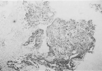
Taking a threshold o f p53 nuclear overexpression in over 20% o f cells, 36/46 specimens (78%) were considered pos- itive and 10/46 (22%) negative. The positive control (co- lon cancer specimen) stained positively in all t u m o u r cell nuclei with a distinct demarcation at the t u m o u r edge in each staining procedure. The normal m e s e n c h y m a l cells in all specimens remained negative and served as an internal negative control. Figures 1 and 2 show staining o f differ- ent tissue specimens.
The correlations between overexpression o f p53, grade and stage were assessed (Table 2). A larger proportion o f tumours with a higher grade or stage overexpressed p53. Fifty per cent of the Ta tumours and 91% of the T 1 tumours overexpressed p53. O f the G2- 71% and of the G3-classi- fied tumours 86% demonstrated positivity. If the c u t o f f was placed at >50% nuclear overexpression, none o f the Ta tu- mours and 50% o f the T1 tumours classified as positive (Table 2). After a median follow-up o f 7 years, 10 o f the 46 patients (22%) progressed to a higher stage or devel- oped metastatic disease. O f these ten progressive bladder tumours, nine (90%) were positive for p53 in over 20% o f the cells, whereas one (10%) did not stain positively.
Overall 36 tumours classified as p53 positive o f which 9 (25%) developed into progressive disease and 27 (75%) did not (Table 3). A p p l y i n g a higher threshold o f over 50% nuclear expression, 16/46 tumours were classified as pos- itive. Seven tumours in the ten patients (70%) with pro- gressive disease classified as positive and three (30%) as negative. O f the 16 p53-positive tumours, 9 (56%) did not develop progressive disease (Table 3). Analysis o f our re- sults shows a sensitivity o f 90% with a specificity o f 25% for progression o f bladder cancer when positivity is de- fined as 20% nuclear overexpression o f p53. Setting the
S 33 cutoff point at 50% results in a decreased sensitivity of 70% and an improved specificity of 75%.
Fig. 1 Immunohistochemical determination of the antibody DO7 for overexpression of p53 in a colon cancer specimen, serving as a positive control
Fig. 2 Intense homogeneous overexpression of p53, as determined by immunohistochemistry with the antibody DO7, in a specimen with superficial transitional cell bladder carcinoma
Table 2 Relationship between overexpression of p53, grade and stage of bladder tumours
Total No. p53 < 20% p53 > 20% p53 > 50% Ta 14 7 (50%) 7 (50%) 0 (0°70) T1 32 3 (9%) 29 (91%) 16 (50%) Grade II 24 7 (29%) 17 (71%) 4 (17%) Grade III 22 3 (14%) 19 (86%) 12 (54%) Total 46 10 (22%) 36 (78%) 16 (35%)
Table 3 Tumour progression in regard to percentage of tumour cell nuclei demonstrating positive immunohistochemical staining
Total Progression No progression
(%) (%) <20% 10 1 (10%) 9 (90%) >20% 36 9 (25%) 27 (75%) <50% 30 3 (10%) 27 (90%) >50% 16 7 (44%) 9 (56%) Discussion
The following conclusions can be drawn from our results: (1) p53 overexpression as determined by immunohisto- chemical determination is frequent in superficial bladder cancer; (2) the results in our group of patients do not sup- port the use of immunohistological p53 expression as a dis- criminating prognostic indicator in superficial cell carci- noma; and (3) p53 mutation m a y be an early event in car- cinogenesis of bladder cancer.
Previous studies have shown changes in the p53 protein to occur in 11-64% of superficial transitional cell bladder tumonrs [3, 5, 7, 16, 21-23]. Interestingly, an exception- ally high proportion (78%) of superficial bladder tumours in our group stained positively for p53 (Ta 50%, T1 91%). In our study we used the murine monoclonal antibody DO7, which recognizes mutant and wild-type p53 for staining procedures. Antigen retrieval was enhanced by target un- masking fluid and according to the microwave procedure. In a comparative study of six c o m m o n antibodies, DO7 demonstrated the highest sensitivity and specificity for the p53 protein [1]. The most c o m m o n l y used antibody, Pabl801, showed a lower sensitivity and specificity in the above-mentioned study. The use of the antibody DO7 could explain the high proportion of tumours with p53 overex- pression in our study in comparison to others at the cutoff point of 20%.
In T1 bladder tumours, Sarkis et al. [17] observed a cor- relation between p53 overexpression and disease progres- sion. Gardiner et al. [7] could not relate p53 overexpres- sion in T1 tumours to patient outcome and therefore did not support p53 immunohistochemical determination as a prognostic indicator in superficial bladder cancer [7]. Thomas et al. [23] demonstrated that significantly higher levels of p53 positivity (> 10% nuclear staining) were found in T1 bladder tumours with disease progression, but that tumour grade remained the most specific single predictor of progression [23]. In our group of patients we find a sen- sitivity of 90% combined with a specificity of 25% for tu- mour progression when both T1 and Ta tumours are in- cluded in the analysis. When analysing only the T 1 tumours the sensitivity remains 90% and the specificity drops to 9%.
A more stringent definition of positivity with a thresh- old at 50% results in positive staining of 50% of T1 tu- mours and none of the Ta tumours. This causes a slight decrease in sensitivity from 90% to 70% and improves specificity from 25% to 59%. These results suggest that, whatever the threshold, overexpression of p53 as deter- mined by immunohistochemistry may not be used as a dis- criminating predictive factor for progression in superficial bladder cancer.
However, by setting the threshold for positivity at 50%, our results become more comparable with other published
results (between 11% and 64% of p53-positive tumours). Interestingly, in these studies cutoff points ranged from 0% to 20% of cells with p53 nuclear overexpression [7, 16, 21-23]. One may speculate that in some instances overex- pression of the wild-type p53 accounts for immunoreac- tivity, especially when particularly sensitive methods are applied. Possibly other mechanisms may influence the ex- pression of the p53 protein such as viral gene products and cellular oncogenic proteins, which are known to stabilize wild-type p53 [11, 13, 15].
How the heterogeneity of p53 staining, found predom- inantly in specimens with a lower percentage of p53 over- expression, should be interpreted remains unanswered. It may be speculated that there are cells staining positively for p53, which are expressing p53 physiologically in re- sponse to damage or representing an early clone of cells with a p53 mutation in an otherwise negative neoplasm.
Another methodological problem in this context is the evaluation of immunohistochemical staining for p53, as there is no standardized form of evaluation and these meth- ods are not quantitative. Furthermore, the intensity of stain- ing, a rarely discussed matter, may influence the interpre- tation of results. Esrig et al. [4] reported that the intensity of staining varies due to the location of mutations. Not only do staining properties vary according to the location of mis- sense mutations, but also different mutant alleles of the p53 gene may have different biological and chemical proper- ties. It has been proposed that cancer patients with differ- ent p53 mutant alleles may have a different prognosis [13]. Esrig et al. [4] reported no evidence of p53 mutations by molecular analysis in 29% of bladder tumours with pos- itive p53 immunoreactivity using the antibody Pabl801. This indicates that either immunohistochemical staining is more sensitive than single-strand conformational polymor- phism analysis in detecting p53 mutations or that discor- dant cases represent tumours with accumulation of wild- type p53 protein without mutations at the
p53
locus.Greenblatt et al. [8] reviewed 84 studies comparing im- munohistochemical determination with sequencing in the same tumour sets and found positive immunohistochemi- cal staining in 44% of tumours, while only 36% contained mutations recognized by single-strand conformational polymorphism analysis [8]. The sensitivity of immunohis- tochemical determination for p53 mutations in these stud- ies was in the range of 36-100% and the positive predic- tive value was 63% (range 8-100%). Thus, results must be interpreted with caution.
p53 nuclear overexpression has been suggested to be an early event, because it occurs in a high percentage of pa- tients with carcinoma in situ [18, 20]. Whether carcinoma in situ does progress to invasive bladder cancer remains unclear. Possibly the high percentage of p53 positivity in carcinoma in situ is more suggestive of a different molec- ular pathway leading to cancerogenesis. This agrees with the theory that carcinoma in situ turnout cells are so undif- ferentiated that they cannot grow invasively, p53 overex- pression has been shown to correlate significantly with grade and stage, which suggests that p53 inactivation might be an early event in bladder cancer [24]. In the present
study the association with grade and stage and the high per- centage of p53 overexpression in Ta tumours further im- plicate that p53 inactivation may occur early in the evo- lution of bladder cancer. Possibly further genetic altera- tions are necessary to induce aggressive tumour growth.
It has been suggested that p53 staining might be predic- tive of chemosensitivity. Doxorubicin treatment can alter the p53 status of bladder tumours and p53 overexpression has been associated with doxorubicin resistance in the his- tocultures of seven superficial Ta/T1 and nine invasive T2-T4 tumours [6]. Further research found p53 overex- pression to be of independent prognostic value for survi- val in patients with invasive bladder cancer treated with neoadjuvant chemotherapy in a cohort of 90 patients [19]. Possibly p53 status will prove to be of prognostic value for therapy forms that influence the cell cycle. Further stud- ies are needed to elucidate the potential value of p53 im- munohistochemistry in predicting outcome and therapeu- tic response in selected patient groups.
Conclusion
Based on our results p53 overexpression is not a single pre- dictive factor in the evaluation of superficial bladder can- cer. In our study population, positive p53 staining was a frequent event, suggesting that p53 mutation may occur early in superficial transitional cell carcinogenesis. Al- though immunohistochemical determination is a simple and readily available procedure, the evaluation can prove difficult and the predictive value is limited. As p53 inac- tivation seems to be one step in a multistage carcinogene- sis, further studies including other tumour suppressor genes and oncogenes are required. Since it seems unlikely that one genetic event is responsible for tumour progres- sion in bladder cancer, this limited information obtained from p53 analysis is not surprising.
References
1. Baas IO, Mulder JWR, Offerhaus GJA, Vogelstein B, Hamilton SR (1994) An evaluation of six antibodies for immunohisto- chemistry of mutant p53 gene product in archival colorectal neo- plasms. J Pathol 172:5
2. Beckstead JH (1994) Improved antigene retrieval in formalin- fixed, paraffin-embedded tissues. Appl Immunohistochem 2:274
3. Dalbagni G, Presti JC Jr, Reuter VE, Zhang ZF, Sarkis AS, Fari WR, Cordon-Cardo C (1994) Molecular genetic alterations of chromosome 17 and p53 nuclear overexpression in human bladder cancer. Diagn Mol Pathol 2:4
4. Esrig C, Spruck III CH, Nichols PW, Chaiwun B, Stevens K, Groshen S, Chen S, Skinner DG, Jones PA, Cote RJ (1993) P53 nuclear protein accumulation correlates with mutations in the p53 gene, tumour grade, and stage in bladder cancer. Am J Path- ol 143:1389
5. Esrig D, Elm@an D, Groshen S, Freeman JA, Stein JR Chen S-C, Nichols PW, Skinner DG, Jones PA, Cote RJ (1994) Accu- mulation of nuclear p53 and progression in bladder cancer. N Engl J Med 19:1259
6. Gan Y, Wientjes MG, Badalament RA, Au JL-S (1996) Pharma- codynamics of doxorubicin in human bladder tumours. Clin Can- cer Res 2:1275
7. Gardiner RA, Walsh MD, Allen S, Rahman S, Samaratunge MLTH, Seymour GJ, Lavin MF (1994) Immunohistochemical expression of p53 in primary pT1 transitional cell bladder can- cer in relation to tumour progression. Br J Urol 73:526 8. Greenblatt MS, Benett WR Hollstein M, Harris CC (1994) Mu-
tations in the p53 tumor suppressor gene: clues to cancer etiol- ogy and molecular pathogenesis. Cancer Res 54:4855
9. Harris CH, Hollstein M (1993) Clinical implications of the p53 tumor-suppressor gene. N Engl J Med 329:1318
10. Kuczyk MA, Serth J, Anton P, H6fner K, Jonas U (1994) Value of p53 immunohistochemistry as an independent prognosticator for superficial bladder cancer. Z Urologic Poster 2/1994 11. Lane DP (1992) P53, guardian of the genome. Nature 358:15 12. Levine AJ (1992) The p53 tumor-suppressor gene. N Engl J Med
326:1350
13. Levine AJ, Momand J, Finlay CA (1991) The p53 tumour sup- pressor gene. Nature 351:453
14. Marshall CJ (1991) Tumor suppressor genes. Cell 64:313 15. Momand J, Zambetti GR Olson DC, George D, Levine AJ (1992)
The mdm-2 oncogene product forms a complex with the p53 pro- tein and inhibits p53-mediated transactivation. Cell 69:1237 16. Nakopoulou L, Constantinides C, Papandropoulos J, Theodoro-
poulos G, Tzonou A, Giannopoulos A, Zervas A, Dimopoulos C (1995) Evaluation of overexpression of p53 tumour suppres- sor protein in superficial and invasive transitional cell bladder cancer: comparison with DNA ploidy. Adult Urol 46:334 17. Sarkis AS, Dalbagni G, Cordon-Cardo C, Zhang Z-F, Sheinfeld
J, Fair WR, Herr HW, Reuter VE (1993) Nuclear overexpres-
S 35 sion of p53 protein in transitional cell bladder carcinoma: a marker for disease progression. J Natl Cancer Inst 85:53 18. Sarkis AS, Dalbagni G, Cordon-Cardo C (1994) Association of
p53 nuclear overexpression and tumor progression in carcino- ma in situ of the bladder. Br J Urol 73:533
19. Sarkis AS, Bajorin DF, Reuter VE, Herr HW, Netto G, Zhang Z-F, Schultz PR, Cordon-Cardo C, Scher HI (1995) Prognostic value of p53 nuclear overexpression in patients with invasive bladder cancer treated with neoadjuvant MVAC. J Clin Oncol
13:1384
20. Schmitz-Dr~iger B J, Roeyen CRC, Grimm M-O, Gerharz C-D, Decken K, Schulz WA, Btiltel H, Makri D, Ebert T, Ackermann R (1994) P53 accumulation in precursor lesions and early stag- es of bladder cancer. World J Urol 12:79
21. Serth J, Kuczyk MA, Bokemeyer C, Hervatin C, Nafe R, Tan HK, Jonas U (1995) P53 immunohistochemistry as an indepen- dent prognostic factor for transitional cell carcinoma of the blad- der. Br J Cancer 71:201
22. Tetu B, Fradet Y, Allard R Veilleux C, Roberge N, Bernard P (1996) Prevalence and clinical significance of HER-2/neu, p53 and Rb expression in primary superficial bladder cancer. J Urol 155:1794
23. Thomas D, Robinson MC, Charlton R0 Wilkinson S, Shenton BK, Neal DE (1993) P53 expression, ploidy and progression in pT1 transitional cell carcinoma of the bladder. Br J Urol 73:533 24. Underwood MA, Reeves J, Smith G, Gardiner DS, Bartlett J, Cooke TG (1996) Overexpression of p53 protein and its signif- icance for recurrent progressive bladder tumours. Br J Urol 77:659