Adaptation of Conduit Artery Vascular Smooth Muscle Tone
to Induced Hypertension
P. F
RIDEZ,
1A. M
AKINO,
2D. K
AKOI,
2H. M
IYAZAKI,
2J.-J. M
EISTER,
3K. H
AYASHI,
2and N. S
TERGIOPULOS1 1Laboratory of Hemodynamics and Cardiovascular Technology, Swiss Federal Institute of Technology, Lausanne, Switzerland; 2Department of Systems and Human Science, Division Mechanical Science, Graduate School of Engineering Science, OsakaUniversity, Japan; and3Laboratory of Biomechanics and Biophysics of Cells, Swiss Federal Institute of Technology, Lausanne, Switzerland
(Received 13 August 2001; accepted 17 May 2002)
Abstract—We studied the changes in vascular smooth muscle
共VSM兲 cell tone during the adaptation of rat common carotids to induced hypertension. Hypertension was induced in 8 week old male Wistar rats by total ligation of the aorta between the two kidneys. Mean blood pressure increased abruptly from 92 ⫾2 mm Hg (mean⫾SE) to 145⫾4 mm Hg and remained con-stant thereafter. Rats were sacrificed 2, 4, 8, and 56 days after surgery and the left common carotid artery was excised for analysis. Pressure-diameter curves were measured in vitro un-der normal, maximally contracted, and totally relaxed VSM. The VSM tone was analyzed in terms of its basal tone共active stress at low strains兲 and its myogenic tone 共increase in active stress at high strains兲. Our results show that the capacity of the VSM to develop maximal active stress is not altered in hyper-tension. Basal tone, however, increases rapidly in the acute hypertension phase共2–8 days postsurgery兲 and drops to nearly control values at 56 days postsurgery. Also, the onset of myo-genic response decreases to lower strains following the step change in pressure, to be restored back to control levels at 56 days postsurgery. We conclude that VSM adaptation is most significant in the acute hypertension phase and acts as a first, rapid defense mechanism for the arterial wall. The VSM tone returns back to normal levels once the slower geometrical and structural remodeling is developed sufficiently to restore the biomechanical environment and function of the arterial wall to control levels. © 2002 Biomedical Engineering Society. 关DOI: 10.1114/1.1507326兴
Keywords—Arterial wall, Remodeling, Adaptation, Myogenic
response, Smooth muscle tone, VSM, Aortic banding, Conduit vessel.
INTRODUCTION
The adaptation of the arterial wall to induced hyper-tension has been studied in great detail in conduit arteries5,6,13,16,24,26 as well as in resistance arteries and arterioles.1,4,12,15,30 In conduit arteries, the focus was on the structural and morphological remodeling of the
arte-rial wall, and there was little attention paid to the changes in vascular smooth muscle共VSM兲 tone, with the exception of the case of spontaneously hypertensive rats 共SHR兲.2,3,20,21
In contrast with the studies on conduit arteries, alterations of VSM tone in resistance vessels exposed to hypertension have been extensively studied.8,17,27
The lack of interest in VSM tone remodeling in con-duit vessels exposed to hypertension is likely to be re-lated to the minor importance accorded to the VSM tone in regulating conduit artery function and to the ambigu-ities on physiological significance of the myogenic re-sponse in large vessels. Yet, conduit arteries may exhibit significant spontaneous tone7 and, depending on the ar-tery, may have a large myogenic contraction capacity.10,25
It is well recognized that conduit arteries use VSM tone to adapt their lumen diameter to changes in flow 共shear stress-dependent mechanism兲19
or to pressure 共myogenic mechanism兲.10,25
It is likely that the same mechanisms are involved in the acute 共hours兲 or early phase 共first few days兲 of arterial wall remodeling in re-sponse to a step change in pressure. However, how the VSM tone develops if the step change in pressure is sustained over longer periods 共days to weeks兲 remains unknown.
The purpose of this study is to obtain a complete tracing of VSM tone adaptation in conduit arteries sub-jected to a sustained step change in pressure. We exam-ine how VSM tone adapts immediately after the pressure rise and how it remodels itself in the long run. We focus not only on the maximal contractile capacity of the VSM but also on normal VSM tone 共often termed resting or residual VSM tone兲, because this reflects the physiologi-cal state of VSM and is thus most indicative of the functional adaptation of the vascular smooth muscle to sustained hypertension.
Address correspondence to Nikos Stergiopulos, Professor, Labora-tory of Hemodynamics and Cardiovascular Technology, Swiss Federal Institute of Technology, 1015 Lausanne, Switzerland. Electronic mail: nikolaos.stergiopulos@epfl.ch
MATERIALS AND METHODS In Vivo Experimental Procedures
The details of the experimental procedures and experi-mental results used in this paper for tone analysis were already published and explained in detail in a previous publication9 and will only be briefly summarized here.
Hypertension was induced in male Wistar rats, aged 56 – 63 days共8 weeks兲 and weighing 270–312 g, by total ligation of the aorta between the two kidneys according to the procedure described in Michel et al.11
Arterial Preparations
Experiments were terminated 2, 4, 8, and 56 days postsurgery (n⫽6 for each group兲. Two control groups were included for the measurement of arterial properties in normotensive rats. One was the presurgery control group共day 0 control兲 and the other one was age matched to the 56 days postsurgery group 共day 56 control; n⫽6 for each兲. We have not included a sham operated group at 8 days since the acute part of these experiments last only 1 week which is a small period in comparison with the age of the animals 共8 weeks兲. Thus, the biomechani-cal properties of the control group would not change substantially within this small period of time. As de-scribed in a previous publication,9 the left common ca-rotid 共LCC兲 artery was carefully exposed, marked, and measured lengthwise to determine the in vivo axial ex-tension ratio. The rats were then killed and the LCC was excised. A segment obtained from the proximal end of the LCC was used for mechanical testing and the re-maining segment was used to measure geometrical prop-erties. All mechanical tests were finished within 9 h postmortem and the specimens were kept in Krebs– Ringer solution during this period 共at 5 °C for storage and slowly warmed up to 37 °C for measurements兲.
Biomechanical Measurements and Analysis Pressure-Diameter Curves. The preparations and the measurement methods used in this study for the inflation/ extension tests were published in a separate paper.9 The excised LCC segment was inflated and deflated at a rate of approximately 170 Pa/s 共1.3 mm Hg/s兲. The external diameter and the intraluminal pressure 共under no flow conditions兲 were measured. Arteries were preconditioned by means of 4 – 6 inflation-deflation cycles 共pressure range 0–26.7 kPa, i.e., 0–200 mm Hg and rate of ap-proximately 170 Pa/s, i.e., 1.3 mm Hg/s兲 until the pressure-diameter ( p – d) curve became reproducible. The ascending limb of the last stable loop was recorded and used for the data analysis under normal VSM tone conditions. The p – d curves under maximal contraction were obtained using NE (5⫻10– 7 mol/L) without
pre-conditioning. Finally, the p – d curves under total relax-ation were obtained using Papaverine共10–4 mol/L兲 with preconditioning.
Geometrical Analysis. Four rings were cut from the middle part of the long distal LCC segment and kept for imaging following the procedures described in a previous paper.9 Then these images were used to determine the internal radius, Ri, and the thickness, H, and these in
turn were used to calculate the wall cross-sectional area at zero load, WCSA⫽(2 RiH⫹H2). Finally, assuming that the wall is incompressible and using the formulas: z(r0
2⫺r i 2
)⫽WCSA and h⫽(r0⫺ri), where z is the
in situ axial extension ratio, the deformed internal radius, ri, and deformed wall thickness, h, are calculated at any
given pressure.
Stress Analysis. The mean circumferencial wall stress,
tot, was calculated using Laplace’s law tot⫽
Piri
r0⫺ri
. 共1兲
The mean circumferencial wall stretch ratio,, indicative of the stretch of the midwall fiber was calculated as follows:
⫽ r0⫹ri
R0⫹Ri. 共2兲 For simplicity, the mean circumferencial wall stretch ra-tio will shortly be referred to as ‘‘strain.’’
Based on the classic Hill model,16 the mean circum-ferential stress 共hoop stress兲 can be written as a sum of its active and passive components
tot⫽pas⫹act. 共3兲
The active component of the hoop stress, act, accounts for the stress borne by the VSM, and therefore will be used as a biomechanical surrogate for VSM tone. The passive component, pas, accounts for the stress borne by the ‘‘passive’’ components of the wall 共i.e., extracel-lular matrix, VSM in completely relaxed state, etc.兲. In Hill’s model, the active and passive stress components are assumed to be independent. Thus, the effect of VSM-extracellular matrix coupling is neglected in this approach.
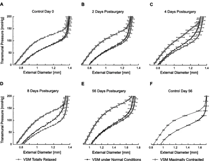
This study focuses on VSM tone and thus, on the characterization of the active stress act. Figure 1 shows how the active stress is assessed. In Fig. 1共A兲, we see a typical set of p – d curves of the rat common carotid artery obtained under total VSM relaxation 共solid line兲,
normal VSM tone共dashed line兲, and maximal VSM con-traction 共dotted line兲. For a given wall distension 共diam-eter d0), the total stress under total relaxation (tot
rel ), the total stress under normal VSM tone (totn ), and the total stress under maximal VSM tone (totmax) are calculated using Laplace’s law
tot rel⫽P relr h , tot n ⫽P nr h , and tot max⫽P maxr h , 共4兲 where r and h are the internal radius and wall thickness corresponding to the external diameter d0, respectively. The superscripts on stress denote the state of VSM tone (n⫽normal tone; max⫽maximal contraction; rel⫽total relaxation兲. The definitions of the passive and total stresses given earlier lead to a stress-strain relationship depicted in Fig. 1共B兲. The subscripts refer to the stress components according to Hill’s model (tot⫽total, pas ⫽passive, act⫽active). Obviously, tot
rel⫽
pas. Subtract-ing the passive stress from the total stress we obtain the active stress under normal VSM tone
act n ⫽
tot n ⫺
pas 共5兲
and under maximal VSM contraction
act max⫽
tot max⫺
pas. 共6兲
Typical active stress-strain curves are shown in Fig. 1共C兲.
Mathematical Description of Active Stress To facilitate the analysis of VSM tone development in hypertension, we use a simple mathematical model for the description of active stress. As seen in Fig. 1共C兲 共see also the Results section兲, active stress under maximal contraction (act
max
) is a linear function of the strain, , and therefore we assume it has the following form:
act max共兲⫽c
0⫹c1. 共7兲
Equation 共7兲 is limited to the ascending portion of the active stress-strain curve, which can be approximated quite well by a straight line. Normally, the active stress at maximal contraction is not a straight line, but a func-tion with a descending part at higher strains, such as a FIGURE 1. „A… Typical pressure-diameter curves from a common carotid belonging to an 8 day hypertension group rat. Data were measuredin vitrounder conditions of total relaxation„solid line…, under normal VSM tone„dashed line…and under maximal contraction „dotted line… of the vessel.„B… Transformation of the pressure-diameter curves of„A… to the stress-strain domain according to Eq.„4….„C…Active stress-strain curves obtained from „B…by subtracting passive stress from total stress under normal VSM tone„dashed line…and from total stress under maximal contraction„dotted line… †Eqs.„5…and„6…‡.„D…Dependence of the VSM tone ratio on strain. VSM tone ratio is obtained from„C…and Eq.„8…. The two separate domains, the first one being the region where basal tone determines the total VSM tone„low strain values, horizontal branch of the curve…and the second domain being the region where the myogenic tone dominate the total VSM tone„higher strain values, increasing branch of the curve…are seen.
bell-shape curve, or a parabolic curve, as been suggested by other authors.4For the sake of simplicity, we chose to simply model the ascending part of the curve with a straight line and not speculate on the form of the curve at higher strains.
For a given level of strain, , and a given level of VSM tone, the smooth muscle develops an active stress,
act n
, which is in the range 0⭐actn ⭐actmax. For a given strain , we define the VSM tone ratio S 共兲 as the ratio between the active stress under normal tone and the active stress under maximal contraction
S共兲⫽ act
n 共兲 act
max共兲, 0⭐S共兲⭐1. 共8兲
Thus, the nondimensional parameter S characterizes the degree of VSM tone, S⫽0 meaning no tone and S⫽1 meaning maximal contraction.
Figure 1共D兲 shows the VSM tone ratio, S(), curve obtained using the data in Fig. 1共C兲 and Eq. 共8兲. There-fore, the S() curve in Fig. 1共D兲 depicts the relative contraction of vascular smooth muscle as a function of the strain under normal VSM tone conditions. We ob-serve that the relative degree of VSM contraction exhib-its two different domains of behavior. In the first domain, at low strain range, S() remains shallow and fairly constant, we refer to this part of the S() curve as the ‘‘basal tone.’’ However, in the second domain, at higher strain values, the VSM tone ratio, S(), increases rap-idly. This represents a strain-activated increase in VSM tone which is characteristic of the myogenic mechanism and this part of S() will therefore be referred to as the ‘‘myogenic tone.’’ Based on the characteristics of the graph in Fig. 1共D兲, we assume that the behavior of the VSM tone ratio in these two distinct domains can be accounted for by a sum of two functions关Sbas, Smyo()] depicting the basal and the myogenic tone, respectively, as follows:
S共兲⫽Sbas⫹Smyo共兲. 共9兲
Sbasis independent of the strain and will be referred to as the basal tone ratio and Smyo() will be referred to as the myogenic tone ratio. As on full strain range, the active stress under normal tone tends to behave as a sigmoid 共see also Fig. 4 in the Results section兲, and the myogenic tone ratio accounts for the inflection domain of the sigmoid curve, we assume it has the following form:
Smyo共兲⫽共1⫺Sbasal兲fmyo共兲
⫽共1⫺Sbasal兲
冉
⫺0 cr⫺0冊
q 1⫹冉
⫺0 cr⫺0冊
q, 共10兲where q cr, and 0 are the parameters of the sigmoid 共see Fig. 2兲. cris the strain at the inflection point of the sigmoid, fmyo(), and it can be viewed as the middle point of the range of values comprising the myogenic response. Thus, we will refer to cr as the myogenic critical strain. The parameter q is proportional to the maximal slope of the VSM tone ratio as a function of strain 共i.e., proportional to the slope at the inflection point, Fig. 2兲. The parameter 0 is the relative length of the circumferencial midwall fiber at zero pressure and thus is entirely determined by the experimental geom-etry: the arterial segments are stretched longitudinally and this causes the length of the circumferencial mid wall fiber to contract to 0. The factor (1⫺Sbas) in the definition of Smyo() accounts for the fact that the myo-genic tone can only operate on the residual tone capacity of the VSM, which is the tone remaining between total contraction (S⫽1) and the basal tone ratio (Sbas).
This description leads to the following representation of active stress under normal VSM tone:
act n 共兲⫽
act
max共兲S共兲 共11兲
withactmaxand S() given by Eqs. 共5兲, 共7兲, 共8兲, and 共10兲. The parameters of model equations derived above leading to a mathematical description of the active stress are determined through fitting the experimental data. This leads to a description of the short- and long-term evolution of VSM tone in response to induced hyperten-FIGURE 2. Shape of the sigmoid function †fmyo„…‡ myo-genic activation. q is proportional to the slope of the sig-moid at the inflection point,cris the strain at the inflection point„myogenic critical strain…, and0is the relative length of the circumferencial midwall fiber at zero pressure.
sion. Then, the evolution of VSM tone is assessed in terms of its maximal contractile capacity共parameters c0,
c1), basal tone ratio (Sbas), and myogenic tone ratio 共parameters cr and q).
Numerical and Statistical Analysis
The five parameters of the model (c0, c1, Sbasal, cr, and q) were determined using a standard Levenberg– Marquardt method 共least square fit兲.
All data were expressed as mean ⫾SE. Comparisons between each group and the control 共0 days postsurgery兲 were made with student’s t test. Differences were con-sidered to be significant for p⬍0.05.
RESULTS
Following aortic ligation mean blood pressure in-creased abruptly from 12.3⫾0.3 kPa (mean⫾SE) to 19.3⫾0.5 kPa (92⫾2 and 145⫾4 mm Hg, respectively兲 and remained constant for the first week postsurgery. At 56 days postsurgery, it had slightly diminished to 18.4 ⫾1.2 kPa (138⫾9 mm Hg) 关Fig. 3共A兲兴. The
experimen-tal results on which the stress-strain calculations are based were explained in detail in a previous publication9 and will only be briefly described here.
Geometrical Adaptation
A gradual increase of the wall thickness at no load state corresponding to 118%⫾4% of the control value at 8 days postsurgery, and 140%⫾2% of the control value at 56 days postsurgery was observed 关Fig. 3共B兲兴. This increase in thickness with respect to control 共day 0兲 found at 8 and 56 days postsurgery was statistically sig-nificant ( p⬍0.05). The changes in the internal radius at mean pressure were not significant during the entire post-surgery period 关Fig. 3共C兲兴. Therefore, we have an eccen-tric hypertrophy, as typically reported in the literature6,22 共hypertrophic which could not be classified as ‘‘inward’’ nor ‘‘outward’’ remodeling兲.33
Time Evolution of Stress-Strain Curves
The evolution of mean wall stress is shown in Fig. 3共D兲. Figure 3 shows the stress-strain curves for the FIGURE 3. „A…Evolution of aortic mean pressure. Error bars indicate SE„nÄ25, 22, 16, 12, 6, 6, from left to right, respectively….
„B…Evolution of wall thickness at zero load state„nÄ6, for each point….„C…Evolution of the internal radius at zero load state
„nÄ6….„D…Evolution of mean wall stress at mean pressure„nÄ6…. In all figures the dashed lines„open squares…and the solid lines „black diamonds…show the evolution of the control and hypertensive groups, respectively. The character ‡ indicates p
Ë0.005 with respect to control at day 0 and day 56; † indicatespË0.005 with respect to control at day 0; and* indicatesp
control group at day 0, the hypertensive groups at 2, 4, 8, and 56 days postsurgery as well as the age-matched control group at day 56. The three curves in each graph represent the stress-strain relation under normal VSM tone 共black diamonds兲, under maximal contraction 共dia-monds兲, and under total relaxation 共circles兲.
For the control groups, we observe that the stress-strain curve under normal VSM tone lies very close to the stress-strain curve under total relaxation. This means that under normotensive conditions, the rat common ca-rotid exhibits very little VSM tone. We may thus con-clude that in the normotensive rat, under physiological conditions, the arterial properties are almost entirely de-termined by the passive components of the wall. How-ever, immediately after exposure to high blood pressure, we observe a significant development in active stress under normal VSM tone. This is clearly seen at 4 and 8 days postsurgery关Figs. 3共C兲 and 3共D兲兴, where the stress-strain under normal VSM tone 共black diamonds兲
‘‘de-parts’’ from a position close to the stress-strain curve under total relaxation 共circles兲 and moves to the middle of the operating stress-strain domain. At 56 days post-surgery 关Fig. 3共E兲兴, however, active stress under normal VSM tone seems to regress and the stress-strain curve under normal conditions moves back to the stress-strain curve under total relaxation.
Adaptation of VSM Tone
The adaptation of the VSM tone to hypertension is seen in Fig. 4共A兲 which shows the evolution of the active components of the stress under normal VSM tone 关act
n , Eq. 共5兲兴 and under maximal contraction 关 act max, Eq. 共6兲兴. Regarding the active stress under maximal con-traction, two points are worth noting: first, it is seen that the maximal active stress,actmax关thick lines, Fig. 4共A兲兴, is linearly proportional to the strain, , for the main part 共0–24 kPa or 0–180 mm Hg兲 of the tested pressures FIGURE 4. Pressure-diameter curves under normal VSM tone „Pn, squares…, under maximally contracted VSM „Pmax, open diamonds…and under totally relaxed VSM„Prel, filled circles….„A…and„F…show the results for the control groups at days 0 and 56.„B…,„C…,„D…, and„E…show data for hypertension groups at 2, 4, 8, and 56 days postsurgery, respectively. Measurements were performed atin situlength. Error bars indicate SE„nÄ6….
range 共0–26.7 kPa or 0–200 mm Hg兲. This is depicted by the linear relationship between actmax and presented in the Methods section 关Eq. 共7兲兴. Second and most im-portant, allactmaxcurves are similar for values less than 1.3, which is the mean working condition under maximal contraction. This similarity implies that the capacity of the VSM to develop maximal active stress is insensitive to hypertension.
In contrast with the active stress under maximal con-traction, which remains unchanged, hypertension leads to profound changes in the active stress under normal VSM tone. This is clearly seen in Fig. 4共A兲 共thin lines兲 where we observe an important increase in basal tone immedi-ately after the onset of hypertension共increase in slope of the initial quasilinear portion of the curves兲. This in-crease continues until 8 days postsurgery. At 56 days postsurgery, however, basal tone returns back near the control levels. The myogenic tone also changes very rapidly in presence of hypertension: the high slope por-tion of the curves shift towards the left, which signifies a reduction of the myogenic critical strain (cr) as part of the VSM adaptation to hypertension. This means that the myogenic response takes place at lower strain共earlier兲 in presence of hypertension. Again, as for the basal tone, the changes incrare transitory, that is, at long term 共56 days postsurgery兲, cr returns back near the control val-ues. There are no significant differences in the slopes of the upstroke curves 共right portion of the thin lines兲 char-acterizing the myogenic response. This suggests that, as a first approximation, the slope parameter q 共see Fig. 2兲 can be considered constant.
Black diamonds in Fig. 4共A兲 indicate the operating point corresponding to the in vivo mean pressure and normal VSM tone. We clearly see that the basal tone does indeed reflect the state of VSM tone for the control group 共0 and 56 days兲. In that sense the somewhat arbi-trary term basal tone used in the present paper is justi-fied. Figure 4共A兲 shows also that with induced hyperten-sion the artery operates under higher pressure and increased stretch. At this state the myogenic mechanism takes part by increasing tone, and therefore the artery is no longer at the basal tone state.
Modeling and Parameter Estimation
To obtain a quantitative description of the adaptation of the VSM tone in response to hypertension, we fitted the simple theoretical model describing active stress un-der maximal contraction 关Eq. 共7兲兴 and under normal VSM tone 关Eqs. 共9兲, 共10兲, and 共11兲兴 to the experimental data.
The active stress under maximal contraction does not appear to change significantly in hypertension 关Fig. 4共A兲兴. Moreover, the mean in vivo strain for maximally contracted curves is less than 1.3 共i.e., near the region
where the curves are more similar兲. Thus, we used a single function 关Eq. 共7兲兴 fit to the entire data set 关thick lines, Fig. 4共A兲兴 and obtained the thick solid line in Fig. 4共B兲. The parameters of the fit are c0⫽⫺44.8 (kPa) and
c1⫽56.6 (kPa) 关Eq. 共7兲兴.
The model equation for the active stress under normal VSM tone 关Eq. 共11兲兴 was fitted to experimental results 关thin lines, Fig. 4共A兲兴 in order to obtain the time evolu-tion of parameters Sbasandcr. Because the slope of the myogenic response does not vary much during adapta-tion, we assume that the slope parameter q is the same for all groups. To determine its value, we used the data obtained at 2, 4, 8, and 56 days postsurgery and control at day 0 and day 56 关six curves shown in Fig. 4共A兲 representing normal VSM tone, thin lines兴. The obtained
act
n () curves are shown in Fig. 4共B兲 共thin lines兲.
The evolution of the model parameters, Sbas andcr, are shown in Figs. 5共A兲 and 5共B兲, respectively. Initially, there is a drastic increase in basal tone ratio, Sbas, reach-ing 716% of the control value at 8 days postsurgery关Fig. 5共A兲兴. This is followed by a gradual decrease near the control value at 56 days postsurgery. We also observe that the myogenic critical strain, cr, undergoes a rapid decrease 共by 7.2%兲 during the first 2 days of hyperten-sion followed by a slower but significant decrease 共up to 10%兲 at 8 days postsurgery compared with the control 关Fig. 5共B兲兴. Thus, similarly to the adaptation of the basal tone ratio, Sbas, the myogenic critical strain,cr, returns to nearly control levels at 56 days postsurgery following a rapid change in the acute hypertension phase. This sheds light on the initial short-term dynamics of the VSM state following induced hypertension, which is qualitatively different than the extensively studied long-term response.
DISCUSSION
We induced hypertension in 8 week old male Wistar rats by total aortic occlusion, which induces a step in-crease in pressure, and we monitored the changes in VSM tone of the left common carotid artery. We studied the early phase of the resulting wall adaptation 共2–8 days postsurgery兲 as well as the remodeling at long-term 共56 days postsurgery兲. We found that the early phase of remodeling is characterized by profound changes in the VSM tone, which mainly result from a large-scale adap-tation of the basal tone and myogenic response of the VSM cells. In the long term, VSM tone appears to return back to control values, as the arterial wall enters a phase of significant geometrical and morphological adaptation. The changes in VSM basal tone and myogenic re-sponse in relation to arterial wall remodeling in hyper-tension have been studied extensively in the microcircu-lation, resistance vessels and in cultured VSM cells.1,15,17,27,28,30,34VSM have also been studied in
con-duit vessels in SHR20,21,23 and in a specific study on rat carotid artery.4The myogenic response of conduit vessels in the pulmonary circulation has also been previously investigated.5The present work is, to our knowledge, the first study in conduit vessels, which shows that acute hypertension leads to profound changes in basal and myogenic VSMC tone. These changes, in turn, contribute considerably to the biomechanical adaptation of the ar-terial wall.
Acute Versus Long-Term Adaptation
Arterial wall remodeling in response to acute changes in the mechanical environment is a fairly rapid process. We observed an 18%⫾4% increase in wall thickness within 8 days after the aortic occlusion, and this
mono-tonic increase reached 40%⫾2% at 56 days postsurgery. It is well recognized that such monotonic increase in wall thickness leads to the restoration of hoop stress to control levels.22 Wall remodeling, on the other hand, tends to restore the elastic and structural 共compliance兲 properties of the arterial wall, although this type of struc-tural adaptation takes place at a much slower pace.6
From a physiological standpoint, however, both the wall thickening and structural remodeling can be consid-ered as long-term 共weeks兲 and permanent remodeling of the arterial wall to hypertension. Our study shows that the wall adapts its VSM tone at a much faster pace than wall thickening and structural remodeling. The myogenic critical strain which characterizes the onset of the myo-genic response undergoes a statistically significant shift from cr⫽1.64 共control, day 0兲 to cr⫽1.47 共8 days FIGURE 5. Stress-strain curves under normal VSM tone„totn , black diamonds…, under maximally contracted VSM„totmax, open diamonds…, and under totally relaxed VSM„totrel, open circles….„A…and„F…show the results for the control groups at days 0 and 56.„B…,„C…,„D…, and„E…show data for groups at 2, 4, 8, and 56 days postsurgery, respectively. Measurements were performed atin situlength. Error bars indicate SE„nÄ6…, and for clarity, error bars are shown at every other two points and in only two directions except in Fig. 3„F…where all error bars are shown for only two directions. The solid lines indicate the result of the theoretical model for normal†Eqs. „9…,„10…, and„11…‡and maximal VSM tone†Eq.„7…‡.
postsurgery兲. In parallel with this, the basal tone ratio exhibits a strong significant increase reaching 527% of the control value at 4 days, and 716% of the control value at 8 days postsurgery关Fig. 5共A兲兴. The time scale of this process indicates that the VSM tone adaptation is the primary defense mechanism that the wall employs to respond to the acute increase in stress and strain.
Physiological Significance of VSM Remodeling The means by which the basal tone ratio (Sbas) is altered and the myogenic mechanism (cr) is displaced to lower distension levels are not known. We observe that for eachactmaxcurve a residual myogenic effect exists and corresponds to the myogenic response on the respec-tive actn curve. This residual effect is the large increase in slope at maximal strain values of actmax curves 共thick lines兲 seen in Fig. 4共A兲. The value of strain at which this increase occurs coincides with the strain value at which the slope of the corresponding actn curve increases共thin lines兲. This suggests that the strain at which the myo-genic mechanism appears (cr) is independent of the amount of basal tone ratio (Sbas). Thus, the adaptation mechanisms ofcrand Sbalseem to follow two indepen-dent pathways 共see Figs. 6 and 7兲.
The increase in basal tone ratio may eventually be related to changes in the ionic balance of intracellular calcium and/or to an increased sensitivity to intracellular calcium concentration.21 The decrease incrrepresents a displacement of the myogenic response toward lower strain values and conceivably implies a change in mem-brane stretch activated channel sensitivity or functionality.30 However, further research is necessary to elucidate these suggestions.
From a functional standpoint, it seems reasonable to assume that it is faster for the VSM to change its con-tractile properties, namely the sensitivity and vigor of the working VSM tone, than to synthesize a sufficient amount of ECM scleroproteins for wall thickening and structural remodeling. Therefore, we may hypothesize that, in the acute phase of hypertension 共immediately after pressure increase up to a few days兲, VSM adapts its contractile properties in order to alleviate excessive strain and improve, to some extent, the apparent wall properties 共compliance兲. This takes place while the slower synthetic activity is being established, after which, stress and elastic properties are optimized through wall thickening and structural remodeling.
Our data provides evidence in support of the above hypothesis. In a previous publication9 we have shown FIGURE 6. „A…Evolution of active stress as a function of strain under maximal contraction„thick lines…and under normal VSM tone„thin lines…. Black diamonds indicate the operating point corresponding to thein vivomean pressure and normal VSM tone.
„B…Results of the model fit to curves shown in„A…. For active stress under maximal contraction the theoretical result„thick line… is found by fitting the model Eq.„7…through the entire set of experimental curves†control at day 0 and 56, and 2,4,8 and 56 days postsurgery, thick lines in Fig. 4„A…‡.
that the VSM tone changes lead to a substantial improve-ment in the apparent structural properties of arterial wall. It was shown that, eight days postsurgery and under normal VSM tone, compliance is significantly larger 共by a factor of 1.6兲 than control at the same operating pres-sure level under normal VSM tone. However, 8 days postsurgery and under total relaxation 共no VSM tone兲, compliance is not significantly different共larger only by a factor of 0.4兲 compared with the control at the same operating pressure level under total relaxation 共no VSM tone兲. Thus, VSM response leads to improved conduit artery function by an increase in its buffer 共Windkessel兲 effect.
Our results show that the VSM tone remodeling is a transitory and reversible process. As shown in Fig. 5, both basal tone ratio and myogenic critical strain change significantly during the acute phase 共2–8 days postsur-gery兲, and in long term 共56 days兲 their values return near the control levels. However, in the SHR, VSM tone un-der normal stimulation remains very high on a permanent basis.9,12,21This means that, in the SHR, VSM maintains a considerable amount of permanent contraction through-out the animal’s life. Therefore, there are significant
dif-ferences in the way VSM is utilized in normotensive and spontaneously hypertensive rats. Thus, simple extrapola-tion of SHR data on to VSM funcextrapola-tion in normotensive rats should be exercised with great caution.
Contractile Capacity
Although basal VSM tone and the myogenic mecha-nism undergo rapid and significant adaptation during the acute hypertension phase, the capacity of the VSM to develop maximal active stress does not change during the entire hypertension follow up period and remains near control values 关Fig. 4共A兲兴. One possible explanation for this would be that wall thickening and VSM hyper-trophy take place in a proportional way. In this case at a given wall section, the ratio of the surface occupied by the VSM to the total surface would remain constant throughout the remodeling period. Since stress is tension 共force兲 per unit area, and the elementary tension devel-oped by each contractile apparatus within the VSM cell is constant, we would expect that the active stress under maximal contraction does not change in hypertension. Further histological measurements may provide support of this hypothesis.
Limitations of the Study
First, we have performed an in vitro assessment of the elastic properties and VSM tone. It has been reported that the in vitro measurements may differ considerably from in situ and in vivo measurements.29
Second, we have neither explicitly tested endothelial function nor included flow in the analysis. Therefore, the influence of endothelium-derived substances on vascular tone and wall properties remains unknown. Previous studies have, nevertheless, shown that in resistance arter-ies, the basal tone is influenced by pressure and flow independently3,14,18 and that in small arterioles the myo-genic response is not modulated by arteriolar blood flow or its attendant shear stress within the physiological range 共wall shear rate 300– 1600 s⫺1).34 Additionally, our in vitro protocol has the advantage of removing a large part of the complications due to neural factors and humeral factors 共isolated artery兲. Also by keeping the wall intact, our in vitro study takes into account the VSMC-extracellular matrix interactions which could be an important modulator of VSM tone.27
Third, we did not expose our arterial segment to a step increase but rather a ramp increase in pressure. However, there are studies31,32 demonstrating that this type of static myogenic response is not necessarily dif-ferent from the response due to a step increase in pres-sure, at least as far as its final amplitude is concerned. In addition, the prompt response of the myogenic effect4 and the slow rate of pressure augmentation used in this FIGURE 7. Evolution of basal tone ratio, Sbas„A…and
myo-genic critical strain, cr „B… in response to hypertension. Bars indicate standard error. Stars indicate statistical signifi-cance„pË0.05… with respect to control„at day 0….
study 共around 170 Pa/s or 1.3 mm Hg/s兲 should allow us to use our pressure-diameter data to study the static myo-genic response of the arterial segment.
Fourth, the hypertension model used in the study is different from the one commonly used in hypertension research 共i.e., renal hypertension兲. The reason we chose the present model is that it produces an immediate step change in pressure, which from a biomechanical model-ing point of view was optimal. Further, it allowed the study of very short adaptation to a step change in pres-sure. The study does not consider the role of angiotensin II on the VSM tone in presence of hypertension mainly because we focused entirely on the mechanical aspects without searching for the biological origin of the VSM tone alterations. It is likely that the renin-angiotensin system contributes to the observed VSM tone increase, without excluding other regulatory 共or even intrinsic to VSM兲 mechanisms. Further studies are needed to answer this question.
Finally, the proposed model focuses on an acutely induced hypertension and any extrapolation of our results to hypertension in man should be made with caution.
Simplified Model of VSM Tone
There is good agreement between experimental data and the result of mathematical model proposed here 关Figs. 3 and 4共B兲兴. Therefore, we suggest that this simple model captures well the characteristics of the evolution of stress-strain curves under normal and maximal VSM tone. In this model we assume that hypertension does not affect 共a兲 the maximal active stress-strain curve and 共b兲 the sensitivity of the myogenic response to strain rate 共slope parameter q). These hypotheses, although plau-sible in the present situation, require further verification. In summary, we observe that the carotid artery of Wistar rats under acute hypertension共days兲 develops en-hanced basal and myogenic tones, the latter adaptation being principally achieved by an augmentation of the sensitivity of the myogenic response to the strain. More-over, the enhanced basal and myogenic tone does not persist in long term 共weeks兲 when the slower structural remodeling comes into play. In contrast with the re-sponse of resistance vessels, this transitory increase in the conduit artery VSM tone is beneficial from a biome-chanical point of view because it leads to increased Windkessel effect and thus reduces heart load and sys-tolic hypertension. Determination of the precise biologi-cal pathways governing the observed VSM adaptation to acute hypertension in conduit arteries will be an inevi-table continuation of this work.
ACKNOWLEDGMENTS
The work presented in this paper is funded in part by the Swiss National Science Foundation 共Grant No. 2100-042321.94/2兲, the Swiss Federal Institute of Technology and by the Japanese Ministry of Education, Science and Culture关Monbusho International Scientific Research Pro-gram: Joint Research, No. 08044147 and Grant-in-Aid for Scientific Research 共B兲 共2兲, No. 10480245兴.
REFERENCES
1Asano, M., K. Masuzawa-Ito, and T. Matsuda. Charybdo-toxin-sensitive K⫹ channels regulate the myogenic tone in the resting state of arteries from spontaneously hypertensive rats. Br. J. Pharmacol. 108:214 –22, 1993.
2Asano, M., Y. Nomura, K. Ito, Y. Uyama, Y. Imaizumi, and M. Watanabe. Increased function of voltage-dependent Ca⫹⫹ channels and Ca共⫹⫹兲- activated K⫹ channels in rest-ing state of femoral arteries from spontaneously hypertensive rats at prehypertensive stage. J. Pharmacol. Exp. Ther. 275:775–783, 1995.
3Belik, J. The myogenic response of arterial vessels is in-creased in fetal pulmonary hypertension. Pediatr. Res. 37:196 –201, 1995.
4Beven, J. A., J. L. Garcia-Roldan, and E. H. Joyce. Resis-tance artery tone is influenced independently by pressure and by flow. Blood Vessels 27:202–207, 1990.
5Cox, R. H. Alterations in active and passive mechanics of rat carotid artery with experimental hypertension. Am. J. Physiol. 237:H597–H605, 1979.
6Cox, R. H. Comparison of arterial wall mechanics in normo-tensive and spontaneously hypernormo-tensive rats. Am. J. Physiol. 237:H159–H167, 1979.
7Davis, M. J., and M. A. Hill. Signaling mechanisms under-lying the vascular myogenic response. Physiol. Rev. 79:387– 423, 1999.
8Davis, M. J., and P. J. Sikes. Myogenic responses of isolated arterioles: test for a rate-sensitive mechanism. Am. J. Physiol. 259:H1890–H1900, 1990.
9Dunn, W. R., S. J. Wallis, and S. M. Gardiner. Remodeling and enhanced myogenic tone in cerebral resistance arteries isolated from genetically hypertensive Brattleboro rats. J.
Vasc. Res. 35:18 –26, 1998.
10Dzau, V. J., and G. H. Gibbons, Cell biology of vascular hypertrophy in systemic hypertension. Am. J. Cardiol. 62:30G–35G, 1988.
11Falcone, J. C., H. J. Granger, and G. A. Meininger. Enhanced myogenic activation in skeletal muscle arterioles from spon-taneously hypertensive rats. Am. J. Physiol. 265:H1847– H1855, 1993.
12
Folkow, B. Comments on endpoints in hypertension: Periph-eral resistance vessels—though mainly on their involvement as starting-points. Blood Press. Suppl. 2:34 –38, 1997. 13Fridez, P., A. Makino, H. Miyasaki, J. Meister, K. Hayashi,
and N. Stergiopulos. Short-term biomechanical adaptation of the rat carotid to acute hypertension: Contribution of smooth muscle. Ann. Biomed. Eng. 29:1–9, 2001.
14Fung, Y. Biomechanics. Motion, Flow, Stress and Growth. New York: Springer, 1990.
15Garcia, S. R., A. S. Izzard, A. M. Heagerty, and S. J. Bund. Myogenic tone in coronary arteries from spontaneously
hy-pertensive rats. J. Vasc. Res. 34:109–116, 1997.
16Hansen-Smith, F., A. S. Greene, A. W. Cowley, Jr., L. Lou-gee, and J. H. Lombard. Structural alterations of microvas-cular smooth muscle cells in reduced renal mass hyperten-sion. Hypertension 17:902–908, 1991.
17Hayoz, D., Y. Tardy, B. Rutschmann, J. P. Mignot, H. Achakri, F. Feihl, J. J. Meister, B. Waeber, and H. R. Brun-ner. Spontaneous diameter oscillations of the radial artery in humans. Am. J. Physiol. 264:H2080–H2084, 1993.
18Kuo, L., W. M. Chilian, and M. J. Davis. Coronary arteriolar myogenic response is independent of endothelium. Circ. Res. 66:860– 866, 1990.
19Langile, B. Blood flow-induced remodeling of the artery wall. In: Flow-Dependent Regulation of Vascular Function, edited by J. A. Bevan and G. M. Rubanyi. New York: Oxford University Press, 1995.
20Laurent, S. Arterial wall hypertrophy and stiffness in essen-tial hypertensive patients. Hypertension 26:355–362, 1995. 21Laurent, S., X. Girerd, J. J. Mourad, P. Lacolley, L. Beck, P.
Boutouyrie, J. P. Mignot, and M. Safar. Elastic modulus of the radial artery wall material is not increased in patients with essential hypertension. Arterioscler. Thromb. 14:1223– 1231, 1994.
22Lee, S., and G. W. Schmid-Schonbein. Biomechanical model for the myogenic response in the microcirculation: Part II— Experimental evaluation in rat cremaster muscle. J. Biomech.
Eng. 118:152–157, 1996.
23
Lee, K. M., K. Y. Tsai, N. Wang, and D. E. Ingber. Extra-cellular matrix and pulmonary hypertension: Control of vas-cular smooth muscle cell contractility. Am. J. Physiol. 274:H76 –H82, 1998.
24Liu, S. Q., and Y. C. Fung. Relationship between hyperten-sion, hypertrophy, and opening angle of zero-stress state of arteries following aortic constriction. J. Biomech. Eng. 111:325–335, 1989.
25Matsumoto, T., and K. Hayashi. Mechanical and dimensional
adaptation of rat aorta to hypertension. J. Biomech. Eng. 116:278 –283, 1994.
26Michel, J. B., A. Bardou, A. Tedgui, and B. Levy. Effect of descending thoracic aorta clamping and unclamping on pha-sic coronary blood flow. J. Surg. Res. 36:17–24, 1984. 27Mulvany, M. J., G. L. Baumbach, C. Aalkjaer, A. M.
Hea-gerty, N. Korsgaard, E. L. Schiffrin, and D. D. Heistad, Vascular remodeling 关letter兴. Hypertension 28:505–506, 1996.
28Nurkiewicz, T. R., and M. A. Boegehold. High dietary salt alters arteriolar myogenic responsiveness in normotensive and hypertensive rats. Am. J. Physiol. 275:H2095–H2104, 1998.
29Osol, G., and W. Halpern. Myogenic properties of cerebral blood vessels from normotensive and hypertensive rats. Am.
J. Physiol. 249:H914 –H1215, 1985; Liu, S. Q., and Y. C.
Fung. Relationship between hypertension, hypertrophy, and opening angle of zero-stress state of arteries following aortic constriction. J. Biomech. Eng. 111:325–335, 1989.
30Pohl, U., and R. Busse. Endothelium-dependent modulation of vascular tone and platelet function. Eur. Heart J. 11 B:35– 42, 1990.
31Schubert, R., and M. J. Mulvany. The myogenic response: established facts and attractive hypotheses. Clin. Sci. 96:313– 326, 1999.
32Stacy, D. L., and R. L. Prewitt. Effects of chronic hyperten-sion and its reversal on arteries and arterioles. Circ. Res. 65:869– 879, 1989.
33Westerhof, N., and M. F. O’Rourke. Haemodynamic basis for the development of left ventricular failure in systolic hyper-tension and for its logical therapy关editorial兴 关see comments兴.
J. Hypertens. 13:943–952, 1995.
34Zanchi, A., N. Stergiopulos, H. R. Brunner, and D. Hayoz. Differences in the mechanical properties of the rat carotid artery in vivo, in situ, and in vitro. Hypertension 32:180–185, 1998.