HAL Id: tel-01127526
https://tel.archives-ouvertes.fr/tel-01127526
Submitted on 7 Mar 2015HAL is a multi-disciplinary open access archive for the deposit and dissemination of sci-entific research documents, whether they are pub-lished or not. The documents may come from teaching and research institutions in France or abroad, or from public or private research centers.
L’archive ouverte pluridisciplinaire HAL, est destinée au dépôt et à la diffusion de documents scientifiques de niveau recherche, publiés ou non, émanant des établissements d’enseignement et de recherche français ou étrangers, des laboratoires publics ou privés.
Staphylococcus Aureus
Juan Song
To cite this version:
Juan Song. Characterization and expression of novel small RNAs in Staphylococcus Aureus. Bacte-riology. Université Claude Bernard - Lyon I, 2012. English. �NNT : 2012LYO10119�. �tel-01127526�
THESE DE L’UNIVERSITE DE LYON délivrée par
L’UNIVERSITE CLAUDE BERNARD LYON 1 et préparée en cotutelle avec
XI’AN JIAOTONG UNIVERSITY ECOLE DOCTORALE
Evolution, Ecosystèmes, Microbiologie, Modélisation (E2M2) DIPLOME DE DOCTORAT
(arrêté du 7 août 2006 / arrêté du 6 janvier 2005)
soutenue publiquement le 31 Août 2012
par
Juan SONG
CHARACTERIZATION AND EXPRESSION OF NOVEL SMALL RNAS IN
STAPHYLOCOCCUS AUREUS
Directeurs de thèse : François VANDENESCH et Yonglie CHU
JURY : M. Michel SIMONET (professeur) M. ZhiKai XU (professeur)
M. Jacques SCHRENZEL (professeur) M. WenYu MA (professeur)
M. François VANDENESCH (professeur) M. YongLie CHU (professeur)
Acknowledgments
At the moment I am about to complete this thesis, I wish to express my sincere gratitude to all those who have made it possible for me.
First and foremost, I would like to thank my supervisor, Professor François VANDENESCH, for providing me a research opportunity in his team, for his excellent guidance, never-ending enthusiastic support and encouragement. I wish to acknowledge equally the assistance of my co-supervisors, Sandrine BOISSET and Thomas GEISSMANN, for the knowledge and experience they have shared with me during this endeavor, and for the many times when their advice and encouragement helped me overcome obstacles to its completion.
I also owe a big thank to Professor Gérard LINA, for encouragements and many useful advices, for his invaluable help with data analysis.
I also acknowledge the technical assistance of numerous staff members and fellow students in INSERM U851, even if I can not put all their names in words. I especially thank Cedric BADIOU, Christophe GINEVRA, Claire LAYS, Florence VINCENT, Oana DUMITRESCU, Yvonne BENITO, for their kind help, for sharing of ideas in the area of small RNA. Thanks also to Genevieve CECILLON, for her kindness and help with administrative things.
I would like to thank my Chinese supervisor - Professor Yonglie CHU, for encouragements and introducing me to the microbiology field.
Furthermore, I wish take the opportunity to thank Chinese Scholarship Council (CSC) and Foudation de la recherché medicale (FRM), for their generous funding my thesis study in France.
Finally, thanks to my husband and my parents, for their love, emotional support through these lengthy years.
RÉSUMÉ
Contexte:
L’expression « petits ARN » est utilisée comme un terme général pour désigner les ARN des bactéries qui ne codent pas des protéines, mais qui sont des éléments clés impliqués dans la régulation de gènes. Cette régulation permet aux bactéries de s'adapter à des environnements complexes en constante évolution. Ils jouent également un rôle dans la régulation de l'expression des facteurs de virulence [1]. Les petits ARN sont divisés en différents groupes selon leurs mécanismes d’action [2-3]: 1) les ARN antisens cis/trans-codés qui possèdent une séquence complémentaire à leurs ARNm cibles; 2) les riboswitches ou thermosenseurs qui modifient leurs propres conformations lors de la détection de divers signaux physiologiques ou des variations de température; 3) les ARNs qui interagissent directement avec les protéines cibles; 4) les CRISPR (Clustered Regularly Interspaced Short Palindromic Repeats) qui interfèrent directement avec l'ADN.
Ces dernières années, avec l’utilisation de nouveaux logiciels de bio-informatique, un grand nombre de petits ARN a été trouvé chez diverses bactéries, y compris Staphylococcus aureus (S. aureus) – un agent pathogène humain important. Un tiers de la population humaine est porteur asymptomatique de S. aureus [4], mais cette bactérie est également responsable de nombreuses infections hospitalières et communautaires. Il s’agit d’infections liées à la sécrétion de toxine (par exemple: intoxication alimentaire, le syndrome du choc toxique et syndrome de la « peau ébouillantée ») ou de maladies invasives (par exemple: furoncles, pneumonie et endocardite). La pathogénie de S. aureus est lié à sa capacité à produire de nombreux facteurs de virulence, telles que les protéines de capsule et de surface, les cytotoxines détruisant les membranes, des toxines superantigéniques et des enzymes. L'expression de ces facteurs de virulence est contrôlée par des systèmes de régulation.
À ce jour, les principaux systèmes de régulation chez S. aureus sont des système à deux composants (TCS), des facteurs de transcription [5] et des ARN régulateurs. Les TCS sont largement présents chez les bactéries, certains sont sous la dépendance de molécules de signalisation dont la concentration est proportionnelle à la densité de la population bactérienne, c’est le système de Quorum Sensing (QS). Agr est un TCS dont l’activation
dépend du quorum sensing [6]. Son effecteur est un ARN régulateur appelé ARNIII. Il est formé de 514 nucléotides et possède une structure secondaire complexe et conservée. En plus de sa capacité de coder l’hémolysine δ, l’ARNIII fonctionne de manière antisens pour réguler l'expression de nombreuses protéines de la paroi cellulaire en phase de croissance post-exponentielle[7-10].
En raison de l'importance de l’ARNIII dans la virulence, notre équipe a recherché d’autres ARN régulateurs impliqués dans la régulation de la virulence. Onze nouveaux petits ARN (ARN Rsa pour RNA from S. aureus) ont été mis en évidence par une approche bio-informatique [11]. Mais leurs fonctions restent encore à définir. Dans ce projet, nous avons entre autre étudier l’expression in vivo (à partir de prélèvements d’infection et de colonisation) de ces petits ARNs afin d’évaluer la pertinence des études in vitro qui montrent leur implication potentielle dans la régulation de la virulence.
Objectifs : Durant ma thèse :
- j’ai participé à la construction des outils génétiques nécessaires à l’étude des fonctions de 4 des ARN Rsa (RsaA, E, G, H) (construction de mutants)
- j’ai étudié la relation entre l'expression in vitro de ces petits ARN et la pression antibiotique
- j’ai analysé pour la première fois l'expression des petits ARN dans différents types de prélèvements issus d’infections aigües ou chroniques à S. aureus et dans des prélèvements de nez de patients colonisés à S. aureus.
- enfin, j’ai étudié l'influence de l'interférence avec Pseudomonas aeruginosa sur l'expression des ARN Rsa et explorer le mécanisme possible.
Méthodes :
1. Construction d'outils génétiques : Chaque ARN a été inactivé par la technique de remplacement allélique en utilisant le vecteur pMAD. Chaque mutant a ensuite été complémenté en électroporant un vecteur exprimant l’ARN inactivé. Ces constructions ont été vérifiées par Northern-blot.
2. Expression des ARN Rsa et la pression antibiotique : La concentration minimale inhibitrice (CMI) pour l'oxacilline et la lévofloxacine ont été déterminées pour les souches RN6390 et HG001. Ensuite, une série de concentrations (de 1/16 CMI à 4 CMI) d’antibiotiques a été ajouté dans les cultures bactériennes en milieu liquide. L’expression de chaque Rsa a été déterminée par RT-PCR en présence et en absence d’antibiotiques.
3. Expression des ARN Rsa en situation clinique : Des prélèvements d’abcès, des crachats de patients atteints de mucoviscidose et des prélèvements de nez de patients colonisés à S.
aureus (20 sujets par condition) ont été collectés. Les souches cliniques correspondantes à
chaque prélèvement ont été isolées et génotypées en utilisant des puces à ADN. L’ARN total a été extrait directement à partir des prélèvements cliniques et de la culture in vitro. Le niveau d’expression des ARN Rsa a été mesuré par RT-PCR.
4. Influence de Pseudomonas aeruginosa (P. aeruginosa) sur l'expression des ARN Rsa : S.
aureus a été co-cultivée avec P. aeruginosa (rapport de volume = 9:1) ou des molécules
chimiques (AHL ou LS) qui sont sécrétées par P. aeruginosa. L'ARN total a été extrait et le niveau d'expression des petits ARN a ensuite été analysé par RT-PCR.
5. Recherche du régulateur impliqué dans la dysrégulation de l’expression des ARN Rsa en présence de P. aeruginosa : Le rôle de plusieurs régulateurs majeurs de S. aureus a été criblé à travers la co-culture de mutants de S. aureus (Δagr, Δarl, Δrot, ΔsarA, ΔsarH1) avec P.
aeruginosa souche de type sauvage (PAO1), suivie par l'analyse de l’expression des petits
ARN. Un extrait protéique total a été extrait de la co-culture de RN6390/PAO1 et la mono-culture de RN6390. Une chromatographie d'affinité ADN a été réalisée pour rechercher d’éventuelles protéines qui peuvent se lier aux petits ARN.
Résultats:
1. Construction d'outils génétiques : les mutants pour 4 petits ARN Rsa ont été construits avec succès dans différents contextes génétiques (RN6390, HG001, et NEWMAN). La complementation a été réalisée en clonant chaque ARN sous le contrôle du promoteur P3. 2. Expression des ARN Rsa et de l’ARNIII en présence d’antibiotiques : les CMI de l’oxacilline et de la lévofloxacine pour RN6390 et HG001 sont identiques, i.e. 0.25 μg/ml, quelque soit le milieu de culture (MH et BH). La croissance bactérienne est inhibée par les deux antibiotiques de manière dose-dépendante de 1/4 CMI à 4 CMI. Les niveaux d’expression des petits ARN diminuent lorsque les concentrations de lévofloxacine/
oxacilline sont ≥ 1/4 CMI. Dans la souche RN6390, 1/16 de la CMI et 1/8 de la CMI de lévofloxacine augmente l’expression de RsaH.
3. Expression des ARN Rsa et de l’ARNIII dans des prélèvements cliniques : Les 5 petits ARN étudiés (RsaA, RsaE, RsaH, RsaG et ARNIII) sont codés et exprimés dans tous les isolats cliniques de S. aureus. L'expression globale de ces petits ARN est extrêmement variée dans les prélèvements d’abcès, plus homogènes dans les crachats de patients atteints de mucoviscidose, et très uniforme dans les prélèvements de nez chez les porteurs sains colonisés à S. aureus. Le niveau d’expression relatif par rapport au gène de ménage gyrB est similaire à celui obtenu en milieu de phase exponentielle de croissance pour l’ARNIII, RsaA et RsaE. Par contre, le niveau d’expression de RsaH est similaire à celui obtenu en fin de phase exponentielle de croissance. Enfin, RsaG présente un niveau d’expression toujours plus élevé in vivo que in vitro.
4. Influence de P. aeruginosa sur l'expression de des ARN Rsa : Le niveau de RsaG dans la souche HG001 est augmenté plus de 8 fois en présence de P. aeruginosa vivants. Le niveau d’expression de RsaH dans RN6390 et HG001 est augmenté de 8 à 15 fois en présence de P.
aeruginosa vivants ou de surnageant de P. aeruginosa. Les molécules sécrétées par P. aeruginosa AHL et LS n’ont aucun effet sur l'expression de RsaG et RsaH.
5. Recherche des régulateurs de S. aureus impliqué dans la dysrégulation de l’expression des ARN Rsa en présence de P. aeruginosa : Le niveau d’expression de RsaH est augmenté dans tous les mutants de S. aureus (Δagr, Δarl, Δrot, ΔsarA, ΔsarH1) en présence de la culture vivante de PAO1. Par contre, aucune augmentation de l’expression de RsaG a été observée dans les mutants de Δagr, Δarl, Δrot et ΔsarH1.
Conclusions:
1. Les mutants et les souches correspondantes complémentaires pour les petits ARN ont été construits avec succès pour les analyses futures transcriptomiques et protéomiques.
2. Des concentrations supérieures à 1/4 de la CMI de lévofloxacine et d'oxacilline ont un effet inhibiteur sur la croissance bactérienne, mais ont aussi tendance à induire une réduction de l'expression des petits ARN. Le facteur σB n'est pas impliqué dans la modification de l'expression de RsaH par certaines concentrations subinhibitrices (<1/8 MIC) de lévofloxacine.
3. L'expression de petits ARN “in vivo” est très différente de leur expression “in vitro”. Surtout dans le cas de la colonisation nasale, il montre un profil d'expression uniforme des petits ARN qui reflète probablement l’état de commensalisme du S. aureus.
4. L’échange d'informations entre S. aureus et P. aeruginosa peut influencer l'expression de RsaG et RsaH. Le mécanisme de cette dysrégulation n'implique pas les molécules AHL et LS. Nous n’avons pas mis en évidence que les systèmes Agr, Rot, SarA, SarH1 participent directement à la régulation de RsaH, mais Agr, Arl, Rot, SarH1 semblent liés à l’augmentation de l’expression de RsaG.
En résumé, dans cette étude nous avons développé plusieurs approches pour étudier le rôle de nouveaux petits ARN régulateurs dans la virulence de S. aureus. L’originalité de nos travaux est l’étude de l’expression de ces petits ARN en condition “in vitro” et durant le processus d'infection ou de colonisation “in vivo”. Cette étude participe à l’enrichissement des connaissances actuelles sur la régulation des petits ARN bactériens.
Mots clés : Staphylococcus aureus; petit ARN; Rsa ARN; ARNIII ; Expression; in vivo; in
CONTENTS
Acknowledgments... III
French abstract...IV
List of Figures and Tables ... XII
Abbreviations...XIV
General Introduction ... 1
Chapter 1 Literature Review... 4
1.1 Small regulatory RNAs in bacteria ...5
1.1.1 Definition...5
1.1.2 Classification of small regulatory RNAs...6
1.1.3 Advantages and limits ...15
1.2 Virulence gene regulation of S. aureus ...16
1.2.1 Pahogenicity of S. aureus ...16
1.2.2 Various virulence factors...20
1.2.3 Regulation systems...30
1.3 sRNA discovery in S. aureus ...43
1.3.1 Computational and experimental approaches...43
1.3.2 Novel sRNAs of S. aureus ...44
1.4 Conclusion...46
Chapter 2 Construction of genetic tools for studying Rsa... 47
2.1 Introduction ...48
2.2 Methods and Materials ...48
2.2.1 Baterial strains and plasmids ...48
2.2.2 Primers and PCR ...51
2.2.3 Vector construction and homologous recombination...51
2.2.4 Complementation ...55
2.3.1 Mutants of Rsa...59
2.3.2 Complementation of RsaE mutant strain...61
2.4 Discussion ...63
2.5 Conclusion...64
Chapter 3 The influence of antibiotics on Rsa expression... 65
3.1 Introduction ...66
3.2 Methods and Materials ...66
3.2.1 Strains and antibiotics ...66
3.2.2 Minimum inhibitory concentration (MIC) test...66
3.2.3 Bacterial culture ...67
3.2.4 RNA extraction...67
3.2.5 Reverse transcription PCR ...67
3.2.6 Data analysis...68
3.3 Results ...69
3.3.1 MIC of the antibiotics...69
3.3.2 Antibiotic effects on bacterial growth ...69
3.3.3 Influence of oxacillin on the expression of sRNA ...72
3.3.4 Influence of levofloxacin on the expression of sRNA ...75
3.4 Conclusion...78
Chapter 4 The Rsa expression in clinical samples... 80
4.1 Introduction ...81
4.2 Results in brief...81
4.3 Article ...82
Chapter 5 Interference between S. aureus and P. aeruginosa ... 83
5.1 Introduction ...84
5.2 Methods and Materials ...84
5.2.1 Strains and chemical molecules ...84
5.2.2 Co-culture P. aeruginosa / S. aureus ...85
5.2.3 S. aureus co-culturing with other bacteria or chemical molecules...86
5.2.4 RNA extraction and RT-PCR...86
5.3 Results ...87
5.3.1 RsaG and RsaH levels are increased by P. aeruginosa and its substrates ...87
5.3.2 RsaH expression is increased by P. aeruginosa but not by other bacteria...90
5.3.3 No molecule tested is responsible ...91
Chapter 6 Search for Rsa regulators ... 94
6.1 Introduction ...95
6.2 Methods and Materials ...95
6.2.1 Strains and media ...95
6.2.2 Baterial interference culture ...96
6.2.3 RNA extraction and RT-PCR...96
6.2.4 DNA biotinylation ...96
6.2.5 Whole protein extraction ...97
6.2.6 DNA affinity-chromatography ...97
6.3 Results ...99
6.3.1 RsaH in all the S. aureus mutants is increased...99
6.3.2 More proteins bind to RsaH in S. aureus/P. aeruginosa co-culture...100
6.4 Conclusion...102
General discussion ... 103
References ... 109
Annex ... 129
Appendix 1 ...130 Appendix 2 ...131 Appendix 3 ...132List of Figures and Tables
Figure 1-1 Regulation mechanism of plasmid-encoded antisense sRNAs...8
Figure 1-2 Mechanmism of riboswitch function ...11
Figure 1-3 (A) The glycine cooperative riboswitch, (B) The GlcN6P ribozyme switch ...12
Figure 1-4 Mechanism of regulation by trans-acting sRNAs ...14
Figure 1-5 Structure of S. aureus ...20
Figure 1-6 Schematic model of MSCRAMMs...22
Figure 1-7 (A) Schematic structure of protein A, (B) Crystal structure of complex protein A, IgG and IgM ...23
Figure 1-8 Global regulation of staphylococcal virulence factors ...30
Figure 1-9 Schematic model of agr response loop...32
Figure 1-10 Secondary structure of RNAIII...34
Figure 1-11 The major targets of RNAIII and mechanisms...36
Figure 2-1 Construction of RsaA inactivation vector...53
Figure 2-2 The schematic of homologous recombination ...56
Figure 2-3 Vector constructs for complementation...57
Figure 2-4 Migration bands of PCR results on 1% and 2% agarose gel, to control the inactivation of Rsa gene ...60
Figure 2-5 Northern blot for RsaE, used to verify complementation in RsaE mutant ...63
Figure 3-1 Growth curves of RN6390 in the presence of oxacillin ...70
Figure 3-2 Growth curves of RN6390 and HG001 in the presence of oxacillin or levofloxacin ...71
Figure 3-3 Relative sRNA expression ratios to gyrB in RN6390 and HG001 under the stress of serial concentration of oxacillin………..73
Figure 3-4 Comparison of the sRNA expression after exposure to oxacillin in RN6390 and HG001 ...74
Figure 3-5 Relative sRNA expression rations to gyrB in RN6390 and HG001 under the stress of serial concentration of levofloxacin ...76
Figure 3-6 Comparison of sRNA expression between RN6390 and HG001 exposed to levofloxacin ...77
Figure 5-1 Comparison of sRNA expression in RN6390 (sigB-) exposed to P. aeruginosa ATCC15442 versus that without ATCC15442 ...87
Figure 5-2 Comparison of sRNA expression in HG001 (sigB+) exposed to P. aeruginosa
ATCC15442 versus that without ATCC15442 ... 89
Figure 5-3 Differential expression of RsaG and RsaH after exposure to various P. aeruginosa strains... 90
Figure 5-4 sRNA expression changes in RN6390 (sigB-) / HG001 (sigB+) exposed to E. coli (A) and E. faecalis (B) ...91
Figure 5-5 RsaG and RsaH expression changes after the interference of AHL molecules (A) and LS (B) ...92
Figure 6-1 Principle of affinity chromatography to search for transcriptional regulators in S. aureus, with RsaH as an example ...99
Figure 6-2 Fold changes of sRNA expression of S. aureus in presence of P. aeruginosa strain PAO1, against to those in absence of PAO1 ...100
Figure 6-3 Electrophoresis on 2% agarose gel to control the biotinylated rsaH sequence with the promotor region ...100
Figure 6-4 The reports of DuoFlow system. ...101
Table 1-1 Major known virulence factors of S. aureus and their regulation by Agr...17
Table 1-2 Small RNAs discovered in S. aureus ...45
Table 2-1 Strains and Plasmids used in this study...49
Table 2-2 Oligonubleotides used in this study ...50
Table 3-1 Primers used in RT-PCR ...68
Table 3-2 MICs of selected antibiotics on RN6390 and HG001...69
Table 5-1 Strains used in this study ...85
Table 6-1 Strains used in this study ...95
Abbreviations
Agr: Accessory gene regulator Arl: Autolysis related locus
BLAST: Basic Local Alignment Search Tool
B. subtilis: Bacillus subtilis
CRISPR: Clustered Regularly Interspaced Short Palindromic Repeats DEPC: Diethylpyrocarbonate
DNA: Deoxyribonucleic acid
E. coli: Escherichia coli
IGR: Intergenic region
MIC: Minimal Inhibition Concentration mRNA: messenger ribonucleic acid
MRSA: Methicillin-resistant Staphylococcus aureus MSSA: Methicillin-sensitive Staphylococcus aureus ncRNA: non-protein coding RNA
PCR: Polymerase Chain Reaction PVL: Panton-Valentin Leucocidin QS: Quorum Sensing
RBS: Ribosome Binding Site RNA: Ribonucleic acid Rot: Repressor of toxins
rRNA: ribosomal ribonucleic acid Rsa: RNA from Staphylococcus aureus
RT-PCR: Reverse Transcription Polymerase Chain Reaction Sae: Staphylococcus aureus exoprotein expression
Sar: Staphylococcal accessory regulator
S. aureus: Staphylococcus aureus
SCCmec: Staphylococcal Chromosomal Cassette mec Spr: Staphylococcus aureus pathogenicity island RNA sRNA: small regulatory RNA
Srr: Staphylococcal respiratory response
tmRNA: transfer-messenger ribonucleic acid, also called SsrA RNA and 10Sa RNA tRNA: transfer ribonucleic acid
Bacterial pathogenicity depends on the expression of numerous virulence factors and stress response ways. Besides proteins, more and more studies have established sRNA as an important role in gene regulation[12]. They could majorly exert regulatory functions through base pairing with mRNA, binding to proteins and metabolites.
Staphylococcus aureus belongs to the commensal flora and is carried by 30% of healthy
human beings in their nasal caves. It could also cause a large diversity of clinical infections as an opportunistic pathogen. Great concern has been aroused about this bacterium due to the appearance of serious drug-resistance [13]. Gene regulation in S. aureus involves a lot of coordinated regulators. One paradigm of them is RNAIII, responsible for the repression of cell-wall associated proteins and activation of transcription of several exoproteins[8,10,14].
Recently, more than 90 sRNAs have been discovered in S. aureus through a combination of bio-informatics and experimental approaches, including newly confirmed Spr RNA and Rsa RNA [11,15-20]. They reside either in the core S. aureus genome or on mobile elements, as single or multiple copies. Among them, RsaA to RsaK were identified by a collaborative work of our team and teams in Strasbourg and Toulouse. Our team focuses on the molecular cloning, to supply mutants and complementation strains for other teams to further study the potential targets and functions of these sRNAs. Our team also performs phenotypic and functional studies in cellular and animal models to characterize the biological functions of these ncRNAs. Until now, RsaE has been proved to down regulate numerous metabolic enzymes by blocking the formation of ribosomal initiation complex [11,15]. Other functional characterization mainly surrounds their responses to simulated stresses, like pH variation, heat/cold shock, nutrient deficiency and oxidative stress. RsaA and RsaE are expressed under various stresses. RsaA is induced by alternative sigma-B factor (σB), while RsaE is agrA-dependant[11].
The clinical importance of S. aureus prompted our team to investigate whether these sRNAs are expressed in such complicated host environment and how they react to various pressures from host, other co-infecting (or colonizing) bacteria and antibiotics in real clinical situations. Last but not least, could we extend those laws obtained in vitro directly to in vivo?
The objective of my thesis is to elucidate the characteristics of 4 Rsa RNAs, namely RsaA, E, G, H, especially their expression levels under various conditions that are encountered in the human host. Firstly, besides the work on genetic construction, effects of several antibiotics on expression of these RNAs have been studied in vitro. Secondly, direct transcript analysis
for these sRNAs has been done for different types of S. aureus infection or colonization. Thirdly, we postulated that other bacteria present in the same niche in the host (i.e P.
aeruginosa in bronchial specimens from cystic fibrosis patients) could interfere with S. aureus, and thereby we analyzed the sRNA expression of S. aureus in the presence of P. aeruginosa. Finally, we tried searching for potential targets of these sRNAs by several
1.1 Small regulatory RNAs in bacteria
After Miescher discovered nucleic acids in 1868, it has not been considered as the major genetic material in cells until the famous Streptococcus pneumoniae conversion test conducted by Avery after 40 years [21]. When the genetic central dogma was established by Crick in 1958 [22], the only recognized role of RNA was to transmit the information from DNA to the functional proteins, which is to realize the transcription and translation process of genetic information. And this RNA was known as messenger RNA (mRNA) later. However, with the graduate discovery of transfer RNA (tRNA), ribosome RNA (rRNA), a hot wave of RNA research was started. In particular, the discovery of ribozymes liberating people’s thought, RNA's biological function is not limited to the transmission of genetic information any more[23].
Cellular RNAs could be divided into two major groups according to their functions, protein-coding RNAs and non-protein-coding RNAs (ncRNA). The former group refers to the mRNAs, taking the classic responsibility of encoding protein synthesis, whereas the members of latter group are unexpectedly numerous, and still increase at high speed in both prokaryotic and eukaryotic cells [24-25]. Usually, the term small RNA – 'sRNA' is predominated for bacterial ncRNAs, although other terms, such as non-messenger RNA (nmRNA) or functional RNA (fRNA), are also occasionally used. Why so large number of sRNAs exist if they are useless? People started to question that and the phrase ‘RNA world’ was put out [26-27].
1.1.1 Definition
Among the enormous sRNAs, some bear with house keeping functions, like 4.5S essential for forming signal recognition particle, tmRNA consisted of both tRNA and mRNA nature, tagging targets the unfinished protein for proteolysis and releases stalled ribosome, RNase P acting as a catalyst on tRNA and other RNAs. Besides house-keeping sRNAs, recent studies revealed that lots of sRNAs are capable to regulate many bacterial physiological activities, either for adapting to the changes of micro-environment or for virulence gene expression
[1,28-29]
.
Small RNAs acting as regulators are major interest, so-called 'small regulatory RNAs', which always are noted by these following features: 1) sizes of them are small, usually from 50 to
500 nt; 2) they are usually not coding for proteins; 3) they could be Hfq-dependant for facilitating the formation of duplex, such as the sRNAs in Escherichia coli[30-31]; 4) last but most important, they are recognized as regulators involved in many biological processes, controlling different steps of target gene expression[28].
1.1.2 Classification of small regulatory RNAs
Different regulatory sRNAs have distinctive biological functions, and they could realize regulatory functions through diverse mechanisms, such as base pairing with target mRNAs (cis- or trans-acting RNA), binding to proteins, changing in their own conformation (riboswitches), interfering with DNA (CRISPR)[2,12].
1.1.2.1 Cis-encoded antisense sRNAs
Most of the discovered sRNAs regulate gene expression through base paring with target mRNAs, and they could be further classified into two categories. The cis-encoded antisense sRNAs are usually transcribed in an inverse direction with their target mRNAs, capable to base pair with them in extensive complementarity; meanwhile, trans-encoded antisense sRNAs are located in the regions distinct from the genes encoding target mRNAs, and their complementarity with targets is more limited[12,32-33].
– Plasmid encoded antisense RNAs
The majority of cis-encoded antisense sRNAs are mainly found in bacteriophages, plasmids and transposons, dedicating to control the copy number of these mobile elements [12,32-33]. They not only help prevent excessive reduction, which leads to loss of the plasmids, but also help prevent excessive replication, which may be toxic and kill the bacteria. Regulation on the plasmid replication by these sRNAs is negative. Increased copy number of these mobile elements will induce an augmentation of the related sRNA transcription, which in turn represses the replication; conversely, decreased copy number of these mobile elements will impair sRNA transcription and then promote the replication of its own [34]. The mechanisms for this copy number regulation include inhibition of replication primer formation and transposase translation, most well-studied examples of which are plasmids ColE1 RNA I, R1 CopA and Tn10 pOUT RNA (Figure 1-1).
ColE1, as the prototype of high copy plasmids, uses a primer RNA – RNAII to control its
conformational changes that will enable it form a persistent hybrid with DNA template strand. This complex hybrid is then cleaved by RNaseH to generate a mature primer that will be used for DNA replication. On the other hand, a 108nt ColE1 antisense RNA, called RNAI, is expressed from the same region and strictly complementary to the 5' region of RNAII. The binding of RNAI and RNAII prevents the conformational change of RNAII, which then can not hybrid with DNA, thus inhibiting the replication of the plasmid [36-37]. During this process, a plasmid-encoded protein – Rom stabilizes the intermediate product, assisting the binding of RNAI and RNAII[35,38](Figure 1-1A).
R1 is representatively well characterized of low-copy number entero-bacterial plasmid. Its replication requires synthesis of a cis-acting protein – RepA, whose expression controls the frequency of replication. The regulation of RepA majorly achieves at two levels: transcriptional inhibition by CopB protein and translational inhibition by CopA antisense RNA. CopA has a strict complementarity with one region of RepA mRNA (CopT). The binding between CopA and CopT blocks translation initiation of protein Tap, and then the complex formed is degraded by RNaseIII [39]. Since the 3' end of tap overlaps the RBS of
repA, the translation of these two transcripts is coupled. Thus CopA – CopT interaction
inhibits the translation of repA indirectly by blocking tap translation[40](Figure 1-1B).
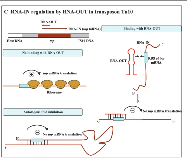
Insertion sequence IS10 is the active element in tetracycline-resistance transposon Tn10, encoding a transposase protein – Tnp. IS10 also encodes three promotors: pIN, the promoter for transposase gene tnp; pOUT, the strong promoter directing transcription outward toward the outside terminus; pIII, the weak promoter symmetrically related to pOUT [41]. The transcript of pOUT is complementary to the 5' end of tnp mRNA transcript from pIN. The complementarity region of 36 nt includes the tnp RBS, then pIN-pOUT pairing directly blocks ribosome binding of this transposase [42]. This negative control prevents the accumulation of these elements in bacteria, thus beneficial for bacterial survival (Figure 1-1C).
No binding with RNAI Binding with RNAI Ori R RNAII Ori R RNase H RNAI RNAII Ori R Ori R
Nascent DNA strand
Ori R Rom
No DNA synthesis A RNAII regulation by RNAI in plasmid ColE1
Ori R RNAII
RNAII (preprimer) RNAI
No binding with RNAI Binding with RNAI
Ori R RNAII Ori R RNase H RNAI RNAII Ori R Ori R
Nascent DNA strand
Ori R Rom
No DNA synthesis A RNAII regulation by RNAI in plasmid ColE1
Ori R RNAII
RNAII (preprimer) RNAI
No binding with RNAI
No binding with RNAI Binding with RNAIBinding with RNAI
Ori R RNAII Ori R RNase H Ori R RNase H RNAI RNAII Ori R RNAI RNAII Ori R Ori R
Nascent DNA strand
Ori R
Nascent DNA strand
Ori R Rom No DNA synthesis Ori R Rom No DNA synthesis A RNAII regulation by RNAI in plasmid ColE1
Ori R RNAII Ori R RNAII RNAII (preprimer) RNAI
B CopT regulation by CopA in plasmid R1
No binding with CopA Binding with CopA
CopT
Couple translation of Tap and RepA
Ribosome
CopA
CopT tap repA
Overlap region of tap/repA
SD
SD
No translation of Tap and RepA CopA
CopT CopB CopT Tap RepA Ori R1
CopA
CopT
B CopT regulation by CopA in plasmid R1
No binding with CopA
No binding with CopA Binding with CopABinding with CopA
CopT
Couple translation of Tap and RepA
Ribosome CopT
Couple translation of Tap and RepA
Ribosome
CopA
CopT tap repA
Overlap region of tap/repA
SD
SD
CopA
CopT tap repA
Overlap region of tap/repA
SD
SD
No translation of Tap and RepA CopA
CopT
No translation of Tap and RepA CopA
CopT CopB CopT Tap RepA Ori R1
CopA
C RNA-IN regulation by RNA-OUT in transposon Tn10
No binding with RNA-OUT
Autologous fold inhibition
5’ RBS of tnp mRNA 3’ RNA-OUT RNA-IN 5’ 3’ No tnp mRNA translation No tnp mRNA translation 5’ 3’
Binding with RNA-OUT
tnp IS10 DNA Host DNA tnp mRNA translation Ribosome RNA-IN (tnp mRNA) RNA-OUT
C RNA-IN regulation by RNA-OUT in transposon Tn10
No binding with RNA-OUT No binding with RNA-OUT
Autologous fold inhibition Autologous fold inhibition
5’ RBS of tnp mRNA 3’ RNA-OUT RNA-IN 5’ RBS of tnp mRNA 3’ RNA-OUT RNA-IN RBS of tnp mRNA 3’ RNA-OUT RNA-IN 5’ 3’ No tnp mRNA translation 5’ 3’ No tnp mRNA translation No tnp mRNA translation 5’ 3’ No tnp mRNA translation No tnp mRNA translation 5’ 3’
Binding with RNA-OUT Binding with RNA-OUT
tnp IS10 DNA
Host DNA tnp IS10 DNA
Host DNA tnp mRNA translation Ribosome tnp mRNA translation Ribosome RNA-IN (tnp mRNA) RNA-OUT
Figure 1-1 Regulation mechanism of plasmid-encoded antisense sRNAs (adapted from Wagner et al., 1994). Regulatory antisense sRNAs are indicated in red and target RNAs in brown. Detailed mechanisms are explained in the text.
– Chromosome encoded antisense RNAs
Fewer antisense RNAs from bacterial genomes are better known compared to those from mobile genetic elements. These RNAs are only partially complementary to their targets. One portion of these sRNAs function as antitoxins repressing translation of mRNAs encoding toxic proteins or inducing their degradation [43-44]. There are many antitoxin sRNAs in bacterial chromosome showing homology to plasmid antitoxin sRNAs. For instance, E. coli strain K–12 has four long directed repeats (LDR) expressing one mRNA (ldr) encoding a toxic protein (LdrD) and an antisense RNA [45]. In bacterial cells, low level of toxins caused by the cis-encoded antitoxin antisense RNA system may only inhibit cell growth or induce stasis. One speculates that this may be beneficial for cells by letting them have sufficient time to self repair or adapt to stresses [46-47].
Besides the toxin-antitoxin modules where antisense sRNAs help maintain low expression of toxin-encoding mRNAs, there is also one sRNA (GadY) which is able to increase the expression of target mRNA (gadX) [28,48-49]. During stationary phase, GadY base pairs with the 3' untranslated region of gadX mRNA and confers increased stability, which allows the GadY dependant accumulation of gadX mRNA [48].
Recently, high-throughput techniques like deep-sequencing and tailing arrays revealed that
cis-antisense RNAs are much more common. It was found that at least 25% of coding
regions are transcripts also in antisense. This leads to a processing of the mRNA-antisense RNA complex by RNaseIII[50]. However, the consequences from this mechanism are not yet well understood.
1.1.2.2 Other cis-acting sRNAs 1.1.2.2.1 Riboswitches
Riboswitches are RNA elements situated in 5' untranslated regions (UTRs) of target mRNAs, which can adopt different conformations in response to various physiological signals. These sRNAs are usually both cis-encoded and cis-acting, however the riboswitches acting in trans do exist [51]. Riboswitches could monitor uncharged tRNAs and a variety of metabolites, such as S-adenosylmethinine (SAM), thiamine pyrophosphate (TPP), flavin mononucleotide (FMN), methionine, lysine, adenine and guanine [52-54]. Usually they consist of two domains, an aptamer which can bind to small metabolites and a regulatory region or so called 'expression platform' that undergoes a conformational shift after the binding of one ligand. Sequence and structure of aptamer domains are highly conserved for each class of riboswitches that sense every particular metabolite. On the contrary, expression platforms can vary in sequence and structure among riboswitches of the same class.
To date, riboswitches have been found to regulate a wide range of genes involved in metabolism, either at the level of transcription attenuation or translation initiation. Leader region of the genes regulated at transcription level contains one intrinsic transcriptional terminator (Rho-independent terminator) composed of G+C rich helix followed by a series of U residues. In the absence of metabolite, an anti-terminator allows the synthesis of mRNA. Once the metabolite binds to the aptamer, a complete stem-loop structure is formed, and the intrinsic transcription terminator blocks the transcription elongation (Figure 1-2A). On the other hand, in the genes regulated at translation level, the binding of the metabolite causes a
conformational change that makes the SD sequence inaccessible to the ribosome. Thus the translation initiation is blocked (Figure 1-2B). Although most riboswitches are found to exert negative regulation on gene expression, there are also mechanisms of positive control. For example, riboswitch of adenine prevents formation of terminator stem, thus activates transcription. In general, the control systems at the transcription termination predominate in Gram-positive bacteria with low GC%, while systems that regulate translation initiation are more common in Gram-negative and Gram-positive bacteria with high GC%[55-56].
Aptamer Expression platform Antiterminator
Antiterminator
Terminator
Without metabolite With metabolite
Aptamer Expression platform SD accessible SD masked
A
B
Aptamer Expression platform Antiterminator
Aptamer Expression platform Antiterminator Antiterminator Terminator Antiterminator Terminator Without metabolite
Without metabolite With metaboliteWith metabolite
Aptamer Expression platform SD accessible Aptamer Expression platform SD accessible SD masked SD masked
A
B
Figure 1-2 Mechanism of riboswitch function [56] MM stands for metabolite. (A) Transcription termination induced by metabolite binding, i.e. guanine riboswitch; (B) Translation initiation blocked by metabolite binding, i.e. TPP riboswitch.
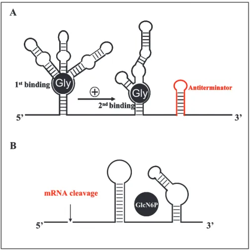
Other novel mechanisms of riboswitches are recently recognized (Figure 1-3). For instance, in Vibrio cholerae, 5' UTR of VC1422 contains two nearly identical conserved domains. After glycine binds to one domain, it will improve the binding efficiency of glycine to the
other domain by 100-1000 folds [57]. One riboswitch residing in the leader region of the glucosamine-6-phosphate synthase gene (glmS) has been demonstrated to control mRNA processing as ribozyme. When GlcN6P bound, a self-cleavage event happens at the specific site at its 5' end, leading to repression of glmS gene [58-59].
GlcN6P 5’ 3’
Gly
Gly
1stbinding 2ndbinding Antiterminator 5’ 3’A
B
mRNA cleavage GlcN6P GlcN6P 5’ 3’Gly
Gly
1stbinding 2ndbinding Antiterminator 5’ 3’Gly
Gly
1stbinding 2ndbinding AntiterminatorGly
Gly
Gly
1stbinding 2ndbinding Antiterminator 5’ 3’A
B
mRNA cleavageFigure 1-3 (A) The glycine cooperative riboswitch (adapted from Lioliou et al., 2010): The first binding of glycine promotes the second binding, and then stabilizes the anititerminator structure. (B) The GlcN6P ribozyme switch: Self cleavage is induced by GlyN6P binding at the 5' UTR of mRNA.
1.1.2.2.2 Thermosensors
To survive in host, bacteria need to response to changing environmental factors and accordingly adjust their gene expression. One major variation they face is the temperature change. RNA thermosensors are those mRNAs responding to temperature changes by altering conformations to allow or prevent binding of the ribosome to translation initiation site, thus modulate the heat-shock or cold-shock protein expression. In Listeria
binding region at lower temperatures (e.g. 30°C). At human body temperature (37°C), another structure is formed where SD region is exposed, allowing the translation start [60]. In contrast, cspA mRNA in E. coli undergoes a structure rearrangement beneficial for translation activation at lower temperature (e.g. 15°C) rather than high temperature (37°C). The structure folded at lower temperature is more stabilized as well, which in turn promotes translation [61].
1.1.2.3 Trans-acting sRNAs
Trans-acting sRNAs are antisense RNAs regulating distant target mRNAs by base pairing to
them in limited complementarity. Usually identified in intergenic regions, they may down regulate target activity through inhibiting translation initiation, or induce complex degradation, or both (Figure 1-4A). They often bind to the 5' UTR of mRNAs and occlude the ribosome-binding site. Then the duplex formed are tended to be degraded by RNaseE. For many trans-acting sRNAs, induced degradation is frequently the major contributor of their negative control on target synthesis, except for few sRNAs, such as RyhB silencing
sodB mRNA and SgrS silencing ptsG mRNA in E.coli [62]. However, positive regulation by
trans-acting sRNAs also exists. An anti-antisense mechanism is adopted during the
activation of target mRNA translation (Figure 1-4B). In this case, base-pairing between sRNA and mRNA could disrupt an inhibitory structure and thereby releasing the ribosome-binding site [63-65]. Sometimes one sRNA can both inhibit and activate target synthesis[65].
Given the partial complementarity between trans-acting sRNAs and targets, the Hfq protein is usually required to facilitate RNA-RNA interactions [30-31]. Hfq, originally discovered in E.
coli, has homology with Sm or Sm-like proteins in eukaryotes and archaea. Hfq mutants
show various stress response related phenotypes, such as slow growth, hypersensitivity to osmotic shock and ultraviolet radiation [66]. It is essential for the expression of sigma factor σS
, global regulator of many genes under stress conditions [67]. Present as a hexamer, it can bind to AU rich single-stranded RNA on the proximal side, poly (A) tail on the distal side. This protein is revealed as a chaperone to accelerate strand exchange and annealing between sRNA and target mRNA. It may also modulate RNAs to melt inhibitory secondary structures. On one hand, it can protect sRNAs from degradation in the absence of base pairing with mRNAs. On the other hand, after the base pairing between sRNAs and mRNAs, it may help
RNasE target and degrade mRNA by forming the RNaseE-Hfq-sRNA complex [68]. In addition, Hfq may also protect mRNA against degradation. For instance, it protects rpoS mRNA from hydrolysis of exoribonucleases and RNaseE by binding to its poly (A) [69].
+sRNA
Translation repression
+sRNATranslation activation
5’ mRNA 3’ 5’ RNase 3’ 5’ 3’ 5’ mRNA 3’ Block of translation initiation DegradationA
B
+sRNA +sRNATranslation repression
Translation repression
+sRNA +sRNATranslation activation
5’ mRNA 3’ 5’ mRNA 3’ 5’ RNase 3’ 5’ RNase 3’ 5’ 3’ 5’ 5’ 3’ 5’ mRNA 3’ 5’ mRNA 3’ Block of translation initiation DegradationA
B
Figure 1-4 Mechanism of regulation by trans-acting sRNAs. (A) Most regulation is negative: sRNA (stem-loops in red) base pairs with SD region (represented in blue box), then makes the translation initiation aborted; or the complex induces irreversible degradation by RNase. (B) Positive regulation: Binding of sRNA and mRNA makes the SD region on mRNA accessible by ribosome.
It has been found that until now all the characterized trans-acting sRNAs in E. coli need the presence of Hfq to exert their regulation roles on targets, however, this requirement for a chaperone protein is not universal among bacteria. For example, in S. aureus, although Hfq binds to RNAIII tightly in vitro, it has no detectable effect on RNAIII-target mRNA complex formation. Another example is VrrA RNA in Vibrio cholerae, it can repress the OmpA protein expression in Hfq mutant strains [70].
1.1.2.4 Protein-binding sRNAs
There is one class of regulatory RNAs having the ability to bind to proteins and regulate their activities. In E. coli, three protein-binding sRNAs are demonstrated to antagonize activities of their target proteins by mimicking the structures of other nucleic acids. They are CsrB sRNA modulating carbon usage related protein CsrA, GlmY sRNA targeting the YhbJ protein, and 6S RNA targeting the
σ
70-containing RNA polymerase [12,71-73]. For example, the RNA-binding protein CsrA normally base pairs with the GGA motifs in 5' UTR of target mRNAs, thus influences its stability or translation. CsrB family of sRNAs, CsrB and CsrC contain several GGA repeats which mimic the binding sites on CsrA target mRNA, and thereby sequester the CsrA from its target mRNA. Homologs of CsrB sRNA have been identified in many other bacteria where they antagonize corresponding CsrA homologs, such as the RsmZ and RsmY antagonize RsmA protein which is involved in carbon metabolism, biofilm formation, motility, virulence in P. aeruginosa[74-76].Worth to mention, the housekeeping sRNAs, as part of ribonucleoprotein complexes (RNPs), have also the protein-binding functions and are crucial for bacterial metabolism and adaptation to stresses. In bacteria, 4.5S RNA and Ffh protein constitute the signal recognition particle (SRP), which target polypeptides to bacterial cell membrane by a co-translational way; tmRNA binds to SmpB protein, then interacts with the translational ribosomal complexes stalled at the 3' end of truncated mRNA, followed by a proteolysis-induced tag added to the truncated protein facilitating rapid degradation for abnormal mRNA; RNase P, consisting of M1 RNA and C5 protein, has the primary role to cleave 5' end of precursor-tRNAs to produce mature 5' end tRNAs.
1.1.3 Advantages and limits
In the last decades, RNAs have been recognized as a key effector of gene regulation among all living bacteria. They have several advantages over proteins. First of all, compared to proteins, RNA regulators are more economic and rapid to produce, since they are much shorter and do not need the step of translation. Secondly, the regulation by RNAs is quite fast, especially for cis-acting sRNAs, such as riboswitches affecting the same mRNA where they reside when sensing appropriate metabolites. Furthermore, one sRNA can simultaneously regulate multiple genes (i.e. RNAIII regulates spa mRNA, hla mRNA and
target in many cases. Moreover, several sRNAs can regulate one single target (i.e. DsrA, RprA and OxyS regulate RpoS translation directly or indirectly), which permit bacteria integrate various environmental signals.
Although many sRNAs are unveiled recently, there are still lots of sRNAs missed, because that they are only expressed under specific conditions, or some cis-encoded antisense sRNAs whose sequences are hard to distinguish from 5' UTR or 3' UTR of bacterial genomes.
1.2 Virulence gene regulation of Staphylococcus aureus
Staphylococcus aureus, a Gram-positive coccal bacterium, could be considered as a
'commensal flora' since about 20-30% of human population are long term carriers of this bacterium on skin or nasal passages [4]. However, it is also a great harm to public health, responsible for many community-acquired and hospital-acquired infections. It can cause a wide variety of diseases, from minor skin infections to severe life-threatening infections. The diversity and severity of S. aureus infection depends on the coordination of different virulence factors expression (Table 1-1), which in turn relies on the fine cooperation of complicated virulence regulators.
1.2.1 Pathogenicity of S. aureus
1.2.1.1 Invasive infection
Once the human skin or mucosal barrier is breached, the bacterial cells from colonization sites enter into the adjacent tissues or bloodstream. Then the combat between S. aureus and host defense system will decide whether this infection localizes or spreads. Several high-risk conditions have been associated with invasive infection. For instance, host immunity is compromised or deficient by underlying diseases, such as newborn, old age, diabetes, HIV infection; or foreign material/equipment is used inside human body, such as intravenous catheter, feeding tube and artificial pacemaker.
Table 1-1 Major known virulence factors of S. aureus and their regulation by Agr (adapted from Novick, 2003)
Gene Location Virulence factor Function/Disease Timea Agr
b
Reference
Capsule and Surface proteins
cap5, 8 chrom Polysacch serotypes 5, 8 Anti-phagocytosis pxp + [77]
spa chrom Protein A Adhesion,
immunomodulation exp _
[78-79]
cna PT island Collagen binding Protein Collagen binding pxp 0 [80]
fnbA, B chrom Fibronectin binding PA, B Fibronectin binding exp - [81]
clfA, B chrom Clumping factor A, B Fibrinogen binding exp 0 [82]
coa chrom Coagulase Adhesion (plasminogen
to plasmin conversion) exp
-[82-83]
Cytotoxins (membrane-damaging)
hla chrom α- haemolysin Cytolysin pxp + [84] hlb chrom β- haemolysin Cytolysin,
sphingomyelinase pxp +
[78,85]
hld chrom δ- haemolysin Cytolysin xp + [86] hlg chrom γ- haemolysin Cytolysin pxp + [87]
lukD/E phage Leucocidin Cytolysin [88]
lukS/F phage P-V leucocidin Cytolysin pxp + [84]
Superantigenic toxins
sea phage Enterotoxin A Food poisoning, TSS xp 0 [89] seb SaPI3 Enterotoxin B Food poisoning, TSS pxp + [84] sec SaPI4 Enterotoxin C Food poisoning, TSS pxp + [90] sed plasmid Enterotoxin D Food poisoning, TSS pxp + [91] eta phage Exfoliatin A Scalded skin syndrome pxp + [92] etb plasmid Exfoliatin B Scalded skin syndrome pxp + [93] tst SaPI1,2 Toxic shock toxin-1 Toxic shock syndrome pxp + [84]
Enzymes
splA-F chrom Serine protease Protease + [85]
sspA chrom V8 protease Diffusion pxp + [94] sspB chrom Cysteine protease Enzyme + [94] scpA Staphopain protease Diffusion, nutrition pxp + [94] geh chrom Glycerol ester hydrolase Diffusion, nutrition pxp + [85] lip chrom Lipase Diffusion, nutrition pxp + [95] aur chrom Metallo protease
(aureolysin) Enzyme pxp +
[96]
nuc chrom Nuclease Nutrition pxp + [97]
hys chrom Hyaluronidase Diffusion xp [98] sak phage Staphylokinase Plasminogen activator pxp + [78] fme chrom FAME Fatty acid esterification pxp + [95] a. exp: early exponential phase, pxp: post exponential phase, xp: exponential phase;
The invasive S. aureus infection has a broad range of clinical presentations from bacteremia with or without primary focus to many complications, such as endocarditis, metastatic infection or sepsis syndrome. Breakthrough towards endothelial cells plays a key role during these pathogenic processes. After adherence to endothelial cells, S. aureus are phagocytized by them [99]. Then the intracellular milieu protects bacteria from both the host defense mechanism and the bactericidal effect of antimicrobial agents. Besides, if the endovascular tissue is invaded, it will become more convenient for bacteria to spread to other tissues.
1.2.1.2 Toxin-mediated disease – Staphylococcal foodborne disease
Foodborne disease (FBD) is the illness caused by consumption of contaminated food (i.e. pathogens, chemical toxins). Each year, the FBD resulted from bacteria contamination takes up more than two thirds of recorded outbreaks [100]. Some kinds of bacteria are particularly arousing attention because of their frequency or seriousness, and S. aureus is one of them. At first, S. aureus could grow in a wide range of temperatures, pH and sodium chloride concentrations. Together with its high carrier rate among normal population, this bacterium can contaminate various food products either during the step of food preparation or processing. Most importantly, the staphylococcal enterotoxins (SEs) secreted by S. aureus contribute to the foodborne disease. Typical symptoms appear rapidly, including nausea, vomiting and abdominal cramps, sometimes followed by diarrhea.
– Toxic shock syndrome
S. aureus is a major cause of toxic shock syndrome (TSS), the dangerous and potentially
fatal illness due to bacterial toxin. Patients with staphylococcal TSS often have high fever, low blood pressure, soon progress to coma and multi-organ dysfunction.
The superantigen toxins (i.e. TSST-1) produced by S. aureus could improperly stimulate host immunology mechanisms and are responsible for TSS. These proteins are resistant to heat denaturation and proteases. They bound firstly with MHC class II molecules then are recognized by an antigen specific T-cell receptor. Formation of this trimolecular complex activates the expansion of T-cells, thus induces a massive cytokine release causing various symptoms of TSS.
– Scalded skin syndrome
Scalded skin syndrome (SSS), also called Ritter disease or Staphylococcal scalded skin syndrome, is a life-threatening infection characterized by skin damage like burn or scald. It usually affects infants and children younger than 5 years or patients with repressed immune system or renal failure. The disease starts with fever and redness of skin, followed by formation of fluid-filled blisters which rupture easily making the top layer of skin peel off in sheets.
Formation of blisters is caused by the production of two exotoxins – exofolitive toxins A and B. They can digest one of the intracellular adhesion molecules, desmoglein1 (Dsg1), whose function is to hold the granulosum and spinasum layers together, thus induce intraepidermal cell-cell dissociation.
1.2.1.3 Positive and adverse effects of host response
Outcomes of infections usually depend on the complex and dynamic interactions between pathogens and host defense mechanisms. During the process of abscess formation, one of the typical S. aureus pathological representations, bacterial cells elicit a series of inflammatory responses [101]. After phagocytosis, endothelial cells express adhesion molecules (vascular-cell adhesion molecules and intercellular adhesion molecules), and release interleukin-1, 6, and 8. Then leukocytes migrate to the site of infection and adhere to these endothelial cells [102]. Macrophage activation occurs after the release of interferon-g by T cells. Cytokines released into bloodstream, from monocytes or macrophages, as well as endothelial cells, contribute to the manifestations of the sepsis syndrome and vasculitis associated with systemic staphylococcal disease [103].
Nevertheless, it has been recognized that the host immune system is like a double edged sword. When working within a proper range, it is essential to protect the host by activation of antimicrobial defenses. If overacting, a bursting swarm of cytokines are released, thus induce severe inflammatory responses which are deleterious to host. However, this uncontrolled inflammatory host response does not take the full responsibility for sepsis mortality. Depressed immune system also plays a crucial role in the prognosis of sepsis [104]. Many studies have reported that leucocytes from sepsis patients may have impaired capacity to release proinflammatory cytokines [105-106]. And in many sepsis death cases, a large number of immune cells, such as B cells, CD4 T cells, dendritic cells, and gastrointestinal
cells, are found lost because of an apoptosis-induced mechanism[107-109].
1.2.2 Various virulence factors
A large number of virulence factors are encoded on the S. aureus core genome and mobile genetic elements, such as surface proteins, exotoxins (Figure 1-5) and extracellular enzymes. They participate in the different steps of staphylococcal infection (adhesion, invasion and escape from host defense) and are responsible for the diversity of diseases.
Figure 1-5 Structure of S. aureus [101]. A. Surface proteins and secreted proteins: surface proteins (also called cell wall-associated proteins) are expressed in early exponential phase whereas the synthesis of secreted proteins (also called exoproteins) is activated in the post-exponential and stationary phase; B. the cross section of cell envelope; C. the typical constitution of clumping factor, like other surface proteins.
1.2.2.1 Capsule and cell wall
Capsule and cell wall are two important structures in pathogenesis of S. aureus. Up to now, 11 capsular polysaccharide types have been identified in this pathogen, most common ones causing human infection are type 5 and type 8 [101,110]. Capsule can not only counterwork phagocytosis, but also promote biofilm constitution and abscess formation [101,111]. S. aureus
has a typical Gram-positive bacterial cell wall composed of many layers of peptidoglycan and teichoic acids, which dedicate to maintain bacterial integrity and mediate evasion of host immune system. For instance, peptidoglycan may covalently link adhesive proteins, whereas lipoteichoic acids play an important role in adhesion and colonization through their hydrophobic part [99,112]. In a rabbit model of endocarditis, the cell wall teichoic acids deficient mutants have been demonstrated to have reduced interactions with endothelial cells
[113]
.
1.2.2.2 Surface proteins – MSCRAMMs
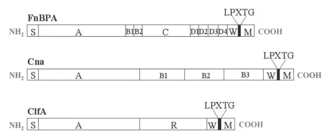
Many staphylococcal surface proteins are those cell wall-anchored proteins with certain structure features. They majorly consist of a long signal sequence at N terminal, and a hydrophobic membrane-spanning domain and a cell-wall anchoring region at C terminal[101]. Some of these surface proteins could function as adhesions to initiate colonization by adhering to components of extracellular matrix (ECM) of host, so named as 'Microbial Surface Components Recognizing Adhesive Matrix Molecules' – MSCRAMMs [114].
MSCRAMMs, secreted via the Sec system, are associated with the peptidoglycan by covalent bonds. Its signal peptide (S) N-terminal position allows the addressing of the protein neo-synthesized at the plasma membrane. C-terminal hydrophobic regions are W and M, separated by the consensus sequence LPXTG (Leu-Pro-X-Thr-Gly). The M-rich region positively charged amino acids corresponds to a region in the cytoplasmic membrane anchor (Figure 1-6). The LPXTG motif is the target of the enzyme sortase, whose role is to anchor the protein adhesin to the cell wall peptidoglycan by a transpeptidation mechanism. The sortase can cleave between threonine and glycine residues of the LPXTG motif and catalyzes the formation of an amide bond between the carboxyl group of threonine and the amino group of the molecules of the wall peptidoglycan [115-117].
NH2 COOH NH2 COOH NH2 COOH NH2 COOH NH2 COOH NH2 COOH
Figure 1-6 Schematic model of MSCRAMMs – domain organization of fibronectin binding protein A (FnBPA), collagen binding protein (Cna) and clumping factor A (ClfA) (from Foster 1998 and thesis of Sandrine 2006). "S" represents the signal sequence, "A" corresponds to the fibrinogen-binding domain, "R" and "D" are involved in binding to fibronectin, the functions of the regions "B" and "C" are unknown, "W" represents the wall-spanning region, "M" represents the membrane-spanning region and positively charged residues. The position of the LPXTG motif is indicated.
More than 20 members of MSCRAMMs have been identified until now, and the most well-studied models are still the fibronectin binding proteins (FnBPA and FnBPB), collagen binding proteins (Cna), fibrinogen binding proteins (clumping factors, ClfA and ClfB)
[118-122]
. However, studies about other surface proteins have also advanced. For example, Serine-aspartate repeat proteins (Sdr) are similar structured with ClfA and ClfB, just different in including an additional region of B repeats between the A-domain and R-domain, and thereby have the same fibrinogen-binding ability[123]. Iron-regulated surface determinant (Isd) can be expressed by S. aureus in iron limited conditions. IsdA is able to adhere to a range of host proteins, such as loricrin and cytokeration K10, whereas IsdB interacts with platelet through directly binding to integrin GPIIb / IIIa[124-126].
– Protein A
Protein A (Spa) is a major surface protein of S. aureus, which is primarily known for its capacity to bind to Fc region of immunoglobulin G. Lately it is referred to as pleiotropic virulence factor, due to its multiple roles in the interaction with the host during infection. Structure organization of this 42-kDa protein has a specific feature, which is a tandem repeat of five homologous domains (E, D, A, B and C) following the N-terminal signal sequence (Figure 1-7A). Polymorphism in the region W, upstream of the LPXTG motif, consists of a
repeating pattern of eight residues. The sequence and number of repetition of this pattern are now used as an epidemiological marker [127]. The extracellular domains (E, D, A, B or C) are responsible for binding to the Fc region of immunoglobulin G or binding to the Fab variable regions of immunoglobulin M [128-129]. The crystal structure of complex Spa-IgM has shown that the helices II and III of Spa domain D interacts directly with VH3 region of IgM. And the residues responsible for Fab binding are separate from those mediating Fc binding (Figure 1-7B)[128].
NH
2COOH
A
B
NH
2COOH
NH
2COOH
A
B
Figure 1-7 (A) Schematic structure of protein A; (B) Crystal structure of complex Spa, IgG and IgM (from Graille et al., 2000). The domain D of protein A (red) binds to VH3 fragment of IgM (blue) and the Fc fragment of IgG (gray).
In addition to its capacity of binding to immunoglobulins, Spa can activate complement[130]. It can also act as an activator of the immune response by its ability to bind to TNFR1, the receptor of TNFα. This interaction is particularly important in respiratory infections such as pneumonia, where the TNFα is essential in the signaling of the infection in respiratory
epithelium. Moreover, Spa plays a role in staph-associated endocarditis by binding to von Willebrand factor, the plasma glycoprotein involved in platelet adhesion to injured vascular endothelium[131]. And these are the same amino acids involved in binding to von Willebrand factor and Fc fragments of IgG [132]. Spa may also interact directly with osteoblasts, preventing proliferation and inducing apoptosis, to accelerate bone weakening in osteomyelitis[133].
– Coagulase and SERAMs
Coagulase is a protein of 60 kDa useful for detection of different staphylococcus isolates, which is always positive for S. aureus. It possesses a conserved region with multiple domain repeats and an N-terminal binding domain of prothrombin. The complex formation between coagulase and prothrombin is called staphylothrombine, causing the polymerization of fibrinogen to fibrin, and then leading to thrombus formation [134]. Its C-terminal domain is demonstrated to be involved in the bacterial adherence to immobilized platelets by using phage display assays [135].
Coagulase belongs to the family of 'Secretable expanded Repertoire Adhesive Molecules' – SERAMs. This family also includes Eap – "extracellular adherence protein", Emp – "extracellular matrix binding protein" and Efb – "extracellular fibrinogen binding protein". No significant homology in structure has been shared among these bacterial proteins, but they do have common functions, which may be illustrated as i) binding to host proteins to mediate bacterial adhesion, and ii) binding to a broad array of host ligands to interfere with host defense mechanisms[136].
SERAMs are fixed by non-covalent bonds to proteins of the extracellular matrix such as fibrinogen, fibronectin, prothrombin, collagen, laminin, sialoprotein, elastin, or vitronectin
[136]
. These bonds could be either hydrophobic, such as Emp (composed of 25% hydrophobic residues) with lipoteichoic acid, or electrostatic, such as Emp and Eap (cationic) interact with the negatively-charged compounds of the wall. Finally, these adhesins may be recognized by receptors on the surface of the bacterium [137].
Except from their functions as adhesins, interaction of these molecules and host components permit them possess immunomodulatory properties. They are involved in the pathogenesis of endo- and extra-vascular staphylococcal diseases [136]. Eap favors the internalization of the bacteria into eukaryotic cells [138], and adherence to the degradation products of ECM. S.
![Figure 1-2 Mechanism of riboswitch function [56] M M stands for metabolite. (A) Transcription termination induced by metabolite binding, i.e](https://thumb-eu.123doks.com/thumbv2/123doknet/14449350.518339/26.892.131.766.384.958/figure-mechanism-riboswitch-function-metabolite-transcription-termination-metabolite.webp)


![Figure 1-5 Structure of S. aureus [101] . A. Surface proteins and secreted proteins: surface proteins (also called cell wall-associated proteins) are expressed in early exponential phase whereas the synthesis of secreted proteins (also called exoproteins](https://thumb-eu.123doks.com/thumbv2/123doknet/14449350.518339/35.892.116.780.379.818/structure-surface-proteins-associated-expressed-exponential-synthesis-exoproteins.webp)

![Figure 1-10 Secondary structure of RNAIII [214-215] . The stem-loop motifs are numbered from 1 to 14](https://thumb-eu.123doks.com/thumbv2/123doknet/14449350.518339/49.892.256.644.361.874/figure-secondary-structure-rnaiii-stem-loop-motifs-numbered.webp)
