HAL Id: hal-02802940
https://hal.inrae.fr/hal-02802940
Submitted on 5 Jun 2020HAL is a multi-disciplinary open access
archive for the deposit and dissemination of sci-entific research documents, whether they are pub-lished or not. The documents may come from teaching and research institutions in France or abroad, or from public or private research centers.
L’archive ouverte pluridisciplinaire HAL, est destinée au dépôt et à la diffusion de documents scientifiques de niveau recherche, publiés ou non, émanant des établissements d’enseignement et de recherche français ou étrangers, des laboratoires publics ou privés.
Isabelle P. Oswald
To cite this version:
Isabelle P. Oswald. Mycotoxins in swine production. Ines Rodrigues and Maximillian Smith, 113 p., 2013, 9781899043415. �hal-02802940�
Mycotoxins
in Swine Production
MYCOTOXINS IN SwINe
prOduCTION
Edited by
BIOMIN Edition
© Copyright 2013 by Erber AG, Austria www.biomin.net
www.erber-group.net
All rights reserved. No part of this publication may be reproduced in any material form (including photocopying or storing in any medium by electronic means and whether or not transiently or incidentally to some other use of this publication) without the written permission of the copyright holder except in accordance with the provisions of the Copyright, Designs and Patents Act 1988. Applications for the copyright holder’s written permission to reproduce any part of this publication should be addressed to the publishers. ISBN: 978-1-899043-41-5
Disclaimer
Every reasonable effort has been made to ensure that the material in this book is true, correct, complete and appropriate at the time of writing. Nevertheless the editors and the authors do not accept responsibility for any omission or error, or for any injury, damage, loss or financial consequences arising from the use of the book.
Cover pictures Copyright: © Erber AG
© Roman Labuda
Published by:
Context products Ltd, 53 Mill Street, Packington, Ashby de la Zouch, Leicestershire, LE65 1WN England, Tel: +44 (0) 1530 411337
fOrewOrd
Mycotoxins are not a new topic and they are certainly not a recent worry. In fact, this year it is a decade since the Council for Agricultural Science and Technology (CAST) released its scientific task force report on mycotoxins and their impacts on plant, animal and human systems. Back then, economic impacts of mycotoxins in the United States alone were estimated to exceed 1.4 billion dollars, through losses in commodity quality and health of livestock. Actually, it is quite surprising to see how these very low molecular weight molecules have affected humans for thousands of years. In the 7th and 8th centuries BC god Robigus was honored in an annual festival aiming to protect cereal crops and trees from rust and mildew. Between 1692 and 1693, the Salem witch trials culminated in dozens of hangings and terrible suffering to hundreds of people accused of witchcraft. Medical explanations based on today’s scientific knowledge suggest ingestion of rye bread infected with Claviceps purpurea fungus and derived ergot alkaloid-mycotoxins as the most plausible causative agent for the convulsive symptoms presented by the presumed witches.
As far as livestock is concerned, general interest in this field arose later on, in the 20th century, as aflatoxins were identified as the cause of death of thousands of turkey poults in England. Since then hundreds of other secondary metabolites of fungi have been identified and swine have been generally acknowledged as one of the most sensitive species to their effects amongst livestock animals. Also, Food and Agriculture Organization of the United Nations (FAO) estimates that by 2050 global agricultural production must grow 60 % above 2005-07 levels in order to meet the increasing demand of an escalating world population. By then, world meat production is expected to double. Livestock systems will have to become more efficient at a time of ever-increasing prices for commodities, labor and natural resources. Nutrition has a crucial role in the maximization of animal genetic potential, representing around 60 to 80 % of total production costs and poses a constant challenge: rations should satisfy the nutritional needs of animals, without posing a risk to their health. As increasing prices of raw materials make cheaper commodities more appealing to the producer, the well-documented increase in mycotoxin occurrence around the world, highlights the risks associated with this strategy.
These were some of the issues behind our choice to combine two topics - swine and mycotoxins - into one publication. This book is intended to be read by those involved in swine production, whether nutritionists or veterinarians, by students aiming to increase their understanding of the topic, or merely by curious minds.
We have gathered the knowledge of several experts and compiled information on a wide range of topics - from general concepts on production of mycotoxins by fungi, to their effects on swine performance, fertility and immunity. We have looked at worldwide existing legislation for mycotoxins, on how to make analysis of these toxic substances more accurate and we have included an overview on forefront mycotoxin management strategies.
I sincerely thank all who have made this book possible, especially Dr. med. vet. Maximilian Schuh, Professor emeritus at the department of Farm Animals and Herd Management, University of Veterinary Medicine, Vienna, Austria and Dr. Isabelle Oswald, research director at the Research Center in Food Toxicology (ToxAlim) in INRA, the leading European agricultural research institute located in France. Besides providing crucial information on specific topics related to their fields of expertise and years of research, they provided valuable inputs to the whole publication. I would also like to acknowledge my colleagues Roman Labuda, Project Leader at the R&D Department of Romer Labs
Holding GmbH, Austria and Georg Häubl R&D Manager at Romer Labs Holding GmbH, Austria, for sharing their expertise in the fields of mycology, microbiology and chemistry. A very big word of appreciation goes to my fellow workers Elisabeth Pichler (Romer Labs, Austria), Roger Berríos and my very good colleague and great team player Karin Nährer for managing this bold project amidst their very busy schedules. Thank you to other contributors to the book including Ursula Hofstetter, Eva Maria Binder and Yin-Jung Liu for their inputs on the whole manuscript and other colleagues for allowing us to use photographs gathered during their regular encounters with mycotoxicoses in the field. Last but not least, thank you Sarah Keeling of Context Products Ltd publishers for handling all my requests and pickiness in such a careful, professional and friendly manner.
It is BIOMIN’s duty as a leader in Mycotoxin Risk Management strategies and a worldwide expert in animal nutrition to identify the enemies hindering efficient animal nutrition and to present you tools to counteract them.
We hope you enjoy reading this book and above all that you find it helpful in your daily activities. Inês Rodrigues
CONTeNTS
fOrewOrd iii
1. MYCOTOXINS – geNeraL CONCepTS 1
Karin Nährer
1.1. What are mycotoxins? 1
1.2. Mycotoxin producing fungi 1
1.3. Conditions for fungal growth and mycotoxin production 4
1.4. Chemical stability of mycotoxins 6
1.5. Masked mycotoxins 7
1.6. Synergistic effects 7
1.7. Mode of action/toxicology/metabolism of mycotoxins 10
1.7.1. Aflatoxins 10 1.7.2. Trichothecenes 14 1.7.3. Ochratoxins 17 1.7.4. Fumonisins 19 1.7.5. Zearalenone 22 1.7.6. Ergot alkaloids 24
1.8. Legislation in the European Union (EU) 26
1.9. Legislation in the United States of America (USA) 28
1.10. Other legislation 30
1.11. Legislation versus safe levels of mycotoxins 30
1.12. Acknowledgements 31
2. MYCOTOXICOSeS IN SwINe 33
Inês Rodrigues and Maximilian Schuh
2.1. Mycotoxicoses 33
2.2. Effects of mycotoxins on swine performance 37
2.2.1. Aflatoxins 37
2.2.2. Trichothecenes 38
2.2.3. Ochratoxins 39
2.2.4. Fumonisins 40
2.2.5. Ergot alkaloids 41
2.3. Effects of mycotoxins on swine fertility 42
2.3.1. Zearalenone and its direct effects on fertility 42 2.3.2. Ergot alkaloids and their direct effects on fertility 44 2.3.3. Other mycotoxins and their direct effects on fertility 45 2.4. Indirect effects of mycotoxins on swine fertility 45 2.4.1. Ochratoxin A and its indirect effects on fertility 45
2.4.2. Deoxynivalenol and its indirect effects on fertility 46 2.4.3. Fumonisins and their indirect effects on fertility 46
2.5. Economic impact of decreased fertility 46
2.6. The impact of mycotoxins on animal immunity 47
3. SwINe IMMuNe SYSTeM 49
Roger Berríos and Isabelle Oswald
3.1. Organs of the immune system 49
3.1.1. Bone Marrow 50 3.1.2. Thymus 50 3.1.3. Spleen 50 3.1.4. Lymph Nodes 50 3.1.5. Peyer’s Patches (PPs) 51 3.1.6. Tonsils 51
3.2. Cells of the immune system 51
3.2.1. Myeloid Cells 51 3.2.2. Neutrophils 52 3.2.3. Basophils 52 3.2.4. Eosinophils 52 3.2.5. Monocytes/Macrophages 52 3.2.6. Dendritic Cells (DCs) 52 3.2.7. Lymphoid Cells 53
3.3. Other components of the immune system 54
3.3.1. The Complement System 54
3.3.2. Host defense peptides (hdps) 54
3.3.3. Cytokines 54
3.3.4. Major Histocompatibility Complex (MHC) 54
3.4. Other cells involved in immune response 55
3.5. Immune system: main mechanisms of action 55
3.5.1. Pathogen Recognition 55
3.5.2. The Inflammatory Response 56
3.6. Mycotoxins and cellular immunity 56
3.6.1. Aflatoxins 57
3.6.2. Trichothecenes 58
3.6.3. Ochratoxin A 60
3.6.4. Fumonisins 61
3.6.5. Multiple mycotoxins 63
3.7. Mycotoxins and humoral immunity 64
3.7.1. Aflatoxins 65
3.7.2. Trichothecenes 65
3.7.3. Fumonisins 66
3.8. Mycotoxins, susceptibility to diseases and vaccine efficacy 66
3.8.1. Aflatoxins 67
3.8.2. Fumonisins 67
3.8.3. Ochratoxins 68
3.9. Conclusions 68
4. aNaLYzINg MYCOTOXIN CONTeNT IN COMMOdITIeS/feedS 71 Inês Rodrigues and Elisabeth Pichler
4.1. Sampling 71 4.2. Sample preparation 73 4.3. Analytical methods 74 4.3.1. Rapid tests 75 4.3.2. Reference Testing 76 5. fIghTINg MYCOTOXINS 79 Inês Rodrigues 5.1. Prevention methods 79
5.1.1. Avoiding contamination in the field and during harvest 79
5.1.2. Avoiding contamination during storage 80
5.2. Elimination of mycotoxins 82
5.2.1. Physical processes 82
5.2.2. Chemical processes 83
5.2.3. Biological methods - Adsorption 84
5.2.4. Testing mycotoxin binders 87
5.2.5. Biological methods - Biotransformation 87
LITeraTure 95
1.
MYCOTOXINS – geNeraL CONCepTS
KARIN NÄHRER
(Adapted from Chapter 2 of Guide to Mycotoxins: Näehrer, 2012)
1.1. what are mycotoxins?
Mycotoxins are naturally occurring secondary metabolites produced by certain moulds/fungi as a result of their organic processes. They are chemical compounds of low molecular weight and low immunogenic capacity (Mallmann and Dilkin, 2007). There are some secondary metabolites of fungal origin with medicinal or industrial applications, for example Penicillin. Unfortunately, most mycotoxins are known to hazardously contaminate crops and consequently animal feeds and animal products, causing significant economic losses associated with their impact on animal and human health, animal productivity and domestic and international trade. In practical terms, the mycotoxins which cause bigger economic impacts in animal production are aflatoxins (Afla), trichothecenes (namely deoxynivalenol (DON) and T-2 toxin (T-2)), zearalenone (ZEN), ochratoxin A (OTA), fumonisins (FUM) and ergot alkaloids (Ergots). These will be the focus in the chapters of this book.
1.2. Mycotoxin producing fungi
The process of mycotoxin production by fungi is, in general, not well known. However, taking into account that 1) fungi, just like any living organism need nutrients to survive and 2) crops subjected to stress (namely droughts, poor fertilization, water excess etc.) usually present higher mycotoxin contamination, a possible explanation is that mycotoxins are produced by fungi so that they are able to prevail in adverse conditions. To put it simply, mycotoxins are produced so that fungi win a competitive advantage on other organisms (Rankin and Grau, 2002). Unfortunately, even if we cannot accurately explain the reasons for their existence, mycotoxins are produced in several stages of production of agricultural products, representing a serious problem of worldwide occurrence.
Despite the fact that proper conditions for growth of fungi can occur at all times during crop growth, harvest and storage, fungal species can be roughly divided into field moulds, which infect crops as parasites, and storage fungi which grow in feedstuffs stored under sub-optimal conditions.
Field fungi are those which in general require higher moisture to grow and produce mycotoxins (> 0.9 water activity), infecting seeds and plants in the field, namely Fusarium sp. Storage fungi are those which require lower water activity, thus being more prominent after harvest and during storage, such as Aspergillus and Penicillium sp.
Infection by Claviceps sp. and Neotyphodium sp. occurs only in the field. Claviceps are plant pathogens that replace plant structures such as grain kernels with hardened fungal tissues called ergots or sclerotia (Tudzynski et al., 2001). These fungal bodies often contain a broad
range of toxic compounds called ergot alkaloids (more than 40 different ergot alkaloids are known) leading to a disease known as ergotism (one of the oldest recognized mycotoxicoses) (CAST, 2003; Flieger et al., 1997). The alkaloid pattern and individual alkaloid contents in sclerotia vary largely, due to differences in the maturity of the sclerotia and to other factors e.g.: fungal strain, host plant, geographical region and general weather conditions (EFSA, 2011). Claviceps genus, mainly Claviceps purpurea, parasitizes more than 600 plants, including some of the economically important cereal grains such as rye, wheat, barley, millet, and oats (Strickland et al., 2011). In addition, ergot contamination in sorghum due to Claviceps africana has been discovered in which the Claviceps spores germinate and grow into the unfertilized seed producing a sclerotium (Krska and Crews, 2008). Toxic alkaloids are also produced by fungal endophytes such as Neotyphodium sp. that colonize vegetative tissue as well as reproductive tissue in certain plant species such as perennial rye grass and tall fescue; thus only a concern for grazing animals (EFSA, 2005).
In Table 1 the most important fungal species are listed together with their respective produced mycotoxins.
Table 1 – The most important mycotoxins and respective producing fungi (Source: www.mycotoxins.info)
Major classes of mycotoxin-producing fungi
Fungi species Mycotoxins
Aspergillus A. flavus A. parasiticus A. nomius A. pseudotamarii Aflatoxin (B1, B2, G1, G2) A. ochraceus Ochratoxin (Ochratoxin A) A. clavatus A. terreus Patulin A. flavus A. versicolor Cyclopiazonic acid Claviceps C. purpurea C. fusiformis C. paspali C. africana Ergot alkaloids: Clavines (Agroclavine) Lysergic acids
Lysergic acid amids (Ergin) Ergopeptines (Ergotamine, Ergovaline)
Fusarium F. verticillioides (syn. F. moniliforme) F. proliferatum Fumonisin (B1, B2, B3) Fusaric Acid Moniliformin F. graminearum F. avenaceum F. culmorum F. poae F. equiseti F. crookwellense F. acuminatum F. sambucinum F. sporotrichioides Type-A Trichothecenes T-2 toxin, HT-2 toxin, diacetoxyscirpenol Type-B Trichothecenes Nivalenol, deoxynivalenol, 3- and 15-acetyldeoxynivalenol, fusarenon-X Moniliformin F. graminearum F. culmorum F. sporotrichioides Zearalenone Penicillium P. verrucosum P. viridicatum Ochratoxin (Ochratoxin A) P. citrinum P. verrucosum Citrinin P. roqueforti Roquefortine PR toxin P. cyclopium P. camemberti Cyclopiazonic acid P. expansum P. claviforme P. roquefortii Patulin Neotyphodium
(formerly Acremonium) N. coenophialum Tall fescue toxins: Ergot alkaloids, lolines, peramine
N. lolii Tall fescue toxins:
Lolitrems, peramine, ergot alkaloid (ergovaline) ©Erber AG
1.3. Conditions for fungal growth and mycotoxin production
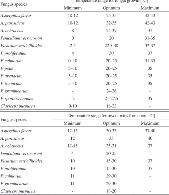
Although there are geographic and climatic differences in the production and occurrence of mycotoxins, exposure to these substances is worldwide (Kuiper-Goodman, 2004). Nevertheless, a preferred pattern in terms of temperature and water activity for fungal growth and mycotoxin production can be established to some extent (Table 2) (Marth, 1992; Sweeney and Dobson, 1998; FAO, 2003; Hussein and Brassel, 2001; CAST, 2003; Sanchis, 2004; Ribeiro et al., 2006).
Table 2 - preferred temperatures and water activity values for fungal growth and mycotoxin production
Fungus species Temperature range for fungal growth [°C]
Minimum Optimum Maximum
Aspergillus flavus 10-12 25-35 42-43 A. parasiticus 10-12 32-35 42-43 A. ochraceus 8 24-37 37 Penicillium verrucosum 0 20 31-35 Fusarium verticillioides 2-5 22.5-30 32-37 F. proliferatum 4 30 37 F. culmorum 0–10 20–25 31-35 F. poae 5–10 20–25 35 F. avenacum 5–10 20–25 35 F. tricinctum 5–10 20–25 35 F. graminearum - 24-26 -F. sporotrichioides -2 21-27.5 35 Claviceps purpurea 9-10 18-22
-Fungus species Temperature range for mycotoxins formation [°C]
Minimum Optimum Maximum
Aspergillus flavus 12-15 30-33 37-40 A. parasiticus 12 33 40 A. ochraceus 12-15 25-31 37 Penicillium verrucosum 4 20-25 -Fusarium verticillioides 10 15-30 37 F. proliferatum 10 15-30 37 F. culmorum 11 29-30 -F. graminearum 11 29-30
-Fungus species Water activity (aw) for fungal growth
Minimum Optimum Maximum
Aspergillus flavus 0.80 0.95-0.99 -A. parasiticus 0.83-0.84 0.95- 0.99 -A. ochraceus 0.77-0.79 0.95-0.99 -Penicillium verrucosum 0.80 0.95 -Fusarium verticillioides 0.87-0.9 - 0.99 F. proliferatum 0.9 - -F. culmorum 0.90-0.91 0.98-0.995 -F. poae 0.90-0.91 0.98-0.995 -F. avenacum 0.90-0.91 0.98-0.995 -F. tricinctum 0.90-0.91 0.98-0.995 -F. graminearum 0.9 - 0.99 F. sporotrichioides 0.88 - 0.99
Fungus species Water activity (aw) for mycotoxins formation
Minimum Optimum Maximum
Aspergillus flavus 0.82 0.99-0.996 0.998 A. parasiticus 0.87 0.99 -A. ochraceus 0.80-0.85 0.98 -Penicillium verrucosum 0.83-0.86 0.90-0.95 -Fusarium verticillioides 0.92-0.93 - -F. proliferatum 0.93 - -F. graminearum 0.9-0.91 0.98 -Table 2 - Contd.
For example, aflatoxins are produced by A. flavus over the temperature range of 12 – 48°C (optimum growth is at 37°C). It is not possible to specify an optimum temperature for the production of the toxins, although production between 25 – 30°C is reported to be significantly greater at higher rather than at lower temperatures. In general, temperature and drought stress are also likely to predispose the plant to increased infection (CAST, 2003).
In the case of F. graminearum growth, the optimum temperature has been estimated at 24 – 26°C and a minimum water activity of 0.90 (Sweeney and Dobson, 1998). Different information is available regarding the most favorable temperature for the production of trichothecenes and zearalenone by this fungus. In general, the production of these mycotoxins is ubiquitous, more prevalent in warm and moderate climates; however, trichothecenes and zearalenone may equally be produced at lower temperatures.
A. ochraceus grows at temperatures ranging from 8 – 38°C (optimum 24 – 37°C). Ochratoxin A is produced by A. ochraceus within the temperature range of 12 – 37°C, with an optimal production at 31°C. P. verrucosum grows within the temperature range of 0 – 31°C (optimum 20°C) and (low) water activity of 0.80; ochratoxin A is produced over the whole temperature range (optimum 20°C). Significant quantities of ochratoxin can be produced at a temperature as low as 4°C and a water activity as low as 0.86 (Sweeney and Dobson, 1998).
In the case of fumonisin production, a study using maize showed an optimum temperature between 15 and 25°C using two major fumonisin-producing Fusarium species Fusarium verticillioides and Fusarium proliferatum (Samapundo et al., 2005).
Favorable environmental conditions such as warm temperatures, high rainfall/humidity and high soil fertility raise the prevalence of fungi (e.g.: Claviceps sp.) and the production of ergot alkaloids (Strickland et al., 2011). Poisonous alkaloid-containing ergot sclerotia are used for sexual reproduction and as a resting structure to enable survival in unfavorable conditions (e.g.: in temperate zones where they overwinter in the ground or during storage and then sexually fruit the following spring when grass hosts are flowering) (Kren and Cvak, 1999; CAST, 2003). Fungal growth continues until the fungus produces this latent structure. Claviceps purpurea occurs in every temperate region and has the widest host range of any other Claviceps species (Kren and Cvak, 1999). Claviceps africana colonizes sorghum in Southern Africa but also in Southeast Asia, South America and USA (Kren and Cvak, 1999). In a study, C. Africana sclerotia could germinate after 1 year of dry storage at ambient temperature (15 – 30°C) (Frederickson et al., 1991). Montes-Belmont et al. (2002) reported optimum climatic conditions for ergot development of a mean day temperature of 25°C, maximum relative humidity of 96 % (maximum temperature 28°C, night relative humidity of 86 %).
1.4. Chemical stability of mycotoxins
Due to their chemical structure and low molecular weight, mycotoxins are chemically stable; they resist high temperatures and several manufacturing processes (Bullerman and Bianchini, 2007). Some review work has been done on the fate of mycotoxins during thermal food processing (Kabak, 2009).
Aflatoxins have a melting point of 268–269ºC and show high stability to dry heat-up temperatures. Temperatures over 150ºC are required to attain partial destruction of the toxin (Samarajeewa et al., 1990).
OTA has a melting point of 169ºC. Dry heating wheat at 100ºC for 40 – 160 min had no effect on OTA. However, wet heating at the same temperature led to a destruction of 50 % OTA after 120 min (Boudra et al., 1995).
FUM may survive most of the commonly used thermal processes as they are also fairly heat stable (up to 100 – 120ºC) (Humpf and Voss, 2004). It is also known that fumonisins can bind to various components of the feed matrix or react with other ingredients of the food, which leads to an underestimation of the potential toxicity of the mycotoxin because of the formation of unknown biologically active decomposition products.
Likewise, DON is a very stable compound, with a melting point of 151 – 153ºC. A significant reduction or destruction of DON was not achieved by thermal processing (CAC, 2003).
ZEN is stable during storage, milling and cooking and possesses a melting point of 164 – 165ºC (EFSA, 2004b). ZEN was shown to survive heat treatments of 140ºC for 4 h (Smith et al., 1994);
a complete reduction of this mycotoxin was observed in aqueous buffer solutions heated at 225ºC for less than 30 min.
Ergot alkaloids were relatively stable during end-use processing of flour into pasta and oriental noodles (Fajardo et al., 1995). Processing flour into pan bread decreased the ergot alkaloid content by approximately 25 % during the baking of a rye roll (Bürk et al., 2006).
It should be kept in mind that in studies carried out regarding the stability of mycotoxins, in general, only the disappearance of the mycotoxin is evaluated. This fact does not necessarily mean that the toxicity risk is reduced. Decomposition or transformation products may be just as dangerous as the parent molecules.
1.5. Masked mycotoxins
The topic of conjugated or masked mycotoxins first became of interest because in some cases of mycotoxicoses, clinical observations in animals did not correlate with the low mycotoxin content determined in the corresponding feed (more interacting factors are explained in the following paragraphs).
Masked mycotoxins are mycotoxins that experienced changes in their chemical structures. Proteins and glucosides, as an example, can be bound to mycotoxins by growing plants in the field to protect themselves from foreign compounds or by microorganisms which may change mycotoxin structures during storage. In rare cases, some mycotoxin conjugates can be excreted directly by fungi (e.g.: 3-acetyldeoxynivalenol and 15-acetyldeoxynivalenol by Fusarium sp.) or by mammals (Berthiller et al., 2009).This phenomenon of altered mycotoxin structures may also occur during food/feed processing, as in the case of FUM reaction with reducing sugars (Lu et al., 2002). More than 50 % of the amount of free mycotoxins (especially zearalenone and deoxynivalenol) is considered to exist in commodities in a masked form (Vendl et al., 2010).Unfortunately these conjugated mycotoxins cannot be detected by any routine analysis. However, during digestion the mycotoxin-ligand bond can be released and the mycotoxin acts as a toxin, thus causing its hazardous effects on animals (Berthiller et al., 2003). A recent in vitro study indicated that deoxynivalenol-3-glucoside is of toxicological relevance as it may become bioavailable again due to hydrolysis. It was suggested to monitor this toxin together with deoxynivalenol in cereals, especially since the part of the masked toxin might increase in the future due to Fusarium resistance breeding efforts (Berthiller et al., 2011). Figure 1 shows scheme representing the example of masked zearalenone-4-glucoside formation and action in the animal.
1.6. Synergistic effects
Because each plant can be contaminated with more than one fungus and each fungus species is able to produce more than one mycotoxin, the probability of co-occurrence of mycotoxins in a feed commodity is very high. On the other hand, the international trading of various feed stuffs also increases this risk. The interaction between mycotoxins often leads to synergistic effects, when the negative effects of one mycotoxin are amplified by the presence of another mycotoxin. Therefore an increase of severity of mycotoxicoses should be taken into account (Speijers and Speijers, 2004).
figure 1- Scheme of the conjugate zearalenone-4-glucoside formation in the plant and subsequent ingestion by the animal followed by hydrolysis and release of the toxic compound.
80 % of pig diseases are related to mis-management due to feed quality, reproduction, housing conditions and biosecurity and only 20 % are due to viral, bacterial or parasitic pathogens. In cases of low levels of mycotoxins in the feed it should be mentioned that interactions between mycotoxins may enhance the toxicological effects. This is especially true in the case of fusariotoxins. Fusarium graminearum and Fusarium culmorum are known to produce several different fusariotoxins under the same conditions, namely ZEN and DON, which are known to interact synergistically in the animal. Analysis of DON often indicates co-occurrence of other fusariotoxins such as trichothecenes (T-2 toxin, nivalenol, diacetoxyscirpenol), zearalenone and fumonisins.
Trenholm et al. (1994) showed that mycotoxin naturally contaminated feed caused more severe toxicity when compared to the same concentration of purified mycotoxins. Another trial confirmed that fusaric acid is able to increase the toxicity of DON in growing pigs (Smith et al., 1997). Zielonka et al. (2009) reported the difficulties examining histopathological lesions caused by DON intoxication due to the common, often synergistic, reaction of this mycotoxin
fumonisin B1 to grower pigs and found negative effects on clinical performance, biochemical, hematological and immunological parameters. The toxic reactions could be described as additive or more than additive particularly regarding the initiation of liver disease.
T-2 toxin synergized the activity of DON with respect to several parameters including weight gain (Friend et al., 1992). A summary of synergistic and additive effects of mycotoxins in pigs is presented in Figure 2 and Table 3.
AFB1 FB1 DON OTA CPA ZEN T-2 toxin DAS MON FA
AFB1 – Aflatoxin B1; FB1 – Fumonisin B1; DON – Deoxynivalenol; OTA –Ochratoxin A; ZEN – Zearalenone; FA –
Fusaric acid; DAS – Diacetoxyscirpenol; CPA – Cyclopiazonic acid; MON – Moniliformin
figure 2 - Synergistic (solid line) and additive (dashed line) effects of mycotoxins in pigs
Table 3 - Mycotoxin combinations in pigs
Mycotoxins Species tested Effect References
AFB1 + OTA Pigs synergistic D’Mello et al., 1999; Huff et al., 1988a AFB1 + FB1 Growing pigs synergistic Harvey et al., 1995; Liu et al., 2002 AFB1 + T-2 toxin Pigs synergistic D’Mello et al., 1999; Schwarzer, 2009 DON + FA Pigs synergistic Raymond et al., 2005; D’Mello et al., 1999 MON + FB1 Pigs additive D’Mello et al., 1999; Schwarzer, 2009 MON + DON Pigs additive D’Mello et al., 1999; Schwarzer, 2009 OTA + DON Weaned piglets synergistic Speijers and Speijers, 2004
OTA + FB1 Weaned piglets synergistic Creppy et al., 2004 ; Speijers et al., 2004 OTA + T-2 toxin Weaned piglets additive Speijers and Speijers, 2004
DON + ZEN Weaned piglets synergistic Zielonka et al., 2009
FB1 + DAS Pigs additive D’Mello et al., 1999; Schwarzer, 2009 FB1 + DON Pigs synergistic D’Mello et al., 1999; Huff et al., 1988a; Speijers and Speijers, 2004 FB1 + T-2 toxin Pigs additive D’Mello et al., 1999; Schwarzer, 2009 DAS + Afla Pigs synergistic D’Mello et al., 1999
AFB1 = Aflatoxin B1; OTA = Ochratoxin A; DAS = Diacetoxyscirpenol; DON = Deoxynivalenol; FB1 = Fumonisin B1; CPA = Cyclopiazonic acid; MON = Moniliformin; PCA = Penicillic acid; FA = Fusaric acid; ZEN = Zearalenone
1.7. Mode of action/toxicology/metabolism of mycotoxins
As chemical structures of different mycotoxins vary widely, they cannot be classified as a group according to their modes of action, toxicology or metabolism. Additionally, as previously explained, additive and synergistic effects can occur in the presence of two or more mycotoxins. Nevertheless, in the following pages the most important mycotoxins and their modes of action, toxicology and metabolism as well as main symptoms and target organs will be described.
1.7.1. AFLATOXINS
Aflatoxins were identified in 1960 and represent one of the most studied mycotoxins. They are mainly produced by certain strains of Aspergillus parasiticus and A. flavus and they occur in agricultural products in tropical and subtropical regions. Aflatoxins are divided into six major toxins (Figure 3) according to their fluorescent properties under ultraviolet light (ca. 365 nm) and their chromatographic mobility (subscripts). Aflatoxin B1 (AfB1) and B2 produce a blue fluorescence while G1 and G2 a green one. Two metabolic products - aflatoxin M1 and M2 - occur in the milk of lactating mammals which have consumed aflatoxin contaminated feed. Aflatoxin B1 is the most toxic and the most prevalent among this family.
figure 3 - Chemical structure of the different aflatoxins
1.7.1.1. Exposure and absorption into the organism
Because of aflatoxins’ common occurrence in feedstuffs, feeds and milk products, these mycotoxins pose a serious threat to humans and animal species. Although the oral route is the main contamination means, inhalation may also occur as a result of people or animals being exposed to the grains’ dust. After respiratory exposure, aflatoxin B1 may appear in the blood more quickly than after oral exposure. Nevertheless, after 4 hours the plasmatic concentration does not differ between the two routes of contamination. Following ingestion, aflatoxin B is
of absorption. Due to the particle’s low molecular weight, the main mechanism of absorption of this mycotoxin, as suggested by several authors, is passive diffusion, in which no efflux pumps or transporters are involved. From the site of absorption, aflatoxin B1 enters the blood stream and is transported to the liver, the major site of metabolism (Gratz, 2007).
1.7.1.2. Metabolism
The metabolism of AfB1 has been extensively reviewed (IARC, 1993; IARC, 2002 and Eaton et al., 2010). AfB1 in liver and other tissues is metabolized by P450 cytochromes enzymes to aflatoxin P1, aflatoxin M1, or aflatoxin Q1 and AfB1-8,9-epoxide (Riley and Voss, 2011). These cytochromes P450 are responsible for activation of aflatoxin B1, aflatoxin M1 and aflatoxin P1 which can form nucleic acid adducts or undergo conjunction to glutathione, conversion to dihydriodiols or binding to serum protein and other macromolecules (Figure 4). Variation in the level of the glutathione transferase system and alterations in the cytochrome P450 system are thought to contribute to the differences observed in the susceptibility of an animal to aflatoxins (Bennett and Klich, 2003).
figure 4 - Aflatoxin B1 pathways (modified from Leeson et al., 1995)
The metabolism of aflatoxin B1 can be described in three phases: I. Bioactivation;
II. Conjugation; III. Deconjugation.
I. Bioactivation
In order for aflatoxin B1 to exert its toxic effects, this phase I bioactivation is needed. In this first stage, aflatoxin B1 is oxidized into several hydroxylated metabolites.
The metabolic pathways for aflatoxin B1 include o-demethylation to aflatoxin P1, reduction to aflatoxicol and hydroxylation to AfB1-8,9-epoxide (acutely toxic, mutagenic, and carcinogenic), aflatoxin M1 (acutely toxic), aflatoxin Q1, or aflatoxin B2a (both relatively non-toxic) (Eaton and Gallagher, 1994).
Aflatoxin B1– 8,9 epoxide is highly unstable, thus several reactions may occur, depending on the second molecule present (Eaton et al., 2010):
• Biological nucleophils (such as nucleic acids) – stable links to RNA and DNA are formed; inducing point mutations and DNA strand breaks. These reactions and the formation of AfB1-DNA adducts are highly correlated with the carcinogenic effect of AfB1 in both animal and human cancer cases.
• Water – in the presence of water molecules, aflatoxin B1– 8,9 epoxide will be hydrolyzed into AfB1– 8,9- dihydrodiol and becomes available to be linked with serum proteins, such as lysine and albumin. This mechanism may explain the toxic effects of aflatoxin. II. Conjugation
Phase I metabolites may undergo phase II biotransformation involving the enzymes glutathione S-transferase (GST), ß-glucuronidase, and/or sulfate transferase which produce conjugates of AfB1-glutathione, AfB1-glucuronide, and AfB1-sulfate, respectively. The major conjugate of AfB1-epoxide identified is the AfB1-glutathione conjugate. This conjugation is the principal detoxification pathway of activated AfB1 in many mammals which is essential in the reduction and prevention of AfB1 induced carcinogenicity. The resulting conjugates are readily excreted via the bile into the intestinal tract. It has been accepted that cytosolic GST activity is inversely correlated to the susceptibility of several animal species to AfB1 carcinogenicity (Figure 5).
Nucleus GSH conjugate Protein binding AFB-guanine adduct (urine) AFB-lysine adduct (serum) Hydroxylation Dihydrodiol Epoxide Toxicity Mutations Repair Reactive oxygen species DNA damage DNA adducts Glucuronides and sulfatiedes Cancer A
III. Deconjugation
This phase can occur in the intestinal tract as a result of bacterial activity. Deconjugation is part of the metabolic role of the large intestinal flora, which results in reabsorption and the establishment of an enterohepatic circulation.
1.7.1.3. Excretion and residues in animal products
The excretion of AfB1 and its metabolites is mainly made through bile and urine. In lactating animals, AfM1 and other metabolites are excreted in the milk. Many studies exist showing the carry-over of aflatoxin into animal products such as porcine tissue, milk and milk products (Völkel et al., 2011).
Besides AfM1, AfB1-DNA adducts and AfB1-albumin adducts are currently available biomarker for aflatoxin B1 exposure (Baldwin et al., 2011).
1.7.1.4. Toxicity
AfB1 belongs to Group 1 of IARC (International Agency for Research on Cancer): carcinogenic to humans and AfM1 to Group 2b: possibly carcinogenic to humans.
In animals, the effects of aflatoxins are variable depending on sex, age, species and even animal breed. The main target organ for aflatoxins is the liver. Nevertheless, due to the toxins’ interference and reactions with nucleic acids, RNA and DNA, proteins and enzymes, their effects on domestic animals are not only hepatotoxic and expressed by toxic hepatitis and jaundice but involve a broad range of organs, tissues and systems (Table 4).
Table 4 - Main systems affected by aflatoxins (all animal species are included in this table. for the effects of mycotoxins in swine, please consult Chapter 2)
Affected system Effects/Signs/Symptoms Genes/gene
expression
Teratogenic effects - Birth defects of the offspring
Carcinogenic effects - Higher incidence of cancer in exposed animals Pathological
changes
Weight variation of the internal organs (liver, spleen, kidneys enlargement, fatty liver syndrome), Bursa of Fabricius and thymus reduction, change in the texture and coloration of the organs (liver, gizzard)
Circulatory
system Hematopoietic effects (hemorrhages, anemia)
Immune system Immunosuppression (decreased resistance to environmental and microbial stressors; increased susceptibility to diseases) Nervous system Nervous syndrome (e.g.: abnormal behavior)
Skin Dermatoxic effects (impaired feathering) Urinary system Kidney inflammation
Digestive system
Impaired rumen function, with decreased cellulose digestion, decreased volatile fatty acid formation, decreased proteolysis, decreased rumen motility, diarrhea
Reproductive
system Decreased breeding efficiency (birth of smaller and unhealthy offspring) 1.7.2. TRICHOTHECENES
Trichothecenes are a family of over 200 structurally related compounds mainly produced by several Fusarium species (Pestka, 2010) but other fungal species such as Stachybotrys and Myrothecium are also producers of selected trichothecenes. The trichothecenes produced by the mould genes Fusarium are mainly classified into two groups:
- Type-a (namely T-2 toxin, HT-2 toxin, diacetoxyscirpenol): characterized by a functional group other than a ketone at C-8 (Figure 6 and Table 5)
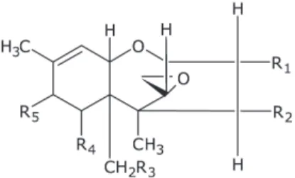
- Type-B (namely deoxynivalenol, 3- and 15-acetyldeoxynivalenol, nivalenol, fusarenon X): characterized by a carbonyl function at C-8 (Figure 7 and Table 6)
Among this group of mycotoxins deoxynivalenol is the most frequently occurring. The structure of trichothecenes is characterized by a sesquiterpene ring and a C-12,13-epoxide ring. T-2 toxin and diacetoxyscirpenol are soluble in non-polar solvents; deoxynivalenol or nivalenol are soluble in polar solvents like alcohol, but also in water.
figure 6 - Structural formula of type-A trichothecenes
Table 5 - Structural formula of type-a trichothecenes
Molecular
formula R1 R2 R3 R4 R5
Diacetoxyscirpenol C19H26O7 OH OAc OAc H H
T-2 toxin C24H34O9 OH OAc OAc H OCOCH2CH(CH3)2
figure 7 - Structural formula of type-B trichothecenes
Table 6 - Structural formula of type-B trichothecenes
Molecular formula R1 R2 R3 R4 Deoxynivalenol C15H20O6 OH H OH OH 3-Acetyldeoxynivalenol C17H22O6 OAc H OH OH 15-Acetyldeoxynivalenol C17H22O6 OH H OAc OH Nivalenol C15H20O7 OH OH OH OH Fusarenon X C17H22O8 OH OAc OH OH 1.7.2.1. Metabolism
Generally, there are three main metabolic pathways: I. Conjugation
II. De-epoxidation III. De-acetylation
The de-epoxidation is the most important step in the detoxification of trichothecenes and can be carried out by microorganisms in the gastrointestinal tract of ruminants.
1.7.2.2. Mechanism of action
Trichothecenes are potent inhibitors of protein, RNA and DNA synthesis and they can interact with the cell membrane. Trichothecenes bind to active polysomes and ribosomes, peptide linkages are interrupted, initiation and termination sequences are reduced, and the ribosomal cycle is disrupted. Their toxicity is primarily based on the 12,13-epoxyde ring. There are two types of the mechanism of protein inhibition:
- Inhibition of the initial step of protein synthesis (e.g.: T-2, HT-2, DAS) - Inhibition of the elongation-termination step (e.g.: DON)
Trichothecenes are especially toxic to tissues with a high cell division rate. Additionally, they are very cytotoxic to eukaryotic cells, causing cell lysis and inhibition of mitosis.
Deoxynivalenol enters the cell and binds to active ribosomes which transduce a signal to RNA-activated protein kinase (PKR) and hematopoeitic cell kinase (HCK). Subsequent phosphorylation of mitogen-actived protein kinases drives transcription factor (TF) activation apoptosis and resultant chronic and immunotoxic effects (modified from Pestka, 2007) (Figure 8).
Proinflammatory
genes
Immune
stimulation/inflammation Chronic effects; anorexia, reduced weight
gain
Tissue
loss / dysfunction Immunosuppression
Active polyribosomes Apoptosis MAPKs TFs Nucleus HCK Ribosomes Deoxynivalenol PKR TRICHOTHECENE DOSE D D D D Endoplasmic reticulum
HCK – Haematopletic cell kinase, PKR – RNA-activated protein kinase, MAPKS – Mitogen-activated protein kinase, TFS – Transcription factor
figure 8 - Mechanism of action of deoxynivalenol (D) (modified from Pestka et al., 2004 and Pestka, 2007)
1.7.2.3. Absorption/Residues
In general, absorption of deoxynivalenol occurs very rapidly within the digestive tract and is widely distributed in many tissues and organs. Residues of DON in pig tissues were reported from several experiments but at very low levels (Döll et al., 2008, Goyarts et al., 2007). 1.7.2.4. Toxicity
Many outbreaks of acute human disease like vomiting, gastrointestinal disorders, diarrhea or headaches have been attributed to consumption of Fusarium-contaminated grains. In animals, decrease in feed consumption (anorexia) and vomiting are two characteristic effects of deoxynivalenol contamination (Table 7).
Table 7 - Main systems affected by trichothecenes (all animal species are described in this table. for the effects of mycotoxins in swine, please see Chapter 2)
Affected system Effects/Signs/Symptoms
Circulatory system Hematopoietic effects (hemorrhages; blood pattern disorders) Immune system Immunosuppression (decreased resistance to environmental and microbial stressors; increased susceptibility to diseases) Digestive system Gastro-intestinal effects (gastroenteritis/inflammation of the rumen; vomiting; feed refusal) Reproductive system Decreased breeding efficiency (birth of smaller and unhealthy offspring) Nervous system Neurotoxic effects (restlessness; lack of reflexes; abnormal wings positioning; nervous syndrome) Skin Dermotoxicity (oral and dermal lesions; necrosis)
Pathological changes Necrosis of the lymphoid and hematopoietic tissues; gizzard lesions 1.7.3. OCHRATOXINS
Ochratoxins are metabolites of Aspergillus ochraceus and Penicillium verrucosum in temperate regions and are present in a large variety of feeds and foods. There are four ochratoxins (A, B, C, and D) and the major mycotoxin among this group is ochratoxin A (OTA). This toxin is a contaminant of cereals, beans and other plant products. The most significant effect of ochratoxins in farm animals is nephrotoxicity (Pfohl-Leszkowicz and Manderville, 2007). Chemically, ochratoxins contain an isocoumarin moiety linked by a peptide bond to phenylalanine.
1.7.3.1. Mechanism of action
OTA primarily affects the enzymes involved in phenylalanine metabolism. It inhibits the enzyme involved in the synthesis of the phenylalanine-tRNA complex. OTA might also interact with other enzymes that use phenylalanine as a substrate, for example, the phenylalanine hydroxylase which catalyzes the irreversible hydroxylation of phenylalanine to tyrosine. In addition, it alters the mitochondrial membrane transportation system; inhibits adenosine triphosphate (ATP) production, enhances membrane lipid peroxidation and superoxide and formation of hydrogen peroxide radicals.
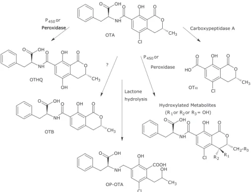
1.7.3.2. Metabolism
The major metabolite of ochratoxin is ochratoxin α, a hydrolysis product by the gut microflora without the phenylalanine moiety. Other metabolites are the hydroxylated derivates 4(R)-, 4(S)- and 10-OH-ochratoxins (Figure 9).
figure 9 - Metabolism of OTA (modified from Pfohl-Leszkowicz et al., 2007)
1.7.3.3. Absorption/excretion/residues
Between 40 and 66 % of ochratoxin is absorbed from the gastrointestinal tract depending on species. The small intestine has been shown to be the major site of absorption and maximal absorption occurs in the jejunum (Pfohl-Leszkowicz and Manderville, 2007).
OTA binds rapidly to serum albumin and is distributed in the blood mainly in the bound form. Generally, the toxin has a long biological half-life due to its high rate of binding to serum protein but differences exist between species. Therefore the ochratoxin protein adduct in serum can be used as a biomarker for ochratoxins’ exposure (Baldwin et al., 2011).
OTA primarily accumulates in the kidneys followed by the liver and muscles but also in whole blood and blood plasma (Battacone et al., 2010).
1.7.3.4. Toxicity
According to IARC ochratoxins are classified as possible human carcinogens (Group 2b). In animals, toxicity varies widely according to animal species and sex. The kidneys are the main target organs. Ochratoxin has been found to be responsible for porcine nephropathy, an extensively studied disease. Apart from the above mentioned effects ochratoxins may also affect other systems (Table 8).
Table 8 - Main systems affected by ochratoxins (all animal species are described in this table. for the effects of mycotoxins in swine, please see Chapter 2)
Affected system Effects/Signs/Symptoms
Circulatory system Hematopoietic effects (hematological disorders, blood in urine and feces) Nephrotoxic effects Increased water consumption; kidney and urinary bladder dysfunction Immune system Immunosuppression (decreased resistance to environmental and microbial stressors; increased susceptibility to diseases) Hepatotoxic effects Liver damage
Digestive system Gastro-intestinal effects (diarrhea) 1.7.4. FUMONISINS
Fumonisins are a group of mycotoxins mainly produced by Fusarium verticillioides and F. proliferatum. They were first isolated from cultures of Fusarium verticillioides in 1988 in South Africa. The most predominant member among this group is Fumonisin B1 (FB1). They mainly occur in maize. Fumonisins are highly polar compounds and soluble in water. 1.7.4.1. Mechanism of action
Fumonisins’ toxicity is based on the structural similarity to the sphingoid bases - sphingosine and sphinganine (Figure 10). They are inhibitors of sphinganine (sphingosine) N-acyltransferase (ceramide synthase), a key enzyme in the lipid metabolism, resulting in disruption of this pathway (Figure 11). This enzyme catalyzes the acylation of sphinganine in the biosynthesis of sphingolipids and also the deacylation of dietary sphingosine and the sphingosine that is released by the degradation of complex sphingolipids (ceramid, sphingomyelin and glycosphingolipide) (Wang et al., 1991).
Sphingolipids are important for the membrane and lipoprotein structure and also for cell regulations and communication (second messenger for growth factors) (Berg et al., 2003). Sphingosine is the backbone of sphingolipids (Merrill et al., 2001).
As a consequence of this disruption many bioactive intermediates are elevated, others reduced, as follows: a rapid increase of sphinganine (sometimes sphingosine), an increase of sphinganine degradation products like sphinganine 1-phosphate and a decrease of complex sphingolipids.
Free sphingoid bases are toxic to most cells by affecting cell proliferation and inducing apoptosis or necrotic cell death or by increasing the sphinganine 1-phosphate concentration in kidney and serum. The accumulation of sphinganine is associated with hepato- and nephrotoxic effects. Complex sphingolipids are important for cell growth regulation and also cell-cell interactions. Fumonisin B1 impairs the barrier function of endothelial cells in vitro. These adverse effects on endothelial cells could indirectly contribute to the neurotoxicity and pulmonary edema caused by fumonisins. Sphingosine 1-phosphate activates the endoplasmic reticulum calcium release and also acts as a ligand for extracellular receptors in the vascular system (S1P receptors) (Eaton et al., 2010).
figure 10 - Structures of fumonisin B1, sphinganine and sphingosine Ceramidase Ceramid Synthase Serine palmitoyltransferase Sphingosine-lyase Sphinganine-1-phosphates lyase Sphinganine-kinase Sphingosine-kinase Ceramide Ceramide FUM Sphingosine Sphingosine-1-phosphates Sphinganine-1-phosphates Free sphinganine Palmitoyl-CoA Serine + Lipid products Sphingomyelin Glycosphingolipids
figure 11 - Lipid metabolism and the inhibition of fumonisins. In order to simplify the graphics, only the main intermediates are described (modified from Merrill et al., 2001 and Voss et al., 2007)
The accumulation of free sphingoid bases (measured by the sphinganine:sphingosine ratio) in the serum and urine is a useful biomarker for the exposure of fumonisins and indicates the degree of sphingolipid metabolism disruption (Riley et al., 1993; Riley et al., 1994). 1.7.4.2. Exposure and absorption into the organism
Several studies indicate that fumonisins are poorly absorbed from the gastrointestinal tract and rapidly cleared from the blood. However, the absorbed fraction seems to undergo a wide distribution, with a high affinity to the liver and kidneys which later on slowly release the toxin (Prelusky et al., 1996). Fumonisins cause apoptosis followed by mitosis in affected tissues (Eaton et al., 2010).
1.7.4.3. Excretion and residues in animal products
Fumonisin carry-over in sow milk and pork meat may only occur after a high level of exposure over a longer period and then this mycotoxin can accumulate in the liver and kidneys (Völkel et al., 2011; Meyer et al., 2003).
1.7.4.4. Toxicity
According to IARC FB1 is classified as possible human carcinogen (Group 2B).
In the case of animals, horses are the most sensitive species to fumonisin toxicity. The mycotoxin causes a disease syndrome which is called equine leukoencephalomalacia (ELEM) and affects the central nervous system. Several studies have indicated that fumonisins cause porcine pulmonary edema (PPE).
Besides severe lung edema, liver injuries were also found in the exposed animals but fumonisins also cause a broad range of effects on other systems (Table 9).
Table 9 - Main systems affected by fumonisins (all animal species are described in this table. for the effects of mycotoxins in swine, please see Chapter 2)
Affected systems Effects/Signs/Symptoms
Immune system Immunosuppression (decreased resistance to environmental and microbial stressors; increased susceptibility to diseases) Digestive system Gastro-intestinal effects (diarrhea)
Circulatory system Hematopoietic effects (hematological disorders; increased concentration of hemoglobulin) Nervous system Neurotoxic effects
Hepatotoxic effects Liver damage
1.7.5. ZEARALENONE
Zearalenone (Figure 12) is an important mycotoxin occurring in warm and temperate climate regions. It is produced mainly by Fusarium graminearum and Fusarium culmorum on a variety of cereal crops.
figure 12 - Chemical structures of ZEN and its derivatives: zearalenone (ZEN), α–zearalenol (α-ZOL), β – zearalenol (β-ZOL), α-zearalanol (α-ZAL) and β-zearalanol (β-ZAL).
1.7.5.1. Metabolism/mechanism of action
Zearalenone is an estrogenic mycotoxin which is often involved in reproductive disorders and hyperestrogenicity in farm animals. The estrogenic effects are based on the structural similarity between zearalenone and estradiol. Estradiol is the most important female sex hormone in the group of estrogens.
The biotransformation of zearalenone takes place in two major pathways (Minervini and Dell’Aquila, 2008):
- Hydroxylation: Formation of α-zearalenol and β-zearalenol, assumed to be catalyzed by 3α- and 3β-hydroxysteroid dehydrogenases.
- Conjugation of zearalenone and its metabolites with glucoronic acid catalyzed by uridine diphosphate glucuronyl transferases.
The reduced form of zearalenone, α–zearalenol, has increased estrogenic effects. Several studies indicated that there are differences in biotransformation of zearalenone in various species, for example, pigs convert zearalenone predominately into α–zearalenol (Malekinejad et al., 2006).
The mycotoxin passes the cell membrane and binds to the estrogen receptor (Figure 13). This complex is transferred into the nucleus and binds there to specific nuclear receptors. Afterwards it generates estrogenic responses via gene activation resulting in the production of mRNAs that code for proteins which are normally expressed by receptor-estrogen complex binding (modified from Riley and Norred, 1996).
Z Z mRNA-ribosome complex Endocrine disruption Inhibitor of hydorxysteroid dehydrogenases Disruption of xeno/endobiotic metabolism Estrogen- or anti-estrogen-like responses Estrogen receptor mRNA Nucleus Z DNA
figure 13 - Simplified view of the mode of action of zearalenone (Z) (reviewed in Zinedine et al., 2008)
1.7.5.2. Exposure and absorption into the organism
Zearalenone is rapidly absorbed and metabolized in intestinal cells. In pigs ZEN and its metabolites show an extensive biliary excretion and enterohepatic cycling (Biehl et al., 1993). Studies investigating the carry-over of ZEN into meat and other edible tissues indicated that there is only limited tissue deposition of this mycotoxin. Transfer of ZEN and its major metabolites into serum was not detected after a administration of 56 μg ZEN per kg feed (Goyarts et al., 2007). 1.7.5.3. Toxicity
Zearalenone poses a relatively low acute toxicity and the oral LD50 values are between 2000– 20000 mg/kg bodyweight. Effects differ between different species (Table 10). Pigs seem to be more sensitive to the effects of zearalenone than others. Furthermore the toxin has shown haematotoxic effects, namely through changes in blood parameters.
Table 10 - Main systems affected by zearalenone (all animal species are described in this table. for the effects of mycotoxins in swine, please see Chapter 2)
Affected system Effects/Signs/Symptoms
Digestive system Gastro-intestinal effects (diarrhea) Reproduction system
Reproductive effects (feminization; enlargement of mammary glands; impaired semen quality; testicular atrophy; swollen prepuce, reddening and swelling of vulva)
Genes/Gene expression Teratogenic effects (splay legs) Pathological changes Atrophy of ovaries; uterus hypertrophy
1.7.6. ERGOT ALKALOIDS
The term ergot alkaloid refers to a diverse group of approximately forty different toxins which are formed by Claviceps sp. on grains such as triticale, corn, wheat, barley, oats, millet, sorghum and rice, and by fungal endophytes such as Neotyphodium sp. in grasses, particularly tall fescue and perennial ryegrass (Scott, 2009; Krska and Crews, 2008).
The ergot alkaloids constitute the largest known group of nitrogenous fungal metabolites and all have a common structure, a tetracyclic ergoline ring system of lysergic acid. Clavines are the simplest ergot alkaloids containing only an ergoline ring sytem (Figure 14 - top), whereas the ergopetides have an additional peptide moiety linked to the basic structure (Figure 14 - bottom). N N H H O CH3 H O N H R1 O N N O H OH R2 H
figure 14 - Structure of ergopeptines (Krska and Crews, 2008)
The main groups of natural ergot alkaloids (CAST, 2003; Panaccione, 2005): • Clavines – e.g.: agroclavine
• Lysergic acids
• Lysergic acid amides – e.g.: ergonovine (ergometrine, ergobasine), ergine
• Ergopeptines – e.g.: ergovaline, ergotamine, ergocornine, ergocristine, ergosine, ergocryptine The amount and pattern vary between fungal strains, host plant and climatic and geographical conditions (Hafner et al., 2008).
Ergot alkaloids appear as colorless crystals that are soluble in various organic solvents, but insoluble or only slightly soluble in water (EFSA, 2005). Moreover most alkaloids can form stereoisomers distinguished by the suffix –inines that are biologically less active, and easily convert back into the genuine –ine form (Flieger et al., 1997).
1.7.6.1. Absorption/excretion/residues
In general, little carry-over of ergot alkaloids into animal tissue has been reported. When a diet of 4 % ergot (containing ergopeptine alkaloids) was fed to pigs there was 90 % absorption
Ergoline ring system
Peptide ring system
}
1.7.6.2. Mechanism of action
The biological activity of the ergot alkaloids in animal systems is mainly due to structural similarities of the ergoline ring structure to the biogenic amines serotonin, dopamine, norepinephrine, and epinephrine (Berde, 1980; Weber, 1980). Due to this structural similarity many ergot alkaloids can bind to biogenic amine receptors and elicit such effects as decreased serum prolactin and vasoconstriction. Ergot alkaloids constitute a very diverse class of chemical compounds with widely different toxicological targets and activities, and potential routes of elimination and rates of clearance (Strickland et al., 2011).
1.7.6.3. Toxicity
Animals can be exposed to complex mixtures of alkaloids in many typical animal agriculture production systems. This exposure results from the fact that the kinds of alkaloids present and their levels can vary widely, depending on the fungal strain, the host plant and environmental conditions. Thus it is practically impossible to relate the exposure to individual toxins. Ergot alkaloid toxicoses in livestock due to consumption of ergot infected grains are widespread and result in disruption of several physiological systems (reproduction, growth, cardiovascular) within the body of an animal (Strickland et al., 2011) (Table 11). Ergot alkaloids exert toxic effects on all animal species, and the most prominent toxic signs can be attributed to the interaction of ergot alkaloids with adrenergic, serotinergic and dopaminergic receptors (EFSA, 2005).
Table 11 - Main systems affected by ergot alkaloids (all animal species are described in this table. for the effects of mycotoxins in swine, please see Chapter 2)
Affected system Effects/Signs/Symptoms Circulatory system
Vasoconstriction symptoms (elevated body temperatures, increased respiration rate), gangrenous changes in tissue of feet, tail and ear, reduced serum prolactin
Immune system Immunosuppression (decreased resistance to environmental and microbial stressors; increased susceptibility to diseases) Digestive system Gastro-intestinal effects (reduced body weight gain, feed refusal, diarrhea) Reproductive system Decreased breeding efficiency (lower conception rates, decreased survival rate of offspring, decreased piglet birth weight, abortions) Nervous system Neurotoxic effects (convulsions, hallucination, anorexia,), lameness Skin Dermatoxicity (oral and dermal lesions; necrosis), rough hair coat
1.8. Legislation in the european union (eu)
In the case of EU member states, Commission Directive 2003/100/EC of 31 October 2003 amending Annex I to Directive 2002/32/EC of the European Parliament and of the Council on undesirable substances in animal feed, and the Commission Regulation (EC) No 1881/2006 of 19 December 2006 setting maximum levels for certain contaminants in foodstuffs, are the basic regulations to be applied. Nevertheless, the regulatory agencies within each country are allowed to set higher standard rules. As for the regulations worldwide, the latter is also applicable. Table 12 shows the European regulations applied to feedstuffs destined for animal consumption.
Table 12 - Maximum concentration of aflatoxin B1 permitted in animal feedstuffs.
Source: Commission Directive 2003/100/EC of 31 October 2003 amending Annex I to Directive 2002/32/EC of the European Parliament and of the Council on undesirable substances in animal feed.
Countries Products intended for animal feed Mycotoxin
Maximum content in mg/kg [ppm] relative to a feedingstuff with a moisture content of 12 % EU member states: Austria, Belgium , Bulgaria, Cyprus, Czech Republic , Denmark, Estonia, Finland, France, Germany , Greece, Hungary, Ireland, Italy, Latvia, Lithuania, Luxembourg, Malta, The Netherlands, Poland, Portugal, Romania, Slovakia, Slovenia, Spain, Sweden, United Kingdom Iceland, Norway and Liechtenstein (following EU legislation)
All feed materials
Aflatoxin B1 0.02 Complementary and complete feed 0.01 With the exception of:
Compound feed for dairy cattle and calves, dairy sheep and lambs, dairy goats and kids, piglets and young poultry animals
0.005
Compound feed for cattle (except dairy cattle and calves), sheep (except dairy sheep and lambs), goats (except dairy goats and kids), pigs (except piglets) and poultry (except young animals)
0.02
In spite of the fact that directives do not exist for other mycotoxins of concern, the European Commission has set up some guidance levels in products intended for animal feeding (Table 13). Recommendations differ between regulations, directives and decisions, in that they are not legally binding for Member States, although they represent an instrument of indirect action
Table 13 – guidelines for the concentration of different mycotoxins in animal feedstuffs.
Source: Commission Recommendation (2006/576/EC) of 17 August 2006 on the presence of deoxynivalenol, zearalenone, ochratoxin A, T-2 and HT-2 and fumonisins in products intended for animal feeding.
Mycotoxin
Products intended for animal feed
Guidance value in mg/kg [ppm] relative to a feedingstuff with a
moisture content of 12 %
Deoxyni
valenol
Feed materials (*)
• Cereals and cereal products (**) with the
exception of maize by-products 8
• Maize by-products 12
Deoxyni
valenol
Complementary and complete feedingstuffs with
the exception of: 5
• Complementary and complete feedingstuffs
for pigs 0.9
• Complementary and complete feedingstuffs
for calves (< 4 months), lambs and kids 2
Zearalenone
Feed materials (*)
• Cereals and cereal products (**) with the
exception of maize by-products 2
• Maize by-products 3
Complementary and complete feedingstuffs • Complementary and complete feedingstuffs
for piglets and gilts (young sows) 0.1
• Complementary and complete feedingstuffs
for sows and fattening pigs 0.25
• Complementary and complete feedingstuffs for calves, dairy cattle, sheep (including lamb) and goats (including kids)
0.5
Ochratoxin A
Feed materials (*)
• Cereals and cereal products (**) 0.25
Complementary and complete feedingstuffs • Complementary and complete feedingstuffs
Mycotoxin
Products intended for animal feed
Guidance value in mg/kg [ppm] relative to a feedingstuff with a
moisture content of 12 % • Complementary and complete feedingstuffs
for poultry 0.1
Fumonisin B
1
+ B
2Feed materials (*)
• maize and maize products (***) 60
Complementary and complete feedingstuffs for:
Fumonisin
B1
+ B
2
• pigs, horses (Equidae), rabbits and pet
animals 5
• fish 10
• poultry, calves (< 4 months), lambs and kids 20 • adult ruminants (> 4 months) and mink 50
(*) Particular attention has to be paid to cereals and cereals products fed directly to the animals that their use in a daily ration should not lead to the animal being exposed to a higher level of these mycotoxins than the corresponding levels of exposure where only the complete feedingstuffs are used in a daily ration.
(**) The term ‘Cereals and cereal products’ includes not only the feed materials listed under heading 1 ‘Cereal grains, their products and by-products’ of the non-exclusive list of main feed materials referred to in part B of the Annex to Council Directive 96/25/EC of 29 April 1996 on the circulation and use of feed materials (OJ L 125, 23.5.1996, p. 35) but also other feed materials derived from cereals in particular cereal forages and roughages.
(***) The term ‘Maize and maize products’ includes not only the feed materials derived from maize listed under heading 1 ‘Cereal grains, their products and by-products’ of the non-exclusive list of main feed materials referred to in the Annex, part B of Directive 96/25/EC but also other feed materials derived from maize in particular maize forages and roughages.
1.9. Legislation in the united States of america (uSa)
In the USA, the Center for Veterinary Medicine of the Food and Drug Administration (FDA) also focuses on the same mycotoxins as the EU. There are action, guidance and advisory levels for aflatoxins, fumonisins and deoxynivalenol, respectively (Tables 14, 15 and 16). No action, guidance or advisory levels for ochratoxin A or zearalenone have been established by the FDA in animal feeds (these two mycotoxins are handled on a case-by-case basis).
Table 14 – action levels for total aflatoxins in livestock feed in mg/kg [ppm].
Source: www.fda.gov (Accessed: 11/01/2013)
Animals Feedingstuffs Aflatoxin levels
Finishing (i.e., feedlot) beef cattle corn and peanut products 0.3 Beef cattle,
swine, or poultry cottonseed meal 0.3
Finishing swine of 100 pounds (45 kg)
or heavier corn or peanut products 0.2
Breeding beef cattle, breeding swine, or mature poultry
corn and peanut products
0.1
Immature animals
corn, peanut products, and other animal feeds and feed ingredients, excluding cottonseed meal
0.02 Dairy animals, for animal species or
uses not specified above, or when the intended use is not known
corn, corn products, cottonseed meal, and other animal feeds and feed ingredients
0.02
Table 15 - guidance levels for total fumonisins in animal feeds in mg/kg [ppm].
Source: www.fda.gov (Accessed: 11/01/2013)
Animal Feedingstuffs and proportion in the diet
Levels in corn and corn
by-products
Levels in complete ration Equids and rabbits Corn and corn by-products (no more than 20 % of diet)** 5 1 Swine and catfish Corn and corn by-products (no more than 50 % of diet)** 20 10 Breeding ruminants breeding
poultry and breading mink*
Corn and corn by-products (no
more than 50 % of diet)** 30 15
Ruminants ≥ 3 months old being raised for slaughter and mink being raised for pelt production
Corn and corn by-products (no
more than 50 % of diet)** 60 30
Poultry being raised for slaughter
Corn and corn by-products (no
more than 50 % of diet)** 100 50
All other species or classes of livestock and pet animals
Corn and corn by-products (no
more than 50 % of diet)** 10 5
* Includes lactating dairy cows and hens laying eggs for human consumption ** Dry weight basis