HAL Id: hal-01764674
https://hal-amu.archives-ouvertes.fr/hal-01764674
Submitted on 29 Oct 2018
HAL is a multi-disciplinary open access archive for the deposit and dissemination of sci-entific research documents, whether they are pub-lished or not. The documents may come from teaching and research institutions in France or abroad, or from public or private research centers.
L’archive ouverte pluridisciplinaire HAL, est destinée au dépôt et à la diffusion de documents scientifiques de niveau recherche, publiés ou non, émanant des établissements d’enseignement et de recherche français ou étrangers, des laboratoires publics ou privés.
Distributed under a Creative Commons Attribution| 4.0 International License
Developmental origin and maintenance of distinct
testicular macrophage populations
Noushin Mossadegh-Keller, Rebecca Gentek, Gregory Gimenez, Sylvain Bigot,
Sebastien Mailfert, Michael Sieweke
To cite this version:
Noushin Mossadegh-Keller, Rebecca Gentek, Gregory Gimenez, Sylvain Bigot, Sebastien Mail-fert, et al.. Developmental origin and maintenance of distinct testicular macrophage populations. Journal of Experimental Medicine, Rockefeller University Press, 2017, 214 (10), pp.2829-2841. �10.1084/jem.20170829�. �hal-01764674�
IntroductIon
Macrophages are innate immune cells that are present in es-sentially all organs of the body. Besides their diverse role in shaping the immune response, they have trophic functions in tissue homeostasis and regeneration and contribute to many organ-specific functions (Wynn et al., 2013), including in the gonads (Shechter et al., 2013; Smith et al., 2015).
In males, the testis are the site of spermatogenesis and male hormone production and can maintain fertility and vi-rility over an extended reproductive period. Testicular mac-rophages (tMφ) represent the most abundant immune cells in the organ (Winnall and Hedger, 2013) and contribute to organ function in several ways. As spermatogenesis is only initiated at puberty and sperm-specific neoantigens become expressed after the education of the immune system, the testis requires a mechanism of self-tolerance to these anti-gens. TMφ contribute to maintaining an immunoprivileged environment in the organ by the production of immuno-suppressive cytokines, such as IL-10 and TGF-β, and by low expression of proinflammatory cytokines (Fijak and Mein-hardt, 2006; Shechter et al., 2013). TMφ are also intimately associated with Leydig cells and play an important role in their development, regeneration, and testosterone production (Hutson, 1998; Smith et al., 2015). Whereas this macrophage population resides in the interstitial space between
seminif-erous tubes, a peritubular population has recently been iden-tified that is associated directly with spermatogonial tubes in proximity to spermatogonial stem cells (SSCs). This has been suggested to have an effect on the SSC niche and a potential functional role in SSC differentiation (DeFalco et al., 2015).
Despite these proposed important functional roles, relatively little is known about the development and main-tenance of tMφ. It was originally thought that tissue mac-rophages were principally derived from blood monocytes developing from hematopoietic stem cells in the BM (van Furth and Cohn, 1968), but this view has changed when dif-ferent genetic-, genomic-, and fate-mapping studies showed that macrophages of different organs can also be derived from embryonic progenitors (Ginhoux et al., 2010; Hoeffel et al., 2012; Schulz et al., 2012; Hashimoto et al., 2013; Yona et al., 2013; Gomez Perdiguero et al., 2015; Hagemeyer et al., 2016; Mass et al., 2016), which seed the organs at different stages of development and self-maintain independently of BM con-tribution into adulthood (Ajami et al., 2007; Hashimoto et al., 2013; Sieweke and Allen, 2013; Gomez Perdiguero et al., 2015). Autonomous macrophage self-renewal capacity de-pends on a network of specific self-renewal genes shared with stem cells (Aziz et al., 2009; Soucie et al., 2016). Depending on the organ, embryonic-derived macrophages can get
par-testicular macrophages (tMφ) are the principal immune cells of the mammalian testis. Beyond classical immune func-tions, they have been shown to be important for organogenesis, spermatogenesis, and male hormone production. In the adult testis, two different macrophage populations have been identified based on their distinct tissue localization and morphology, but their developmental origin and mode of homeostatic maintenance are unknown. In this study, we use genetic lineage–tracing models and adoptive transfer protocols to address this question. We show that embryonic pro-genitors give rise to the interstitial macrophage population, whereas peritubular macrophages are exclusively seeded postnatally in the prepuberty period from bone marrow (BM)–derived progenitors. As the proliferative capacity of inter-stitial macrophages declines, BM progenitors also contribute to this population. once established, both the peritubular and interstitial macrophage populations exhibit a long life span and a low turnover in the steady state. our observations identify distinct developmental pathways for two different tMφ populations that have important implications for the further dissection of their distinct roles in organ homeostasis and testicular function.
Developmental origin and maintenance of distinct
testicular macrophage populations
Noushin Mossadegh-Keller,
1Rebecca Gentek,
1Gregory Gimenez,
1,2Sylvain Bigot,
1Sebastien Mailfert,
1and Michael H. Sieweke
1,21Centre d'Immunologie de Marseille-Luminy, Aix Marseille University, Centre National de la Recherche Scientifique, Institut National de la Santé et de la Recherche
Médicale, Marseille, France
2Max-Delbrück-Centrum für Molekulare Medizin in der Helmholtzgemeinschaft, Berlin, Germany
Correspondence to Michael H. Sieweke: [email protected]
Abbreviations used: HS/PC, hematopoietic stem and progenitor cell; IF, immu-nofluorescence; SSC, spermatogonial stem cell; TAM, tamoxifen; tMϕ, testicular macrophages.
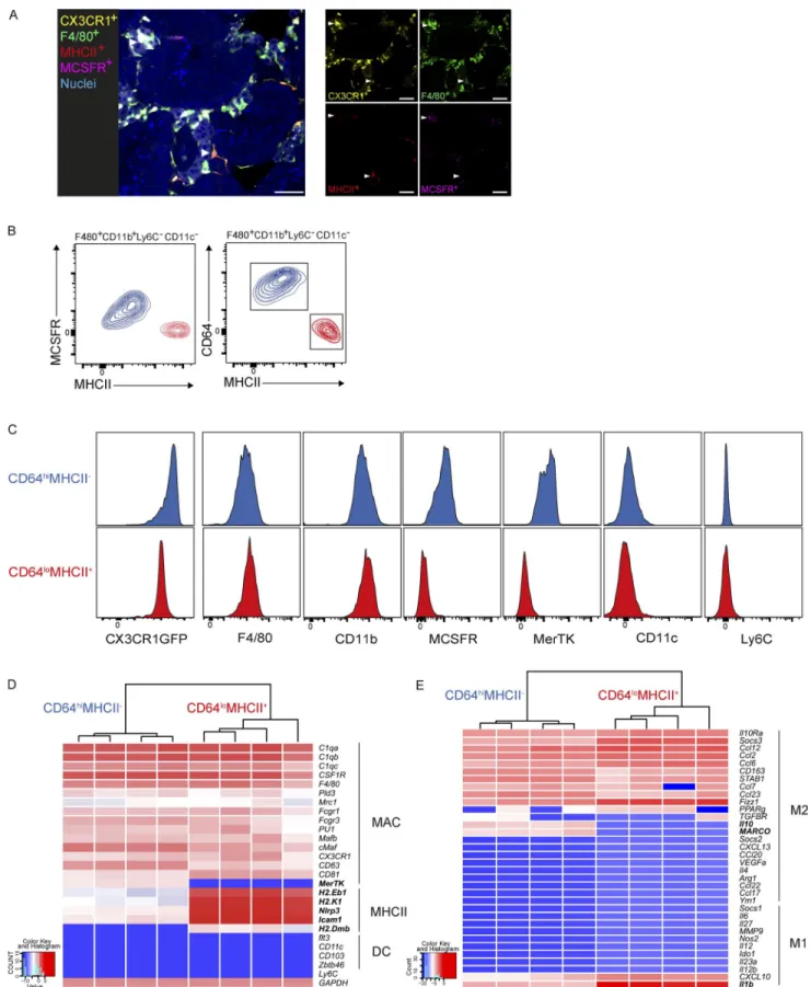
Figure 1. Phenotypic characterization of two tMφ populations by cell-surface marker and gene expression profiling. (A) IF imaging of 16-wk-old CX3CR1-GFP mouse testis showing morphology, localization, M-CSFR, and MHC II staining of interstitial tMφ and peritubular tMφ, indicated with arrow-heads. Bars, 40 µm. (B) Representative FACS profiles of adult mouse F4/80+CD11b+Ly6C−CD11c− tMφ populations showing that both CD64 and M-CSFR
tially or fully replaced by BM-derived macrophages in the adult tissues (Tamoutounour et al., 2012; Bain et al., 2014; Epelman et al., 2014; Molawi et al., 2014). For example, we have shown previously that embryo-derived cardiac macro-phages loose self-renewal capacity with age and are succes-sively replaced by BM-derived macrophages in adulthood (Molawi et al., 2014). Here, we have now characterized post-natal development and the origin of tMφ.
During the initial phases of testis morphogenesis in early embryonic development from E10.5 to E14.5, yolk sac–derived macrophages are associated with nascent gonadal and mesonephric vasculature and are required for vascular re-modeling (DeFalco et al., 2014). In the adult testis, two dif-ferent macrophage populations have been distinguished by their morphology, their M-CSFR and MHC II expression profiles, and their localization in the interstitium or the sur-face of seminiferous tubules (DeFalco et al., 2015). However, it is not known whether these two macrophage populations are derived from early embryonic tMφ or whether they have distinct origins and arise independently. Here, we examined the postnatal development dynamics, the turnover, and the specific origin of these two tMφ populations. Combining ge-netic lineage tracing and a neonatal adoptive transfer model, we demonstrate that embryo-derived macrophages persist into the postnatal testis, where they selectively contribute only to the interstitial population but are partially replenished from BM-derived progenitors at later stages, when local ex-pansion has declined. Peritubular tMφ, however, only emerge postnatally from BM-derived progenitors in the prepuberty period. Once established, the two populations exhibit long life span and low turnover in the adult testis at steady state.
results And dIscussIon
We initially characterized the macrophage populations pres-ent in testis by immunofluorescence (IF). Consistpres-ent with previous observations (DeFalco et al., 2015), we observed that tMφ were positive for CX3CR1 and F4/80 but that two populations could be distinguished based on their per-itubular and interstitial locations, which were M-CSFR pos-itive and MHC II negative or M-CSFR negative and MHC II positive, respectively (Fig. 1 A). We further characterized these two interstitial (M-CSFR+MHC II−) and peritubular
(M-CSFRloMHC II+) tMφ populations by flow cytometry
(FACS), using an extended antibody panel that excluded infiltrating leukocytes (Fig. S1). Within the testicular leuko-cytes fraction (CD45+), resident macrophages were defined as
Ly6C− CD11c− F4/80+ CD11b+ cells (Fig. S1). We also
ana-lyzed the expression of additional tissue-resident macrophage markers CX3CR1, CD64, and MerTK (Gautier et al., 2012). We found that interstitial (MCS FR+MHC II−) and
peritu-bular (M-CSFRloMHC II+) tMφ could be interchangeably
and clearly distinguished by differential expression of CD64 and MHC II (Fig. 1, B and C). The two populations also ex-pressed differential levels of MerTK but were both positive for CX3CR1. Although both populations were negative for CD11c (Fig. 1 B), the absence of the core macrophage markers CD64 and MerTK in peritubular CD64loMHC
II+ macrophages prompted us to analyze macrophage- and
DC-specific gene expression in the two populations in more depth. We investigated the expression of several core macro-phage-associated factors (C1qa, C1qb, C1qc, CSF1R, F4/80, Pld3, Mrc1, Fcgr1, Fcgr3, Pu1, Mafb, cMaf, CXC3CR1, CD81, CD63, and MerTK), factors regulating antigen pro-cessing and presentation (H2.Dmb, H2.Eb1, Nlrp3, H2.K1, and Icam1; Epelman et al., 2014), and DC markers (CD103, Zbtb46, Flt3, and CD11c), using nanofluidic Fluidigm array real-time PCR (Fig. 1 D). Both M-CSFR+CD64hiMHC II−
interstitial and M-CSFRloCD64loMHC II+ peritubular
mac-rophages expressed all analyzed macrophage markers, except MerTK, which was absent in CD64loMHC II+ macrophages.
In contrast, the expression of genes regulating antigen pro-cessing and presentation was up-regulated in this population. The DC markers tested were not expressed by either pop-ulation, clearly identifying both of them as macrophages. As spermatogenesis occurs after the maturation of the immune system and the establishment of systemic tolerance, the devel-oping gametes in the testis need to be protected from auto-immune attack. Establishment and maintenance of auto-immune tolerance in the testis has, at least in part, been attributed to an antiinflammatory phenotype of tMφ (Fijak and Meinhardt, 2006; Shechter et al., 2013), but it has not been established whether both tMφ populations have an immunosuppressive phenotype and which pathways might be differentially en-gaged. Therefore, we analyzed CD64hiMHC II− interstitial
and CD64loMHC II+ peritubular tMφ populations for the
expression of proinflammatory genes characteristic of a classi-cal M1-type activation or several immunosuppressive IL-10–, TGF-β-, and IL-4–dependent alternative M2-type activation
pathways (Murray et al., 2014). The two populations showed highly similar gene expression profiles with the absence of proinflammatory gene expression but the expression of a large number of immunosuppressive and alternative M2-type
acti-staining distinguishes CD64hi or M-CSFR+MHC II− (interstitial; blue) and CD64lo or M-CSFR−MHC II+ (peritubular; red) populations in adult testes. (C) Analysis
of expression levels of core macrophage cell-surface markers in CD45+F4/80+CD11b+ tMφ for CX3CR1-GFP levels and in CD45+CX3CR1GFP+ for F4/80,
CD11b, MCS FR, and MerTK in interstitial CD64hiMHC II− and peritubular CD64loMHC II+ tMφ. (A–C) Data are representative of two independent experiments.
(D) Gene expression analysis of interstitial (CD64hiMHC II−) and peritubular (CD64loMHC II+) tMφ isolated from four individual wild-type mice. Expression
lev-els of macrophage-related genes, genes regulating antigen processing and presentation, and DC-related genes were determined using nanofluidic Fluidigm array real-time PCR. MAC, macrophage. (E) Gene expression analysis on the same populations as in D for M2 and M1 macrophage activation-related genes by nanofluidic Fluidigm array real-time PCR. Genes in bold show the difference between the two testicular populations.
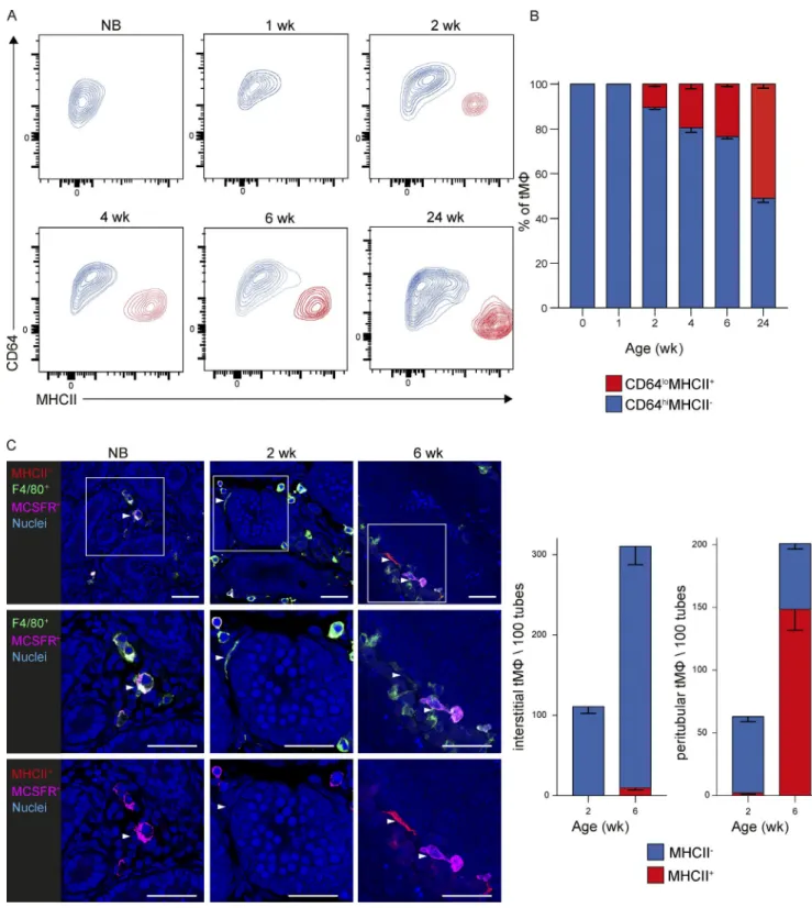
Figure 2. tMφ populations dynamically change during postnatal development. (A and B) Analysis of the proportion of CD64hiMHC II− interstitial and
CD64loMHC II+ peritubular tMφ from mice of the indicated ages, showing representative FACS plots (A) and mean percentage (B). Error bars represent SEM. n
= 4–5. All data are representative of at least two independent experiments with four to eight individual mice each. NB, newborn. (C) Analysis of localization and relative numbers of interstitial and peritubular tMφ by IF for M-CSFR and MHC II expression during postnatal development from newborn to 2- and 6-wk-old mice. Nuclear staining by Sytox blue and antigens is shown by the indicated colors. Bars, 40 µm. Representative images (left) and quantification of relative numbers and MHC II expression in interstitial and peritubular populations (right) are shown. Arrowheads show an interstitial macrophage localized in the interstitial space with expression by IF of F4/80 and MCS FR (newborn; left), show peritubular macrophage localization surrounding the tube and
vation genes in both populations (Fig. 1 E). As an exception to this, the expression of only a few genes differed, such as the immunosuppressive M2-type genes IL-10 and macrophage receptor with collagenous structure (MAR CO), which were more highly expressed in CD64hiMHC II− interstitial
macro-phages, and IL-1b, which was increased in CD64lo MHC II+
peritubular macrophages. Thus, phenotypic characterization by cell-surface markers and mRNA gene expression profiling clearly and consistently identified two distinct interstitial and peritubular tMφ identities.
To investigate the postnatal development of the intersti-tial and peritubular tMφ populations, we analyzed CD64 and MHC II expression in tMφ from newborn to 24-wk-old mice (Fig. 2, A and B). We observed that all tMφ present at birth were uniformly CD64hiMHC II− (Fig. 2, A and B). This was
consistent with IF analysis showing exclusive interstitial local-ization and expression of M-CSFR but absence of MHC II in tMφ both at birth and at later time points (Fig. 2 C). MHC II+
macrophages emerged only after 2 wk of life and further in-creased thereafter both by FACS and IF analysis (Fig. 2, A–C). IF and confocal imaging analysis allowed a clear distinction of the interstitial macrophage populations with a spherical shape from the peritubular population with an elongated morphol-ogy. Interestingly, peritubular macrophages emerged at 2 wk but mostly did not express MHC II yet, whereas a majority was positive at 6 wk, suggesting a potential maturation step after localization to this niche (Fig. 2 C).
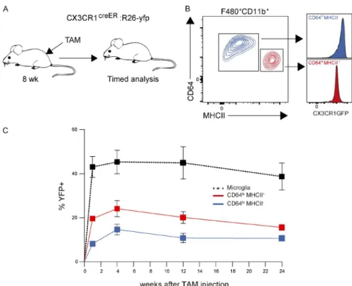
Because our data revealed two clearly distinct tMφ pop-ulations with different tissue localization, morphology, and surface marker expression emerging at different times during postnatal developmental, we hypothesized that they might also have a distinct developmental origin. To address this question and investigate a potential embryonic origin of the differ-ent tMφ populations, we used a genetic fate–mapping model based on CX3CR1-driven tamoxifen (TAM)-inducible Cre recombinase (CreER) and R26-yfp reporter mice (Fig. 3 A;
Yona et al., 2013; Molawi et al., 2014). TAM-induced pulse labeling at E9 in this model allows identifying cells derived from CX3CR1+ yolk sac macrophage progenitors
(Hage-meyer et al., 2016) before the onset of definitive hematopoi-esis. As a positive control, we used YFP+ labeling in microglia,
which are exclusively derived from yolk sac macrophage pro-genitors, remain CX3CR1+ throughout development, and
self-maintain without contribution of hematopoietic stem cell–derived cells (Ginhoux et al., 2010; Kierdorf et al., 2013). They therefore represent an internal control for maximal CX3CR1 labeling efficiency. E9 pulse labeling with TAM showed a median contribution of 5% from embryonic macro-phage progenitors to tMφ in newborn mice, compared with a median of 25% in microglia cells (Fig. 3 B). Using values
normalized to microglia to control for interindividual varia-tion in labeling efficiency, we observed that embryonic pro-genitor contribution to tMφ after birth declined from 20% in neonates to 5% in 6-wk-old mice (Fig. 3 B and S3), similar to what has been reported for other organs (Bain et al., 2014; Molawi et al., 2014; Ensan et al., 2016). IF imaging confirmed that YFP+ embryonic progenitor-derived tMφ decreased over
time (Fig. 3 C). To determine whether this decline was caused by a decrease in macrophage self-renewal potential over time, as has been observed before in other organs (Bain et al., 2014; Molawi et al., 2014), we determined the proportion of tMφ in S-phase by BrdU incorporation. We observed a strong de-cline in proliferative capacity of tMφ with age from 8% in neonates to 0% at adulthood (Fig. 3 D). By analyzing BrdU incorporation in YFP+ tMφ from E9 pulse-labeled mice, we
confirmed that this was also the case specifically for embry-onic-derived macrophage progenitor tMφ, which showed a decline from 3% in 2-wk-old mice to 0.5% in 6-wk-old mice of cells in S-phase (Fig. 3 E). Next, we determined to which tMφ subpopulation embryonic macrophage progeni-tors could contribute. Interestingly, all embryo-derived YFP+
tMφ at 6 wk exclusively belonged to the CD64hiMCH II−
population and showed no contribution to CD64loMCH II+
tMφ in FACS analysis (Fig. 3 F). IF confirmed that YFP+
cells contributed only to the interstitial M-CSFR+MCH II−
population but not to M-CSFRloMCH II+ peritubular tMφ
(Fig. 3 G). These data demonstrated that embryo-derived macrophages persisted in mouse testis after birth but were restricted to the interstitial CD64hiM-CSFR+MCH II− tMφ
population and declined with age.
The lack of embryonic progenitor-derived macro-phages in the CD64loMCH II+ peritubular population (Fig. 3)
and their emergence only 2 wk after birth (Fig. 2) suggested that this population developed from definitive hematopoiesis in the juvenile animal. Furthermore, the decline of the em-bryonic progenitor-derived CD64hiM-CSFR+MCH II−
in-terstitial tMφ population could also be caused by a successive replenishment from definitive hematopoiesis-derived cells in the growing animal. To investigate such a potential BM origin of tMφ populations, we established a novel adoptive transfer protocol in neonatal mice (Fig. 4 A). Because the peritubular population appears only 2 wk after birth in the prepuberty period of organ development (Fig. 3 A), classical methods such as BM transplantation into myeloablated mice or parabiosis were not feasible. Therefore, we took a different approach and exploited the fact that the neonatal liver still significantly contributes to hematopoiesis around birth be-fore hematopoietic stem cells transition from this organ to the BM (Orkin and Zon, 2008). We transplanted BM cells directly into the neonatal liver (Hoeffel et al., 2012) without
expressing F4/80 but not MCS FR (2 wk; middle), and display one peritubular macrophage expressing F4/80 and MHC II and an interstitial macrophage with expression of F4/80 and MCS FR (6 wk; right). Data are based on counting 100–1,000 F4/80+ cells normalized to 100 seminiferous tubules and represented
Figure 3. embryonic macrophage contribution to tMφ populations in the postnatal testis. (A) Experimental design. CX3CR1CreER :R26 -yfp embryos
were treated with TAM at E9 and analyzed on the day of delivery (newborn [NB]) or 2 or 6 wk after birth. (B) Relative abundance of lineage-traced YFP+
conditioning, which represents largely physiological condi-tions during a period of normal organ growth. We transferred BM from mice expressing dTomato from the ubiquitin locus (Ghigo et al., 2013) intrahepatically into newborn pups and analyzed the contribution of BM to tMφ populations 2, 6, and 20 wk after transplantation (Fig. 4 A). 1 wk after trans-plantation, dTomato+ cells contributed to the hematopoietic
stem and progenitor cell (HS/PC) compartment in the BM, confirming the potential of the adoptive transfer protocol to contribute to BM hematopoiesis (Fig. 4 B). We found that transplanted dTomato+ BM already contributed to 14% of
CD64loMHC II+ peritubular macrophages in 2-wk-old
mice and to 30% in 6-wk-old mice and further increased in 20-wk-old mice (Fig. 4 C). In contrast, contribution of transplanted dTomato+ BM to the CD64hiMHC II−
intersti-tial population was negligible at 2 wk, only at 3% by 6 wk, and at 5% at 20 wk (Fig. 4 C). Confocal imaging analysis confirmed that transplanted BM-derived macrophages local-ized only to the peritubular location at 2 wk further increased at this location by 6 wk and were nearly exclusively MHC II+. No contribution to interstitial macrophages was
ob-served at 2 wk, and only minimal numbers were obob-served at 6 wk (Fig. 4 D). Together, these observations confirmed that CD64loMHC II+ peritubular tMφ were exclusively BM derived
and that the decline of early embryo-derived macrophages in the CD64hiMHC II− interstitial population was partially
com-pensated by replenishment from BM-derived cells.
Next, we determined the turnover rate of the two tMφ populations once they were stably established in the adult testis. As both CD64hiMHC II− interstitial and CD64loMHC II+
peritubular populations are CX3CR1 positive, we could use CX3CR1CreER :R26 -yfp mice to pulse label both populations
in adult mice at 8 wk of age by TAM injection (Goldmann et al., 2013) and follow YFP+ cell contribution to both tMφ
populations over time (Fig. 5, A and B). FACS showed a nearly constant contribution of YFP+ cells to both CD64hiMCH II−
interstitial and CD64loMCH II+ peritubular tMφ up to 24 wk
after labeling, similar to the stable contribution to microglia cells that served as a positive control (Figs. 5 C and S2).
Here, we have characterized the origin, postnatal devel-opment, and turnover of two testis-resident macrophage
pop-ulations. We identified clearly distinct developmental pathways for the peritubular and interstitial tMφ populations that have been associated with distinct support roles for the function of SSCs (DeFalco et al., 2015) and testosterone-producing Ley-dig cells (Hutson, 1998; Smith et al., 2015). Macrophages have also been suggested to contribute to the immunosuppressive environment in the testis (Fijak and Meinhardt, 2006; Shech-ter et al., 2013). Here, we demonstrate that both tMφ popula-tions lack the expression of proinflammatory M1-type genes and express a subset of M2-type polarization genes. Despite the overall similarity, interstitial tMφ might make a stronger contribution because of significantly higher IL-10 expression. We have shown that early embryonic progenitors contribute selectively to the interstitial but not to the peritubular pop-ulation. Similar to other resident macrophages (Bain et al., 2014; Molawi et al., 2014), the interstitial population shows a decline in self-renewal and a reduction in the contribution of early embryo-derived cells with age. This loss is partially compensated by replenishment from BM-derived macro-phages. As this contribution remains relatively small in the adult, the replacement of early embryo-derived cells might already occur early after birth from fetal sources of hemato-poiesis. In contrast, the peritubular population only becomes established at 2 wk of age in the prepuberty period and is derived exclusively from BM progenitors. Once established, both populations exhibit a slow turnover and extraordinary long life span in the steady state.
Our observations on peritubular tMφ provide a unique example of a functionally and phenotypically distinct resi-dent macrophage population that develops exclusively from BM-derived progenitors to populate a dedicated niche that only becomes available after birth. Most macrophage pop-ulations are of mixed origin because of successive waves of seeding of the developing organ during embryogenesis and after birth from different yolk sac, fetal liver, and BM sources of hematopoiesis (Sieweke and Allen, 2013; Ginhoux and Guilliams, 2016; Perdiguero and Geissmann, 2016). The inter-stitial tMφ described here fall into this category with mixed embryonic and BM origin. At one extreme end of this spec-trum, microglia cells of the brain are exclusively derived from early yolk sac–derived progenitors and do not receive
addi-cells normalized to microglia labeling efficiency at the indicated time points (right). Bars show the median. n = 6–8. (C) Analysis of the contribution of fate-mapped YFP+ cells to F4/80+ tMφ by IF, showing a representative example from a 2-wk-old mouse (left) and quantification of contribution in 2- and
6-wk-old mice (right). Data are based on counting 100–200 YFP+F4/80+ cells normalized to 100 seminiferous tubules. Arrowheads show one YFP+F480+
testicular macrophage (top) and the same cell with only YFP labeling, proving that we can visualized our fate-mapping cells (bottom). Bars, 40 µm. (D) Analysis of proliferation rate of tMφ at the indicated age by BrdU incorporation and flow cytometry 4 h after BrdU injection (i.p), showing representative FACS plots (left) and quantification (right). Bars show the median. n = 4–12. NB, newborn. (E) Analysis of the proliferation rate of fate-mapped YFP tMφ (F4/80+CD11b+Ly6C−CD11c−) in 2- and 6-wk-old mice by BrdU incorporation and flow cytometry 4 h after BrdU injection (i.p.). Bars show the median.
n = 8. (F) Analysis of contribution of fate-mapped YFP+ cells to CD64hiMHC II− interstitial and CD64loMHC II+ peritubular macrophages in 6-wk-old mice,
showing a representative FACS plot (left) and quantification expressed as the median (right). n = 8. (B and D–F) Data were pooled from two independent experiments. (G) Contribution of fate-mapped YFP+ cells to interstitial and peritubular tMφ populations in 6-wk-old mice determined by IF showing
representative examples (left) and quantification of cells normalized to 100 seminiferous tubules. Arrowheads show an interstitial fate-mapped YFP+
macrophage localized in the interstitial space with expression by IF of F4/80 and M-CSFR. Bars, 40 µm. Error bars represent SEM. P-values were obtained by Mann-Whitney U tests. *, P ≤ 0.05; ***, P ≤ 0.0007.
Figure 4. BM contribution to tMφ populations. (A) Experimental design. Total dTomato+ BM cells were adoptively transferred by intrahepatic injection
into newborn mice and analyzed for contribution to tMφ populations 2, 6, and 20 wk after transplantation. (B) Contribution of dTomato+ BM cells to the
hema-tional input from other hematopoietic sources in the steady state. Here, we now show that the other extreme also exists for a macrophage niche that only becomes available about 2 wk after birth, when all sources of embryonic hematopoiesis have subsided, and then becomes exclusively populated from BM-derived macrophages. This late seeding is not associated with a short half-life or high turnover that has been described for some short-lived monocyte-derived macrophages that have been labeled as passenger macrophages (Perdiguero and Geissmann, 2016). Therefore, despite their late emergence and BM origin, peritubular tMφ have to be considered a long-lived resident macrophage population.
The testis is unique in that functional maturation and important aspects of organ development only occur after birth in the prepuberty period. M-CSF–deficient mice have reduced numbers of tMφ, defective spermatogenesis, and are infertile (Cohen et al., 1996). Diphtheria toxin–mediated depletion experiments also suggest that macrophages might promote spermatogenesis, possible by directly acting on SSCs found in close proximity to peritubular macrophages (DeFalco et al., 2015). Peritubular macrophages might also
play a unique tolerizing role against meiotic germ cell auto-antigens that are shed during spermiation. Because a subset of them can pass the Sertoli cell barrier into the intersti-tial space (Tung et al., 2017), the close proximity of per-itubular tMφ and their high expression levels of MHC II might predispose them to present autoantigen to regulatory T cells. We now observed that macrophages only start to populate this specific peritubular location when spermato-genesis is initiated at 2–3 wk of age, although the physical structures of seminiferous tubules are already present long before. This suggests that macrophages might not only affect spermatogonial differentiation and tolerance against autoan-tigens, but also that, reversely, initiation of spermatogenesis might induce a specific environment enabling the recruit-ment of a dedicated macrophage population. Together, this highlights the power of the microenvironment in con-trolling macrophage identity and numbers (Sieweke, 2015; Guilliams and Scott, 2017). It also suggests an intimate link between specific support macrophage and parenchymal cell populations of the organ that reciprocally influence each other’s identity and function.
topoietic stem cell; KSL, Kit+Sca-1+Lineage−. (C) Contribution of neonatally transplanted dTomato+ BM cells to CD64hiMHC II− interstitial and CD64loMHC II+
peritubular macrophage populations, assessed by flow cytometry at 2, 6, and 20 wk of age, showing FACS plot examples (left) and quantification (right). Ly6C+ blood monocyte and BM hematopoietic stem cell contributions are shown as references. Data were pooled from two independent experiments for
2- and 6-wk-old mice (n = 7–9) and are representative of two independent experiments for 20-wk-old mice (n = 4). (D) IF imaging analysis of transplanted dTomato+ BM contribution to interstitial and peritubular tMφ in 2- and 6-wk-old mice showing representative images (left) and quantification of MHC II
expression (right). Arrowheads show peritubular macrophages after BM transplantation, labeled in tomato and showing or not expressing MHC II. Data are based on counting 100–300 dTomato+ F4/80+ cells normalized to 100 seminiferous tubules. Errors bars represent SEM. Bars, 40 µm.
Figure 5. tMφ populations are long
lived. (A–C) Analysis of tMφ turnover in adult mice using TAM pulse labeling of CD64hiMHC II− interstitial and CD64loMHC
II+ peritubular macrophage populations in
8-wk-old CX3CR1CreER :R26 -yfp mice, showing
experimental design (A), CX3CR1 expression in both populations (B), and contribution of labeled YFP+ cells to CD64hiMHC II− and
CD64loMHC II+ populations at the indicated
time points, with microglia contribution shown as a positive control (C). Error bars represent SEM. n = 4–5. Data are representative of two independent experiments.
MAterIAls And Methods Mice
Wild-type C57BL/6J mice were purchased from Janvier laboratories. All transgenic mice used in this study have a C57BL/6 background. CX3CR1GFP/+ mice (Jung et al.,
2000), Rosa26-yfp and CX3CR1creER mice (JAX stock no.
20940 B6J.129-Cx3cr1<tm1.1[cre/ ERT2]Jung>/J; Yona et al., 2013), and Ubow mice, provided by M. Bajenoff (Centre d'Immunologie de Marseille-Luminy, Marseille, FranceGhigo et al., 2013) were used. Experiments were performed on mice at the indicated age. In vivo procedures were performed under specific pathogen–free conditions following protocols approved by the Ethics Committee of Marseille in accor-dance with institutional, national, and European regula-tions (approval nos. APA FIS 3292-2015 1221 09359224 and APA FIS 10545-2017 0710 08253541).
Genetic fate mapping
To genetically fate map progeny of cells expressing CX3CR1, CX3CR1CreER mice were crossed onto R26-yfp mice, and
pregnant CX3CR1CreER :R26 -yfp females were administered
a single dose of TAM (Sigma) at E9. TAM was complemented with progesterone (Sigma), dissolved in ethanol and diluted 10-fold in corn oil (Sigma). The freshly prepared solution was injected intraperitoneally at a dose of 0.1 mg TAM and 0.05 mg progesterone per gram body weight. For induction of Cre recombinase in 8-wk-old CX3CR1CreER R26-yfp mice,
4 mg TAM, dissolved in 200 µl of corn oil, was injected twice subcutaneously 48 h apart.
neonatal hematopoietic cell transplantation
Total BM was obtained from long bones according to stan-dard procedures. Adult (8–12 wk) Ubow mice that express dTomato under the control of the human ubiquitin–C pro-moter (Ghigo et al., 2013) were used as donors for BM cells. RBCs from total dTomato BM were lysed using RBC lysis buffer (CliniSciences). Five million cells were injected in 20 µl PBS intrahepatically into newborn pups using a micro-fine syringe with 8-mm needles (Micro-Fine 0.3 ml; BD).
In vivo Brdu incorporation
To assess DNA synthesis as readout for proliferation, mice were pulsed with BrdU (Sigma) for 4 h before euthaniza-tion and organ harvest. Mice were administered 0.1 mg BrdU per gram body weight either subcutaneously (newborn pubs) or intraperitoneally. BrdU incorporation was mea-sured using a BrdU Flow kit (BD) according to the manu-facturer’s instructions.
leukocyte isolation from mouse tissues
The tunica albuginea of resected testes was opened, and cells were transferred into RPMI medium containing 1 mg/ml Collagenase II (Worthington Biochemicals) and 0.15 mg/ ml DNase I (Sigma). Testis-resident leukocytes were obtained through enzymatic digestion at 37°C for 40 min and passage
through 70-µm cell strainers (BD Falcon). Brain tissue was macerated and taken up in HBSS containing 0.5% d-glucose (Sigma) and 15 mM Hepes (Life Technologies). The resulting cell suspension was passed through 70-µm cell strainers and subjected to a 70/37/30% Percoll gradient, from which mi-croglia were isolated. Peripheral blood leukocytes were isolated by density centrifugation using Lymphoprep reagent (Stem Cell Technologies) following the manufacturer’s instructions.
Flow cytometry
Single-cell suspensions were preincubated with an anti-body blocking Fc receptors (TruStain Fx anti-CD16/32; clone 2.4G2; Biolegend). Subsequently, fixable live/dead staining reagents were used according to the manufacturer’s instructions (Zombie Dyes; Biolegend). Staining for expres-sion of surface antigens was performed for 20 min at 4°C. The following antibodies were used throughout this study: anti-CD45–BV421 (clone 30F11; Biolegend), anti-F4/80– BV785 (clone BM8; Biolegend), anti-CD11b–BV605 (clone M1/70; BD), anti-CD64–PerCP/Cy5.5 (X54-5/7.1; Bioleg-end), anti-CD14–APC (Sa2-8; eBioscience), anti-MerTK– biotin (RD Systems), anti-CD11c–BV711 (clone N418; Biolegend), anti-CD115–BV421 (clone AFS98; Biolegend), anti-Ly6C–APC/Cy7 (clone HK1.4; Biolegend), and anti–I-A/I-E (MHC II)–PE/Cy7 (clone M5/114.15.2; Biolegend). Biotinylated antibodies were detected with streptavidin-PE and -BV786 (BD). For HS/PC isolation, total BM cells were depleted from mature cells by staining with biotinylated rat anti–mouse lineage antibody cocktail, followed by strepta-vidin immunomagnetic microbeads (Miltenyi Biotec). Lineage-negative cells were stained with HS/PC markers: anti-CD117–APC-H7 (clone 2B8; BD), anti-Sca1–PE-Cy5 (clone D7; Biolegend), streptavidin-APC (eBioscience), and LIVE/DEAD Fixable Violet Dead cell dye (Invitrogen) as a viability marker. Data were acquired on an LSR II instru-ment (BD) and analyzed using FlowJo software (Tree Star). Flow cytometry cell sorting was performed on ARIA and ARIA SORP systems (BD).
IF and confocal imaging
Resected testes were fixed in Antigen-Fix reagent (Diapath) for 2 h at 4°C, washed in phosphate buffer, and dehydrated in a 30% sucrose solution overnight at 4°C. Samples were embedded in TissueTek medium (Sakura Finetek), frozen at −80°C, and cut into 20-µm sections for the entire organ using a cryostat (CM3050S; Leica). Slides were blocked 20 min at room temperature in PBS/2% BSA/1% don-key serum/1% FCS/0.1% saponin and stained overnight at 4°C with the indicated antibodies. The following antibod-ies were used throughout this study: anti-F4/80–Alexa Fluor 647 (clone BM8; Biolegend), anti-MHC II–efluor450 (clone M5/114.15.2; ebioscience), and anti–M-CSFR (C-20; Santa Cruz). After three washes in PBS/0.05% saponin, anti–M-CSFR antibody was detected with anti–rabbit–Alexa Fluor 594 (Jackson Immunoresearch), and visualization of YFP+
cells was enhanced using an anti-GFP antibody (Life Tech-nologies). Slides were mounted in medium containing Sytox blue dye (Prolong Sytox; Life Technologies). Confocal micros-copy acquisitions were performed on a confocal microscope (LSM780; Zeiss) at room temperature, and slides were imaged with a 40×/1.4 oil differential interference contrast objective (Plan-Apochromat). Different lasers were used (405, 488, 561, and 633 nm) to excite the fluorophores (Sytox blue, eFluor 450, Alexa Fluor 488, Alexa Fluor 594, and Alexa Fluor 647). Fluorescence was recorded in individual channels acquired in a sequential mode to avoid cross talk using a highly sen-sitive 32-channel gallium arsenide phosphide detector. The pinhole was set to 1 airy unit. Image processing was done with ImageJ (National Institutes of Health). Only a median filter was performed on the images to remove salt and pep-per noise. For quantification in wild-type mice, macrophages corresponding to a total of 450–680 tubes were counted per individual mouse and scored for localization and expression of M-CSFR and MHC II. Quantification of fate mapped or BM transplantation–derived macrophages was based on scor-ing 100–200 YFP+ or 100–500 Tomato+ cells, respectively.
Gene expression analysis by microfluidic real-time Pcr
Total mRNA was extracted from FACS-sorted bulk popu-lations of 5,000 cells. RNA extraction and cDNA synthe-sis were performed with a µMACS one step T7 template kit (Miltenyi). Specific target gene expression was detected ac-cording to Fluidigm protocols as previously described (Soucie et al., 2016). In brief, after 10 cycles of cDNA preamplifica-tion, microfluidic real-time PCR was performed using Dy-namic Array integrated fluidic circuits (Biomark; Fluidigm) with TaqMan gene expression assays (ABI) or primer assay in 96.96 Dynamic Arrays on a BioMark System (Fluidigm). Ct values were calculated from the system’s software (BioMark Real-time PCR Analysis; Fluidigm).
Primers used for microfluidic real-time PCR were: IL10Ra forward, 5′-CAC CAC TGA GAC TCG CTT CA-3′ and reverse, 5′-CGT CCA TTG CTT TCA GAG TCAC-3′; Soc3 forward, 5′-CCT TCA GCT CCA AAA GCG AG-3′ and reverse, 5′-GCT CTC CTG CAG CTT GCG-3′; Ccl12 for-ward, 5′-TCC GGA AGC TGA AGA GCT AC-3′ and reverse, 5′-GTC AGC ACA GAT CTC CTT ATC CA-3′; Ccl2 forward, 5′-AGC AGC AGG TGT CCC AAA-3′ and reverse, 5′-TTC TTG GGG TCA GCA CAG AC-3′; Ccl6 forward, 5′-ATC GTC GCT ATA ACC CTC CA-3′ and reverse, 5′-CAT GGG ATC TGT GTG GCA TAA-3′; CD163 forward, 5′-GCC ATA ACT GCA GGC ACA AA-3′ and reverse, 5′-GTT GGT CAG CCT CAG AGA CA-3′; STAB1 forward, 5′-GTA CGG TAC CAC ATC TAC AACC-3′ and reverse, 5′-TGG TGA GGA CAC GTC CTT TA-3′; Ccl7 forward, 5′-TCT GTG CCT GCT GCT CATA-3′ and reverse, 5′-CAT AGC AGC ATG TGG ATG CA-3′; Ccl23 forward, 5′-GCT GCA CGT CCT TTA TTT CCAA-3′ and reverse, 5′-GCA GCT TGG GGT CAG TACA-3′; Fizz1 forward, 5′-ACT TCT TGC CAA TCC AGC TAAC-3′ and reverse, 5′-CAA GCA CAC CCA
GTA GCA GT-3′; PPARg forward, 5′-AAG ACA ACG GAC AAA TCA CCA-3′ and reverse, 5′-GGG GGT GAT ATG TTT GAA CTTG-3′; TGF BR forward, 5′-TCT GCA TTG CAC TTA TGC TGA-3′ and reverse, 5′-AAA GGG CGA TCT AGT GAT GGA-3′; IL10 forward, 5′-GGC GCT GTC ATC GAT TTC TC-3′ and reverse, 5′-ATG GCC TTG TAG ACA CCT TGG-3′; MAR CO forward, 5′-CCA GGA CTT TCA GGT GCC AA-3′ and reverse, 5′-TGG CCA GAA GAC CCT TTC AT-3′; Socs2 forward, 5′-GGT TGC CGG AGG AAC AGTC-3′ and reverse, 5′-GAG CCT CTT TTA ATT TCT CTT TGGC-3′; CXCL13 forward, 5′-GTG TGA ATC CTC GTG CCA AA-3′ and reverse, 5′-AGC TTG GGG AGT TGA AGA CA-3′; Ccl20 forward, 5′-TGG GTG AAA AGG GCT GTG AA-3′ and reverse, 5′-TTG GGC TGT GTC CAA TTC CA-3′; VEGFa forward, 5′-CCA GCA CAT AGG AGA GAT GAG-3′ and reverse, 5′-CTG GCT TTG TTC TGT CTT TCTT-3′; IL4 forward, 5′-ACG GAG ATG GAT GTG CCA AA-3′ and reverse, 5′-GAA GCA CCT TGG AAG CCC TA-3′; Arg1 forward, 5′-GGA TTG GCA AGG TGA TGG AA-3′ and reverse, 5′-CGA CAT CAA AGC TCA GGT GAA-3′; Ccl22 forward, 5′-CCT TCT TGC TGT GGC AAT TCA-3′ and reverse, 5′-GGC AGC AGA TAC TGT CTT CCA-3′; Ccl17 forward, 5′-CAG GAA GTT GGT GAG CTG GTA-3′ and re-verse, 5′-CTT GCC CTG GAC AGT CAG AA-3′; Ym1 for-ward, 5′-TGG CCC ACC AGG AAA GTA CA-3′ and reverse, 5′-GAC CTC AGT GGT CCT TCA TTC-3′; Socs1 forward, 5′-CAA CGG AAC TGC TTC TTC GC-3′ and reverse, 5′-AG C TCG AAA AGG CAG TCG AA-3′; IL6 forward, 5′-CGG AAG GGA GAC TTA CCT TGAA-3′ and reverse, 5′-GCT TTG ACT GGC AAT CAG GAA-3′; IL27 forward, 5′-CCC AAT GTT TCC CTG ACT TTCC-3′ and reverse, 5′-CGA A GT GTG GTA GCG AGG AA-3′; MMP9 forward, 5′-TCC CCA AAG ACC TGA AAA CC-3′ and reverse, 5′-GGG TGT AAC CAT AGC GGT AC-3′; Nos2 forward, 5′-GCC ACC AAC AAT GGC AAC AT-3′ and reverse, 5′-TCG ATG CAC AAC TGG GTG AA-3′; IL12 forward, 5′-AAA CCA GCA CAT TGA AGA CC-3′ and reverse, 5′-GGA AGA AGT CTC TCT AGT AGCC-3′; Ido1 forward, 5′-ACT TTG TGG ACC CAG ACA CG- 3′ and reverse, 5′-GCA GGA GAT TCT TTG CCA GC- 3′; IL23a forward, 5′-ACC AGC GGG ACA TAT GAA TCT-3′ and reverse, 5′-AGA CCT TGG CGG ATC CTT TG-3′; IL12b forward, 5′-GGA AGC ACG GCA GCA GAA TA-3′ and reverse, 5′-AAC TTG AGG GAG AAG TAG GAA TGG-3′; CXCL10 forward, 5′-GGG CCA TAG GGA AGC TTG AA-3′ and reverse, 5′-GGA TTC AGA CAT CTC TGC TCA TCA-3′; IL1B forward, 5′-TGG CAA CTG TTC CTG AAC TCA-3′ and reverse, 5′-GGG TCC GTC AAC TTC AAA GAAC-3′; and GAP DH forward, 5′-GGC CCT CTG TGT GCT CAAG-3′ and reverse, 5′-CTG ATA AAA TCT ACA GTC ATA GGA ATG GA-3′.
online supplemental material
Figs. S1 and S2 describe the gating strategy of tMφ and mi-croglia, respectively. Fig. S3 shows relative contribution of embryonic macrophages to tMφ populations.
AcknoWledGMents
We thank Dr. Bajenoff for Ubow mice and the Centre d'Immunologie de Marseille- Luminy (CIML) flow cytometry and mouse house facilities for support. We acknowl-edge the PIC SL imaging facility of the CIML (ImagImm), member of the national infrastructure France-BioImaging supported by the French National Research Agency (ANR-10-INBS-04).
This work was supported by institutional grants from Institut National de la Santé et de la Recherche Médicale, Centre National de la Recherche Scientifique, and Aix-Marseille University to the CIML and grants to M.H. Sieweke from the Agence Nationale de la Recherche (ANR-11-BSV3-0026), Fondation pour la Recherche Médi-cale (DEQ. 20110421320), and the European Research Council (ERC) under the Euro-pean Union’s Horizon 2020 research and innovation program (grant agreement number 695093 MacAge). M.H. Sieweke is a Berlin Institute of Health–Einstein fellow and Institut National de la Santé et de la Recherche Médicale–Helmholtz group leader.
The authors declare no competing financial interests.
Author contributions: N. Mossadegh-Keller designed and performed experi-ments, analyzed data, and wrote the manuscript. R. Gentek contributed to data anal-ysis and writing. G. Gimenez performed qPCR gene expression analanal-ysis. S. Bigot contributed to FACS experiment design and analysis. S. Mailfert contributed to mi-croscopy experiment design and analysis. M.H. Sieweke conceived and supervised the study, analyzed data, and wrote the manuscript.
Submitted: 8 May 2017 Revised: 16 June 2017 Accepted: 13 July 2017
reFerences
Ajami, B., J.L. Bennett, C. Krieger, W. Tetzlaff, and F.M. Rossi. 2007. Local self-renewal can sustain CNS microglia maintenance and function throughout adult life. Nat. Neurosci. 10:1538–1543. http ://dx .doi .org /10 .1038 /nn2014
Aziz, A., E. Soucie, S. Sarrazin, and M.H. Sieweke. 2009. MafB/c-Maf deficiency enables self-renewal of differentiated functional macrophages.
Science. 326:867–871. http ://dx .doi .org /10 .1126 /science .1176056
Bain, C.C., A. Bravo-Blas, C.L. Scott, E.G. Perdiguero, F. Geissmann, S. Henri, B. Malissen, L.C. Osborne, D. Artis, and A.M. Mowat. 2014. Constant replenishment from circulating monocytes maintains the macrophage pool in the intestine of adult mice. Nat. Immunol. 15:929–937. http ://dx .doi .org /10 .1038 /ni .2967
Cohen, P.E., O. Chisholm, R.J. Arceci, E.R. Stanley, and J.W. Pollard. 1996. Absence of colony-stimulating factor-1 in osteopetrotic (csfmop/ csfmop) mice results in male fertility defects. Biol. Reprod. 55:310–317. http ://dx .doi .org /10 .1095 /biolreprod55 .2 .310
DeFalco, T., I. Bhattacharya, A.V. Williams, D.M. Sams, and B. Capel. 2014. Yolk-sac-derived macrophages regulate fetal testis vascularization and morphogenesis. Proc. Natl. Acad. Sci. USA. 111:E2384–E2393. http ://dx .doi .org /10 .1073 /pnas .1400057111
DeFalco, T., S.J. Potter, A.V. Williams, B. Waller, M.J. Kan, and B. Capel. 2015. Macrophages contribute to the spermatogonial niche in the adult testis.
Cell Reports. 12:1107–1119. http ://dx .doi .org /10 .1016 /j .celrep .2015
.07 .015
Ensan, S., A. Li, R. Besla, N. Degousee, J. Cosme, M. Roufaiel, E.A. Shikatani, M. El-Maklizi, J.W. Williams, L. Robins, et al. 2016. Self-renewing resident arterial macrophages arise from embryonic CX3CR1+
precursors and circulating monocytes immediately after birth. Nat.
Immunol. 17:159–168. http ://dx .doi .org /10 .1038 /ni .3343
Epelman, S., K.J. Lavine, A.E. Beaudin, D.K. Sojka, J.A. Carrero, B. Calderon, T. Brija, E.L. Gautier, S. Ivanov, A.T. Satpathy, et al. 2014. Embryonic and adult-derived resident cardiac macrophages are maintained through distinct mechanisms at steady state and during inflammation. Immunity. 40:91–104. http ://dx .doi .org /10 .1016 /j .immuni .2013 .11 .019 Fijak, M., and A. Meinhardt. 2006. The testis in immune privilege. Immunol.
Rev. 213:66–81. http ://dx .doi .org /10 .1111 /j .1600 -065X .2006 .00438 .x
Gautier, E.L., T. Shay, J. Miller, M. Greter, C. Jakubzick, S. Ivanov, J. Helft, A. Chow, K.G. Elpek, S. Gordonov, et al. Immunological Genome Consortium. 2012. Gene-expression profiles and transcriptional regulatory pathways that underlie the identity and diversity of mouse tissue macrophages. Nat. Immunol. 13:1118–1128. http ://dx .doi .org /10 .1038 /ni .2419
Ghigo, C., I. Mondor, A. Jorquera, J. Nowak, S. Wienert, S.P. Zahner, B.E. Clausen, H. Luche, B. Malissen, F. Klauschen, and M. Bajénoff. 2013. Multicolor fate mapping of Langerhans cell homeostasis. J. Exp. Med. 210:1657–1664. http ://dx .doi .org /10 .1084 /jem .20130403
Ginhoux, F., and M. Guilliams. 2016. Tissue-resident macrophage ontogeny and homeostasis. Immunity. 44:439–449. http ://dx .doi .org /10 .1016 /j .immuni .2016 .02 .024
Ginhoux, F., M. Greter, M. Leboeuf, S. Nandi, P. See, S. Gokhan, M.F. Mehler, S.J. Conway, L.G. Ng, E.R. Stanley, et al. 2010. Fate mapping analysis reveals that adult microglia derive from primitive macrophages. Science. 330:841–845. http ://dx .doi .org /10 .1126 /science .1194637
Goldmann, T., P. Wieghofer, P.F. Müller, Y. Wolf, D. Varol, S. Yona, S.M. Brendecke, K. Kierdorf, O. Staszewski, M. Datta, et al. 2013. A new type of microglia gene targeting shows TAK1 to be pivotal in CNS autoimmune inflammation. Nat. Neurosci. 16:1618–1626. http ://dx .doi .org /10 .1038 /nn .3531
Gomez Perdiguero, E., K. Klapproth, C. Schulz, K. Busch, E. Azzoni, L. Crozet, H. Garner, C. Trouillet, M.F. de Bruijn, F. Geissmann, and H.R. Rodewald. 2015. Tissue-resident macrophages originate from yolk-sac-derived erythro-myeloid progenitors. Nature. 518:547–551. http ://dx .doi .org /10 .1038 /nature13989
Guilliams, M., and C.L. Scott. 2017. Does niche competition determine the origin of tissue-resident macrophages? Nat. Rev. Immunol. 17:451–460. http ://dx .doi .org /10 .1038 /nri .2017 .42
Hagemeyer, N., K. Kierdorf, K. Frenzel, J. Xue, M. Ringelhan, Z. Abdullah, I. Godin, P. Wieghofer, M.J. Costa Jordão, T. Ulas, et al. 2016. Transcriptome-based profiling of yolk sac-derived macrophages reveals a role for Irf8 in macrophage maturation. EMBO J. 35:1730–1744. http ://dx .doi .org /10 .15252 /embj .201693801
Hashimoto, D., A. Chow, C. Noizat, P. Teo, M.B. Beasley, M. Leboeuf, C.D. Becker, P. See, J. Price, D. Lucas, et al. 2013. Tissue-resident macrophages self-maintain locally throughout adult life with minimal contribution from circulating monocytes. Immunity. 38:792–804. http ://dx .doi .org /10 .1016 /j .immuni .2013 .04 .004
Hoeffel, G., Y. Wang, M. Greter, P. See, P. Teo, B. Malleret, M. Leboeuf, D. Low, G. Oller, F. Almeida, et al. 2012. Adult Langerhans cells derive predominantly from embryonic fetal liver monocytes with a minor contribution of yolk sac–derived macrophages. J. Exp. Med. 209:1167– 1181. http ://dx .doi .org /10 .1084 /jem .20120340
Hutson, J.C. 1998. Interactions between testicular macrophages and Leydig cells. J. Androl. 19:394–398.
Jung, S., J. Aliberti, P. Graemmel, M.J. Sunshine, G.W. Kreutzberg, A. Sher, and D.R. Littman. 2000. Analysis of fractalkine receptor CX3CR1 function
by targeted deletion and green fluorescent protein reporter gene insertion. Mol. Cell. Biol. 20:4106–4114. http ://dx .doi .org /10 .1128 / MCB .20 .11 .4106 -4114 .2000
Kierdorf, K., D. Erny, T. Goldmann, V. Sander, C. Schulz, E.G. Perdiguero, P. Wieghofer, A. Heinrich, P. Riemke, C. Hölscher, et al. 2013. Microglia emerge from erythromyeloid precursors via Pu.1- and Irf8-dependent pathways. Nat. Neurosci. 16:273–280. http ://dx .doi .org /10 .1038 /nn .3318
Mass, E., I. Ballesteros, M. Farlik, F. Halbritter, P. Günther, L. Crozet, C.E. Jacome-Galarza, K. Händler, J. Klughammer, Y. Kobayashi, et al. 2016. Specification of tissue-resident macrophages during organogenesis.
Science. 353:aaf4238. http ://dx .doi .org /10 .1126 /science .aaf4238
Molawi, K., Y. Wolf, P.K. Kandalla, J. Favret, N. Hagemeyer, K. Frenzel, A.R. Pinto, K. Klapproth, S. Henri, B. Malissen, et al. 2014. Progressive
replacement of embryo-derived cardiac macrophages with age. J. Exp.
Med. 211:2151–2158. http ://dx .doi .org /10 .1084 /jem .20140639
Murray, P.J., J.E. Allen, S.K. Biswas, E.A. Fisher, D.W. Gilroy, S. Goerdt, S. Gordon, J.A. Hamilton, L.B. Ivashkiv, T. Lawrence, et al. 2014. Macrophage activation and polarization: nomenclature and experimental guidelines.
Immunity. 41:14–20. http ://dx .doi .org /10 .1016 /j .immuni .2014 .06 .008
Orkin, S.H., and L.I. Zon. 2008. Hematopoiesis: an evolving paradigm for stem cell biology. Cell. 132:631–644. http ://dx .doi .org /10 .1016 /j .cell .2008 .01 .025
Perdiguero, E.G., and F. Geissmann. 2016. The development and maintenance of resident macrophages. Nat. Immunol. 17:2–8. http ://dx .doi .org /10 .1038 /ni .3341
Schulz, C., E. Gomez Perdiguero, L. Chorro, H. Szabo-Rogers, N. Cagnard, K. Kierdorf, M. Prinz, B. Wu, S.E. Jacobsen, J.W. Pollard, et al. 2012. A lineage of myeloid cells independent of Myb and hematopoietic stem cells. Science. 336:86–90. http ://dx .doi .org /10 .1126 /science .1219179 Shechter, R., A. London, and M. Schwartz. 2013. Orchestrated leukocyte
recruitment to immune-privileged sites: absolute barriers versus educational gates. Nat. Rev. Immunol. 13:206–218. http ://dx .doi .org /10 .1038 /nri3391
Sieweke, M.H. 2015. Waddington’s valleys and Captain Cook’s islands. Cell
Stem Cell. 16:7–8. http ://dx .doi .org /10 .1016 /j .stem .2014 .12 .009
Sieweke, M.H., and J.E. Allen. 2013. Beyond stem cells: self-renewal of differentiated macrophages. Science. 342:1242974. http ://dx .doi .org /10 .1126 /science .1242974
Smith, L.B., P.J. O’Shaughnessy, and D. Rebourcet. 2015. Cell-specific ablation in the testis: what have we learned? Andrology. 3:1035–1049. http ://dx .doi .org /10 .1111 /andr .12107
Soucie, E.L., Z. Weng, L. Geirsdóttir, K. Molawi, J. Maurizio, R. Fenouil, N. Mossadegh-Keller, G. Gimenez, L. VanHille, M. Beniazza, et al. 2016. Lineage-specific enhancers activate self-renewal genes in macrophages and embryonic stem cells. Science. 351:aad5510. http ://dx .doi .org /10 .1126 /science .aad5510
Tamoutounour, S., S. Henri, H. Lelouard, B. de Bovis, C. de Haar, C.J. van der Woude, A.M. Woltman, Y. Reyal, D. Bonnet, D. Sichien, et al. 2012. CD64 distinguishes macrophages from dendritic cells in the gut and reveals the Th1-inducing role of mesenteric lymph node macrophages during colitis. Eur. J. Immunol. 42:3150–3166. http ://dx .doi .org /10 .1002 /eji .201242847
Tung, K.S., J. Harakal, H. Qiao, C. Rival, J.C. Li, A.G. Paul, K. Wheeler, P. Pramoonjago, C.M. Grafer, W. Sun, et al. 2017. Egress of sperm autoantigen from seminiferous tubules maintains systemic tolerance. J.
Clin. Invest. 127:1046–1060. http ://dx .doi .org /10 .1172 /JCI89927
van Furth, R., and Z.A. Cohn. 1968. The origin and kinetics of mononuclear phagocytes. J. Exp. Med. 128:415–435. http ://dx .doi .org /10 .1084 /jem .128 .3 .415
Winnall, W.R., and M.P. Hedger. 2013. Phenotypic and functional heterogeneity of the testicular macrophage population: a new regulatory model. J. Reprod. Immunol. 97:147–158. http ://dx .doi .org /10 .1016 /j .jri .2013 .01 .001
Wynn, T.A., A. Chawla, and J.W. Pollard. 2013. Macrophage biology in development, homeostasis and disease. Nature. 496:445–455. http ://dx .doi .org /10 .1038 /nature12034
Yona, S., K.-W. Kim, Y. Wolf, A. Mildner, D. Varol, M. Breker, D. Strauss-Ayali, S. Viukov, M. Guilliams, A. Misharin, et al. 2013. Fate mapping reveals origins and dynamics of monocytes and tissue macrophages under homeostasis. Immunity. 38:79–91. http ://dx .doi .org /10 .1016 /j .immuni .2012 .12 .001