1
CRISPR-mediated interrogation of small cell lung cancer
bySheng Rong Ng B.A., Natural Sciences University of Cambridge, 2011
Submitted to the Department of Biology
in Partial Fulfillment of the Requirements for the Degree of Doctor of Philosophy
at the
MASSACHUSETTS INSTITUTE OF TECHNOLOGY June 2018
© 2018 Massachusetts Institute of Technology. All rights reserved
Signature of Author ... Department of Biology May 25, 2018 Certified by ... Tyler Jacks Professor of Biology Thesis Supervisor Accepted by ... Amy Keating Professor of Biology Chair, Biology Graduate Committee
2
CRISPR-mediated interrogation of small cell lung cancer
bySheng Rong Ng
Submitted to the Department of Biology on May 25, 2018 in Partial Fulfillment of the Requirements for the Degree of Doctor of Philosophy in Biology
ABSTRACT
Small cell lung cancer (SCLC) is a highly aggressive neuroendocrine lung carcinoma that remains among the most lethal of solid tumor malignancies. Despite decades of research, treatment outcomes for SCLC remain very poor, highlighting the need for novel approaches to target the disease. Recent genomic sequencing studies have identified multiple recurrently altered genes in human SCLC tumors, many of which remain to be functionally validated. Genetically engineered mouse models
(GEMMs) of SCLC have been developed that recapitulate many key features of human SCLC. These models have been used extensively to investigate various aspects of SCLC biology, including tumor initiation, progression and metastasis.
The development of the CRISPR-Cas9 system has greatly facilitated genome editing in mammalian cells, leading to its widespread adoption for various applications in cancer biology. We have utilized this system in two complementary ways to investigate the molecular mechanisms involved in SCLC initiation, progression and maintenance. Firstly, we have adapted the CRISPR-Cas9 system for use in GEMMs of SCLC, to enable rapid modeling and functional validation of candidate tumor suppressor genes in vivo. Using this system, we have demonstrated that p107, a member of the
retinoblastoma family that is mutated in a significant fraction of human SCLC tumors, is a functional tumor suppressor in SCLC. Notably, loss of p107 in SCLC tumors resulted in significant phenotypic differences compared with loss of its close relative, p130. We also demonstrated that CRISPR-induced mutations can be used to infer lineage relationships between primary and metastatic tumors in the same animal.
Secondly, we have performed a CRISPR-based genetic screen, utilizing a custom sgRNA library targeting the druggable genome, to identify novel SCLC-specific genetic vulnerabilities. We found that SCLC cells displayed enhanced sensitivity
towards disruption of several key metabolic pathways, including the de novo pyrimidine biosynthesis pathway. Pharmacological inhibition of Dhodh, a key enzyme in this
pathway, reduced the viability of SCLC cells in vitro and strongly suppressed SCLC tumor growth in vivo, validating this pathway as a promising therapeutic target in SCLC.
Taken together, the work presented here demonstrates the utility of the CRISPR-Cas9 system for performing functional interrogation of SCLC.
Thesis Advisor: Tyler Jacks Title: Professor of Biology
3
CURRICULUM VITAE SHENG RONG NG
Address: Koch Institute for Integrative Cancer Research Massachusetts Institute of Technology
77 Massachusetts Avenue, 76-453 Cambridge, MA 02139
Email address: [email protected]
EDUCATION
2012 – present PhD Candidate in Biology
Massachusetts Institute of Technology, USA
2008 – 2011 Bachelor of Arts (First Class Honours) in Natural Sciences (Biochemistry) University of Cambridge, United Kingdom
RESEARCH EXPERIENCE
2013 – present PhD Candidate
Advisor: Prof. Tyler Jacks, Koch Institute for Integrative Cancer Research, Massachusetts Institute of Technology
Investigating tumor progression and metastasis in small cell lung cancer.
2011 – 2012 Research Officer
Advisor: Dr. Leah Vardy, Institute of Medical Biology, A*STAR, Singapore
Investigated the role of ribosomal proteins in mouse embryonic stem cell pluripotency and differentiation.
2011 Undergraduate Research Student
Advisor: Prof. Austin Smith, Centre for Stem Cell Research, University of Cambridge, UK
Investigated the role of a transcription factor in mouse embryonic stem cell commitment to differentiation.
2010 Summer Research Student
Advisor: Dr. Rick Livesey, Gurdon Institute, University of Cambridge, UK
Studied the effects of microRNAs on the temporal order of neurogenesis by neocortical stem cells in mice.
2009 Research Intern
Advisor: Dr. Lian-Hui Zhang, Institute of Molecular and Cell Biology, A*STAR, Singapore
Contributed to the characterization of a protein involved in regulation of quorum sensing in Pseudomonas aeruginosa.
2008 Research Intern
Advisor: Dr. Gerald Udolph, Institute of Medical Biology, A*STAR, Singapore
Contributed to the study of fetal cell migration from fetal to maternal tissues during pregnancy.
4
TEACHING EXPERIENCE
Fall 2015 Teaching Assistant, Introductory Biology (7.012), MIT Spring 2014 Teaching Assistant, Genetics (7.03), MIT
AWARDS
2017 Koch Institute Marlena Felter Bradford Research Travel Fellowship 2017 MIT School of Science Fellowship in Cancer Research
2012 A*STAR National Science Scholarship (PhD) 2011 A*STAR Roll of Honor
2010, 2011 Simon Wilson Prize, Christ’s College, University of Cambridge
2010 J.B. & Millicent Kaye Fund Travel Grant, Christ’s College, University of Cambridge
2009, 2010 S.W. Greig Prize, Christ’s College, University of Cambridge 2009 Valerie Barker Prize, Christ’s College, University of Cambridge 2006 A*STAR National Science Scholarship (BS)
PUBLICATIONS
2015 Li CM, Gocheva V, Oudin MJ, Bhutkar A, Wang SY, Date SR, Ng SR,
Whittaker CA, Bronson RT, Snyder EL, Gertler FB, and Jacks T. (2015) Foxa2 and Cdx2 cooperate with Nkx2-1 to inhibit lung adenocarcinoma metastasis.
Genes Dev 29 (17), 1850-1862.
2014 Wong QWL, Li J, Ng SR, Lim SG, Yang H, and Vardy LA. (2014) RPL39L is an example of a recently evolved ribosomal protein paralog that shows highly specific tissue expression patterns and is upregulated in ESCs and HCC tumors. RNA Biology 11:1, 33-41.
PRESENTATIONS
2017 Ng SR, Rideout WM 3rd, Wagner BL, and Jacks T. (2017) CRISPR-mediated
modeling and functional validation of candidate tumor suppressor genes in small cell lung cancer. Frontiers in Cancer Science 2017. Singapore.
Selected for poster and oral presentations.
2017 Ng SR, Rideout WM 3rd, Wagner BL, and Jacks T. (2017) CRISPR-mediated
modeling and functional validation of candidate tumor suppressor genes in small cell lung cancer. AACR Special Conference: Advances in Modeling
Cancer in Mice: Technology, Biology, and Beyond. Orlando, Florida. Selected for poster presentation.
2016 Ng SR, Rideout WM 3rd, Wagner BL, Yu JK, and Jacks T. (2016)
CRISPR-mediated interrogation of recurrent genomic alterations in small cell lung cancer. 81st Cold Spring Harbor Symposium on Quantitative Biology:
Targeting Cancer. Cold Spring Harbor, New York. Selected for poster presentation.
5
ACKNOWLEDGEMENTS
I have had an incredibly enriching experience throughout my six years at MIT, including five with the Jacks Lab. Many people have helped and supported me along the way, enabling me to develop both scientifically and personally.
First and foremost, I would like to thank Tyler for giving me the opportunity to join his lab. Tyler has been extremely supportive throughout my whole time here, and I am very grateful to him for providing me with the necessary time and space to grow, particularly during my first couple of years in the lab when I was still fumbling around with projects and trying to find my way. He has never pressured me to start producing results immediately, which has definitely allowed me to develop into a better scientist-in-training. In addition, I have always remained very impressed by his high level of engagement with our projects in spite of his numerous other commitments outside of the lab. Perhaps most importantly, he has fostered a conducive lab environment filled with people who make it enjoyable to come to lab every day. I have learnt so much from Tyler both scientifically and personally, and I am sure he will continue to be an excellent role model for me and many others in the years and decades to come.
I would like to thank my thesis committee members, Richard Hynes and Mike Hemann, for their continuous guidance and support throughout my scientific journey. I really appreciate all the useful scientific advice they have given me, especially in areas outside my expertise, as well as their constant encouragement through various challenging phases of my projects. I would also like to thank Nick Dyson for agreeing to serve as the external member on my thesis defense committee, and contributing to a very stimulating discussion of the broader implications of my thesis work.
I have had the privilege of sharing this journey with over 60 members of the Jacks Lab since I started five years ago, and it is probably fair to say that each and every member of the lab has helped me in at least one way or another during my time in the lab. Many of these people have become close friends of mine, and I certainly hope that we will continue to stay in touch in the future. There are a number of people whom I would like to specially acknowledge below.
I want to thank Judy, Karen, Kate, Margaret, Kim and Ines, who form the bedrock of our lab, and without whom the lab would never be able to function as smoothly as it does. Judy and Karen, in particular, do so much work in the background that I think we will never be able to fully appreciate their importance to the lab. Judy has also become a really great friend over the years, and never fails to go out of her way to help with matters big or small.
Carman took me on as a rotation student and really reinforced my decision to join the lab. Francisco was the senior graduate student in the lab when I started, and he was always very supportive and helpful even while he was busy with his own projects, often spurring me on through challenging times. Other graduate students in the lab – Pan, Talya, Leah – have served as inspirations for me at various points in time. The graduate student experience in the Jacks Lab is a unique and often challenging one, and to see all of them succeed and move on to greater things always gave me hope that things would eventually work out.
Thales was the one who really helped me to get started once I joined the lab, introducing me to the world of SCLC together with David McFadden and Anna Farago. My first two bay mates, Wen and Tuomas, were always available to provide advice and technical expertise whenever I needed them. Nik and Mandar, even though they worked on projects quite distinct from and
6
unrelated to my own, never failed to offer me useful advice for my work whenever I approached them, as well as after my group meeting presentations. I want to thank all of these people for welcoming me into the lab and helping me to establish my projects.
Among the people who have joined more recently, Britt, Leanne and Caterina, my current SCLC teammates, have contributed to all aspects of my projects. Rodrigo, Santiago and Will, the leading CRISPR experts in the lab, have also contributed significantly to the development of many of my project ideas. It has been very helpful to have people in the lab with similar areas of interest to bounce ideas back and forth with, as well as to plug holes in my knowledge of the literature.
Amy, the current senior graduate student in the lab, started in the lab at around the same time as I did, and has been a good friend throughout this whole time. It has been great to have her in the lab as a fellow graduate student going through the same trials and tribulations together, even if we have been working on largely unrelated projects. It is only appropriate, then, that we are both defending our theses at the same time – certainly a reason for double celebration. Santiago, Carla, Caterina – these are my daily lunch buddies as well as my closest friends in the lab. At various times, they have provided me not just with scientific and technical help, but also with much-needed emotional support and a listening ear. Their friendship and support have been instrumental in keeping me going and thriving through various stresses and problems, both in and out of lab. It would be difficult to overstate how much this has meant to me. I have had the chance to work with many collaborators outside of our lab, especially from the Manalis Lab – Bashar, Emily, Chris, Lucy and Kelsey, among others. In particular, Bashar has really been a wonderful collaborator, driving the CTC project through many highs and lows, and it has been a pleasure to work with him, as well as to get to know him as a close friend.
Outside of the lab, many Singaporean friends, both old and new, have provided me with a welcome dose of home from time to time, whether it is during catch-up sessions over Singaporean/Malaysian food at Royal East, or at the various activities organized by the MIT Singapore Students Society. Many of us share similar yet distinct experiences as graduate students in various departments, and it is refreshing to be able to discuss non-work-related subjects from time to time.
Last but definitely not least, I want to thank my family – my father, mother and brother – as well as my girlfriend, Shue Li, for their constant support from home throughout this long sojourn of mine. My brother, who was also a graduate student at MIT, really paved the way for me to pursue a career in research, and also helped me to adapt quickly to life in Boston. My parents have always been supportive of my work and, among other things, often send over local snacks and delicacies to keep me going. My girlfriend has been a constant source of strength for me, and I am forever thankful that she has chosen to stick with me throughout this whole time that we have been apart. I look forward to returning home soon to resume our journey together.
7
TABLE OF CONTENTS
ABSTRACT ··· 2 CURRICULUM VITAE ··· 3 ACKNOWLEDGEMENTS ··· 5 TABLE OF CONTENTS ··· 7 CHAPTER 1 INTRODUCTION ··· 9Part I: Functional interrogation of cancer ··· 10
1. The genetic basis of cancer ··· 10
1.1 Cancer as a genetic disease ··· 10
1.2 Cancer in the genomics era ··· 13
1.3 Comprehensive molecular profiling of cancer ··· 14
1.4 Genetic screens and functional genomics ··· 16
2. Modeling cancer in mice··· 20
2.1 Transgenic mouse models ··· 21
2.2 Targeted gene modifications in mice ··· 23
2.3 Spatial and temporal control of gene knockout/activation ··· 26
2.4 Speeding up generation of new mouse models ··· 34
3. CRISPR-Cas systems in cancer biology ··· 38
3.1 Discovery of CRISPR-Cas systems ··· 38
3.2 CRISPR-Cas9 as an RNA-guided DNA endonuclease ··· 40
3.3 Expansion of the CRISPR-Cas toolbox ··· 43
3.4 Use of CRISPR-Cas9 in cancer biology ··· 46
Part II: Small cell lung cancer ··· 50
4. Background ··· 50 4.1 Characteristics of SCLC ··· 51 4.2 Treatment of SCLC ··· 51 5. Genetics of SCLC ··· 53 5.1 TP53 in cancer ··· 54 5.2 RB1 in cancer ··· 58
6. Genetically engineered mouse models of SCLC ··· 64
6.1 Trp53/Rb1 double knockout model ··· 65
6.2 Derivatives of the Trp53/Rb1 model ··· 67
6.3 Biological insights from mouse models of SCLC ··· 68
Conclusion ··· 72
References ··· 73
CHAPTER 2 CRISPR-mediated modeling and functional validation of candidate tumor suppressor genes in small cell lung cancer ··· 97
ABSTRACT ··· 98
INTRODUCTION ··· 99
RESULTS ··· 102
Strategy for in vivo CRISPR-mediated targeting of genes in mSCLC ··· 102
8
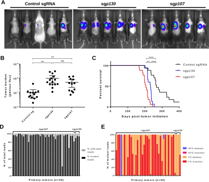
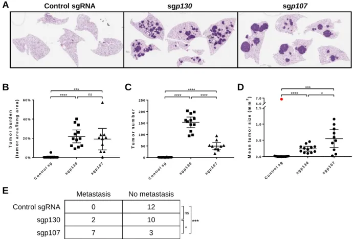
Differential effects of loss of p107 and p130 on tumor progression ··· 106
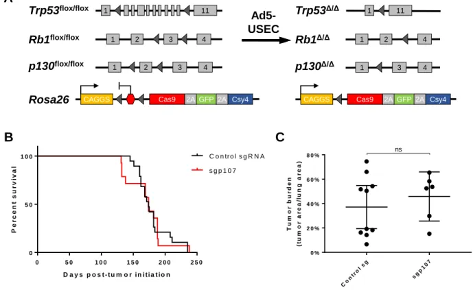
Loss of p107 in the Trp53/Rb/p130-null background does not accelerate tumor progression ··· 108
Inferring lineage relationships between primary and metastatic tumors ··· 108
DISCUSSION ··· 113
MATERIALS AND METHODS ··· 117
ACKNOWLEDGEMENTS ··· 122
SUPPLEMENTARY TABLES AND FIGURES ··· 123
REFERENCES ··· 126
CHAPTER 3 Identification of novel therapeutic targets in small cell lung cancer through CRISPR-based genetic screens ··· 131
ABSTRACT ··· 132
INTRODUCTION ··· 133
RESULTS ··· 135
Design of druggable genome sgRNA library ··· 135
Identification of SCLC-specific genetic vulnerabilities··· 137
SCLC cells exhibit increased sensitivity to Dhodh inhibition ··· 141
Dhodh inhibition suppresses growth of SCLC tumors in vivo ··· 143
DISCUSSION ··· 145
MATERIALS AND METHODS ··· 149
ACKNOWLEDGEMENTS ··· 156
SUPPLEMENTARY TABLES AND FIGURES ··· 157
REFERENCES ··· 166
CHAPTER 4 DISCUSSION AND FUTURE DIRECTIONS ··· 169
Rapid functional validation of candidate genes ··· 171
Significance of low-frequency genetic alterations in cancer ··· 172
Alternatives to GEMMs for modeling cancer ··· 174
Functional profiling of cancer ··· 175
Targeting metabolism in cancer ··· 176
Final perspective ··· 177
References ··· 178
APPENDIX 1 An optofluidic real-time cell sorter for longitudinal CTC studies in mouse models of cancer ··· 180
ABSTRACT ··· 181
INTRODUCTION ··· 182
RESULTS ··· 184
DISCUSSION ··· 194
MATERIALS AND METHODS ··· 196
ACKNOWLEDGEMENTS ··· 207
SUPPLEMENTARY FIGURES ··· 208
9
CHAPTER 1
10
Part I:
Functional interrogation of cancer
1.
The genetic basis of cancer
1.1 Cancer as a genetic disease
The discovery of the first oncogene some forty years ago laid the foundations for much of modern cancer biology research today. Prior to this, there were already
numerous factors associated with the development of various cancers that hinted at a genetic basis for cancer. For example, certain occupations were long known to be associated with elevated incidences of specific cancers, such as chimney sweeps and scrotal cancer, and dye-manufacturing workers and bladder cancer, although the actual causes were not known at that time. Yamagiwa and Ichikawa first demonstrated that chemicals could act as carcinogens by experimentally inducing metastatic tumors in rabbits through the application of coal car to their ears (Yamagiwa and Ichikawa, 1918). Many years later, Ames and colleagues established that many well-known carcinogens, including aflatoxin, polycyclic hydrocarbons such as benzo[a]pyrene (found in chimney soot and coal tar), and aromatic amines such as naphthylamine (found in dyes),
functioned as mutagens upon activation by mammalian liver homogenate, and therefore were likely to cause cancer by inducing somatic mutations in the genome (Ames et al., 1973b, 1973a).
In apparently unrelated studies, many viruses had been discovered to induce tumors in animals, with the most well-known example being the Rous sarcoma virus in chickens (Rous, 1911). This gave rise to the idea that perhaps cancer was an infectious disease, leading many to begin searching for potential infectious agents in human tumors, in order to establish whether this was relevant to human cancers. Eventually, in
11
a landmark finding that transformed the field, Bishop, Varmus and colleagues demonstrated that DNA related to the segment of the Rous sarcoma virus genome responsible for its ability to transform cells, the v-src oncogene, could be detected in the genomes of normal uninfected avian cells (Stehelin et al., 1976), as well as cells from more distantly related vertebrates (Spector et al., 1978). This showed that retroviral oncogenes were, in fact, modified counterparts of normal cellular genes, or proto-oncogenes, that are likely to have essential cellular functions.
Discovery of cellular oncogenes
Subsequently, multiple groups demonstrated that retroviruses were completely dispensable for the transformation process. Segments of human DNA isolated from tumor cell lines, when transfected into untransformed mouse fibroblast cells, were sufficient to cause their transformation in vitro (Goldfarb et al., 1982; Perucho et al., 1981; Pulciani et al., 1982; Shih and Weinberg, 1982; Shih et al., 1981). These segments were later found to correspond to the previously discovered viral ras gene (Der et al., 1982; Parada et al., 1982; Santos et al., 1982), thereby validating the
relevance of earlier studies of retroviral oncogenes. Furthermore, the version of the ras gene isolated from human tumors differed from the normal version by just a single point mutation that modified a single amino acid, demonstrating conclusively that mutations in endogenous cellular genes can transform a cell (Reddy et al., 1982; Tabin et al., 1982; Taparowsky et al., 1982). Collectively, these studies and others showed that cancer was a result of alterations in normal genes present in the genome, generating defective versions of these genes that confer the ability to transform normal cells into neoplastic cells.
12
Tumor suppressor genes
Despite these groundbreaking findings, there remained observations that could not be explained by the concept of gain-of-function oncogenic mutations in normal cellular genes. In a series of experiments conducted by Harris and colleagues, it was observed that the fusion of a malignant cell with a non-malignant counterpart resulted in daughter cells that were non-malignant (Harris, 1971), which suggested that malignancy was a recessive phenotype, in contrast to the apparent dominant nature of mutated oncogenes. In addition, using elegant statistical analyses of patients who developed unilateral versus bilateral retinoblastoma tumors, Knudson had hypothesized that retinoblastoma formation requires two mutational events (the two-hit hypothesis), with patients who developed bilateral retinoblastomas having already inherited the first mutation (Knudson, 1971). This hypothesis was subsequently validated by experiments demonstrating that patients with hereditary predisposition to retinoblastoma harbored loss-of-function mutations in a specific genomic locus, and that retinoblastoma tumors that developed in these patients frequently lost the second wild-type copy of that locus (Cavenee et al., 1983). The gene at this locus was eventually isolated (Friend et al., 1986) and aptly named the retinoblastoma gene, or RB1, proving to be the first example of a tumor suppressor gene. Tumor-predisposing mutations in this second class of cancer-associated genes confer a recessive phenotype, rather than a dominant phenotype observed with mutations in proto-oncogenes.
Ever since the discoveries of the first human oncogene and the first tumor suppressor gene, numerous other examples of both classes of genes have been discovered in various cancers. The advent of whole-genome sequencing technologies,
13
which made possible the cataloging of all the genes in the human genome as well as the detection of the vast repertoire of mutations in human tumors, has significantly accelerated this endeavor.
1.2 Cancer in the genomics era
One of the major research efforts in the 1990s and early 2000s was the Human Genome Project, which culminated in the unveiling of the sequence of the entire human genome (Collins et al., 2004; Lander et al., 2001; Venter et al., 2001). For the first time, it was possible to begin annotating the complete catalog of human genes, as well as to study their roles in various diseases in a comprehensive fashion. Combined with the subsequent development of next-generation sequencing technologies, which vastly increased the speed of sequencing while hugely decreasing costs, this rapidly ushered in the genomics era in cancer research.
It has long been recognized that cancer is a multistep process, requiring the accumulation of multiple genetic alterations, a paradigm that is best illustrated in colorectal cancer (Vogelstein and Kinzler, 1993). However, recent sequencing studies have shown that human tumors harbor an average of 30-60 non-silent coding mutations each (Lawrence et al., 2013; Vogelstein et al., 2013), which is far higher than the 4-7 genetic events that are thought to be required for tumor progression (Kinzler and Vogelstein, 1996; Renan, 1993). There is considerable heterogeneity in mutational frequencies across different cancer types, with lower frequencies in pediatric cancers such as rhabdoid tumors and Ewing sarcoma, and much higher frequencies in cancers commonly associated with carcinogens, such as melanoma (UV irradiation) and lung
14
cancer (cigarette smoke) (Lawrence et al., 2013; Vogelstein et al., 2013). To complicate matters further, there often exists a large range of mutational frequencies even within each cancer type (Lawrence et al., 2013).
With such high mutation rates in many tumors, it becomes a significant challenge to distinguish between driver mutations, which are mutations in genes that confer a selective growth advantage to the cancer cell, and passenger mutations, which are mutations that have no significant effect on cancer cell growth. This is especially
challenging in cancers with high mutation rates such as melanoma and lung cancer, as well as subsets of cancers that harbor mutations in key DNA repair genes. For example, a subset of colorectal tumors harbor mutations in genes involved in DNA mismatch repair, such as MSH2 and MLH1, which result in tumors that contain thousands of non-synonymous mutations each (Vogelstein et al., 2013). It is not technically feasible to test each mutation individually to determine which of these mutations are important for tumor progression.
1.3 Comprehensive molecular profiling of cancer
Large-scale whole-exome and whole-genome cancer sequencing studies aim to overcome this problem, based on the idea that functionally important driver events are likely to occur in a significant fraction of independent tumors, while passenger mutations that result from random mutational processes should occur at much lower frequencies. Such studies have grown to include national and international collaborations, such as The Cancer Genome Atlas (TCGA), which is itself part of the International Cancer Genome Consortium. To date, the TCGA project has sequenced over 11,000 human
15
tumor samples across 33 tumor types, with many of these samples also having
matched gene expression profiles, as well as clinical data such as overall survival and progression-free interval. This has provided a rich dataset for identifying key cancer driver genes and pathways in various cancer types.
To illustrate the utility of this approach, comprehensive molecular profiling of lung adenocarcinoma revealed that the majority of these tumors harbored known driver mutations within the RTK/RAS/RAF pathway, such as EGFR, KRAS and BRAF, as well as previously identified fusions involving ALK, ROS1 and RET (Cancer Genome Atlas Research Network, 2014). However, among the remaining RTK/RAS/RAF pathway-negative tumors, unique focal amplifications in ERBB2 and MET, as well as mutations in NF1 and RIT1, were found to be significantly enriched. In addition to providing potential new targets for the development of novel therapies, the discovery of these genetic events in these tumors opens up the possibility of using existing therapies, such as MET and ERBB2 inhibitors, in lung adenocarcinoma patients.
The success of comprehensive cancer profiling studies has, ironically, resulted in a different problem – how to prioritize candidate genes for subsequent functional
validation. Even with rigorous statistical analyses to account for variable mutation rates and heterogeneity in mutational processes in different cancers, there remains a long list of genes that are altered at significant frequencies (Lawrence et al., 2013). Efforts have begun to analyze sequencing data from across different cancer types to identify “pan-cancer” genetic drivers (Bailey et al., 2018), which may aid in the prioritization of important genes. Future efforts will likely require the development of better tools to functionally characterize the consequences of specific genetic alterations, as well as
16
improved tools to predict the effects of gain or loss of gene function at the global level in tumor cells.
1.4 Genetic screens and functional genomics
An alternative approach to identify novel cancer-specific therapeutic targets involves the use of genetic screens. This approach employs genetic tools that are used to perturb gene function in a large-scale, unbiased fashion, followed by functional
assays to assess the resulting phenotypes of the perturbation. Depending on the design of the experiment, phenotypes can include changes in rates of cell proliferation,
activation of reporter genes, secretion of protein products, and many more. Such an approach has been termed functional genomics, which underscores the emphasis on characterizing gene function.
Reverse genetics to identify gene function
The concept of perturbing the function of a gene to assess the resulting phenotypes is known as reverse genetics. This approach has long been used in the field of yeast genetics, where a systematic effort to generate yeast deletion strains comprising the entire genome was undertaken, culminating in the Saccharomyces Genome Deletion Project (Giaever et al., 2002; Winzeler et al., 1999). The motivation behind this effort was the sequencing of the complete genome of the yeast
Saccharomyces cerevisiae, which revealed that a significant proportion of the roughly 6,000 open reading frames present in the yeast genome had not been previously studied, and thus were of unknown function (Dujon, 1996). The collection of yeast knockout strains has been used for many different functional studies, including
17
identification of factors involved in cell growth, mating, sporulation and germination, as well as response to environmental stresses (reviewed in Giaever and Nislow, 2014).
Similar efforts have been carried out to study gene functions in mammalian systems, such as mice. The International Mouse Phenotyping Consortium (IMPC), which builds upon programs such as the Knockout Mouse Project (KOMP), aims to generate knockout mutations for every gene in the mouse genome, so as to discover and ascribe biological functions to each gene (Brown and Moore, 2012). However, it is much more time-consuming and expensive to generate knockout mice than to generate knockout yeast strains. Furthermore, it is simply not feasible to characterize every single knockout mouse strain each time one wishes to interrogate a specific biological
question. Therefore, the mainstay of functional genomics in mammalian systems has been genetic screens in cell-based systems, where genetic perturbations can be performed in a large-scale, highly parallel fashion.
Genetic screens in mammalian cells
Performing genetic screens in mammalian cells is complicated by the diploid nature of most mammalian cell lines. As most loss-of-function mutations generate recessive phenotypes, this means that both alleles of a particular gene need to be mutated or deleted before any phenotype becomes apparent. This limits the use of insertional mutagenesis methods, such as retroviruses and transposons, to certain cell lines that are mostly haploid, such as the human chronic myeloid leukemia cell line KBM-7 (Carette et al., 2009; Kotecki et al., 1999).
The development of RNA interference (RNAi)-based methods transformed the way loss-of-function genetic screens could be performed. RNAi, also known as
post-18
transcriptional gene silencing (PTGS), was first observed in plants and subsequently in animals such as the nematode worm Caenorhabditis elegans. Fire and colleagues first determined that RNAi was mediated by double-stranded RNA in cells (Fire et al., 1998). Subsequent work showed that these double-stranded RNAs were processed into 21-nucleotide single-stranded RNAs that mediated gene silencing (Elbashir et al., 2001a; Hamilton and Baulcombe, 1999; Hammond et al., 2000; Zamore et al., 2000). Elbashir and colleagues then showed that 21-nucleotide RNA duplexes could suppress gene expression in a sequence-specific manner in mammalian cells (Elbashir et al., 2001b). At around the same time, multiple groups identified endogenous small RNAs, or
microRNAs (miRNAs), that form hairpin structures in the cell and can regulate gene expression in a broad range of organisms (Lagos-Quintana et al., 2001; Lau et al., 2001; Lee and Ambros, 2001). Collectively, these discoveries led to the development of short hairpin RNA (shRNA)-based RNAi systems, which can be designed for targeted gene repression (Brummelkamp et al., 2002; Paddison et al., 2002; Paul et al., 2002). Because RNAi represses gene expression post-transcriptionally, this bypasses the need to generate mutations in both copies of a gene in mammalian cells.
Large-scale RNAi-based libraries have been designed and cloned into retroviral and lentiviral vectors for performing genetic screens in mammalian systems, both in vitro (Berns et al., 2004; Moffat et al., 2006; Paddison et al., 2004; Silva et al., 2005) as well as in vivo (see, for example, Zender et al., 2008). However, there are several issues associated with the use of RNAi in genetic screens. These include incomplete knockdown of gene expression, as well as significant off-target effects (Kaelin, 2012). Many of these shortcomings have been overcome by the use of CRISPR-Cas-based
19
systems, which have begun to complement, and in some cases supersede, the use of RNAi-based systems for genetic screens. This will be discussed in greater detail later in this chapter.
Gain-of-function genetic screens have traditionally been far more challenging to perform compared with loss-of-function genetic screens. Unlike in the case of RNAi, where 21-nucleotide sequences can be designed to repress expression of essentially any gene in the genome, there has, until recently, been no equivalent method to activate gene expression. Large-scale lentiviral libraries expressing open reading frames (ORFs) have been developed for gain-of-function screens (Yang et al., 2011), but these do not offer the same level of flexibility as RNAi knockdown libraries. As will be discussed, CRISPR-Cas systems have been modified to enable transcriptional activation of genes in a highly flexible fashion, allowing such systems to be adapted for gain-of-function screens as well.
Identification of context-specific genetic vulnerabilities
The power of genetic screens is perhaps best illustrated in its use for identifying context-specific genetic vulnerabilities in cells. Loss-of-function screens have been performed in many different cell lines to identify genes that, when mutated, result in decreased cell proliferation or death (Aguirre et al., 2016; Hart et al., 2015, 2017; Wang et al., 2015). Such screens have identified genes that appear to be essential across all cell lines; as expected, these genes encode proteins that are involved in essential
cellular processes such as translation, transcription and DNA replication, and have been termed “core essential genes”. However, other genes appear to be essential only in
20
certain cell lines and do not result in any proliferative defects when lost in other cell lines. These are referred to as context-specific essential genes (Hart et al., 2014).
In the context of cancer, genetic screens can be used to identify potential therapeutic targets that are essential in cancer cells, but non-essential in normal, untransformed cells. This principle underlies the concept of synthetic lethality, in which mutation of a particular gene is lethal to a cell only when it occurs together with mutation of a second gene (Kaelin, 2005). For cancer cells, this could be mutations in oncogenes or tumor suppressor genes, which are not present in normal cells. Loss-of-function screens have been performed to identify such synthetic-lethal interactions. For example, Wang and colleagues performed genome-wide CRISPR-based genetic screens in a panel of acute myeloid leukemia (AML) cell lines, half of which harbored a mutant RAS allele, and found that loss of the guanine nucleotide exchange factor PREX1 was selectively essential in RAS-mutant AML lines and not in RAS-wild-type AML lines (Wang et al., 2017). More broadly, both RNAi-based and CRISPR-based screens have been used to profile cancer type-specific vulnerabilities across large panels of hundreds of cancer cell lines (McDonald et al., 2017; Meyers et al., 2017; Tsherniak et al., 2017). In combination with cancer genome sequencing studies, such efforts have enabled the discovery and prioritization of genes for downstream functional analyses.
2.
Modeling cancer in mice
Many groundbreaking discoveries in cancer biology have been made using in vitro cell culture-based systems, as discussed in the previous section. In vitro systems offer many advantages for studying specific mechanisms of oncogene or tumor
21
suppressor gene function. For example, such systems provide a well-controlled environment where experimental variables can be minimized, allowing the results of a specific perturbation to be interpreted more easily. However, in vitro systems are unable to fully replicate certain key features of cancer. To illustrate this, of the six hallmarks of cancer described by Hanahan and Weinberg (Hanahan and Weinberg, 2000), two of these – sustained angiogenesis, and tissue invasion and metastasis – cannot be fully recapitulated in cell culture-based systems. Furthermore, the recent resurgence of cancer immunotherapy has ignited huge interest in understanding the interactions between tumor cells and the immune system, which can only be effectively studied in whole-organism systems.
Mouse models of cancer have long been utilized to bridge the gap between in vitro systems and humans. Many aspects of the laboratory mouse make it a suitable model system for studying cancer, including similarities with humans at the genomic and physiological levels. The use of mouse cancer models has been reviewed
extensively elsewhere (see, for example, Frese and Tuveson, 2007). In this section, I will focus on genetically engineered mouse models (GEMMs) of cancer. In particular, I will highlight the key technological advances that have enabled the development of advanced GEMMs that allow precise spatial and temporal control of tumorigenesis.
2.1 Transgenic mouse models
The development of methods to generate transgenic mice was the first step towards creating mouse models of cancer. Jaenisch and Mintz first demonstrated the successful transmission of simian virus 40 (SV40) DNA in mice. They microinjected
22
SV40 DNA into preimplantation mouse blastocysts, allowed the blastocysts to develop into adults, then showed that SV40-specific DNA sequences could be detected in DNA extracted from various adult organs and tissues, providing confirmation that the injected SV40 DNA had been successfully transmitted throughout mouse development
(Jaenisch and Mintz, 1974). In a subsequent study, Jaenisch demonstrated successful germline transmission of Moloney murine leukemia virus DNA in mice (Jaenisch, 1976), paving the way for generating heritable changes to the mouse genome in vivo.
Many transgenic mouse models of cancer were developed using this approach. For instance, Brinster, Palmiter and colleagues generated mice carrying SV40 large and small T antigens under the control of the SV40 enhancer, and found that these mice developed choroid plexus tumors in the brain (Brinster et al., 1984; Palmiter et al., 1985). Some level of tissue specificity could be conferred by generating hybrid constructs driven by tissue-specific regulatory elements. Mice carrying the myc gene under the control of the mouse mammary tumor virus (MMTV) promoter specifically develop mammary adenocarcinomas (Stewart et al., 1984). Likewise, expression of the SV40 large T antigen under the control of the rat insulin promoter in mice results
specifically in pancreatic β-cell tumors (Hanahan, 1985).
Although these models allowed tumorigenesis and tumor progression to be studied in vivo, there were several problems with transgenic models. In many cases, the inserted gene is expressed at high levels that do not reflect physiological conditions, due to the use of ectopic promoters without endogenous regulatory elements.
23
variable gene expression. More importantly, transgenic models have limited utility for modeling loss-of-function mutations in tumor suppressor genes.
2.2 Targeted gene modifications in mice
The next generation of genetically engineered mouse models resulted from a confluence of two separate technological developments in the 1980s – the use of homologous recombination to generate targeted genetic modifications in cells, and the derivation of mouse embryonic stem (ES) cells.
Targeted gene modifications by homologous recombination
Homologous recombination is a process that involves the exchange of genetic material between two DNA strands with regions of sequence homology. In eukaryotic cells, this process occurs as part of normal meiosis, and is responsible for the
phenomenon of gene conversion in yeast (Holliday, 1964; Meselson and Radding, 1975; Szostak et al., 1983).
The first demonstration that homologous recombination could be used to target an exogenous DNA sequence to a specific locus in the eukaryotic genome was
performed in yeast (Hinnen et al., 1978). Hinnen and colleagues introduced a plasmid carrying the yeast LEU2+ gene into a leu2- yeast strain, and obtained colonies at low
frequencies that could grow in medium lacking leucine. Further characterization revealed that some of the transformed colonies had replaced the original leu2- locus
with the incoming LEU2+ sequence. Subsequently, Orr-Weaver and colleagues showed
that recombination efficiency could be greatly enhanced by using linear rather than circular plasmids (Orr-Weaver et al., 1981).
24
Building on the work in yeast, the Smithies and Capecchi groups later reported the successful targeting of genes to specific loci in mammalian cells via homologous recombination, showing that this method was not limited to just yeast cells (Smithies et al., 1985; Thomas et al., 1986). It was now possible not just to replace an endogenous gene with an altered version of the gene to modify its function while retaining locus-specific regulation of the gene, but also to “knock out” an endogenous gene by inserting a sequence within the gene to disrupt its coding sequence.
Derivation of mouse embryonic stem cells
The relatively low efficiency of homologous recombination events compared with random integration events (Thomas et al., 1986) meant that it was not feasible to
generate targeted gene modifications directly in blastocysts or zygotes, as had been done with transgenic approaches. Fortunately, Evans and Kaufman had developed a method to isolate and propagate murine embryonic stem (ES) cells from mouse preimplantation blastocysts (Evans and Kaufman, 1981). These cells exhibited
pluripotency, or the ability to differentiate into cells from all three germ layers, and gave rise to teratocarcinomas when injected subcutaneously into syngeneic mice.
Importantly, unlike the previously derived embryonal carcinoma cell lines that had similar differentiation capabilities (Martin and Evans, 1975; Papaioannou et al., 1975), ES cells were karyotypically normal and stable. Subsequently, ES cells were shown to contribute to chimera formation with high efficiency when injected into donor
blastocysts, with ES cell-derived progeny contributing to all tissues in the mouse, including the germ line (Bradley et al., 1984).
25
Generation of mice with targeted gene modifications
The availability of ES cells meant that low-frequency events such as gene targeting by homologous recombination could be performed in vitro, followed by a selection step to isolate correctly targeted cells before blastocyst injections. It was not long before germ line transmission of targeted genetic alterations in mice was
successfully achieved (Koller et al., 1989; Thompson et al., 1989). These initial studies involved the correction of a defect in the hypoxanthine guanine phosphoribosyl
transferase gene (HPRT); successful targeting events could be selected for and enriched by culturing cells in hypoxanthine-aminopterin-thymidine medium, in which only HPRT-positive cells survive (Doetschman et al., 1987). Subsequently, Mansour and colleagues developed a generalizable method, known as positive-negative selection, to selectively enrich for cells that have undergone successful homologous recombination-mediated targeting over cells that have undergone random integration (Mansour et al., 1988). This is especially useful in cases where the targeted genes do not have an easily selectable phenotype. With the obvious utility of this approach for studying the effects of loss of tumor suppressor genes in vivo, it was no surprise that multiple groups quickly reported the generation of mice with targeted disruptions in key tumor suppressor genes, such as Rb1 (Jacks et al., 1992; Lee et al., 1992), p53
(Donehower et al., 1992; Jacks et al., 1994a) and Nf1 (Jacks et al., 1994b). Although this approach was useful in uncovering the roles of these tumor suppressor genes in various cancers, some of which were not previously appreciated, one pattern that quickly emerged was that homozygous loss of such genes in vivo often resulted in embryonic lethality. This reflected the fact that many these genes play crucial
26
roles in normal cellular function; however, it also posed a problem for modeling their roles in cancer progression. The use of heterozygous knockout mice partially overcame this problem, but this required sporadic loss-of-heterozygosity events to occur before tumors could develop, resulting in variable penetrance of tumor phenotypes.
Furthermore, in cases where loss of the gene, or a combination of genes, results in multiple tumor types forming in the mouse, these tumors often progress at different rates, causing the mice to succumb to one type of tumor before another type has had time to develop. For example, in mice with combined Rb1+/-;p53-/- mutations,
approximately 40% of animals developed small hyperplastic foci of neuroendocrine cells in the bronchi and bronchioles of the lung. However, these hyperplastic foci did not develop further due to the animals succumbing to other tumors by two to six months of age (Williams et al., 1994). Given the frequent loss of RB1 and TP53 in small cell lung cancer (SCLC), which is a subtype of neuroendocrine lung tumors, it is likely that these lesions represented SCLC precursors that did not have sufficient time to develop into frank tumors. Thus, these problems highlighted the need for methods to control when and where genetic modifications occur in the mouse.
2.3 Spatial and temporal control of gene knockout/activation
Further advances in genetic tools led to the development of the next generation of mouse models of cancer. These tools enabled spatial and temporal control of gene activation or disruption, overcoming a number of the problems associated with
27
Site-specific recombinase-based systems
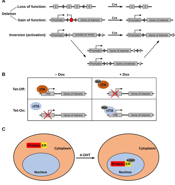
Site-specific recombinases, such as the Cre-loxP system derived from the bacteriophage P1 and the Flp-FRT system derived from yeast, were one of the earliest tools used to enable spatial and temporal control of genetic modifications in the mouse. These systems involve the use of a recombinase (Cre or Flp) that recognizes and mediates strand exchange between specific sequences of DNA (loxP or FRT sites). Depending on the location and relative orientation of the two recognition sites, site-specific recombinases can mediate excision of a sequence (two directly repeated sites on the same DNA strand), inversion of a sequence (two inverted sites on the same strand), integration of a circular strand into a linear strand (two sites on separate strands), or exchange of sequences between two linear strands (two sites on separate strands) (Fig. 1A). Such systems are most commonly used for deletion of genes or portions of genes to disrupt gene function, as well as for gene activation via the deletion of a transcriptional termination sequence between a gene and its promoter (Lakso et al., 1992). The recombinase can be placed under the control of tissue-specific or
developmental-specific promoters, or be introduced via a viral vector to specific sites in the mouse, to allow gene deletion or activation to be spatially and temporally controlled.
The first use of the Cre-loxP system for tissue-specific gene modification was demonstrated by two different groups. Lakso and colleagues generated transgenic mice carrying the SV40 large T antigen driven by the lens-specific αA-crystallin promoter, but separated by a transcriptional stop sequence flanked by loxP sites, and crossed them to mice carrying Cre recombinase expressed from either the αA-crystallin promoter or the
28 Nucleus Cytoplasm Nucleus Cytoplasm A B Gene of interest TRE tTA Gene of interest TRE rtTA Dox rtTA Gene of interest TRE tTA Dox Gene of interest TRE – Dox + Dox Tet-Off: Tet-On: Loss of function: 1 2 3 4 1 4 Cre Gene of interest
Promoter Promoter Gene of interest
Gain of function: Deletion
Inversion (activation): Promoter st tere f in o ne Ge
Gene of interest Promoter Gene of interest Promoter Gene of interest Promoter Cre Cre C Protein ER 4-OHT Protein ER 4-OHT
Figure 1: Genetic tools for spatial and temporal control of gene expression.
(A) Use of the Cre-loxP system as an example of site-specific recombinase systems. Triangles
represent loxP sites.
(B) Tetracycline-regulated systems. tTA: tetracycline-controlled transactivator; rtTA: reverse
tetracycline-controlled transactivator; TRE: tetracycline-response element; Dox: doxycycline.
(C) Hormone receptor fusion systems. ER: estrogen receptor hormone-binding domain; 4-OHT:
29
human cytomegalovirus promoter (Lakso et al., 1992). These mice went on to develop lens tumors, whereas mice without Cre recombinase did not develop tumors,
demonstrating tissue-specific activation of T antigen. Orban and colleagues generated mice expressing a loxP-flanked (floxed) β-galactosidase transgene and crossed them to mice expressing Cre recombinase driven by a thymocyte-specific promoter, showing that the β-galactosidase transgene was specifically deleted in thymocytes (Orban et al., 1992).
Subsequently, Gu and colleagues demonstrated the utility of this approach for studying tissue-specific deletion of an endogenous gene (Gu et al., 1994). They generated mice harboring floxed alleles of DNA polymerase β, crossed them to mice harboring a T cell-specific Cre recombinase gene, and demonstrated that they were able to delete DNA polymerase β specifically in T cells with no effects on the rest of the mouse, in contrast to constitutive deletion of DNA polymerase β throughout the mouse, which resulted in embryonic lethality. This study validated the use of Cre-loxP-mediated conditional deletion for studying tissue-specific loss of genes that are essential for mouse development.
An alternative method of achieving spatially restricted Cre expression was demonstrated by Shibata and colleagues, who generated an adenoviral vector
expressing Cre recombinase, and injected the virus into the colon of mice harboring a modified Apc allele that contained loxP sites flanking exon 14. This resulted in colon-specific inactivation of Apc, leading to the formation of colorectal adenomas and adenocarcinomas (Shibata et al., 1997). In contrast, in an earlier model involving the use of a constitutive loss-of-function Apc allele, mice that were heterozygous for the
30
allele developed tumors predominantly in the small intestine rather than the colon (Fodde et al., 1994).
Cre-loxP-based and Flp-FRT-based mouse models have now become one of the mainstays of murine models of cancer. Complex models combining multiple conditional alleles have been developed to model multiple genetic events simultaneously,
recapitulating what is often observed in human cancers. In addition, because of the ability to restrict Cre expression to specific tissues, the same combination of alleles can be used to model multiple types of cancer. As an example, a mouse model harboring a conditional oncogenic allele of Kras and a conditional mutant allele of Trp53 has been used to model lung adenocarcinoma (Jackson et al., 2005), pancreatic ductal
adenocarcinoma (Hingorani et al., 2005), and soft tissue sarcoma (Kirsch et al., 2007).
Tetracycline-regulated systems
In addition to site-specific recombinase-based systems, an alternative system was developed that allowed gene expression to be reversibly toggled on or off. This system was based on the tetracycline resistance operon present in bacteria. In the absence of the antibiotic tetracycline, a repressor protein (TetR) binds to an operator sequence (tetO) to repress transcription of the entire operon. When tetracycline is present, it binds to TetR, preventing it from binding to tetO and resulting in the transcription of genes that mediate tetracycline resistance.
To adapt this system for use in mammalian cells, Gossen and Bujard fused the TetR protein with the C-terminal domain of the VP16 transactivation domain from the herpes simplex virus (HSV) to form the tetracycline-controlled transactivator (tTA) (Gossen and Bujard, 1992). When combined with a tetracycline-response promoter
31
comprising multiple copies of the tetO sequence fused to a minimal promoter, tTA was able to bind to the promoter and activate expression of a transgene in the absence of tetracycline. Expression of the transgene was repressed when tetracycline was added to the cells (“Tet-Off”). A variation of this system was generated by mutation of the TetR gene sequence, generating a version that binds to tetO only in the presence of
doxycycline, a derivative of tetracycline (Gossen et al., 1995). This version was known as the reverse tetracycline-controlled transactivator (rtTA), and is used in “Tet-On” systems (Fig. 1B). Both versions were rapidly adopted for in vivo applications (Furth et al., 1994; Kistner et al., 1996). Spatial control of gene expression can be achieved by placing the tTA or rtTA transgene under the control of a tissue-specific promoter (Kistner et al., 1996).
Because of the ability to reversibly switch gene expression on or off, both Tet-Off and Tet-On systems have been used to investigate the requirement for oncogene expression in tumor maintenance. For instance, Felsher and Bishop generated mice expressing human MYC in hematopoietic cells using the Tet-Off system, resulting in the development of malignant T cell lymphomas and acute myeloid leukemias. Treatment of tumor-bearing animals with doxycycline to inactivate MYC expression caused rapid regression of these tumors (Felsher and Bishop, 1999). Similarly, Chin and colleagues expressed oncogenic H-RasG12V in melanocytes using the Tet-On system, resulting in
melanoma formation in a doxycycline-dependent fashion. Withdrawal of doxycycline resulted in rapid regression of these tumors (Chin et al., 1999). Both of these examples demonstrate that, at least in certain cancers, sustained expression of the initiating oncogene is required for tumor maintenance.
32
Hormone receptor fusion systems
Another method of achieving reversible control of gene expression involves the fusion of proteins of interest with the hormone-binding domain of nuclear hormone receptors, most commonly the estrogen receptor. Nuclear hormone receptors normally reside in the cytoplasm of the cell in the absence of their cognate hormone. Upon hormone binding, these receptors translocate to the nucleus, where they mediate
changes in gene expression. Fusion proteins containing the hormone-binding domain of such receptors can be regulated in a similar manner (Fig. 1C). The first demonstration of this method, performed by Picard and colleagues, involved the fusion of the
hormone-binding domain of the glucocorticoid receptor to the adenovirus E1A gene product, resulting in E1A activity being regulated by dexamethasone (Picard et al., 1988). Subsequently, Eilers and colleagues fused the hormone-binding domain of the estrogen receptor (ER) to c-myc, and showed that when introduced into rat fibroblast cells, the fusion protein was able to mediate transformation of the cells in a reversible manner, depending on the presence of β-estradiol (Eilers et al., 1989).
Use of the ER hormone-binding domain for in vivo applications was initially complicated by the presence of high levels of circulating estrogens in mice. To overcome this problem, Danielian and colleagues generated a mutant version of the murine ER harboring a G525R mutation, which drastically reduced its affinity for endogenous estrogens but not for the synthetic steroid 4-hydroxytamixofen (4-OHT) (Danielian et al., 1993). The equivalent mutation in the human ER (G521R) similarly abolished binding to endogenous estrogens (Feil et al., 1996). Subsequently, additional mutations to improve the efficiency of activation by 4-OHT resulted in the generation of
33
the ERT2 version of the ER hormone-binding domain, which is currently widely used
(Feil et al., 1997; Indra et al., 1999).
The first demonstration of the use of hormone receptor fusion systems in vivo was in combination with the Cre-loxP system (Feil et al., 1996). Fusion of Cre
recombinase with the ER hormone-binding domain enables precise temporal control of Cre activity via the administration of tamoxifen, which is converted to 4-OHT in vivo. In addition, by placing expression of the CreER fusion gene under the control of a tissue-specific promoter, both spatial and temporal control of Cre activity can be achieved, as first demonstrated in B lymphocytes (Schwenk et al., 1998) and the embryonic neural tube (Danielian et al., 1998).
As with Tet-On and Tet-Off systems, ER domain fusions have also been used to control proto-oncogene and tumor suppressor gene activity in vivo. Pelengaris and colleagues generated a transgenic mouse model in which a fusion c-MycERTM protein
was placed under the control of the human involucrin promoter to confer epidermal-specific expression. Upon topical application of 4-OHT to the skin, these mice developed premalignant squamous cell neoplasia, which regressed back to normal morphology upon withdrawal of 4-OHT (Pelengaris et al., 1999).
To enable regulation of p53 activity, Christophorou and colleagues generated a knock-in mouse in which the endogenous Trp53 allele was replaced with one encoding p53ERTAM, rendering p53 activity dependent on tamoxifen administration (Christophorou
et al., 2005). In the absence of tamoxifen, the allele functions as a null allele. This allele has been used to study the consequence of p53 restoration in various tumor models,
34
such as lymphoma and lung adenocarcinoma (Christophorou et al., 2006; Junttila et al., 2010; Martins et al., 2006).
2.4 Speeding up generation of new mouse models
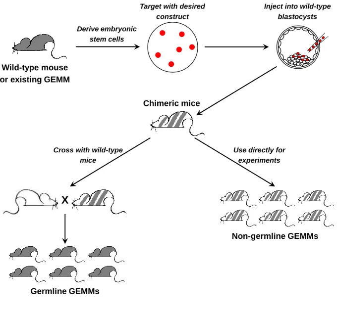
One of the biggest limitations of modeling cancer in mice is the time required to generate new models, as well as the concomitant cost (reviewed in Huijbers et al., 2011). To functionally interrogate a gene of interest in a specific cancer, one needs to generate ES cells with the desired modification (insertion of a transgene or a targeted knockout/knock-in). Following injection of the modified ES cells into blastocysts and implantation into surrogate female mice, chimeric pups are obtained. These are crossed to unmodified mice to determine whether germline transmission has occurred (Fig. 2). Mice with successful germline transmission are then crossed into an existing model of interest, which typically requires at least an additional two generations of breeding before a suitable-sized cohort is produced for actual experiments. The whole process takes one to two years and involves significant cost at each step. This has made it a significant challenge to carry out functional validation of the ever-growing lists of
candidate genes identified through large-scale sequencing studies or functional genetic screens.
Non-germline mouse models
One way to reduce the time required for generating new mouse models is to bypass the need to introduce genetic modifications into every single cell in the mouse. Traditionally, only chimeric mice in which the modified ES cells have contributed to the germ line are deemed useful, as these mice are subsequently bred to generate mice
35
Wild-type mouse or existing GEMM
Derive embryonic stem cells
Target with desired construct
Inject into wild-type blastocysts
Chimeric mice
Cross with wild-type mice
X
Use directly for experiments
Non-germline GEMMs
Germline GEMMs
Figure 2: Generation of targeted genetic modifications in mice.
36
with the desired genetic modification in all the cells in the body. However, several groups have demonstrated the utility of chimeras themselves as models for experimental manipulation, rather than simply as an intermediate step in model generation (Huijbers et al., 2014; Zhou et al., 2010). These have been referred to as non-germline GEMMs (Fig. 2).
Instead of starting with ES cells with a single genetic modification, this approach requires the generation of ES cells with all of the desired genetic modifications. Upon injection into wild-type blastocysts and subsequent development into adult mice, each tissue will contain a subset of cells that harbor the full complement of genetic
modifications. These chimeric mice are then used for experiments in the same manner as conventional germline models. Two different approaches have been used to
generate such ES cells. The first involves the sequential modification of wild-type ES cells, with in vitro and in vivo validations at various steps in the process. This approach has been used to rapidly generate and compare different models of lung
adenocarcinoma driven by distinct oncogenic events, such as HER2V659E, PIK3CAmyr,
EGFRL858R, and KRASG12V (Zhou et al., 2010).
The second approach involves the derivation of ES cells from existing well-established GEMMs of cancer (GEMM-ESCs), thereby bypassing the need to re-target these genes. These GEMM-ESCs are then used for introducing new genetic
modifications. To further speed up the process of gene targeting, a docking site
containing recognition sites for site-specific recombinases can be introduced into the ES cells, which facilitates rapid insertion of DNA constructs expressing genes of interest in a process known as recombinase-mediated cassette exchange (RMCE) (Baer and
37
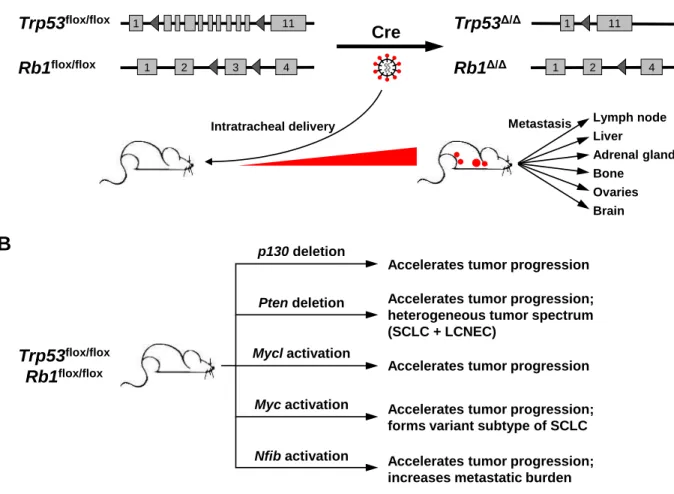
Bode, 2001; Bode et al., 2000). Using this technique, Huijbers and colleagues have demonstrated that overexpression of Mycl1, Nfib, or both Mycl1 and Nfib together, accelerates tumor progression in an established model of small cell lung cancer, thereby validating their oncogenic functions (Huijbers et al., 2014; Semenova et al., 2016).
Generating somatic genetic alterations
Another way to speed up generation of new mouse models of cancer is to bypass the need for ES cell manipulation and chimera generation entirely, by
generating genetic alterations directly in the somatic cells of tissues of interest. This can be accomplished, for example, using lentivirus-based vectors. Because lentiviruses stably integrate in the host genome of non-dividing mammalian cells, they can mediate long-term expression of genes of interest in target tissues. This method has been used to model various cancers in vivo. For example, a lentiviral vector expressing a Cre-inducible H-RasV12 allele was injected into the brains of mice that expressed Cre under the control of the GFAP promoter, which is expressed in neural stem and progenitor cells, as well as terminally differentiated astrocytes. This resulted in the development of glioblastoma multiforme in these mice (Marumoto et al., 2009).
In addition to lentiviral vectors, transposon-based systems have also been used to manipulate somatic cells in vivo (reviewed in Tschida et al., 2014). This approach has been successfully used to model hepatocellular carcinoma (Wangensteen et al., 2008) as well as glioblastoma multiforme (Wiesner et al., 2009).
Most recently, the development of the CRISPR-Cas9 system has greatly expanded the ability to precisely target genes of interest in mammalian cells, and has
38
paved the way for the use of other transient, non-integrating delivery systems, such as adenoviral vectors, for somatic gene editing. This and other aspects of CRISPR-Cas systems will be discussed in greater detail in the next section.
3.
CRISPR-Cas systems in cancer biology
Advances in scientific knowledge are often driven by the development of new technological tools. This has been illustrated in cancer biology by the development of ever-improving models to study the disease. The development of the CRISPR-Cas9 system has proven no different in this regard. However, the CRISPR-Cas9 system has arguably transformed the field more rapidly and broadly than any other preceding technological advance, due to the remarkable simplicity of the system that makes it accessible to practically every aspect of biological research.
In this section, I will discuss key advances in the development of CRISPR-Cas systems that have facilitated their use in cancer biology research.
3.1 Discovery of CRISPR-Cas systems
Clustered regularly interspaced short palindromic repeats, or CRISPR for short, were first observed as an incidental finding in a study by Ishino and colleagues, who were sequencing the iap gene in the bacterium Escherichia coli (Ishino et al., 1987). They noted that “Five highly homologous sequences of 29 nucleotides were arranged as direct repeats with 32 nucleotides as spacing”, but the biological significance of these sequences was not investigated. It was only much later that similar arrays of repeats were identified in a wide variety of prokaryotic genomes across many phylogenetic
39
groups in both Archaea and Bacteria (Mojica et al., 2000). Initially named Short Regularly Spaced Repeats (SRSRs), these arrays were later named CRISPR by
Jansen and colleagues in a study that also identified the first CRISPR-associated (cas) genes. Such genes were located adjacent to CRISPR loci and were present in CRISPR-positive prokaryotes, but absent from CRISPR-negative prokaryotes (Jansen et al., 2002).
The first clue of the biological function of CRISPR arrays came in 2005, when the spacer sequences between the direct repeats were identified to have homology to foreign genetic elements such as bacteriophages and conjugative plasmids (Bolotin et al., 2005; Mojica et al., 2005; Pourcel et al., 2005), leading to the proposal that CRISPR systems function as an adaptive immune system in prokaryotic cells (Makarova et al., 2006). In a series of experiments in Streptococcus thermophilus, Barrangou and colleagues demonstrated conclusively that this was indeed the case. When wild-type strains of S. thermophilus were challenged with two different phages to generate phage-resistant mutants, analysis of the CRISPR loci in the mutant strains showed that they had incorporated new spacer sequences, which corresponded to the regions within the genomes of the respective phages used in the challenge. When these spacer
sequences were deleted from the CRISPR locus, the resulting bacterial strain once again became sensitive to infection by that specific phage. Likewise, when the
corresponding spacer sequences were introduced into a S. thermophilus strain that was sensitive to the same phage, the bacterial strain now gained resistance to infection by that phage (Barrangou et al., 2007).
40
The identification of an adaptive immune system in prokaryotes was by itself a groundbreaking discovery, but subsequent experiments that elucidated the mechanistic basis of CRISPR-mediated immunity would serve to unleash its true transformative potential.
3.2 CRISPR-Cas9 as an RNA-guided DNA endonuclease
First, it was shown that transcription of the CRISPR array results in the formation of a precursor CRISPR RNA (crRNA), which is then processed by CRISPR-associated (Cas) proteins to form individual crRNAs. These crRNAs then associate with other Cas proteins to mediate antiviral responses (Brouns et al., 2008). Next, it was demonstrated that CRISPR-Cas systems mediate cleavage of double-stranded bacteriophage and plasmid DNA in a sequence-specific fashion (Garneau et al., 2010).
Although a huge diversity of CRISPR-Cas systems have been discovered across prokaryotic species (Koonin et al., 2017), the most widely used is the type II CRISPR-Cas system, due to its simplicity compared with other subtypes. Sapranauskas and colleagues demonstrated that in type II systems, a single Cas protein, Cas9, was sufficient to mediate CRISPR-encoded interference (Sapranauskas et al., 2011), in contrast to other subtypes that required multiple different Cas proteins to mediate binding and cleavage of the target DNA locus (Brouns et al., 2008; Hale et al., 2012; Sinkunas et al., 2011; Zhang et al., 2012). As a result, type II CRISPR-Cas systems are also commonly referred to as CRISPR-Cas9 systems.
CRISPR-Cas9 systems encode a separate trans-activating CRISPR RNA (tracrRNA) that is required for crRNA maturation as well as for cleavage of target DNA