• Aucun résultat trouvé

A critical benefit analysis of artificial gravity as a microgravity countermeasure

N/A
N/A
Protected

Academic year: 2021

Partager "A critical benefit analysis of artificial gravity as a microgravity countermeasure"

Copied!
187
0
0

Texte intégral

(1)

A Critical Benefit Analysis of Artificial Gravity as a

Microgravity Countermeasure

by

Justin David Kaderka

B.S. Mechanical Engineering

B.S. Materials Science and Engineering

University of California at Berkeley, 2006

SUBMITTED TO THE DEPARTMENT OF AERONAUTICS AND ASTRONAUTICS IN

PARTIAL FULFILLMENT OF THE REQUIREMENTS FOR THE DEGREE OF

MASTER OF SCIENCE IN AERONAUTICS AND ASTRONAUTICS

AT THE

MASSACHUSETTS INSTITUTE OF TECHNOLOGY

June 2010

© 2010 Massachusetts Institute of Technology

All rights reserved

Signature of Author: ____________________________________________________________

Department of Aeronautics and Astronautics

May 21, 2010

Certified by: ___________________________________________________________________

Laurence R. Young

Apollo Program Professor of Astronautics

Professor of Health Sciences and Technology

Thesis Supervisor

Accepted by: __________________________________________________________________

Eytan H. Modiano

Associate Professor of Aeronautics and Astronautics

Chair, Committee on Graduate Studies

(2)
(3)

3

A Critical Benefit Analysis of Artificial Gravity as a Microgravity Countermeasure

By

Justin David Kaderka

Submitted to the Department of Aeronautics and Astronautics

on May 21, 2010 in partial fulfillment of the

requirements for the degree of

Master of Science in Aeronautics and Astronautics

Abstract

Human physiological systems, especially the cardiovascular and musculo-skeletal systems,

are well-known to decondition during spaceflight. Several countermeasures that are in use today

have been rigorously developed over the decades to combat this deconditioning. However, these

countermeasures are system specific and have proven to be only partially effective. Artificial

gravity has been persistently discussed as a countermeasure that potentially has salutary effects

on all physiological systems, though few ground-based studies have been performed in

comparison to other countermeasures. The current analysis attempts to elucidate the

effectiveness of artificial gravity by directly comparing results of previously published and

unpublished deconditioning studies with those of more traditional, ground-based

countermeasures (i.e. resistive exercise, aerobic exercise, lower body negative pressure, or some

variation of these). Animal studies were also evaluated to supplement the knowledge base and to

fill gaps in the human countermeasure literature. Designs of published studies, such as study

duration, deconditioning paradigm, subject selection criteria, measurements taken, etc., were

confounding variables; however, studies that had some measure of consistency between these

variables were compared, although notable differences were cited in the analysis and discussion.

Results indicate that for prolonged spaceflight an artificial gravity-based countermeasure may

provide benefits equivalent to traditional countermeasures for the cardiovascular system. Too

few comparable, human studies have been performed to draw any conclusions for the

musculo-skeletal system, although animal studies show some positive results. Gaps in the current

knowledge of artificial gravity are identified and guidance for future deconditioning studies is

offered. Based on the results of this study, a comprehensive artificial gravity protocol is

proposed and future research topics using this countermeasure are addressed.

Thesis Supervisor:

Dr. Laurence R. Young

Title:

Apollo Program Professor of Astronautics

Professor of Health Sciences and Technology

Department of Aeronautics and Astronautics

This work was supported by the NASA Johnson Space Center contract number NNX09AE50G

as well as by the W. A. Brookshire Foundation.

(4)
(5)

5

Acknowledgements

Firstly, I would like to thank Dr. John Charles for funding this research through NASA contract

number NNX09AE50G. This research would not be possible without his support.

To my advisors:

Professor Laurence Young: Thank you for giving me the opportunity to work on this project.

It has truly been a pleasure having you as a mentor, a professor, and traveling companion. I

greatly value your guidance and I look forward to working with you in the future.

Professor Bill Paloksi: For almost half of this research, I had only known you by your voice

through our weekly telecons until we finally met in person. Thank you for your wealth of

knowledge and guidance that you have contributed to this project.

To Liz: Thank you for always being there to answer all the administrative questions I have had

over the past two years.

To Dr. Alan Natapoff: Thank you for the long hours we spent together trying to figure out how

to analyze such a hodge-podge collection of data. Also, thank you for all the trivia you have

imparted to me; the volume of interesting information you know never seizes to amaze me.

To my officemates: Thank you for not getting too upset when my papers and books somehow

make it onto your desk space.

To the students of the MVL: I can honestly say that I have never had such a great work

environment as the MVL. Plus, the extracurricular activities have been fantastic…beer brewing,

sailing, squash playing, and, of course, warfish. It has been great getting to know every one of

you and I look forward to staying in touch in the future.

To Mom, Dad, Kendra, and Aunt Sharon: Thank you for being so supportive of me and my

endeavors. Your words of encouragement have kept me going through the thick and thin.

And finally, to my love, Tiffany: Thank you for always being there for me, even when I have

been difficult to reach the past few weeks as the semester came to a close. You always have a

way of putting a smile on my face. I can‟t wait for our travels this summer and where this road

takes us in the future.

(6)
(7)

7

Table of Contents

1.0

Introduction ... 13

1.1

Effects of Spaceflight on Human Physiology ... 13

1.1.1

Cardiovascular System and Deconditioning ... 13

1.1.2

Muscular Physiology Deconditioning ... 17

1.1.3

Bone Physiology and Deconditioning ... 19

1.1.4

Vestibular Physiology and Deconditioning ... 21

2.0

Background ... 23

2.1

Evolution of Countermeasures ... 23

2.1.1

Countermeasures from the Advent of Spaceflight through the 1990s ... 23

2.1.2

Current Countermeasures and Protocols on the International Space Station .. 27

2.2

Artificial Gravity ... 28

2.2.1

Continuous, Rotating Spacecraft ... 28

2.2.2

Short-Radius Centrifuge ... 29

2.3

Motivation ... 31

2.4

Ground-Based Analogs ... 31

2.4.1

Human Ground-based Deconditioning Paradigms ... 32

2.4.2

Animal Ground-based Deconditioning Paradigm ... 33

2.4.3

Ground-based Countermeasures ... 34

2.5

Measurement Techniques of Ground Studies ... 36

2.5.1

Cardiovascular Measurements ... 36

2.5.2

Skeletal Muscle Measurements ... 38

2.5.3

Bone Measurements ... 39

3.0

Methods ... 41

3.1

Artificial Gravity Studies ... 42

3.2

Traditional Countermeasure Studies ... 48

3.3

Statistics ... 52

4.0

Results ... 55

4.1

Cardiovascular System... 55

4.2

Muscle Results ... 69

4.3

Bone Results ... 75

5.0

Discussion ... 83

5.1

Cardiovascular Results... 84

5.2

Muscle Results ... 86

(8)

8

5.3

Bone Results ... 87

5.4

Vestibular Motivations for Artificial Gravity ... 89

5.5

Nutritional Aspects ... 90

5.6

Implementation of Artificial Gravity in Space ... 90

5.7

A Comprehensive Artificial Gravity Protocol ... 91

5.8

Future Work on Artificial Gravity ... 94

6.0

Conclusion ... 97

7.0

References ... 99

Appendix A: World Centrifuges ... 111

(9)

9

List of Figures

Figure 1. Circulation schematic of the human cardiovascular system (Guyton and Hall 2006) . 14

Figure 2. Vasculature composition and cross-section... 15

Figure 3. Fluid shift in space. a) Normal 1G fluid distribution, b) Initial fluid shift in space, c)

fluid shift from long duration spaceflight, d) fluid distribution upon return to Earth (Vernikos

1996) ... 16

Figure 4. Leg muscles of the left leg (anterior view) ... 18

Figure 5. Bone changes due to A) normal aging – periosteal and endosteal expansion and B)

spaceflight – only endosteal expansion... 20

Figure 6. Rate of bone loss per month at different locations of the body (Buckey 2006) ... 20

Figure 7. Tilt-translation ambiguity of the otoliths. Backward head tilt has the same effect as

forward acceleration (Previc and Ercoline 2004) ... 21

Figure 8. Skylab upright cycle ergometer (Diamandis 1997) ... 24

Figure 9. MK-I exercise device and exercises. MK-II was similar in design to MK-I. (Thornton

and Rummel 1977) ... 25

Figure 10. Skylab passive treadmill device (Thornton and Rummel 1977) ... 25

Figure 11. Deployed EDO cycle ergometer (Sawin et al. 1999) ... 26

Figure 12. Collapsible LBNP device (Sawin et al. 1999) ... 26

Figure 13. Penguin suit (Clement 2003) ... 27

Figure 14. Advanced resistive exercise device (Bentley et al. 2006) ... 28

Figure 15. Concepts for continuously rotating spacecraft. A) Werner von Braun‟s rotating torus,

B) Jooston 2007 rotating truss. ... 29

Figure 16. Example of a short radius centrifuge at the University of Texas Medical Branch

(NASA) ... 30

Figure 17. Hindlimb suspension model (Morey-Holton et al. 2005) ... 34

Figure 18. Flywheel exercise device for performing supine squats and calf presses ... 35

Figure 19. LBNP device with a vertical treadmill (Lee et al. 2007) ... 35

Figure 20. Specially designed resistive exercise device for high intensity training for a

long-duration study (Shackelford et al. 2004) ... 35

Figure 21. Orthostatic tolerance time for AG studies (A) and traditional countermeasures (B). 56

Figure 22. Plasma volume for AG studies (A) and for traditional countermeasure studies (B) .. 57

Figure 23. Hematocrit measurement ... 58

Figure 24. Stroke volume for AG studies (A) and for traditional countermeasure studies (B). .. 59

Figure 25. Cardiac Output... 60

Figure 26. High frequency heart rate. ... 60

Figure 27. High frequency / low frequency heart rate. ... 61

Figure 28. Total Peripheral Resistance.. ... 61

Figure 29. CSA of anterior tibial artery. ... 62

Figure 30. CSA of the media thickness of the anterior tibial artery. ... 62

Figure 31. Resting heart rate treatment effect for AG studies (A) and traditional countermeasure

studies (B) plotted over time ... 64

Figure 32. Collapsed treatment effect for resting heart rate and grouped by countermeasure. ... 64

(10)

10

Figure 34. VO

2

max treatment effect for AG studies (A) and traditional countermeasure studies

(B) plotted over time ... 66

Figure 35. Collapsed treatment effect for VO

2

max and grouped by countermeasure ... 66

Figure 36. VO

2

max for AG studies (A) and traditional countermeasure studies (B). ... 67

Figure 37. Exercise time to exhaustion. ... 68

Figure 38. Minute ventilation, Ve. ... 68

Figure 39. Respiratory exchange ratio ... 69

Figure 40. Soleus fiber cross-sectional area (Caiozzo et al. 2009) and soleus type I fiber

diameter (Trappe et al. 2008).. ... 70

Figure 41. Animal soleus type I fiber cross-sectional area. ... 70

Figure 42. Vastus lateralis fiber CSA or diameter in humans (A) and rhesus monkeys (B). ... 71

Figure 43. Muscle volume of the knee extensors. ... 72

Figure 44. Soleus wet weight for AG / AG analog countermeasures (A) and traditional

countermeasures (B). ... 73

Figure 45. Gastrocnemius wet weight for AG / AG analog countermeasures... 74

Figure 46. Knee extensor maximum voluntary contraction... 75

Figure 47. Peak force, P

o

, of the soleus muscle in animals (A) and humans (B). ... 75

Figure 48. Bone mineral density in the lumbar spine (A), femoral neck (B), trochanter (C), and

total hip (D). ... 76

Figure 49. Animal (rat) femur density. ... 77

Figure 50. Animal (rat) femur maximum load (A) and elastic load (B). ... 78

Figure 51. Parathyroid hormone. ... 79

Figure 52. Calcium in the urine (A) and serum (B). ... 79

Figure 53. Bone resorption markers. (A) n-telopeptide crosslink; (B) Pyridinoline crosslink; (C)

Deoxypyridinoline. ... 81

Figure 54. Bone formation markers. (A) 25(OH)-Vitamin D; (B) 1,25(OH)-Vitamin D; (C)

Osteocalcin; (D) Bone Specific Alkaline Phosphate ... 82

(11)

11

List of Tables

Table 1. Bone markers of the body ... 40

Table 2. Human AG deconditioning studies ... 42

Table 3. Animal AG deconditioning studies... 44

Table 4. Human AG training (no deconditioning) studies ... 47

Table 5. Human deconditioning studies using traditional countermeasures... 48

Table 6. Animal deconditioning studies using traditional countermeasures ... 51

Table 7. Comprehensive results of AG studies for the different physiological systems. Red cells

– ineffective protocol, blue – effectiveness could not be determined, grey – protocol not

attempted. ... 92

Table 8. A hypothetical AG protocol that might counteract deconditioning of all physiological

systems. ... 94

(12)

12

Acronyms

AG

Artificial gravity

ARED

Advanced resistive exercise device

ATV

Automated transfer vehicle

BMD

Bone mineral density

BSAP

Bone specific alkaline phosphate

CEVIS Cycle ergometer with vibration isolation system

CO

Carbon monoxide

CSA

Cross-sectional area

DPD

Deoxypyridinoline

DXA

Dual energy X-ray absorptiometry

EDO

Extended duration orbiter

EKG

Electrocardiogram

ESA

European Space Agency

HDBR

Head-down bed rest

HF

High frequency

ISS

International space station

JAXA

Japanese aerospace exploration agency

LBNP

Lower body negative pressure

LEO

Low earth orbit

LF

Low frequency

MPLM Multi-purpose logistics module

MRI

Magnetic resonance imaging

MVC

Maximum voluntary contraction

NTX

n-telopeptide cross-link

OC

Osteocalcin

OTTR

Otolith tilt-translation reinterpretation

PPG

Photoplethysmograph

pQCT

Peripheral quantitative computed tomography

PTH

Parathyroid hormone

PYD

Pyridinoline

RER

Respiratory exchange ratio

SRC

Short-radius centrifuge

SUS

Suspension (Hindlimb unloading)

TPR

Total peripheral resistance

TVIS

Treadmill with vibration isolation system

ULLS

Unilateral lower limb suspension

UTMB University of Texas Medical Branch

(13)

13

1.0 Introduction

As humankind prepares to venture from the safety of low-earth orbit (LEO) to more

ambitious and novel destinations, countermeasures for microgravity and partial gravity

deconditioning of astronauts will play a vital role in mission success. For a hypothetical three

year, round-trip mission to Mars, astronauts would need to maintain nearly a pre-flight level of

fitness on the outbound one-year segment in order to safely endure a Martian landing and

gravitational stress (3/8G). In addition, astronauts would also need to maintain a high level of

fitness on the return one-year voyage in order to endure a more violent Earth reentry and

gravitational stress. Regardless of which celestial destination humans will next embark,

countermeasures to physiological deconditioning of humans will undoubtedly remain a high

priority.

1.1 Effects of Spaceflight on Human Physiology

Spaceflight deconditioning has been a well-documented problem since orthostatic

hypotension was first observed following the final two Mercury missions. This deconditioning

affects many physiological systems with effects that manifest at different time scales for the

astronaut; some are realized upon entering microgravity while others take weeks in microgravity

to become noticeable. The following sections give a brief introduction to body systems and how

these systems are affected by spaceflight.

1.1.1 Cardiovascular System and Deconditioning

The cardiovascular system is a profoundly intricate body system that has several critical

functions. Its primary tasks are to perfuse the body with oxygenated blood (the brain and central

nervous system receive highest priority), to act as a communicative medium to different parts of

the body, to remove waste and to meet metabolic needs of the human system. In an

oversimplified model, the cardiovascular system can be thought of as the heart, which acts as the

pump, the pulmonary exchange, and the peripheral vasculature (Figure 1). Within the heart,

blood enters the right atrium from the vasculature and then enters the right ventricle. Blood then

(14)

14

proceeds through the pulmonary loop to conduct gas exchange with the alveoli in the lungs and

then flows to the left atrium and left ventricle and exits to the peripheral vasculature. This

vasculature consists of the collective arteries, which are – in order of decreasing size – the aorta,

arteries, arterioles, pre-capillaries, and capillaries, and carry oxygenated erythrocytes, and the

veins, which are – in order of increasing size – the venules, veins, and vena cava, and carry

carbon dioxide waste. Vasculature structure from the inside-out consists of an endothelium

lining, which is a permeable membrane, smooth muscle cells, which can constrict and relax the

vessel, and a collagen outer lining. All of the vasculature, except the true capillary and venule,

has these structures in varying degrees, which is shown in Figure 2.

Figure 1. Circulation schematic of the human cardiovascular system (Guyton and Hall

2006)

(15)

15

Figure 2. Vasculature composition and cross-section

The cardiovascular system has several methods of coping with systematic stress. One of the

primary mechanisms of mediating stress, such as moving from a supine to an upright position, is

the baroreceptor reflex (also known as the baroreflex). The baroreceptors are pressure sensors

located in several areas of the body, including the aortic arch and the carotid arteries. Unloading

of these receptors in response to standing triggers the baroreflex, which initiates an autonomic

nervous system response that increases heart rate, vasoconstriction and venous constriction, in

order to maintain arterial blood pressure.

Within minutes of entering the microgravity environment, blood and body fluids, which are

normally pooled in the lower extremities in a gravitational environment, shift into the thorax and

upper extremities (Figure 3). This fluid shift is the reason astronauts exhibit „chicken legs‟ and

„puffy face‟ and causes an increase in central venous pressure, which in turn increases cardiac

stroke volume and cardiac output. An increased transmural pressure in microgravity along with

this cardiac distension results in plasma volume migrating from intravascular to extravascular

(16)

16

compartments (Beckers et al. 2006). Plasma volume reduction on the order of 17% has been

observed on the first day of spaceflight, thus leading to an increase in hematocrit. This triggers a

decrease in the level of erythropoietin, which is a regulating hormone of red blood cells, and

results in a decrease in hematocrit over the first few days to a week (Buckey 2006). However,

hematocrit ultimately remains elevated from pre-flight levels.

Figure 3. Fluid shift in space. a) Normal 1G fluid distribution, b) Initial fluid shift in space,

c) fluid shift from long duration spaceflight, d) fluid distribution upon return to Earth

(Vernikos 1996)

Long term adaptation in space has several implications for the astronaut. Loss of plasma

volume and elevated hematocrit persist throughout the spaceflight. The cardiac muscle

atrophies, which results in lower stroke volume and lower cardiac output. For the arterial

(17)

17

vasculature, the lack of a gravitational stress results in increased vasoconstriction in space, which

results in a reduction of vasoconstrictive reserve upon return to Earth. This adaptation could

contribute to the lower total peripheral resistance rise seen upon standing in astronauts post-flight

versus pre-flight (Zhang et al. 2008).

Orthostatic intolerance is observed in a number of astronauts returning from spaceflight;

depending on how the orthostatic tolerance is defined, the incident rate of astronauts is as high as

63% (Buckey 2006). The exact mechanism for orthostatic intolerance has yet to be determined;

however, it is most likely a combination of the adaptive changes above, including decreased

plasma volume, stroke volume, and increased total peripheral resistance. Moreover, data has

shown that astronauts who are orthostatic intolerant exhibit a significantly lower norepinephrine

response to standing than their tolerance counterparts (Fritsch-Yelle et al. 1996).

Finally, aerobic capacity is affected by spaceflight. Upon return to Earth, astronauts have

exhibited a decrease in maximal oxygen uptake despite maintaining fitness in space with the use

of countermeasures. This decrease in oxygen uptake may occur as a result of the detrimental

changes listed above.

1.1.2 Muscular Physiology Deconditioning

Skeletal muscle fibers are divided into predominately three groups: slow twitch (type I),

fast-twitch non-fatigueable (type IIa), and fast-twitch fatigueable (type IIx). Muscle groups that

have a majority of slow twitch fibers are usually postural muscles that support the weight of the

body in a 1G environment (e.g. soleus and gastrocnemius), whereas muscle groups with a

majority of fast-twitch fibers are explosive muscles that are useful, for example, in sprinting (e.g.

vasti group and rectus femoris).

Postural muscles play a small role in the locomotion of astronauts in space. In the absence

of gravity, locomotion is performed by small bursts of muscle activation, followed by „flying‟

through the spacecraft, and then again small muscle bursts to arrest the astronaut. Thus,

antigravity muscles, which are predominantly extensor muscles such as the plantar flexors

(soleus and gastrocnemius) and quadriceps (vastus lateralis, vastus intermedius, vastus medialis,

and rectus femoris), atrophy initially to a greater extent than flexor muscle groups. After

(18)

long-18

duration flight, the extensors and flexors exhibit similar levels of atrophy (Fitts et al. 2000).

Whole muscles also show evidence of a reduction of peak force and power after spaceflight. The

anatomical location of these muscle groups is shown in Figure 4, with the rectus femoris (not

shown) located just anterior to the vastus intermedius.

Figure 4. Leg muscles of the left leg (anterior view)

Atrophy of the muscles begins with an imbalance of muscle protein homeostasis. During

spaceflight, there is a decrease in rate of protein synthesis while the rate of breakdown remains

the same. This imbalance causes a decrease in fiber cross-sectional area (CSA), which

ultimately results in atrophy of the whole muscle.

Several changes take place within the muscle fiber itself. Contractile proteins are lost

disproportionately compared to other cellular proteins and actin molecules are lost to a larger

extent than myosin molecules (Fitts et al. 2000). This results in a decrease in force per CSA and

an increase in the shortening velocities of the fibers. In addition, slow-twitch fibers have a

tendency to transition from a slow type I to fast type II fiber.

(19)

19

1.1.3 Bone Physiology and Deconditioning

The human skeleton is composed of a collagen protein matrix as well as a mineral matrix of

calcium carbonate. Long bones of the body have an outer, dense region called the cortical bone

while the inner trabecular region is of a much lower density matrix. In a 1G environment, bone

health is maintained through a process called remodeling, where osteoblast (used in bone

formation) and osteoclast (used in bone resorption) activities are coupled and are constantly

occurring. Conversely, modeling is the adaptive process of bone that – according to Frost‟s

mechanostat – tries to maintain bone mass to keep bone strain within an acceptable daily range

(Frost 1997). Modeling usually takes place on the periosteal, or outer surface of the bone, while

the majority of remodeling occurs on the endosteal, or inner surface of the bone. The aging

process of bone of a normal ambulatory human results in an expansion of the endosteal as well

as an expansion (to a lesser extent) of the periosteal so the section modulus of the bone is

constant. This process is shown in Figure 5A.

During spaceflight, normal bone homeostasis is disturbed due to the absence of gravity.

Osteoclast activity has been found to increase, while osteoblast activity decreases. This

imbalance results in a net loss of bone mineral density (BMD). Furthermore, without gravity and

stress on the bone, no modeling takes place in space. The aging process of bone in space is an

expansion of the endosteal while the periosteal remains constant, which results in a decrease in

section modulus and decrease in bone strength. This process is shown graphically in Figure 5B.

Because of the absence of gravity in space, the normal load bearing sections of the skeleton

will have the greatest loss of bone mineral density (BMD). These sections include the hip,

lumbar spine, and calcaneus. Bone loss rates for various anatomical regions of the body are

shown in Figure 6. It is interesting to note that the upper regions of the body may increase bone

mass. This phenomenon might result from an increase in extracellular pressure due to fluid shift,

which in turn increases osteaoblast activity in the region.

(20)

20

Figure 5. Bone changes due to A) normal aging – periosteal and endosteal expansion and

B) spaceflight – only endosteal expansion

Figure 6. Rate of bone loss per month at different locations of the body (Buckey 2006)

Pre-spaceflight

one

Post-spaceflight

Young bone

Aged bone

A

(21)

21

1.1.4 Vestibular Physiology and Deconditioning

The vestibular system plays a crucial role in balance control of the body, such as in normal

locomotion or running. It lies in the inner ear and is comprised of two organs: the semicircular

canals and the otoliths. The semicircular canals are three, roughly orthogonal, fluid-filled canals

that detect angular acceleration of the head; however, due to the inherent design of the canals, the

signal transmitted to the brain closely follows angular velocity. The otoliths consist of the utricle

and the saccule, which are two roughly orthogonal planes of calcium carbonate stones embedded

in a sensory epithelium. The otoliths detect linear acceleration, which includes gravity as well as

linear acceleration from translational movement. Because of this dual role, certain ambiguities –

somatogravic illusions – arise that the otoliths cannot resolve. For instance, pitching the head

backwards to 45° past vertical will have the same otolith response as accelerating forward at a

specific acceleration with the head upright, as seen in Figure 7. This ambiguity is resolved by a

number of mechanisms, including any visual cues, prolonged acceleration (interpreted as tilt), or

a history of prior motions.

Figure 7. Tilt-translation ambiguity of the otoliths. Backward head tilt has the same effect

as forward acceleration (Previc and Ercoline 2004)

In spaceflight, one important adaptation of the vestibular system is the otolith tilt-translation

reinterpretation (OTTR). Because there is an absence of gravity in space, the otoliths must adapt

(22)

22

by interpreting any acceleration as translation of the body instead of tilt. While this adaptation is

perfectly suitable for spaceflight, it has detrimental effects with the reintroduction of a gravity

field. Upon return to Earth, several astronauts have experienced translational acceleration with a

given head tilt (Parker et al. 1985). In addition to OTTR during spaceflight, the central nervous

system may also reduce the weighting of the otoliths and rely more heavily on the visual scene to

determine orientation. Post-flight, experiments performed on a rotating chair have determined

that astronauts tend to overestimate tilt (Clement et al. 2003).

Finally, there exists some evidence that the sensitivity of the otolith afferents increases after

spaceflight. This phenomenon was observed in oyster toadfish that flew on STS-90 (Neurolab)

and STS-95. Otolith sensitivity was reduced to the pre-flight baseline 30 hours post-flight

(Boyle et al. 2001). The full extent of afferent adaptation due to spaceflight is still an open area

of research.

(23)

23

2.0 Background

2.1 Evolution of Countermeasures

Countermeasures for spaceflight deconditioning have continually evolved throughout the

human experience in space. This evolution has in part followed the continual refinement of how

the human body adapts to life in space. The following two sections present an overview of

countermeasures on spacecraft through the 1990‟s and the countermeasures used in the

International Space Station (ISS) era.

2.1.1 Countermeasures from the Advent of Spaceflight through the 1990s

The first manned spaceflights of Project Mercury witnessed medical advisors who were

concerned not with how the astronauts‟ bodies would adapt to spaceflight, but rather if the

human body could even survive in space. There persisted a debate among the scientific

community on whether astronauts could breathe in space, swallow in space, and so forth. The

first few Mercury spaceflights, however, quelled these concerns.

Following Mercury-Atlas 8 and more dramatically after Mercury-Atlas 9, astronauts Walter

Schirra and Gordon Cooper, respectively, experienced an exaggerated increase in heart rate and

decrease in blood pressure upon standing from their capsule. This orthostatic hypertension after

the final two flights of the Mercury Project gave scientists their first indication that the human

body changes with spaceflight. Projects Gemini and Apollo soon followed Mercury, and

astronauts were given a bungee device with which to exercise in space. Because of the small

size of the capsules, this bungee exercise was performed from the astronaut‟s seat by pushing on

the bungee with the feet and restraining it with the hands. This „exercise‟ was not intended to be

a countermeasure, but rather a diagnostic tool to obtain a time-course history of the

cardiovascular changes in space. In addition, bone mineral density of Apollo astronauts was

measured pre- and post-flight in the hand and foot by single-photon densitometers and was found

to have decreased substantially, which indicated bone adaptations might take place during flight.

(24)

24

Skylab was the first American spacecraft to truly have countermeasures for spaceflight

deconditioning. Though because astronauts did not want to feel like guinea pigs in medical

experiments and due to potential personal inconvenience, they strongly objected to a prescribed

exercise plan. Exercise was therefore not regulated on Skylab, but astronauts were still required

to report how long and how hard they exercised and they were required to undergo periodic

fitness tests (Compton and Benson 1983). Skylab 2 was equipped with an upright cycle

ergometer device as the primary countermeasure, which could be peddled with either the hands

or feet (Figure 8). Though the cycle ergometer was adequate for aerobic exercise, it did not

provide the appropriate forces to maintain muscle mass. Skylab 3 introduced additional

countermeasure devices, the MK-I and MK-II (Figure 9), to perform isokinetic exercises. These

exercises provided appropriate forces to the upper body; however, the force transmitted to the

legs was still less than 1G equivalent (Thornton and Rummel 1977). Skylab 4 added a treadmill

to the countermeasure gamut, although this treadmill was passive due to severe weight

constraints. The astronaut was restrained by bungees and ran on a low friction Teflon sheet, as

shown in Figure 10. Skylab also had a Lower Body Negative Pressure (LBNP) device, which

was used to periodically test the integrity of the cardiovascular system. A subsequent analysis of

the Skylab data revealed that the periodic LBNP use did not significantly improve the post-flight

performance of astronauts (Nicogossian et al. 1988). [LBNP is a device that fits around the

lower half of the body and evacuates some air to create a partial vacuum around the lower

extremities. This in turn causes blood to pool in the lower extremities as it would normally in

1G to create cardiovascular stress.]

(25)

25

Figure 9. MK-I exercise device and exercises. MK-II was similar in design to MK-I.

(Thornton and Rummel 1977)

Figure 10. Skylab passive treadmill device (Thornton and Rummel 1977)

Early shuttle missions had only a few countermeasures because of the small size of the cabin

as well as the short duration (8-10 days) of the missions. Astronauts made use of a treadmill,

although the tread length was extremely short due to stowage requirements, and a cycle

ergometer. When a series of Extended Duration Orbiter (EDO) missions began with STS-50 in

1992, a broader range of countermeasures was developed to assess their effectiveness in 16-day

missions. These countermeasures included a cycle ergometer (Figure 11), an EDO treadmill,

which had a longer running surface and more comfortable harness than the shuttle treadmill, a

(26)

26

collapsible LBNP device (Figure 12), and a rowing device, which was similar to the MK-I and

MK-II devices of Skylab.

Figure 11. Deployed EDO cycle ergometer (Sawin et al. 1999)

Figure 12. Collapsible LBNP device (Sawin et al. 1999)

The Russians have also developed a series of countermeasures to deal with the problem of

space deconditioning, especially countermeasures with respect to long-duration spaceflight since

that has been the focus of their space program. The devices developed for their space stations

include: a treadmill with bungee cords and restraints for running, walking, or jumping; a cycle

ergometer; the „Penguin‟ suit, which is a suit that exerts forces on the body through elastic cords

and is worn throughout the 8-hour workday (Figure 13); an LBNP device; and the „Tonus-2‟

system, which is a multichannel device for electrically stimulating muscle (Kozlovskaya et al.

1995). The intensity and use of the countermeasure regime has depended in which of the three

(27)

27

flight segments the astronauts were. During the first flight segment, exercise was relatively light

and mainly performed on the cycle ergometer. The second phase of flight utilized all the

countermeasures except for the LBNP device and exercise was performed on a 3+1 schedule (3

days of exercise, 1 day of rest). During the final stage of flight (4-6 weeks before landing),

cosmonauts performed exercise at the highest intensity, and, in addition, a steadily increasing

LBNP regiment was applied (Kozlovskaya et al. 1995).

Figure 13. Penguin suit (Clement 2003)

2.1.2 Current Countermeasures and Protocols on the International Space

Station

From this evolution of space exercise, a suite of countermeasures has been developed for use

on the ISS. Current facilities include: the advanced resistive exercise device (ARED), which

replaced the interim resistive exercise device and is capable of exercising the arms and legs

(Figure 14); the cycle ergometer with vibration isolation system (CEVIS), which is very similar

to the EDO ergometer; and the T-2 treadmill with vibration isolation system (TVIS) (also known

as COLBERT). In addition, Russian cosmonauts continue to wear the Penguin suit and also

continue to utilize the LBNP device that they maintain. Finally, some non-traditional

(28)

28

countermeasures have made a debut on the ISS; namely, a trial phase has begun using the

bisphosphonates alendronate and zoledronic acid as a pharmaceutical countermeasures for bone.

Figure 14. Advanced resistive exercise device (Bentley et al. 2006)

2.2 Artificial Gravity

One potential countermeasure approach that has been persistently discussed is artificial

gravity (AG), which was first conceived by Konstantin Tsiolkovsky for application in space well

before the space age (Clement and Bukley 2007). AG has the potential to be a comprehensive

multi-system countermeasure for human deconditioning because it directly supplies the missing

stimulus – gravity – in space. AG can be supplied via two distinct methods: 1) a large,

continuously rotating spacecraft or 2) a short-radius centrifuge (SRC) within a conventional

spacecraft.

2.2.1 Continuous, Rotating Spacecraft

A rotating spacecraft would be ideal from a countermeasure standpoint because it could

provide a continuous, 1G acceleration, just as humans experience on Earth. This rotating

spacecraft could take the form of a rotating torus, such as one envisioned by Werner von Braun

with the crew contained on its outside perimeter (Figure 15A), or the spacecraft could be a

(29)

29

rotating truss with the crew habitability module at one extremity (Figure 15B). Recent studies

have shown that rotating vehicles like Figure 15B need to only be 130 meters in diameter to

create 1G while maintaining a tolerable rotation rate (Joosten 2002). In addition, these designs

are technically feasible and could be developed for costs comparable to conventional vehicle

designs; however, this approach has not been popular with program managers.

Figure 15. Concepts for continuously rotating spacecraft. A) Werner von Braun’s rotating

torus, B) Jooston 2007 rotating truss.

2.2.2 Short-Radius Centrifuge

AG can also be supplied by a short radius centrifuge, which typically has a radius in the

range of two or three meters (Figure 16). This rotating platform would apply artificial gravity

only intermittently, with astronauts experiencing weightlessness for the remainder of the day.

Astronauts could be nominally rotated at 1G, or they could be rotated in hypergravity (>1G) to

try to make up for the lack of gravity in space. Because the acceleration level is dependent on

radius in a rotating environment,

𝐚

𝐜

= 𝛚

𝟐

𝐫

(1)

there exists a near 100% gravity gradient on an SRC as the head is near the axis of rotation.

(30)

30

Figure 16. Example of a short radius centrifuge at the University of Texas Medical Branch

(NASA)

AG via SRC was first investigated by White et al. in 1965 (White et al. 1965), and SRC

research has since continued intermittently in multiple countries around the globe. To date, only

one human centrifuge has flown in space on the STS-90 Neurolab mission, although this was an

off-axis centrifuge and not primarily intended to be used to prevent deconditioning. In addition,

some Russian biosatellites (Cosmos series) have flown centrifuges that contained mice; however,

these centrifuges did not supply AG intermittently, but rather continuously. Despite this

research, the questions of „how many Gs?‟, „how long for centrifugation?‟, and „how often to

apply centrifugation?‟ to prevent human deconditioning still remain due primarily to the overall

low number of AG studies performed.

Several SRCs exist throughout the world that are still operational. On some centrifuges, the

subject only passively rides, such as the centrifuge at the University of Texas Medical Branch

(UTMB), while others have the capability for subjects to perform exercise while undergoing

centrifugation, such as cycling or squats. One example of this type is the centrifuge at the

Massachusetts Institute of Technology, Man Vehicle Laboratory. A list of known short radius

centrifuges in the world and operational details are compiled in Appendix A: World Centrifuges.

(31)

31

2.3 Motivation

Despite over four decades of optimization to the „traditional countermeasures‟, a high rate of

deconditioning is still observed in astronauts returning from ISS Expeditions. Specifically, a

study by Trappe et al. has shown that calf muscle maximum voluntary contraction (MVC) and

muscle volume was significantly decreased after six months aboard the ISS (Trappe et al. 2009).

Of particular concern, however, is the precipitous loss in bone mineral density (BMD) after six

month ISS expeditions, with losses of 1.2–1.5%/month (p < 0.0001), 0.4–0.5%/month (p < 0.01),

and 2.2–2.7%/month (p<0.001) observed in the hip integral, cortical, and trabecular regions,

respectively (Lang et al. 2004). Some individuals from this study incurred losses equivalent to

one-half the BMD loss that they would experience in a lifetime of normal aging. While these

rates of deconditioning are deemed acceptable by NASA for low earth orbit and perhaps even

6-month excursions to the Moon, they will certainly not be acceptable for exploration-class

missions (e.g. a Mars mission currently has an estimated mission time of three years). The need

for improved comprehensive countermeasures from the status quo is clear.

Space agencies have invested far more resources into studies using traditional

countermeasures, which have resulted in advanced, although only partially effective, protocols

that are well past a definition phase. Before increasing investment in an AG-based

countermeasure approach, a study is needed to investigate scientific merit of pursuing an AG

countermeasure instead of only continuing with system specific countermeasures. The present

study addresses this problem and is an exploratory analysis on comparing the effectiveness of

AG-based studies to the effectiveness of traditional countermeasure-based studies. The

hypotheses for this analysis is that AG will be as effective as the traditional countermeasures for

the cardiovascular system, while not enough data exists to make a judgment on the

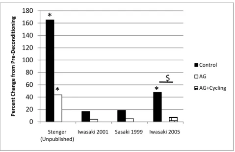
musculoskeletal system. Moreover, AG coupled with exercise is hypothesized to be a greater

benefit to physiological systems than AG alone.

2.4 Ground-Based Analogs

One of the greatest hindrances for evaluating countermeasures during spaceflight is the lack

of rigorous controls, whereas in the laboratory investigators can control almost all aspects of

research. For instance, falling behind the flight plan often led to abbreviating or completely

(32)

32

skipping exercise sessions in early spaceflight. Only since the ISS era has the exercise block

been completely preserved and only until the installation of ARED and T-2 in 2009 can the

actual workload and ground reaction forces be recorded and correlated to each astronaut (no

results yet of this endeavor). In addition, the lack of a control group in space also serves to

hinder evaluations of countermeasures as there is no one to which the astronauts can compare,

save for their own fitness level pre-flight. Indeed, having a control group in space that did not

exercise would present a major ethical dilemma, which is most likely the reason it has not been

executed.

Because of these difficulties in evaluating countermeasure effectiveness in space (not to

mention the cost of performing iterative studies), it became necessary to develop methods for

ground-based studies to represent an analog environment.

2.4.1 Human Ground-based Deconditioning Paradigms

Methods to create an analog space environment began almost with the dawn of spaceflight.

Some method was needed to immobilize the subject as well as reduce the effect of gravity, if

possible. A few deconditioning paradigms met these goals.

2.4.1.1 Wet or Dry Immersion

Immersion in water seemed like a logical choice as water offloads much of the effect of

gravity. The Russians have had vast experience with this method and first utilized wet

immersion, in which the subject is submerged to the neck, as a deconditioning measure.

However, subjects cannot stay submerged in water for extended periods of time and this method

was only used for very short studies on the order of a few days or less. A suitable modification

to wet immersion for longer duration studies is dry immersion in which the subject dons a rubber

suit and thus remains dry throughout the experiment.

2.4.1.2 Bed Rest

Another method developed to immobilize subjects is through the bed rest model, in which

subjects are confined to a bed for the duration of the study. The only exceptions to bed rest

would be for countermeasure exercise (ideally it would still be in a recumbent position). In order

(33)

33

to elicit appropriate responses, such as fluid shift, Russian investigators adjusted the paradigm to

a head down bed rest (HDBR) regime and experimented with angles until 6° head down was

deemed optimal (Pavy Le Traon et al. 2007). A 6° HDBR has since become the standard of bed

rest deconditioning.

2.4.1.3 Unilateral Lower Limb Suspension

Another method to immobilize subjects is through unilateral lower limb suspension (ULLS).

In this paradigm, the subject uses crutches and moves about on only one leg with a shoe that has

a large sole. Thus, the other leg does not receive any weight support. Because this paradigm

only immobilizes one leg and does not mimic the fluid shift and other deconditioning aspects

associated with spaceflight, it is not commonly employed.

2.4.2 Animal Ground-based Deconditioning Paradigm

Animals are extremely valuable to medical research because they are much cheaper than

human studies, and animals have the added benefit of enabling the use of invasive or destructive

techniques. Large bipedal primates, such as monkeys, can still utilize the HDBR model and

produce a similar response to humans. Quadrupeds, such as mice or rats, on the other hand

cannot use the HDBR model because they would still be standing and not immobilized. A

head-down, hindlimb unloading model (tail suspension, or SUS) was developed by E. R. Morey in

1979 to simulate a fluid shift and hindlimb immobilization (Morey-Holton et al. 2005). In this

model, the rat, for instance, is suspended from its tail so the hindlimbs are suspended and the

head is angled sharply downward. The rat usually retains some mobility with its front limbs; the

bar to which the tail is attached can swivel 360°. Since its inception in 1979, the hindlimb

unloading model has become the standard space analogue for rodents. One example of the

model is shown in Figure 17.

One problem does arise with animal experiments that is not present in human experiments.

In taking most physiological measurements, animals must be sacrificed because many techniques

(discussed later in 2.5) require the removal of parts of the anatomy. Because of this nuance,

animal experiments require an additional experimental „control‟ group that are killed at the

beginning of the experiment.

(34)

34

Figure 17. Hindlimb suspension model (Morey-Holton et al. 2005)

2.4.3 Ground-based Countermeasures

Countermeasures that have been assessed in human, ground-based studies have tended to

closely parallel countermeasures used in space. For example, the cycle ergometer, LBNP, and

isokinetic exercise of ground studies closely resemble the cycle ergometer, LBNP, and

MK-I/MK-II, respectively, which have been used in space. Other countermeasures, such as squats

and calf presses, have been performed on the ground with a flywheel device, such as that shown

in Figure 18. In addition, some countermeasures for ground studies have been developed that are

a combination of these countermeasures. For instance, an LBNP device with a vertical treadmill

(Figure 19) has been developed in California. This device uses LBNP to pull the horizontal

subject onto the vertical treadmill with the force equivalent to 1G. Another example is the

specially designed resistive exercise device for rigorous strength training as shown in Figure 20.

This device was used only in the Shackelford et al. 2004 study and had the capability to exercise

the lower and upper body for a total body workout.

(35)

35

Figure 18. Flywheel exercise device for performing supine squats and calf presses

Figure 19. LBNP device with a vertical treadmill (Lee et al. 2007)

Figure 20. Specially designed resistive exercise device for high intensity training for a

long-duration study (Shackelford et al. 2004)

(36)

36

As previously mentioned, ground-based studies investigating AG as a countermeasure have

either had subjects passively ride the centrifuge or have coupled centrifugation with exercises

such as cycling. In addition to centrifugation per se, some investigations have used standing or

walking (in 1G) to simulate the AG countermeasure.

A variety of countermeasures have also been developed for animals, with many being

similar to their human counterpart, such as passive centrifugation (in the –Gx direction), standing

or walking in 1G, or head-up tilt. On the other hand, some countermeasures to simulate resistive

exercise differ substantially from their human counterpart, such as dropping the animal from

58cm (Hauschka et al. 1987), climbing an 85° grid (Herbert et al. 1988), mechanical stimulation

where the rat‟s leg undergoes external loading (Innman et al. 1999), or electrical stimulation to

trigger muscle contraction (Haddad et al. 2006). In addition to these countermeasures, animal

testing is also very valuable for pharmaceutical countermeasures, where effectiveness and dose

rates can be investigated before progressing to human trials. These studies will be detailed in the

following chapter.

2.5 Measurement Techniques of Ground Studies

The measurement techniques used for assessing subjects in ground-based studies are vast.

Notable diagnostic techniques are discussed in the following sections, which are portioned into

body systems that the measurements diagnose. Special attention is given to where and how the

measurement is performed. This discussion of techniques will greatly aid in comprehending the

results of a number of studies.

2.5.1 Cardiovascular Measurements

2.5.1.1 Orthostatic Tolerance

Orthostatic tolerance is measured through a test that stresses the cardiovascular system.

This test usually takes the form of a head-up tilt to 60°-80° from a supine starting position.

However, Russian and Japanese investigators have used a +3G

z

(measured at the feet) overload

to provide cardiac stress, while other investigators have used a graded LBNP test as the stressor.

These tests are terminated when either the subject reaches a preset time limit or the subject

undergoes presyncope. Presyncope itself is defined in a variety of manners, though the most

(37)

37

common definition is any occurrence of the following: sudden drop in heart rate (>15 beats per

minute), systolic blood pressure (>25mmHG), or diastolic blood pressure (>15mmHG),

sweating, nausea, or clammy skin. Orthostatic tolerance time is the measured by the time

elapsed in the stress test.

2.5.1.2 Vital Signs / Cardiac Measurements

Vital signs that were typically measured in experiments are resting heart rate and blood

pressure. If the experiment involved an orthostatic tolerance test or an exercise test, these vital

signs were typically measured at the termination of the test as well. Heart rate was measured by

a standard electrocardiogram (EKG). Blood pressure was measured either by a

sphygmomanometer and cuff at the arm, by a photoplethysmographic method (PPG) in the ear

lobe, or by a finger blood pressure cuff. The PPG and finger blood pressure cuff could be used

to obtain continuous blood pressure measurements. Blood pressure was presented in the

literature by either systolic/diastolic pressure or by the mean arterial pressure, which is the

weighted average pressure of an artery over one heart cycle.

Cardiac function was measured through an EKG analysis. Stroke volume was calculated as

the product of the aorta cross sectional area (CSA) and the integral of the beat-to-beat aortic

outflow (measured by ultrasound). Cardiac output was generally calculated as the product of the

stroke volume and heart rate. Total peripheral resistance (TPR), which is the sum of resistances

in the systemic vasculature, was calculated as the mean arterial pressure divided by the cardiac

output. The degree of change in TPR with cardiovascular stress is indicative of the baroreflex.

Blood samples drawn from subjects provided the means to measure hematocrit, which is the

fraction of packed erythrocyte volume of blood. Blood assays could also be performed to

measure catecholamines, such as epinephrine and norepinephrine. Plasma volume – the liquid

component of blood – is also measured in a variety of manners. Older investigations used the

Evan‟s blue dye dilution method; however, this method has now been banned in many countries

because it involved the use of a radioisotope. Plasma volume is also estimated through

hematocrit measurements, although this method yields only percentage change of plasma

volume. A newer method to measuring plasma volume is the carbon monoxide (CO) rebreathing

method, which has been used extensively in recent studies.

(38)

38

2.5.1.3 Aerobic Capacity

Aerobic capacity was measured by a graded exercise test – either graded cycle ergometer or

graded treadmill – until the subject underwent volitional fatigue. Total exercise time, which is a

measure of endurance, was the total elapsed time of the exercise test. Gaseous exchange studies

were performed on these tests and included measurements such as VO

2

– the rate of oxygen

uptake per minute, V

E

– the volume of air inhaled in one minute (minute ventilation), and

Respiratory Exchange Ratio (RER) – the ratio between exhaled CO

2

and inhaled O

2

. Several

degrees of these measurements are made with the exercise test, including a resting measurement,

possible submaximal measurements, and maximum measurements (the maximum measurements

usually occur close to volitional fatigue).

2.5.1.4 Spectral Power Analysis

A relatively new technique to measure the autonomic nervous system is through heart rate

variability. A frequency analysis of heart rate variability divides the response into low frequency

(LF) and high frequency (HF). The high frequency (0.15 – 0.04 Hz) is indicative of

parasympathetic activity, while the low frequency is affected by both sympathetic and

parasympathetic activity. The ratio LF/HF reveals the sympathetic activity alone.

2.5.1.5 Measurements from Biopsies

Invasive techniques used for animals have involved the excision of sections of the basilar

and femoral arteries. These sections were used to analyze the artery CSA, media (i.e. smooth

cells and elastin fiber layers) thickness, intraluminal diameter, CSA of smooth cells, and number

of smooth cells. In addition, contractile responses of the samples were also obtained with the

addition of 100 mM potassium chloride (KCl).

2.5.2 Skeletal Muscle Measurements

The primary concern with muscle deconditioning lies with the antigravity muscles.

Therefore, measurements in experiments are almost exclusively performed on the soleus,

gastrocnemius, and quadriceps.

(39)

39

2.5.2.1 Non-invasive Techniques

Non-invasive techniques for assessing human muscle are widespread. Magnetic resonance

imaging (MRI) was used to determine the muscle CSA as well as the whole muscle volume. A

dynamometer was used to obtain muscle strength and maximum voluntary contraction (MVC)

data.

2.5.2.2 Muscle Fiber

Muscle biopsies were performed to determine muscle fiber characteristics. In many studies,

the biopsy was stained to differentiate between type I, type IIa, and type IIx fibers. From these

biopsies, fiber CSA (or diameter) and fiber type ratios could be obtained. In addition, fiber

performance characteristics were determined, such as V

o

, fiber shortening velocity, and P

o

, peak

force. Finally, a common measurement with animal experiments – since the whole muscle is

excised – is the wet weight of the muscle.

2.5.3 Bone Measurements

2.5.3.1 Direct Bone Measurements

Dual energy X-ray absorptiometry (DXA) scans have been used extensively throughout

manned spaceflight for BMD measurements. Many experiments still utilize this method for the

ability to compare to historical data. The major inhibitor to DXA scans, however, is that density

[g/cm

3

] is not really measured, but rather it is an areal density [g/cm

2

]. Another measurement

technique that has been used (often in conjunction with DXA) is peripheral quantitative

computed tomography (pQCT), which can make volumetric measurements and distinguish

between cortical and trabecular bone. Measurements are usually performed on the weight

bearing bones, with special attention given to areas of the hip such as the femoral neck and

greater trochanter. Other sights of interest are the calcaneus, lumbar spine, and tibia.

Because the bone (specifically the femur) is removed in animal experiments, mechanical

testing can be performed. Measurements that have been obtained are wet weight, dry weight,

elastic load, and maximum load.

(40)

40

2.5.3.2 Bone Markers

Bone can also be assessed through an array of bone markers found in blood serum and urine,

although they tend to be extremely variable depending on the time of day the sample is taken.

To reduce variability as much as possible, a 24-hour sampling period is performed. One

downside of bone marker measurements is that they represent the entire skeletal homeostasis and

are not capable of translating site-specific information. Bone markers can represent either

osteoblast or osteoclast activity; a detailed list is found in Table 1.

Table 1. Bone markers of the body

BONE FORMATION

Acronym

Name

Location

25(OH)D

25-Hydroxy vitamin D

Serum

1,25(OH)2D

1,25-dihydroxy vitamin D

Serum

OC

Osteocalcin

Serum

BSAP

Bone specific alkaline phosphate

Serum

PTH

Parathyroid hormone

Serum

BONE RESORPTION

NTX

n-telopeptide cross-link

Urinary

PYD

Pyridinoline

Urinary

DPD

Deoxypyridinoline

Urinary

Calcium

Urinary

(41)

41

3.0 Methods

An exhaustive review of AG literature – published and unpublished studies using either

human or animal subjects – was performed. Owing to the absence of long-radius centrifuge

experiments that investigate countermeasures to deconditioning, this study was limited to

analyzing results from intermittent AG experiments (via SRC). Paradigms of standing, walking,

or head-up tilt were also included as a variant of AG.

A review of the literature was also performed on traditional countermeasures, which are

defined as the types of countermeasures, or some variation, currently available on the ISS

(Section 2.4.3). It was impossible to perform an exhaustive review of this body of literature, as

there are a vast number of studies that have been performed. To bring some order to the

literature search, a method was adopted to start with the most recent studies and progress

backwards to older studies. This method was justified by reasoning that the most recent studies

will have the most advanced countermeasure protocols, and thus theoretically have the greatest

salutary effect on the subjects.

This literature was compiled into a master database that details the experimental methods as

well as the results of each study. The following tables are abridged versions of the master

database and serve to inform the reader of the experiments that were included in this study. AG

deconditioning studies that use human subjects are listed in Table 2, while those that use animals

as subjects are listed in Table 3. AG training studies (no deconditioning) with humans are found

in Table 4. Traditional countermeasure deconditioning studies using human subjects are found

in Table 5 and that use animals are found in Table 6. The complete database is shown in

Appendix B: Master Database.

Figure

Figure 5.  Bone changes due to A) normal aging – periosteal and endosteal expansion and  B) spaceflight – only endosteal expansion
Figure 7.  Tilt-translation ambiguity of the otoliths.  Backward head tilt has the same effect  as forward acceleration (Previc and Ercoline 2004)
Figure 9.  MK-I exercise device and exercises.  MK-II was similar in design to MK-I.
Figure 15.  Concepts for continuously rotating spacecraft.  A) Werner von Braun’s rotating  torus, B) Jooston 2007 rotating truss
+7

Références

Documents relatifs

Tools for co-construction and sustainable implementation of innovations in dryland Africa Atelier Inter- national APPRI : Apprentissage, Production et Partage d’Innovations :

information on the characteristics, connected with relevant spectral, time and space resolutions, of the radiation emission from objects considered as rotating black

Ce projet avait pour objectif de développer la riziculture biologique dans les principaux bassins rizicoles européens, où le riz est principalement cultivé dans des

Le propane sort au fond de la colonne à une température de 62 °C se dirige directement vers le premier préchauffeur du fractionnateur, passe ensuite vers les

Ecris la fin des phrases puis colorie de la bonne couleur en suivant les instructions de

Determinez la valeur de la position, et la valeur de chaque chiffre

Ensuite, il faut renforcer le programme d’éducation sanitaire concernant les AES (entrepris depuis 2006) en veillant sur son respect et sa continuité, ce programme

Par ailleurs, ce site a subi au tout début du projet (janvier-février 2014) une séquence sismique comprenant deux chocs de magnitude M6+ et de très nombreuses