HAL Id: tel-03080958
https://tel.archives-ouvertes.fr/tel-03080958
Submitted on 18 Dec 2020
HAL is a multi-disciplinary open access archive for the deposit and dissemination of sci-entific research documents, whether they are pub-lished or not. The documents may come from teaching and research institutions in France or abroad, or from public or private research centers.
L’archive ouverte pluridisciplinaire HAL, est destinée au dépôt et à la diffusion de documents scientifiques de niveau recherche, publiés ou non, émanant des établissements d’enseignement et de recherche français ou étrangers, des laboratoires publics ou privés.
Investigation into the role of human RAP1 in telomere
protection
Liudmyla Lototska
To cite this version:
Liudmyla Lototska. Investigation into the role of human RAP1 in telomere protection. Human genetics. COMUE Université Côte d’Azur (2015 - 2019), 2018. English. �NNT : 2018AZUR4240�. �tel-03080958�
Le rôle de la protéine RAP1 dans la protection des
télomères humains
Liudmyla LOTOTSKA
IRCAN – CNRS UMR 7284 – Inserm U1081
Telomere, senescence and cancer
Présentée en vue de l’obtention du grade de docteur en Sciences
De l’Université Côte d’Azur – UFR Sciences
Mention: Interactions moléculaires et cellulaires
Dirigée par le Pr. Eric GILSON
Co-encadrant Dr. Aaron Mendez-Bermudez
Soutenue le 17 Décembre 2018, devant le jury composé de :
Corine BERTOLOTTO
Directrice de Recherche, C3M, Nice Présidente du Jury,
Examinatrice
Paula MARTINEZ
Chargé de recherche, CNIO, Madrid
Rapportrice
Stéphane MARCAND
Directeur de Recherche, CEA, Fontenay-aux-roses Rapporteur
Eric GILSON
PU-PH, Professeur, IRCAN, Nice
Directeur de thèse
Investigation into the role of human RAP1 in
telomere protection
Liudmyla LOTOTSKA
IRCAN – CNRS UMR 7284 – Inserm U1081
Telomere, senescence and cancer
Presented to obtain the PhD degree
University of Côte d’Azur – UFR Sciences
Specialty: Molecular and Cellular Interactions
Supervised by Prof. Eric GILSON
Co-superviser Dr. Aaron Mendez-Bermude
z
Defended on the 17th of December 2018 in front of the committee:
Corine BERTOLOTTO
Director of Research, C3M, Nice President of the committee,
Inspector
Paula MARTINEZ
Staff scientist, CNIO, Madrid
Referee
Stéphane MARCAND
Directeur de Recherche, CEA, Fontenay-aux-roses Referee
Eric GILSON
PU-PH, Professor, IRCAN, Nice Director of the thesis
Résumé
Les télomères sont des séquences d’ADN, généralement répétées en tandem, localisées à l’extrémité des chromosomes linéaires. Une des fonctions principales des télomères est de différencier l’extrémité des chromosomes des cassures double-brin, et ainsi de prévenir l’activation des voies de réparation de l’ADN. Chez les mammifères, cette fonction est plus spécifiquement assurée par le complexe shelterin. Il s’agit d’un complexe hétérogène composé de six protéines distinctes: TRF1, TRF2, POT1, RAP1, TPP1 et TIN2, qui interagit spécifiquement avec l’ADN télomérique. Au sein de ce complexe, les protéines RAP1 et TRF2 coopèrent afin d’empêcher l’extrémité des chromosomes d’être perçue comme un dommage de l’ADN, ce qui autrement aboutirait à des fusions inter-chromosomiques suite au processus de réparation. La protéine TRF2 se lie directement à la molécule d’ADN dans laquelle elle s’enroule de façon spécifique. Cette propriété est primordiale pour générer une structure d’ADN en forme de boucle, appelée t-loop, et dont le bon fonctionnement des télomères dépend.Les travaux effectués au cours de cette thèse ont mis en évidence deux scenari indépendants dans lesquels la protéine RAP1 assure un rôle critique dans la stabilité des télomères. Premièrement, RAP1 peut prévenir les fusions inter-chromosomiques dans des cellules exprimant une forme altérée de TRF2 incapable de former des t-loops. Deuxièmement, l’inhibition de RAP1 dans des cellules en sénescence réplicative conduit à l’activation des voies de réparation de l’ADN et à la formation de fusions inter-chromosomiques. Ces observations font écho à des résultats précédents obtenus dans des cellules HeLa traitées avec l’inhibiteur de la télomérase BIBR1532, et dont l’expression de la protéine RAP1 était abolie par shRNA. De plus, j’ai montré que les fusions inter-chromosomiques engendrées par la perte de RAP1 sont dépendantes de la ligase IV, qui est un acteur principal de la voie de réparation de l’ADN par recombinaison non-homologue (NHEJ).
Dans l’ensemble, ces travaux démontrent l’importance de la protéine RAP1 dans la stabilité des télomères lorsque la protéine TRF2 est non fonctionnelle, mais aussi dans des situations physiologiques telles que la sénescence réplicative.
Mots-clés: Télomères, RAP1, TRF2, fusions inter-chromosomiques, recombinaison non-homologue (NHEJ), sénescence réplicative
Abstract
In mammals, the shelterin complex is the guardian of telomere stability. It operates through a set of six proteins (TRF1, TRF2, POT1, RAP1, TPP1 and TIN2) that binds telomeric DNA and protects it from being recognized as DNA double-strand breaks and therefore control DNA repair and DNA damage response pathways.
Among them, RAP1 and TRF2 cooperate and together protect chromosome extremities from end-to-end fusions. TRF2 is seen as a major factor to control telomere DNA topology by wrapping DNA around itself in a right handed manner. This property of TRF2 is required to promote the formation of t-loops, special DNA structures at telomeres that are considered as protective barriers to DNA damage response and fusion.
Here we demonstrate two independent situations where RAP1 dysfunction is critical for telomere protection. First, in cells expressing a wrapping-deficient TRF2 allele that cannot form t-loops, RAP1 appears as a backup anti-fusion mechanism. Second, RAP1 downregulation in replicative senescent cells leads to telomere fusions and DNA damage response activation. This is consistent with similar observations in HeLa cells treated with the telomerase inhibitor BIBR1532, and in which RAP1 expression was abolished by an inducible shRNA system. In addition, we show that fusions triggered by RAP1 loss are dependent upon ligase IV, which is a key player of the classical non-homologous end-joining (c-NHEJ) repair pathway.
Altogether, these results indicate that RAP1 takes over telomere protection when TRF2 cannot properly function or in the normal physiological situation, such as replicative senescence.
“Somewhere, something incredible is waiting to be known.”
Sharon Begley
Acknowledgements
First of all, I would like to thank the members of my Thesis committee, Dr. Corine Bertolotto, Dr. Paula Martinez and Dr. Stéphane Marcand, for their courage to accept my invitation and willingness to evaluate my thesis thoroughly. I greatly appreciate your efforts and time spent on my work.
Then I would like to extend my gratitude to my PhD supervisor Pr. Eric Gilson. It was a rare luck and privilege to hit the jackpot twice: being granted a Signalife PhD fellowship and being accepted into your team after project rotations. Thank you very much for allowing me to work on RAP1 and senescence and even more, thank you for believing in my work despite all the project ups and downs we faced together throughout the PhD.
My very special thanks go to my co-supervisor Dr. Aaron Mendez-Bermudez. I thank you enormously for being always by my side during this extreme but surely unforgettable PhD journey. I always admire your optimism and hard work, and I owe you a lot in terms of professional and personal skills I acquired while working with you. I must admit I always felt very privileged and happy to work with you. Thank you for all the amazing discussions and team work, and I’m looking forward to seeing our work published any time soon.
Many many thanks to my lab colleagues, past and present, for welcoming me in the team and providing unbelievable care and support during my PhD. Specifically, I thank Rita for sharing so many memorable moments together, for your helping hand with literally everything, for being my elder sister even though I don’t have a real one. Sabrina, my dear “mommy” friend, thank you for taking care of me in very difficult times and for your very big heart. Delphine, thanks a lot for always supporting and encouraging me, especially during my last year. Marie-Jo, I thank you for teaching me a hardcore biochemistry, such as EMSA and overhang assay. Nadir, thank you for being very patient with my orders and thanks a lot for always willing to help. Julien, I would like to thank you for being always a witty and positive person and I learned a lot from you about cancer and immunology. Alex, thanks for reminding me to buy some groceries and for your fantastic lemon cakes, which I will definitely miss in the future. Many thanks to our PhD crew, Alice, Martin, Sol, Charlene, for many great and sometimes crazy moments in the lab and not only. Alice, thanks for your interesting talks about corals, Martin, I learned from you a few cool methods to work with neurons, Sol, you’re my autophagy expert, Charlene, you are the one
who knows all the challenges to work with senescent cells. I also thank Claire, Melanie, Serge and many others for contributing to a good atmosphere in the team. I would like to mention also the contribution of Mounir and Marta during my first year of PhD.
I would like to express my gratitude also to our “floor-mates”, teams Liti and Cristofari. Thank you for being amazing neighbours and for sharing with me reagents when I needed the most. Special thanks go to my “stay-late-in-the-lab” mates, Arpita and Ben, for their presence during sometimes late working hours. Arpita, I will never forget our iconic trip to Berlin together with Rita. Ben, I am very grateful for your kind help with a lot of issues I had, no matter whether it’s personal or professional. Thank you also for providing me with an excellent French language expertise.
Many thanks go also to Ludo from the cytometry platform and Sabine for taking care of my radioactivity orders and being so supporting and cheerful person.
I would like to thank my high school teachers, Kavetska Tamara Borysivna and Vasyliuk Tetiana Ivanivna for always believing in me and being my real motivation to study biology and always stay curious. Thanks a lot to all my NaUKMA University teachers, for absolutely intense and fruitful 6 years of studies. Many thanks to Doan Svitlana Ivanivna and Kondratov Oleksandr, my Bachelor and Master thesis supervisors, who motivated me to pursue scientific career.
I absolutely thankful to all my friends who supported me during my PhD and not only. Special thanks to Natalia and Maksym, as well as Daria. I will never forget our trips and the 10th Nice-Cannes marathon in which we participated, they are among the best memories I had during my PhD. Many thanks to Sergii and Tania, for their enthusiasm in cheering me up always when I needed the most.
I am grateful to all the Signalife Labex community that became my second family. Among many people I met thanks to this amazing PhD Program, I would like to acknowledge Dr. Konstanze Beck and Anna Bliznyuk. I greatly appreciate your help with all the administrative procedures and for making me integrate into new culture and country smoothly. Many thanks to Lenka, Ramona and Nikita for many wonderful moments we shared and for your presence in the most difficult times. I feel very blessed to have you as my friends. My very warm wishes and special thanks go also to my Signalife buddy and flatmate Denisa. Words are not enough to express my gratitude to you. Hope we will all cross our paths in the future.
Oleksiy, among many things I am grateful to you, I would like to thank you especially for always believing in me and being my motivation to apply for PhD grants. I wouldn’t be here writing these acknowledgements without your love and support. I admire your scientific approach in our daily life, and I am very grateful to all the wonderful moments we spent together and looking forward to many more to come.
Last, but not least, I would like to thank my family. Specifically, my mom and dad, I absolutely adore you for being so loving and caring parents, for so much support I got from you during all my life and especially during my PhD.
Дякую усім, хто підтримував мене протягом моєї роботи над дисертацією, і особлива подяка моїй сім’ї, батькам, за любов і підтримку.
Table of contents
Page Résumé ……….. x Abstract ……….. xi Acknowledgements ………... xii Table of contents ………... 1 List of abbreviations……….. 3 Introduction……… 5Chapter 1. Telomere fusion: control, mechanisms and consequences……… 7
1. Telomere basic: structure and replication ……… 7
2. Factors that maintain and protect telomeres ……….. 10
3. Chromosome fusion and DNA repair ……… 13
3.1. The “good” and the “bad” of fused chromosomes: lessons from evolution ……… 13
3.2. The DNA double-strand break repair mechanisms promoting chromosome fusions……….. 16
3.2.1. Homologous recombination repair……… 17
3.2.2. Non-homologous end-joining repair………. 20
3.2.3. Alternative non-homologous end-joining pathway………. 23
3.3. DNA damage response at the sites of a double-strand break……… 26
3.4. Anti-fusion mechanisms at telomeres………. 29
3.4.1. Telomere factors controlling fusions………. 29
3.4.2. How cell cycle controls telomere fusion……… 31
Chapter 2. Research project……….. 36
1. Objectives of the study……….. 36
2. Article 1……….. 37
3. Article 2……….. 85
Chapter 3. General discussion and future perspectives……… 106
Bibliography……… 114
List of abbreviations
ALT alternative lengthening of telomeres a-NHEJ alternative non-homologous end-joining
ATM ataxia-telangiectasia mutated
ATR ATM- and Rad3-Related
BFB cycle breakage-fusion-bridge cycle
BLM Bloom helicase
BRCA 1/2 Breast-Cancer 1/2
c-NHEJ classical/canonical non-homologous end-joining
DDR DNA damage response
dHJ double Holiday junction
DNA-PK DNA-dependent protein kinase
DNA-PK cs DNA-dependent protein kinase, catalytic subunit
DSB double-strand break
dsDNA double-strand DNA
EXO1 Exonuclease 1
HHR homologous reombination repair
lncRNA Long non-coding RNA
MHEJ microhomologous end-joining
NHEJ non-homologous end-joining
POT1 Protection of telomeres 1
PARP1 Poly (ADP-ribose) polymerase
RAP1 Repressor Activator Protein1
RPA Replication protein A
ssDNA single-strand DNA
SSA single-strand annealing
TIN2 TERF1-interacting nuclear factor 2 TRF1/2 Telomeric repeat binding factor 1/2
TERC telomerase RNA component
Introduction
Over the course of evolution, mammalian telomeres developed the shelterin complex, a very elegant way to protect chromosome extremities from various DNA damaging insults. This complex is comprised of six proteins that can employ and control different interactors with one final goal: keep the telomeres functional.
One of highly dangerous events in the life of telomeres is chromosome fusion. Chromosome fusions usually occur when two chromosomes fuse end-to-end forming dicentric or ring chromosomes. Fusion can be executed by several different DNA repair pathways with or without telomere loss as an outcome. Non-homologous end-joining repair (NHEJ) and homologous recombination repair (HRR) are two common DNA repair pathways that promote telomere fusions.
In human, there are two key telomeric proteins that inhibit chromosome fusions: TRF2 and RAP1. TRF2 recruits RAP1 to telomeres and thus is considered as the master regulator of telomere protection, whereas the role of RAP1 at mammalian telomeres has been debatable for a long time.
Therefore, this Thesis manuscript sheds light on human RAP1 and its role in telomere protection.
In Chapter 1, the literature overview is a resume of the most recent and relevant findings in the field of DNA repair and DNA damage in connection to telomere fusion.
Chapter 2 represents the actual results of the thesis project. It is grouped into two articles. Article 1 is the published manuscript, which focuses mainly on how TRF2 protects telomeres in the context of DNA topology. We demonstrate that TRF2 can wrap DNA around its homodimerization domain (TRFH). The TRFH-dysfunctional mutant of TRF2 (called Top-less) is not able to wrap telomeric DNA, has decreased ability to promote t-loop formation and does not protect against DNA damage response (DDR), whereas it was able to rescue chromosome fusions. As part of the PhD project, we show that Top-less was not able to protect chromosomes from NHEJ upon RAP1 dysfunction. The latter result was an inspiration for Article 2, where we focus solely on the RAP1 role in telomere protection in the context of replicative senescence. Specifically, we found that in senescent cells RAP1 becomes essential to protect telomeres from DDR checkpoint and NHEJ repair. In Article 2 (the manuscript in preparation for publication) the main findings of my PhD work are described.
Chapter 3 is the final chapter of this dissertation. It discusses the main findings and suggests future work that would be important to better understand the mechanism of RAP1-dependent telomere protection in senescent cells.
During my PhD training, I have contributed to another research project on the role of TRF2 in pericentromere function. The results were recently published. Since this is not my main research project, the results are not discussed in this manuscript, but the article is attached in Appendix I. Article 3.
Chapter 1
Telomere fusion: control, mechanisms and
consequences
1. Telomere basic: structure and replication
Our understanding of telomere functions somehow started 80 years ago by the work of Herman Muller and Barbara McClintock. Muller was studying chromosome damage in Drosophila melanogaster upon ionizing radiation. He was the first to introduce the name “telomere”, which originates from the Greek words telos (end) and meros (part), and he used this term to describe the end parts of chromosomes [Muller HJ., 1938]. Simultaneously, McClintock highlighted the importance of telomeres during her studies of plant chromosomes in corn cells. She noted that the loss of natural chromosome ends (telomeres) destabilizes cellular genomes, causing chromosomes to become “sticky” and undergo adhesion and fusion at their ends, with consequent formation of dicentric chromosomes. She also demonstrated that the ends could be restored if chromosomes acquired a new telomere [McClintock B., 1939; McClintock B., 1941].
When the Watson-Crick double helical structure of DNA was resolved in 1953, it immediately suggested a mechanism of its replication – each strand in the duplex acts as a template to guide the synthesis of its complement. However, understanding the mechanism of the semi-conservative DNA replication [Meselson M. and Stahl FW., 1958] identified the “end replication problem”, consisting of the inability of cells to completely replicate the linear ends of DNA [Gilson E. and Ségal-Bendirdjian, E., 2010]. The first formulation of the end replication problem was focused on the lagging strand synthesis process where the gap generated by removal of the RNA primer at the 5’-end cannot be filled at the end of the chromosomal DNA, resulting in shortening of the newly synthesized strands with each round of DNA replication. This lagging strand problem was revisited later on by Cech and colleagues on the basis of the structure of the parental telomere extremity that corresponds to a 3’-overhang: the lagging chromatid is expected to somehow reproduce the 3’ overhang while the leading chromatid ends as a blunt DNA if the DNA polymerase synthesizes until the last nucleotide and by a 5’-overhang if the polymerase stop before, but
in any case the genetic information of the 3’ overhang of the parental DNA is lost. Thus, it was postulated that the end replication is more a leading strand than a lagging strand problem [Lingner J. et al., 1995]. Watson also predicted the existence of a protective mechanism to prevent the chromosomal shortening [Watson JD. et al., 1972].For Olovnikov, the terminal replication problem was the cause of a progressive telomere shortening, which also acted as an internal clock to determine the number of divisions that a cell can undergo during its lifespan. Therefore, telomere shortening could not only control the process of ageing but also acts as a molecular clock that counts the number of cycles that the cell can support [Olovnikov AM., 1973]. This is also consistent with the “Hayflick limit”, an observation made in the early sixties showing that cultured primary fibroblasts have a limited number of divisions [Hayflick L, 1965].
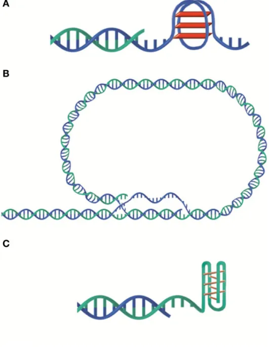
Structure and length of telomeres vary greatly among different species. The basic units of telomeres are tandem repeats, for instance, T2AG3 in mammals. Telomeric DNA is double-stranded with a short 3’-tail (150-300 kb) in the very end of the chromosome. However, plants can have blunt-ended telomeres [Kazda A. et al., 2012]. In species with relatively long telomeres the 3’-overhang can fold backwards and invade the double-stranded telomere DNA forming t-loops (Figure 1). Interestingly, t-loops have been discovered among different species. For example, Trypanosoma form very tiny t-loops, less than 1 kb in length, whereas field pea harbours extremely large t-loops, up to 50 kb in size [de Lange T., 2004]. T-loops are considered as structural barriers that protect telomeric DNA from being recognized by DDR machinery [Van Ly D. et al., 2018].
Both G-rich and C-rich telomeric strands may form additional complex DNA structures. For example, the G-rich strand can adopt a four-stranded G-quadruplex structure involving planar G-tetrads of guanine, while the C-rich strand can form the so-called i-motif with intercalated C·C+ base pairs (Figure 1). Different G-quadruplex structures exist, and they may be important to protect 3’-tails [Phan AT. et al., 2002].
Chromosomal DNA extremities can be recognized as accidental double strand breaks (DSBs) and treated as such by the cell leading to cell cycle arrest (DDR checkpoint) and recombination (DDR repair) [Shay JW., 2004]. Therefore, the natural ends of chromosome must be protected both from DDR checkpoint and repair.
Altogether, telomeres have to deal with two major problems: end replication and end protection. It turns out, they can do so with a help from different proteins that are described in the next section.
Figure 1. Different telomere structures. A. G-quadruplex. B. t-loop. C. i-motif. Illustration
2. Factors that maintain and protect telomeres
To overcome the end protection problem, cells developed a few strategies to keep the equilibrium of the telomere length, such as telomerase and alternative lengthening. At the same time, end protection problem can be effectively solved by means of the capping proteins.
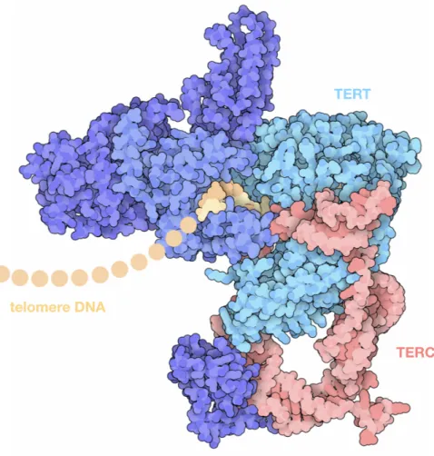
Telomerase was discovered in Tetrahymena thermophila by Greider and Blackburn[Greider CW. and Blackburn EH., 1985]. This protein is composed of two essential components: TERT (telomerase reverse transcriptase) and TERC (telomerase RNA component). TERC binds to the 3’-tail and serves as a template for TERT, which elongates telomeres (Figure 2) [Schmidt JC. and Cech TR., 2015].
Figure 2. Structure of the telomerase holoenzyme. This structure includes a reverse transcriptase (TERT) and associated proteins, an RNA template (TERC), and a short piece of the telomere DNA [Illustration from Protein Data Bank, http://pdb101.rcsb.org/motm/227].
Interestingly, in human and several other vertebrate species, but not all, TERT expression and telomerase activity are severely shut down in somatic tissues at the end of embryogenesis with the exception of progenitor or stem cells but to a level insufficient to fully replenish telomeric DNA ends at each round of cell division [Cong YS. et al., 2002].
An alternative way to counteract telomere attrition is based on homologous recombination and is called alternative lengthening of telomeres (ALT) pathway. Whereas approximately 85-90% of tumours utilize telomerase to elongate their telomeres [Kim NW. et al., 1994], some cancers (notably tumours of mesenchymal origin) use the ALT pathway that relies on homologous recombination[Apte MS. and Cooper JP., 2017].
Several telomere capping proteins, protecting telomeres from unwanted DDR activation, exist among different organisms. The prototypes of telomere capping protein complexes were identified in budding yeast, consisting mainly in two complexes: shelterin and CST (Cdc13-Stn1-Ten1). Shelterin is restricted to the RAP1 protein, which specifically binds telomeric DNA repeats to protect telomere DNA from fusion, while CST plays a key role against telomeric DNA degradation, checkpoint activation and telomere replication [Giraud-Panis MJ. et al., 2010].
The equivalents of the shelterin and CST complexes are found in many (if not all) eukaryotic organisms but with a great diversity of protein composition (Figure 3) [Giraud-Panis MJ. et al., 2013]. In mammals, shelterin is comprised of six proteins: TRF1 and TRF2 that through a Myb-like domain named Telobox bind to double-stranded DNA (dsDNA), TPP1 binding to POT1, which binds to the 3’-overhangs, TIN2 making a protein bridge between TRF1/TRF2 and TPP1 and finally RAP1 that, in contrast to budding yeast, binds indirectly to telomeric DNA via a direct interaction with TRF2 (Figure 3) [de Lange T., 2005; Giraud-Panis MJ. et al., 2013].
Figure 3. Telomere-associated proteins among different species. Modified from
3. Chromosome fusions and DNA repair
3.1. The “good” and the “bad” of fused chromosomes: lessons from evolution
Over the years, eukaryotic chromosomes acquired certain differences in their shape, size, composition, and number. These features made species distinguishable among each other, therefore, they appear to be important targets of evolution.
Simply, two ways of chromosome number evolution exist: fusion and fission, which lead to two different consequences for the genome: either reduction or amplification in the number of the existing genetic material [Schubert I., 2007].
In terms of evolution, there is a large body of evidence that end-to-end fusions lead to reduction in the total number of chromosomes. For example, fusion of two ancestral primate chromosomes created human chromosome 2 [Ijdo W. et al., 1991]. Fusions were also a c o m m o n c a u s e o f r e d u c e d c h r o m o s o m e n u m b e r a m o n g a n t s p e c i e s
Mycetophylax conformis and Mycetophylax morschi [Cardoso DC. et al., 2014], and plant
Arabidopsis thaliana [Lysak MA. et al., 2006]. Evolution of the budding yeast genome is
characterized by the whole-genome duplication (from n=8 to n=16 chromosomes). However, it has been observed that in some other yeast species, such as
Zygosaccharomyces, Kluyveromyces, Lachancea, and Ashbya, the number of chromosomes
varies from 6 to 8. The most common event in reducing chromosome number among those yeast is telomere end-to-end fusions [Gordon JL. et al., 2011]. Recently, two independent groups created Saccharomyces cerevisiae strains with dramatically reduced number of chromosomes [reviewed in Liti G., 2018]. Luo et al. engineered yeast with n=2 chromosomes, and Shao and colleagues fused all the chromosomes into a single chromosome in a functional yeast [Shao Y. et al., 2018; Luo J., 2018]. Both studies concluded that reduced chromosome number causes no major growth defects when cells are grown under various conditions and stresses. The groups showed that the n = 1 and n = 2 strains can undergo sexual reproduction, albeit with reduced efficiency compared with wild-type yeast, and produce spores that are slightly less viable. Therefore, these engineered yeast strains constitute powerful resources for studying fundamental concepts in chromosome biology [Liti G., 2018].
Probably the most impressive example of natural chromosome reduction is the Indian muntjac (Muntiacus muntjak), whose females only have 6 chromosomes, and its males only 7
[Wurster DH. and Benirschke K., 1970]. By means of comparative mapping and sequencing approach, Tsipouri and colleagues characterized the sites of ancestral chromosomal fusions in the Indian muntjac genome [Tsipouri V. et al., 2008].Specifically, they screened an Indian muntjac bacterial artificial chromosome library with a telomere repeat-specific probe. They found that all seven Indian muntjac sequences, that were analyzed, contained centromeric satellite I repeat sequences immediately adjacent to the telomeric-repeat block [Tsipouri V. et al., 2008]. Furthermore, high frequency of tandem fusions, which arise from telomere and centromere repetitive elements, has been proposed as the main mechanism of stasipatric (rapid) speciation that is common among muntjacs [Wang W. and Lan H., 2000].
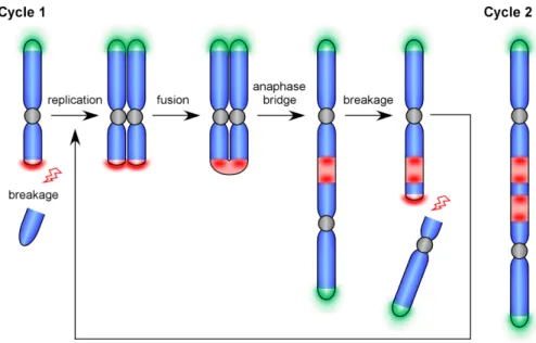
Presence of telomere and centromere or pericentromere repeats and their duplication at the fusion site is a proof of DNA damage repair by non-homologous end-joining. This type of repair is usually error-prone and can give rise to certain types of genetic instability through initiation of ‘breakage-fusion-bridge’ (BFB) cycles, first discovered by McClintock in Zea mays [McClintock B.,1939; McClintock, 1941]. Such cycles start with the loss of telomeres at the ends of the chromosomes (Figure 4). Then DNA is replicated, and sister chromatids with fused ends are formed. During anaphase, centromeres of those sister chromatids are pulled in the opposite directions forming bridges as the ends are fused. While pulling centromeres apart from each other, a break of the bridge occurs at any point in a way that a daughter cell receives an uneven chromosome without telomeres. Telomeres can be restored by telomerase, but if the chromosome still lacks telomeres at the ends, the BFB cycle will continue during the next cell division.
BFB cycles cause duplications, deletions, inversions as secondary rearrangements in the chromosomes. Genetic instability that occurs becomes a driving force of evolution. As described above, it can lead to appearance of new species (which can be considered as "good"). On the other hand, it can be a cause of establishing and promoting different malignancies (which for a normal cell and the whole organism is usually considered as "bad") [Selvarajah S. et al., 2006; Kwei KA. et al., 2010; Martínez P. and Blasco MA., 2017; Maciejowski J. and de Lange T., 2017]. Therefore, detailed studies of the mechanisms that lay behind genetic instability are needed to better understand how the switch between “good” and “bad” occurs.
The next part of this manuscript is focused on DNA repair mechanisms that are in connection to telomere fusion.
3.2. The DNA double-strand break repair mechanisms promoting chromosome fusions
In normally functioning cells, chromosome fusion must be prevented in order to maintain genome stability. In this regard, cells developed several mechanisms that inhibit fusion at natural chromosome ends. This is one of the main function accomplished by telomeres. Among the telomere strategies to prevent fusion, one can cite peculiar DNA structures (t-loops, 3’-overhangs), shelterin and other associated telomere factors.
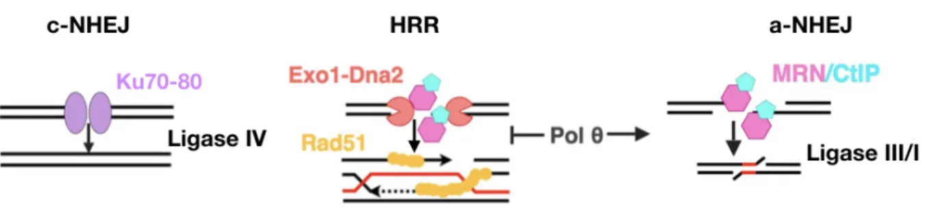
In the absence of a proper anti-fusion activity, chromosome extremities can fuse by different recombinational repair mechanisms: homologous recombination repair as well as classical and alternative non-homologous end-joining (Figure 5).
In the next section of this manuscript, the mentioned pathways will be presented in detail with a focus on their relationship to chromosome fusions.
3.2.1. Homologous recombination repair. Homologous recombination repair (HRR) or homology-directed repair (HDR) is a high-fidelity pathway of DSB repair. Although many different proteins and some of the non-coding RNAs are implicated in HRR, and several different mechanisms exist, the basic principles are conserved among prokaryotes and eukaryotes. This type of repair relies on homologous recombination, where a homologous DNA template is used to repair and restore the sequence around the break. DSB repair by HRR in mitotic cells favours the use of the sister chromosome over the homologous chromosome as a template donor [Kadyk LC. and Hartwell LH., 1992]. Notably, repair of DSBs by means of HRR can lead to two different consequences: crossover and non-crossover. For example, crossover occurs during meiosis and can be also used to generate genetic diversity [Baudat F. and de Massy B., 2007; Heyer WD. et al., 2010]. However, the primary mechanism of HRR, gene conversion, does not result in the crossover, which makes it a faithful DNA repair process. Also, synthesis-dependent strand annealing pathway (SDSA) does not result in crossovers and is important to preserve genomic integrity [Verma P. and Greenberg RA., 2016]. When DSBs cannot be processed by the conventional mechanism of HRR, cells decide between SDSA, double-strand break repair (DSBR), break-induced replication (BIR), or single-strand annealing (SSA) (Figure 6) [Chapman JR. et al., 2012; Verma P. and Greenberg RA., 2016; Wright WD. et al., 2018].
Regardless of which choice has been made, initial steps of HRR share the same principles. First, after a DSB occurs, broken DNA ends undergo nucleolytic end resection to generate 3'-ssDNA overhangs. Generation of 3'-overhangs can be characterised by a two-step mechanism. First, in higher eukaryotes, an immediate recruitment of Mre11-Rad50-Nbs1 (MRN) along with CtIP complex occurs at the sites of DSB [Lamarche BJ. et al., 2010; Langerak P. et al., 2011]. MRN-CtIP removes small oligonucleotides to generate a short protruding end [Muraki K. and Murnane P., 2018]. Next, several other enzymes are recruited to produce long single-stranded overhangs by resection, for instance, Exonuclease 1 (EXO1), DNA replication helicase/nuclease 2 (DNA2) [Mimitou EP. and Symington LS., 2009; Jasin M. and Rothstein R., 2013]. Furthermore, Bloom helicase (BLM) can be important for long-range resection of DNA ends [Nimonkar AV. et al., 2011], as well as Werner helicase (WRN). The latter can substitute BLM in DNA2-mediated resection [Sturzenegger A. et al., 2014]. Therefore, BLM and WRN act epistatically and ensure the single-strand 3'-overhang formation on both strands of the break.
Figure 6. Different pathways of homologous recombination repair in human. Modified
from [Heyer WD. et al., 2010].
Moreover, breast cancer suppressor BRCA1 can also take part in the initial steps of HRR, since it has been shown to interact with MRN [Zhong Q. et al., 1999] and CtIP [Yu X. et al., 1998] and promote HRR [Moynahan ME. et al. 1999; Stark JM. et al. 2004], as does CtIP [Sartori AA. et al. 2007; Bennardo N. et al. 2008]. Interestingly, BRCA1 may control the CtIP-dependent recruitment of DNA2 to DNA damage sites for subsequent DSB resection [Hoa NN. et al., 2015]. It has been demonstrated that BRCA1-A complex comprised of ubiquitin interacting motif (UIM) containing protein RAP80, adapter protein Abraxas, MERIT40 (mediator of RAP80 interactions and targeting 40 kDa, also known as NBA1), BRCC45, and deubiquitinylating enzyme BRCC36 guides BRCA1 to the sites of DSB through interaction with UIMs of RAP80 [reviewed in Greenberg RA., 2008; Daley JM. et al., 2014; Her J. et al., 2016]. Also, a proper recruitment of BRCA1 to the DSB sites is controlled by lncRNA DDSR1 [Sharma V. et al., 2015].
After 3'-overhangs are generated, they are immediately covered by ssDNA-binding replication protein A (RPA). This binding prevents the formation of unwanted secondary structures on ssDNA [Chen H. et al., 2013]. The next step is to load Rad51 on 3'-overhangs. In humans, several proteins can be important to replace RPA with Rad51. Among them, Rad52 appears to be essential to physically replace RPA with Rad51 and promote, therefore, formation of the nucleoprotein filament [Sugiyama T. and Kowalczykowski SC., 2002; Plate I. et al., 2008]. Notably, it has been also shown that BRCA2 interacts directly with Rad51 and recruits it to the RPA-coated ssDNA at the DSB site [Her J. et al., 2016]. Therefore, several mechanisms exist for the proper functioning of initial steps of HRR.
When the filament is formed, Rad51 initiates the search for a homologous template followed by the donor DNA strand invasion, formation of a D-loop and subsequent DNA synthesis mediated mainly by DNA polymerase δ in eukaryotes [Maloisel L. et al., 2008; McVey M. et al., 2016]. Strand invasion and formation of the D-loop is mediated by Rad54 (a protein that belongs to the SNF2/SW12 family in humans), which removes Rad51 from the filament [Kanaar R. et al., 1996; Li X. and Heyer WD., 2009; Mazin AV. et al., 2010].
To complete HRR, three different scenarios are possible (Figure 6). First, if the second DNA end is present, mitotic cells mainly follow the SDSA pathway [Andersen SL. and Sekelsky J., 2016]. Therefore, either of 3'-overhangs or even both of them can invade the donor template. The invading strand is further displaced during the D-loop migration and the newly formed DNA strand anneals back to the ssDNA overhang of the second end, resulting in a non-crossover product [Heyer WD. et al., 2010]. However, a second possibility is the creation of a double Holiday junction (dHJ), which can result either in the crossover or non-crossover outcome depending on the proteins involved in the processing. For example, BLM together with topoisomerase 3 alpha (Top3A) process dHJs in a way that crossover does not occur [Wu L. and Hickson ID., 2003].
BIR takes place when there is only one accessible DNA end. The available 3'-overhang invades the homologous DNA and then extends to the end of the chromosome. In higher eukaryotes, BIR is an important mechanism to repair and restart broken replication forks, as well as it can contribute to the alternative lengthening of telomeres [reviewed in Verma P. and Greenberg RA., 2016].
SSA is another type of DSB repair, which can be considered as an alternative pathway of HDR [Verma P. and Greenberg RA., 2016]. SSA is initiated when DSB occurs between homologous direct repeats. These repeats first are resected bidirectionally, then nucleases cleave off unpaired 3'-overhangs. The final step is annealing and ligation of the DSB. Remarkably, SSA does not require Rad51 filament, therefore, is Rad51-independent. Because the nuclease cleavage can result in deletion of repeats, SSA is a mutagenic process [Verma P. and Greenberg RA., 2016; Bhargava R. et al., 2016].
In summary, homologous recombination repair is represented by several pathways. All these pathways are conserved among different organisms, and some of them are redundant. However, given how complex is the interaction among different proteins within one pathway, new approaches emerge in order to better dissect the mechanism of HRR. One of these approaches relies on super-resolution microscopy methods to study the process at single-molecule resolution [Kaniecki K. et al., 2018].
If HRR is not properly executed, this can lead to rapid telomere resection and loss followed by appearance of telomere-free ends and massive telomere-free chromosome fusions. Since HDR relies on the presence of homologous DNA template, it favours formation of sister chromatid fusions and can promote unequal sister chromatid exchange that will create fragile chromosomes [Rudd MK. et al., 2007].
3.2.2. Non-homologous end-joining repair. Non-homologous end-joining is a second type of repair that cells employ on a regular basis. Described as a “willy-nilly” end-joining [Deriano L. and Roth DB., 2013], it relies on joining damaged DNA strands together. It can be either very robust and precise if the ends do no miss nucleotides or do not require further processing; otherwise, it can lead to certain genetic instability or diversity [Lieber MR., 2010; Chang HHY. et al., 2017]. For example, V(D)J recombination in immune cells absolutely requires NHEJ and is considered as a normal physiological process [Malu S. et al., 2012], whereas incongruous NHEJ may promote cancer formation [Sishc BL. and Davis AJ., 2017]. The latter is due to formation of dicentric chromosomes that initiate BFB cycles or chromotrypsis [Maciejowski J. and de Lange T., 2017]. If NHEJ acts between two telomeres, it fuses chromosomes as an immediate outcome [Marcand S., 2014]. Therefore, NHEJ is the prime mechanism to create both intra- and inter-chromosome fusions.
Nowadays many different proteins involved in classical or canonical NHEJ have been characterized (which is often referred to as c-NHEJ), however, the basic principles on how
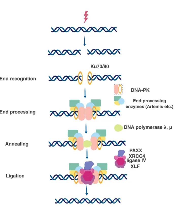
this pathway is executed are the same among various species. In general, the c-NHEJ can be divided into three very general steps: DSB recognition, processing, and ligation (Figure 7) [Lieber MR., 2010; Yang K. et al., 2016].
There are two essential components to make NHEJ work: DNA-PK and the ligase IV complex [Waters CA. et al., 2016]. However, presence of those two complexes can be enough only if the DSB forms blunt ends and do not require further processing. If the DSB is followed by incompatible DNA ends, direct ligation cannot be performed, and therefore several end-processing and annealing proteins are on call before the ligation can occur [Chang HH. et al. 2016; Chang HHY. et al., 2017].
DSB recognition. The first protein, which is recruited within seconds to the sites of DSB, is Ku. In human, Ku is very abundant (500 000 molecules per cell) and demonstrates the strong affinity for DNA binding [Fell VL. and Shild-Poulter C., 2015]. Nevertheless, both
in vitro and in vivo studies show that just two molecules of Ku are enough to cover the sites in
the vicinity of the damaged DNA, presumably, each one covering the broken ends [Roberts SA. and Ramsden DA., 2007; Britton S. et al., 2013]. In eukaryotes, Ku is present as a heterodimer, which is called Ku70/80. After being recruited to the sites of damage, Ku70/80 promotes sequestration of several other NHEJ factors for the appropriate repair (nucleases, polymerases, ligases), thus, Ku acts as a hub or scaffold protein [Fell VL. and Shild-Poulter C., 2015]. In yeast, there are Yku70/80 orthologs for mammalian Ku proteins. Strikingly, Yku is not an essential protein in yeast, whereas loss of human Ku86 leads to massive telomere loss and cell death [Wang Y. et al., 2009].
Ku forms the DNA-PK complex together with the DNA-dependent protein kinase catalytic subunit (DNA-PKcs) [Spagnolo L. et al., 2006]. DNA-PKcs has been discovered only in higher eukaryotes so far. Recently, the cryo-EM structure of human DNA-PK has been solved. Two research groups independently demonstrated that DNA-PKcs and Ku70/80 together form a DNA-binding bridge or tunnel. DNA-PKcs is relatively proximal, and Ku70/80 is distal, to the free DNA end. DNA-PKcs and Ku70/80 both wrap around one and a half turn of the DNA duplex with the blocked DNA end flanking outside of the complex [Yin X. et al., 2017; Sharif H. et al., 2017]. Notably, DNA-PKcs alone barely binds to DNA but strongly binds to DNA in the presence of Ku70/80 [Yin X. et al., 2017]. DNA-PKcs can be autophosphorylated or trans-phosphorylated by ATM. These two states of phosphorylation regulate the switch between recruitment of Artemis or ligase reaction [Uematsu N. et al., 2007; Jiang W. et al., 2015].
DSB processing. Components of the DNA-PK complex can recruit to the sites of DSB DNA end-processing factors suchas Artemis [Riballo E. et al., 2004], Werner [Chen L. et al., 2003; Shamanna RA. et al., 2016], polynucleotide kinase-phosphatase (PNKP) [Shimada M.
et al., 2015], APTX–polynucleotide kinase-phosphatase-like factor 1 (APLF) [Macrae CJ. et al., 2008; Grundy GJ. et al., 2013], DNA polymerases Pol λ and Pol μ [Capp JP et al., 2006; Chayot R. et al., 2012], terminal deoxynucleotidyl transferase (TdT) [Boubakour-Azzouz I. et al., 2012]. Depending on how complex is the DSB, the mentioned factors can be required for the accurate DNA end cleavage and annealing in order to facilitate further ligation [Yang K. et al., 2016]. Interestingly, mammalian DNA-PKcs and the rest of the mentioned processing factors (except DNA polymerases) do not have orthologs in budding yeast S.cerevisiae [Dudásová, Z. et al., 2004]. Instead, MRX complex becomes of outstanding importance to execute NHEJ [Emerson CH. and Bertuch AA., 2016]. Regarding the DNA polymerases in yeast, Pol4 is a Pol X family polymerase (related to mammalian polymerases λ and μ). Moreover, yeast employs also Pol3 (mammalian Polδ) [Ramsden D., 2011].
DSB ligation. Importantly, c-NHEJ is distinct in this regard, because it relies on the ligase IV function [Wang H. et al., 2001]. In mammals, ligase IV forms a complex with XRCC4 and XLF [Ahnesorg P. et al., 2006]. XRCC4 and XLF are particularly important for bridging DNA molecules and therefore promoting ligase IV activity [Andres SN. et al., 2012].
PAXX (XRCC4 paralogs) is a regulator of XRCC4 [Xing M. et al., 2015]. Besides XRCC4-ligase IV complex, it interacts with Ku70 directly and promotes Ku accumulation at the break [Ochi T. et al., 2015; Liu X. et al., 2017]. An emerging view is that PAXX is an additional protein recruited to hard-to-repair DSBs [Tadi SK. et al., 2016], where it can promote DNA polymerase λ activity [Craxton A. et al., 2018].
In yeast, ligation occurs due to the activity of DNA ligase IV or Dnl4 in S. cerevisiae. Dnl4 is strongly associated with Lif1. If Lif1 is dysfunctional, Dnl4 becomes unstable [Herrmann G. et al., 1998]. Mrx and Yku were reported to promote association of the Dnl4-Lif1 complex to the DSB, as well as Nej [Emerson CH. and Bertuch AA., 2016].
3.2.3. Alternative non-homologous end-joining pathway. It has been reported that critically short telomeres tend to fuse end-to-end via non-canonical end-joining that requires microhomology [Letsolo BT. et al., 2010].
Alternative NHEJ (a-NHEJ) in certain literature reviews can also be referred to as alternative end-joining (a-EJ). An early evidence for the existence of alternative end-joining pathways came from studies in Ku-deficient budding yeast [Boulton SJ. and Jackson SP., 1996]. In mammals, similar observations were made in p53 knockout mice lacking the
components of the NHEJ machinery, but yet supporting insertions, deletions and microhomology [Zhu C. et al., 2002].
A-NHEJ is distinct from c-NHEJ and HRR on several counts. First, it does not necessarily require homology to function as HRR does. However, certain types of fusions that occur through a-NHEJ can use microhomology, which makes it similar to the SSA pathway [Verma P. and Greenberg R., 2016; Sallmyr A. and Tomkinson AE., 2018]. The latter pathway is called MMEJ or MHEJ (microhomology-mediated repair). In contrast to SSA, MMEJ relies on very short homologies, less than 20 bp [Pannunzio NR. et al., 2014; Mladenov E. et al., 2016]. The final step in the repair is ligation of DNA, but in comparison with c-NHEJ it is ligase IV independent process, which is executed via either ligase III or I [Wang H. et al., 2005; Simsek D. et al., 2011; Masani S. et al., 2016].
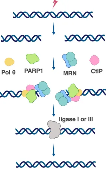
The distinct characteristics of a-NHEJ are the key players involved in the pathway: poly(ADP-ribose) polymerase1 (PARP1), DNA polymerase θ (POLQ), and Ligase III/I [Chang HHY. et al., 2017] (Figure 8). The main role of PARP1 is to catalyse the polymerization of ADP-ribose units — derived from the ADP donor NAD+ — resulting in the attachment of either linear or branched PAR polymers to itself or other target proteins. PARP1 is therefore believed to be a sensor of DNA damage [Ray Chaudhuri A. and Nussenzweig A., 2017]. By means of several biochemical and super-resolution microscopy approaches, it has been demonstrated that PARP1 competes with KU for DNA DSB repair. KU can be removed from the sites of DSBs by PARylation that is performed by PARP1 [Wang M. et al., 2006; Yang G. et al., 2018]. Furthermore, PARP1 recruits MRN complex to the repair centre (Figure 8) [Haince JF. et al., 2008]. In analogy to HRR, MRN together with CtIP may be necessary for end processing and removal/recruitment of other proteins [Lamarche BJ. et al., 2010]. Contrary to HRR, MMEJ does not require γ-H2AX, neither BLM nor EXO1 for end processing [Truong LN. et al., 2013].
After DNA is recognized and processed, DNA polymerase θ is needed for a stable annealing of DNA strands. Polθ uses short microhomology (2-6 bp) for annealing. Notably, if this microhomology is not present, due to its transferase activity, Polθ can add several nucleotides to create microhomology at the site of the break [Kent T. et al., 2015]. Remarkably, polymerase θ was found to promote a-NHEJ at dysfunctional telomeres in cooperation with PARP1 [Mateos-Gomez PA. et al., 2015]. Recently, another DNA polymerase β was reported to participate in a-NHEJ [Ray S. et al., 2018].
Figure 8. Alternative NHEJ in human.
The final step of a-NHEJ is ligation of the annealed DNA. End ligation is ligase IV-independent and relies on the activity of either ligase III or I. Ligase III seems more efficient in comparison to Ligase I [Lu G., 2016]. Ligase III can form a complex with XRCC1, which was found to co-exist with MRN in a-NHEJ [Caldecott KW. et al., 1994; Della-Maria J. et al., 2011].
Whether this pathway is a backup of the main c-NHEJ or acts independently, there is a body of evidence that a-NHEJ is employed by the cell to create genetic diversity [Ottaviani D. et al., 2014]. Nevertheless, there are still many outstanding questions that have to be explored for better understanding how a-NHEJ works and what is the prime importance of this pathway.
3.3. DNA damage response at the sites of double-strand break
In eukaryotes, DNA damage response (DDR) is a cascade of signalling events within the cell as a response to DNA damage. Like a classical signal transduction pathway, it is comprised of signal sensors, transducers, mediators and effectors. One of the peculiarities of this signalling pathway is that instead of ligand-receptor interactions, DDR machinery upstream events rely on the direct recognition and further processing of damaged DNA molecules. The sensors of this pathway are the proteins that directly recognize damaged DNA and activate upstream DDR kinases. Then the signal is amplified through the activation of different mediator kinases, and the final effectors spread the signal that will determine the fate of the cell.
DNA repair and DDR pathway are tightly connected with each other. MRN complex is essential in this regard because it appears to be in the front line, where different repair choices and DNA damage sensing merge [Williams RS. et al., 2007]. In the context of DDR, MRN is believed to play a role as a sensor of the damage. It is activated immediately at the damaged sites and directly binds dsDNA. Also, MRN acts as the main factor required for the rapid localization of ATM (ataxia-telangiectasia mutated) to DSBs [Lee JH. and Paull TT., 2005]. It is important to mention that ATM and ATR (ATM- and Rad3-Related) pathways are d i s t i n c t f r o m e a c h o t h e r. AT M r e s p o n d s t o D S B s , w h e r e a s AT R - t o both ssDSBs and dsDSBs with a greater preference to ssDSBs, and is particularly important to repair the DNA lesions that occur during replication [Maréchal A. and Zou L., 2013].
At the sites of DSBs, ATM is a transducer protein kinase, which becomes activated by phosphorylation of its serine residue Ser1981 in human [Bakkenist CJ. and Kastan MB., 2003]. It has been shown that ATM activation is impaired in cells with MRN deficiencies [Uziel T. et al., 2003]. Furthermore, the carboxyl terminus of Nbs1 (a protein of the MRN complex) is known to interact with ATM [Falck J. et al., 2005]. Moreover, recently it was demonstrated that several proteins can enhance ATM signalling via direct interaction with the MRN complex. For instance, a signalling mediator, p53-binding protein 1 (53BP1) through it BRCT domain binds to MRN complex directly and regulate ATM phosphorylation of its substrates [Lee JH. et al, 2010]. Rad17, a replication checkpoint protein, also binds directly to MRN and is required for the early recruitment of the MRN complex to the DSB site, and it contributes to ATM activation [Wang Q. et al., 2014]. Smad7 interacts with Nbs1
and enhances the interaction between ATM and Nbs1 upon DNA damage response, leading to phosphorylation of downstream substrates [Park S. et al., 2015].
Among substrates that are phosphorylated by ATM are BRCA1, Chk2, p53, H2AX, MDC1. The latter two act in cooperation. It has been described that H2AX phosphorylation is performed by ATM as one of the upstream events of DDR activation. The phosphorylated histone is called γH2AX. γH2AX, in turn, acts as a hub for nuclear foci formation, the DDR centres where many DNA repair proteins and chromatin remodelling factors are accumulated [Iijima K. et al., 2008; Clouaire T. et al., 2017; Podhorecka M. et al., 2010].
Formation of γH2AX foci is one of the key steps in DDR signalling and repair in the context of chromatin. MDC1 was found to directly interact with γH2AX and therefore contribute to the γH2AX foci formation. At the same time, MDC1 interacts with ATM. Thus, it acts as a mediator between ATM and γH2AX and helps spread phosphorylation of γH2AX by ATM over long chromosome distances[Stewart GS. et al., 2003; Lee JH. et al., 2005; Stucki M. et al., 2005].
Although there is no doubt that phosphorylation of H2AX is essential for the DDR pathway, it has been documented that many other chromatin modifications occur, such as DNA methylation, different histone modifications etc., which require specific chromatin remodelling factors [reviewed in Polo SE. and Jackson SP., 2011]. Notably, γH2AX triggers cascades that rely on ubiquitylation and SUMOylation in order to recruit BRCA1 and 53BP1 to the damaged sites [reviewed in Daley JM. and Sung P., 2014; Muraki K. and Murnane JP., 2017].
In heterochromatin repair, ATM through its substrate Chk2 phosphorylates KAP1 and also stimulates further dissociation of heterochromatin protein HP1-β from H3K9me3 around DSBs [Goodarzi AA. et al., 2008; Bolderson E. et al., 2012]. Also, cells that do not form 53BP1 foci, fail to form phosphorylated KAP1 foci [Noon AT. et al., 2010]. Interestingly, changes in the chromatin structure upon DDR activation have been reported to increase chromosome mobility [reviewed in Hauer MH. and Gasser SM., 2017; Smith MJ. and Rothstein R., 2017; Marnef A. and Legube G., 2017]. This phenomenon is believed to be common in yeast, where damaged DNA becomes highly mobile and moves within the nucleus to the repair centres [Lisby M. et al., 2003]. Notably, DSBs that are unable to be repaired move to the yeast nuclear periphery [Nagai S. et al., 2008]. In higher eukaryotes, chromosome mobility is relatively weaker compared to yeast, however, it does occur. For
example, increased chromosome movement of uncapped telomeres in mouse cells has recently been associated with the 53BP1 repair protein and LINC-domain complex [Dimitrova N. et al., 2008; Lottersberger F. et al., 2015].
As mentioned before, a second transducer kinase pathway can be activated as a response to DNA damage. This pathway relies on ATR. In the DSB repair, ATR is activated when the resection of DNA ends takes place, and therefore ssDNA overhangs of certain length are present [Shiotani B. and Zou L., 2009]. In this process, RPA that coats ssDNA, is required for the recruitment of the ATR-ATRIP complex to the sites of DNA damage [Zou L. and Elledge SJ., 2003]. In order to be activated at the site of ssDNA, ATR-ATRIP interacts with several other proteins. For example, TopBP1 is one of the best characterized proteins that contains an ATR-activation domain to promote ATR kinase activity through interaction with both ATR and ATRIP [Kumagai A. et al., 2006; Mordes DA. et al., 2008]. Interestingly, TopBP1 can be activated through phosphorylation by ATM [Yoo HY. et al., 2007]. Apart from that, ATM may also promote the recruitment of of TopBP1 to sites of DNA damage through γH2AX and Mdc1 [Wang J. et al., 2011].
It turned out that the MRN complex (through its subunit Nbs1) is important for activation of ATR [Shiotani B. et al., 2013]. In line with this, MRN can also recruit TopBP1 to ssDNA-to-dsDNA junctions [Duursma AM et al., 2013]. Recently another TopBP1-independent way to activate ATR was described. Human RPA-binding protein ETAA1 can directly bind to RPA and propagate ATR signalling [Haahr P., et al., 2016; Lee YC. et al., 2016]. Last but not least, ATR can be activated via autophosphorylation [Liu S. et al., 2011]. Altogether, activation and recruitment of ATR-ATRIP complex to the sites of DSBs involves several factors and yet more to be discovered. The key substrate in the ATR pathway is Chk1. Activation of Chk1 triggers important pathways in cell homeostasis, such as response to replication stress, apoptosis and many others [Flynn RL. and Zou L., 2011; Blackford AN. and Jackson SP., 2017].
3.4. Anti-fusion mechanisms at telomeres
3.4.1. Telomere factors controlling fusions. In budding yeast, several different mechanisms to prevent fusions have been described. One of them relies on the protein Rap1 (Repressor Activator Protein 1). In 1985, this protein was initially identified as a DNA binding factor which interacts specifically with the 5’-upstream region of three yeast genes, TEF1, TEF2 and RP51A, whose products are part of the translation apparatus [Huet J. et al., 1985]. At that time, this DNA binding factor was temporarily called TUF, for translational upstream factor [Huet J. et al., 1985]. Although there was no known connection at that time, another study identified this factor to bind telomeric repeats directly [Berman J. et al., 1986]. The link between the two has been established later when Shore and Nasmyth purified the same protein than TUF and described it as a transcriptional regulator that can play a role in either repression or activation of transcription, and therefore dubbed it Rap1 [Shore D. and Nasmyth K., 1987]. Important discoveries were then to show that Rap1 is localized on telomeric DNA in budding yeasts [Conrad MN. et al., 1990; Klein F. et al., 1992], covers the entire length of telomeric DNA [Gilson E. et al., 1993] and regulates telomere length [Lustig AJ. et al., 1990]. Many more outstanding findings were observed later on, which broaden the spectrum of yeast Rap1 functions in heterochromatin formation, telomerase regulation and senescence [Moretti P. et al., 1994; Hecht A. et al., 1995; Marcand S. et al.,1996; Maillet L. et al., 1996; Marcand S. et al., 1997; Platt JM. et al., 2013].
Rap1 is a key protein to protect against c-NHEJ in yeast [Pardo B. and Marcand S., 2005]. It can do so either directly through its RCT domain or via recruitment of two other proteins, Sir4 and Rif1 [Marcand S. et al., 2008]. In addition to Rap1, Nej1 in a complex with Lif1 and Dnl4 prevent telomere fusions due to telomerase dysfunction [Liti G. and Louis EJ, 2003]. Notably, yeast Ku heterodimer (Yku) rapidly associates with the DNA at the damaged sites and prevents resection of DNA through inhibition of MRX complex, which as a consequence prevents fusions [Bertuch AA. and Lundblad V., 2003; Celli GB. et al., 2006].
Finally, higher order telomeric chromatin conformation could play a role in budding yeast to prevent fusion. Although it was not possible to detect conventional t-loops, it was reported that yeast telomeres can form fold-back structures through Rif2-mediated Rpd3L recruitment to telomeres [Poschke H. et al., 2012].
The identification of human RAP1 was obtained thanks to a yeast two-hybrid screen of HeLa cells with TRF2 as a bait [Li B. et al., 2000]. Comparison of RAP1 structure within
different species (H.sapiens, S.cerevisiae, K.lactis) revealed a high degree of domain conservation; however, the sequence similarities are surprisingly low [Li B. et al., 2000]. Importantly, in contrast to budding yeast, mammalian RAP1 does not bind telomeric DNA directly but through its direct interaction with TRF2 [Li B. et al., 2000]. Some of the yeast Rap1 functions were confirmed in mice and humans. For instance, it was demonstrated that both mouse and human RAP1 binds to telomeric and extra-telomeric sites and regulates the transcription of its target genes, specifically those involved in the metabolism control [Martinez P. et al., 2010; Yang D. et al., 2011; Yeung F. et al., 2013; Martinez P. et al., 2013]. Interestingly, a cytoplasmic fraction of RAP1 was found to regulate NF-κB signalling pathway [Teo H. et al., 2010]. Some early studies also reported that RAP1 can negatively regulate the telomere length [Li B. and de Lange T., 2003; O'Connor MS. et al., 2004], although, this was not confirmed by means of TALEN RAP1 knockout [Kabir S. et al., 2014]. Since different cell lines were used to measure the length of telomeres upon RAP1 downregulation, this may suggest that RAP1 controls the length in cell type-dependent fashion.
Although yeast RAP1 is a key anti-fusion protein, conflicting results regarding its role as an anti-fusion factor in mammals were reported. Indeed, mouse telomeres lacking RAP1 did not develop DNA damage response activation [Sfeir A. et al., 2010; Kabir S. et al., 2014] but can lead to telomere recombination by HDR [Sfeir A. et al., 2010]. As an outcome, this can trigger telomere resection and fusion [Rai R. et al., 2016]. In vitro, human RAP1 has been shown to protect against NHEJ either in cooperation with TRF2 or upon tethering to the telomeric DNA when TRF2 is removed [Bae NS. and Baumann P., 2007; Sarthy J. et al., 2009; Bombarde O. t al., 2010]. However, none of the studies in mice revealed RAP1 role as anti-NHEJ factor [Martinez P. et al., 2010; Sfeir A. et al., 2010] except one observation where upon telomerase dysfunction, RAP1-defficient mice are characterized by progressive telomere shortening, telomere end-to-end fusions and telomere loss [Martinez P. et al., 2016].
In this regard, Rai and co-workers identified that BRCT and Myb domains of RAP1 are important to prevent telomere-free fusions and signal-free ends [Rai R. et al., 2016]. They showed that RAP1 in cooperation with TRF2 are required to fully repress PARP1 and SLX4 localization at telomeres and further t-loop resolution and telomere loss due to circle-mediated excision [Rai R. et al., 2016].
Importantly, the anti-fusion properties of yeast Rap1 are expected to depend on its interacting partner TRF2. Interestingly, in addition to be the RAP1 recruiter at telomeres,
TRF2 exhibits potent anti-fusion activities independently of RAP1. TRF2 dysfunction leads to massive end-to-end-fusions, which are ligase IV-dependent [van Steensel B. et al., 1998; Smogorzewska A. et al., 2002].
What is the mechanism of telomere protection that depends on TRF2?
One mechanism relies on t-loops, which are the terminal loops that results from invasion of the 3’ overhang into the duplex part of telomeric DNA forming a lasso-like structure [Doksani Y. et al., 2013; Benarroch-Popivker D. et al., 2016]. TRF2 promotes the formation and stabilization of t-loops and protects them from cleavage by resolvases [Poulet A. et al., 2009; Doksani Y. et al., 2013; Schmutz I. et al., 2017]. It does so through either basic N-terminal domain [Saint-Leger A. et al., 2014] or by means of homodimerization domain (TRFH) [Benarroch-Popivker D. et al., 2016, presented in this manuscript].
TRF2 also interacts with other proteins to prevent NHEJ, like Ku, in order to repress initial steps of NHEJ [Ribes-Zamora A. et al., 2013]. TRF2 also cooperates with Apollo to protect from fusions and aberrant telomere recombination [Lenain C. et al., 2006; van Overbeek M. and de Lange T., 2006; Lam YC. et al., 2010]. Apollo is a Artemis-like nuclease that has 5ʹ to 3ʹ exonuclease activity, which can be regulated by TRF2 [Ye J. et al., 2010]. Topoisomerase III alpha was shown to influence chromosome stability in cooperation with BLM and TRF2 because its dysfunction results in formation of anaphase bridges and degradation of the 3’-overhangs [Temime-Smaali N. et al., 2008]. Additionally, ERCC1/XPF complex interacts with TRF2 and is important for the maintenance of the 3’-overhang, which per se is sufficient to prevent telomere fusion, even when TRF2 is inhibited [Zhu XD. et al., 2003].
Outside mammals, Taz1, a functional homolog of TRF2 in fission yeast [Deng W. et al., 2015], interact with a RAP1 homolog to prevent telomere fusion [Miller KM. et al., 2005]. It has been reported that a-NHEJ is activated in cells lacking Ku and is enhanced by further TPP1-POT1 and TRF2 removal [Sfeir A. and de Lange T., 2012]. In addition, DNA-PK inhibits a-NHEJ in vitro [Bombarde O. et al., 2010].
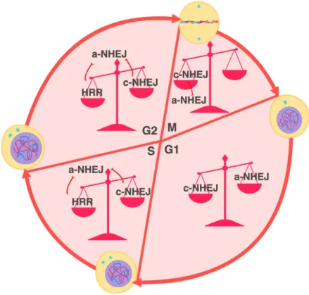
3.4.2. How cell cycle controls telomere fusion. DDR at telomeres is controlled by several shelterin factors. TRF2 has been shown to prevent ATM activation, whereas TPP1-POT and TRF1 - ATR [Guo X. et al., 2007; Denchi EL. and de Lange T., 2007; Sfeir A. et al., 2009]. Besides excessive DNA damage, TRF1 dysfunction is characterized by multiple telomere