HAL Id: hal-00321709
https://hal.archives-ouvertes.fr/hal-00321709
Submitted on 15 Sep 2008
HAL is a multi-disciplinary open access archive for the deposit and dissemination of sci-entific research documents, whether they are pub-lished or not. The documents may come from teaching and research institutions in France or abroad, or from public or private research centers.
L’archive ouverte pluridisciplinaire HAL, est destinée au dépôt et à la diffusion de documents scientifiques de niveau recherche, publiés ou non, émanant des établissements d’enseignement et de recherche français ou étrangers, des laboratoires publics ou privés.
Magalie Faivre, Clément Campillo, B. Pepin-Donat, Annie Viallat
To cite this version:
Magalie Faivre, Clément Campillo, B. Pepin-Donat, Annie Viallat. Responsive Giant Vesicles filled with Poly(N-isopropylacrylamide) Sols or Gels. Progress in Colloid and Polymer Science, Springer Verlag, 2006, 133, pp.41. �10.1007/2882_050�. �hal-00321709�
Enhanced resistance and reversible
temperature-triggered volume change of giant vesicles enclosing
Poly(N-isopropylacrylamide) solutions or gels.
MAGALIE FAIVRE1, CLEMENT CAMPILLO1, BRIGITTE PEPIN-DONAT2(✉), ANNIE VIALLAT1(✉) 1 Laboratoire de Spectrométrie Physique, UMR C5588 (CNRS) - Université Joseph Fourier
38402 Saint Martin d’Hères, France.
2 Laboratoire de Physique des Métaux Synthétiques, UMR 5819, DRFMC, CEA-Grenoble,
France
viallat@marseille.inserm.fr, fax : 33 491 75 73 28, phone : 33 491 74 40 47; donat@drfmc.ceng.cea.fr, fax : 33 438 785 113, phone : 33 491 74 40 47
presented in the 42nd Meeting of the German Colloid Society September 26 - 28, 2005, Aachen, Germany
Abstract We prepared giant unilamellar vesicles (GUVs) enclosing solutions or covalent gels of Poly(N-isopropylacrylamide) (PolyNipam). Concentrated suspensions of GUVs were prepared by applying an alternative field on a lipid film hydrated by a monomer solution containing N-isopropylacrylamide, crosslinker (N,N’-methylene-bis-acrylamide), initiator and sucrose. Vesicle inner medium was polymerised and cross-linked by U.V.irradiation of the suspension, yielding viscous vesicles enclosing a solution of linear PolyNipam chains (when no bisacrylamide was used) or elastic vesicles filled with a covalent PolyNipam gel. We show that gel-filled vesicles are responsive systems triggered by the temperature: they shrink, reducing by a factor eight their volume below the critical temperature (32°C in water, lower in presence of sucrose) and re-swell in a reversible and reproducible way upon decreasing temperature. In both cases, we show that the vesicle lipid membrane interacts with the internal polymer, resulting in an strong resistance of the vesicles to external mechanical stresses (enhanced tension of lysis). Key words Vesicles polymer gels Poly(N-isopropylacrylamide) volume transition
Introduction
Giant unilamellar vesicles (GUVs) are fluid and semi-permeable soft lipid shells. They have been extensively studied because their sizes and curvatures are similar to that of living cells and their lipid bilayer membrane exhibits basic properties of biological membranes[1,2]. However, their poor mechanical properties due to the absence of cytoplasm and cytoskeleton, limit their relevance as minimal models for living cells. The goal of this study is to prepare viscoelastic GUVs with controlled mechanical properties, which confer to them an enhanced resistance to external stresses, thus allowing to address open physical questions relative to the behaviour of
deformable soft shells submitted to external stresses (flow, pressure..). A second objective is to obtain responsive systems, triggered by an external parameter, and able for instance to release material or to move by themselves in successive contraction /extension stages. Hydrophilic polymers are well-suited candidates to confer such properties to vesicles although only a few works have been devoted to combinations of polymer networks and liposomes. Apart studies concerning gel beads coated with lipids[3-9], lipid vesicles were mainly combined with physical gels, used as external coating (rigid actin filaments [10]) or internalized in the vesicle volume (rigid actin networks [11,12], agarose gels [13], solutions of aggregated
Poly(N-isopropylacrylamide) (PolyNipam)[14]). A recent work dealt with controlled covalent PolyNipam gels but concerned submicronic liposomes [15,16].
Here, we report the preparation, first characterisation and the responsive behaviour of GUVs enclosing PolyNipam solutions or gels.. PolyNipam is known to present a volume transition at 32°C, see for example [17].
Experimental section
The lipid is 1,2-dioleolyl-sn-glycero-3-phosphocholine (DOPC) (Sigma; St Quentin-Fallavier, France). One experiment was performed using 5% biotynilated 1,2-dioleoyl-sn-glycero-3-phosphoethanolamine-n-(biotinyl) (Sigma; St Quentin-Fallavier, France) lipid, incubated with FITC-streptavidin in order to make the membrane fluorescent. The monomer intended to constitute the polymer gel is N-isopropylacrylamide (NIPAM) (Acros Organics; Janssens Pharmaceuticalaan 3A, Belgium). The crosslinker is N,N’-methylene-bis-acrylamide (MBA) (Sigma) and the initiator is 2,2-diethoxyacetophenone (DEAP) (Acros Organics).
Vesicles were electroformed [18] in a pregel aqueous solution containing sucrose (100 mM), NIPAM (300 mM), MBA (0 mM,3 mM, 9 mM or 15 mM) and DEAP (1µl/ml). A small volume of the obtained vesicle suspension was injected in small chambers filled with a solution of glucose (100 mM), NIPAM (300 mM), DEAP (1µl/ml), but which did not contain the
crosslinker. This former solution i) allows vesicle sedimentation since the density of sucrose is smaller than that of glucose, and makes easier the observation of vesicles from phase contrast microscopy (sucrose and glucose have different refractive indexes); ii) ensures osmotic
lipid membrane); iv) prevents gelation of the external solution (since no crosslinker is present), which would trap gel-filled vesicles in the external medium.
All solutions were deoxygenated by bubbling with dry argon for 30 mn and kept in a glove bag under argon atmosphere. Finally, the chambers were covered by a quartz coverslip and placed over an ice pack (Medicool coolpack MC-15) to keep temperature below 30°C. UV irradiation was performed during a period of time ranging from five minutes to one hour using a UV lamp (UV-B Mid-range 300nm Sunlight-Erythemal AH 68-1091 F6T5E, Harvard Apparatus,). Photo-illuminated vesicles were then diluted in a 100 mM glucose solution and observed by phase contrast or fluorescence (inverted microscope Olympus IX71, CCD camera). Image analysis was performed using the image software (NIH, 1.62c). A detergent (Decon90, Prolabo, France) was used to dissolve the gel-filled vesicles membrane and Rhodamine 6G (Sigma) wasused as fluorophore for membrane labelling.
Results
The vesicles observed after photo-illumination are well contrasted and generally present very regular spherical shapes of radii in the range 10 µm - 60 µm (Fig. 1a).
Characterisation of vesicles enclosing a gel or a polymer sol.
The presence of the lipid membrane is clearly demonstrated from fluorescence observations of vesicles prepared with biotynilated lipids bearing FITC-streptavidin, as illustrated in Fig. 1b.
Figure 1. Gel-filled vesicle observed from phase contrast microscopy (a) and fluorescence microscopy: FITC-streptavidin fixed on biotynilated membrane(b).
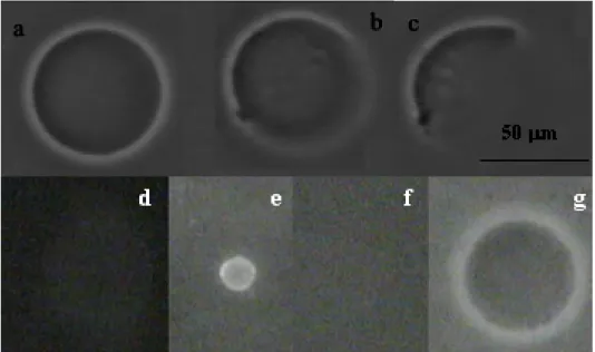
The sol or gel nature of the inner vesicle volume was characterised after breakage of the membrane upon injection of a detergent close to the vesicle. The behaviour of a polymer sol filled-vesicle is illustrated in Fig. 2a, b, c. Upon Decon injection, the membrane of the vesicle (initial state, Fig. 2a) breaks and the inner polymer solution progressively vanishes, since it is fully soluble (Fig. 2b-2c). The behaviour of a gel-filled vesicle is shown in Fig. 2d, e, f. First, rhodamine is added to the solution. It concentrates in the hydrophobic lipid bilayer and permits to see the membrane by fluorescence microscopy (initial state: fig. 2d). After Decon injection, the inner volume of the vesicle shrinks and keeps its shape (Fig. 2e), thus disclosing the presence of a 3D gel network. Shrinkage is attributed to the gel collapse at the contact of PolyNipam with Decon, (non-solvent of PolyNipam) after the breakage of the membrane. Concomitantly, rhodamine accumulates in the collapsed hydrophobic gel. Finally, glucose solution is injected close to the gel, which reswells (fig. 2f) and releases rhodamine. No more fluorescence is detected on the object: the lipid membrane has been destroyed. The final size of the gel bead is larger than the initial vesicle size (fig. 2g), which indicates that when the gel was enclosed in a vesicle, it was not swollen at its maximal degree.
Figure 2. Attack of a vesicle by a detergent (Decon). Vesicle filled with a PolyNipam solution observed by Phase contrast microscopy: 1a: initial state, 1b-1c: the membrane is broken and the vesicle progressively vanishes; gel-filled vesicle (MBA concentration 9 mM) observed by fluorescence microscopy (d,e,f) : 1d: initial state; 1e: gel collapse after membrane rupture; 1f:
re-swelling of the gel after Decon dilution. Gel after membrane destruction observed by phase contrast microscopy : 1g:diameter of the gel is greater than the initial vesicle diameter.
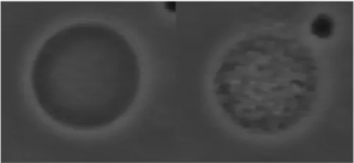
Responsive vesicles: temperature-induced phase transition
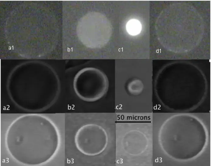
PolyNipam solutions and gels are known to exhibit a reversible microphase separation and a sharp volume change respectively, which are triggered by the temperature upon crossing the lower critical solution temperature (T=32°C in water, 29 °C, 25 °C and 21 °C in 0.1 M, 0.25 M and 0.5 M glucose solutions respectively)..Indeed, microphase separation upon heating with formation of large aggregates in the inner volume of linear PolyNipam-filled vesicles is observed, as shown in Fig. 3 and recently reported by Jesorka et al[14]. More interesting is the behaviour of gel-filled vesicles. Figure 4 depicts selected time frames from movies that show the typical size changes induced by a cycle of temperature (25°C-40°C-25°C) of three gel-filled vesicles. Above the transition temperature, vesicles contract rapidly (Fig. 4a and b) while keeping their shape. They reach a stationary size when the temperature is maintained at 40°C (fig. 4c). Finally, when the temperature is lowered back to 25°C, vesicles re-swell and retrieve their initial size, (Fig. 4d). The first vesicle (a1- d1) is observed by fluorescence after addition of rhodamine. It is clearly seen that the membrane is not destroyed during both gel contraction and expansion stages. It also indicates that the membrane is coupled to the gel during the volume transition and is believed to crumple onto the surface. The mechanisms of crumpling and the origin of PolyNipam-lipid interactions have still to be studied. The cycle of temperature was successively repeated six times without apparent vesicle damage.
Figure 4. Heating above the PolyNipam demixion temperature and cooling stages of gel-filled vesicles: fluorescence observation of a gel-filled vesicle (MBA: 9 mM) incubated with rhodamine: (a1) initial state T=20°C, (b1) heating T= 40°C, (c1) final state at T=40°C, (d1) final state after cooling T=20°C; phase contrast observations of a gel-filled vesicle (MBA: 3 mM): (a2) initial state T=20°C, (b2) heating T= 40°C, (c2) final state at T=40°C, (d2) final state after cooling T=20°C), phase contrast observations of a gel-filled vesicle (MBA: 9 mM): (a3) initial state T=20°C, (b3) heating T= 40°C, (c3) final state at T=40°C, (d3) final state after cooling T=20°C
Gel membrane interaction.
In order to test the vesicle membrane strength, we performed micropipette suction experiments to produce a uniform membrane tension and stretch bilayers of sol and gel-filled vesicles. The applied pipette suction (up to 104Pa) is small compared to osmotic driving forces (105Pa)
required to displace water on the time scale of the experiment and small compared to the osmotic activity of the trapped sucrose (2 105 Pa). Thus, the vesicle volume remains fixed during the test.
A typical experiment is shown in Fig. 5 on a sol-filled vesicle. The pipette suction is increased up to more than 104 Pa without membrane lysis occurring. The corresponding tension, measured from a geometric relation based on the pipette caliber and the diameter of the vesicle spherical segment exterior to the pipette [19] reached ~0.1 N.m. Gel-filled vesicles present a similar behavior under pipette suctions of 104 Pa and higher, though the aspirated projection length inside the pipette is comparatively smaller. In particular, no separation of the membrane from the gel is detected..
The value of reached for the membrane tension and pipette suction are about one order of
magnitude higher than that obtained for the lysis tension and maximal applied pressure for simple DOPC vesicles[20]. This result clearly suggests that the membrane of PolyNipam filled vesicles is strongly strengthened, likely due to lipid/PolyNipam interactions.
Figure 5. Micropipette aspiration of a sol-filled vesicle held by micropipette suction (104 Pa).
Conclusion
We prepared GUVs containing solutions and elastic gels of PolyNipam. The concentration and the nature of components in the inner volume of the vesicles can be varied to modulate both their mechanical properties and the phase transition temperature. We showed that gel-filled vesicles are responsive systems, which exhibit a reversible volume transition triggered by temperature. The membrane of both sol and gel-filled vesicles is strongly strengthened compared to that of simple vesicles and is coupled with the inner gel. These systems appear as excellent candidates to isolate and mimick certain cell mechanical features.
REFERENCES
1. Menger FM, Angelova MI (1998) Acc Chem Res 31: 789
2. Lipowsky R, Sackmann E (1995) Structure and dynamics of Membrane. Elsevier, North-Holland 3. Gao K, Huang L (1987) Biochim Biophys Acta 897: 377
4. Jin T, Pennefather P, Lee PI (1996) FEBS Let 397: 70
5. Kiser PF, Wilson G, Needham D (1998) Nature (London) 394: 459
6. Yang Q, Liu X, Ajiki S, Hara M, Lundahl P, Miyake V (1998) J Chromatog. B. 707: 131 7. Hara M, Yuan H, Miyake M, Iijima S, Yang Q, Miyake (2000) J Mater Sci Eng C. 1:3: 117. 8. Collier JH, Messersmith PB (2001) Annu Rev Mater Res 31: 237
9. Kazalov S, Kaholek M, Teraoka I, Levon, K (2002) Macromol 35: 1911
10. Helfer E, Harlepp S, Bourdieu L, Robert J, MacKintosh FC, Chatenay D(2001). Phys Rev E 63: 021904 11. Haeckl W, Baermann M, Sackmann E (1998) Phys Rev Lett 80: 1786
12.Limozin L, Sackmann E, (2002) Phys Rev Lett 89: 1681031 13. Viallat A, Dalous J, Abkarian M, (2004) Biophys J 86: 2179 14. Jesorka A, Amarkström M, Orwar O, (2005) Langmuir 21(4): 1230
15. Stauch O, Uhlmann T, Fröhlich M, Thomann R, El-Badry M, Kim YK, Schubert R (2002) Biomacromolecules 3: 324 16. Stauch O, Schubert R, Savin G, Burchard W (2002) Biomacromolecules 3: 565
17 Matsuo E. S, Tanaka T. (1988) J. Chem. Phys. 89:1695–1703.
18. Angelova MI, Soléau S, Méléard P, Faucon J, Bothorel P (1992) Progr Coll Pol Sci 89: 127 19. Kwok R, Evans EA (1981) Biophys J 35: 637