Evolution and diversity of Rickettsia bacteria
Lucy A Weinert*
1, John H Werren
2, Alexandre Aebi
3, Graham N Stone
1and
Francis M Jiggins
1Address: 1Institute of Evolutionary Biology, University of Edinburgh, Edinburgh, EH9 3JT, UK, 2Biology Department, University of Rochester, Rochester, NY 14627, USA and 3Agroscope Reckenholz-Tänikon, Research Station ART, Reckenholzstrasse 191, 8046 Zürich, Switzerland Email: Lucy A Weinert* - [email protected]; John H Werren - [email protected]; Alexandre Aebi - [email protected]; Graham N Stone - [email protected]; Francis M Jiggins - [email protected]
* Corresponding author
Abstract
Background: Rickettsia are intracellular symbionts of eukaryotes that are best known for infecting
and causing serious diseases in humans and other mammals. All known vertebrate-associated Rickettsia are vectored by arthropods as part of their life-cycle, and many other Rickettsia are found exclusively in arthropods with no known secondary host. However, little is known about the biology of these latter strains. Here, we have identified 20 new strains of Rickettsia from arthropods, and constructed a multi-gene phylogeny of the entire genus which includes these new strains.
Results: We show that Rickettsia are primarily arthropod-associated bacteria, and identify several
novel groups within the genus. Rickettsia do not co-speciate with their hosts but host shifts most often occur between related arthropods. Rickettsia have evolved adaptations including transmission through vertebrates and killing males in some arthropod hosts. We uncovered one case of horizontal gene transfer among Rickettsia, where a strain is a chimera from two distantly related groups, but multi-gene analysis indicates that different parts of the genome tend to share the same phylogeny.
Conclusion: Approximately 150 million years ago, Rickettsia split into two main clades, one of
which primarily infects arthropods, and the other infects a diverse range of protists, other eukaryotes and arthropods. There was then a rapid radiation about 50 million years ago, which coincided with the evolution of life history adaptations in a few branches of the phylogeny. Even though Rickettsia are thought to be primarily transmitted vertically, host associations are short lived with frequent switching to new host lineages. Recombination throughout the genus is generally uncommon, although there is evidence of horizontal gene transfer. A better understanding of the evolution of Rickettsia will help in the future to elucidate the mechanisms of pathogenicity, transmission and virulence.
Background
Rickettsia bacteria are intracellular symbionts of eukaryo-tes. The genus is classified in the family Rickettsiaceae within the alpha-proteobacteria, and is closely related to
the genera Erlichia and Wolbachia [1,2]. Rickettsia are most noted for causing human diseases, including Rocky Mountain spotted fever and epidemic typhus, which has been a major source of mortality at times in human
his-tory [3]. However, all known vertebrate-associated Rickett-sia are vectored by arthropods as part of their life-cycle, and many other Rickettsia are found exclusively in arthro-pods with no known secondary host (for convenience, we will refer to the former as 'vertebrate Rickettsia' and the lat-ter as 'arthropod Rickettsia'). In recent years, arthropod Rickettsia have been discovered in a diverse range of hosts, suggesting that they are more common than had been sus-pected [4-16]. Nevertheless, research effort has tended to concentrate on the medically important vertebrate Rickett-sia, or on the more common arthropod endosymbionts, such as Wolbachia and Cardinium, and so we know little about the biology of arthropod Rickettsia. Even less is known about the closely related bacteria that have been recently discovered in organisms such as leeches and pro-tists, and in metagenomic studies sequencing all DNA in an environmental sample [17-25]. This neglect is unfortu-nate, because comparing the vertebrate pathogens with related species can help to elucidate the mechanisms of pathogenicity, transmission and virulence [26,27]. How-ever, this requires a robust phylogeny for the genus. Historically, Rickettsia were classified into three major groups based on serological characteristics, namely the 'typhus group', 'spotted fever group' and 'scrub typhus group', although subsequent DNA sequencing led to the latter being reassigned to the related genus Orientia [28]. The relationship of species within the remaining two groups of Rickettsia has been the subject of intensive study over the last decade as progressively more informative genes have been sequenced [29-32] culminating in a multi-genic approach [33]. As a result it has been sug-gested that the spotted fever group consists of two sister clades, one of which is now designated 'transitional' [34] (although see [35]). A fourth so-called 'ancestral' clade, including Rickettsia bellii and Rickettsia canadensis, is thought to be basal to the other groups and is largely non-pathogenic to vertebrates. However, the position of R. canadensis remains uncertain [33].
While many studies have helped to clarify the relation-ships between the vertebrate Rickettsia, only one recent study has explored the relationship of the well classified groups to the newly discovered arthropod Rickettsia [36]. The authors found that most arthropod Rickettsia are basal to the vertebrate Rickettsia and that the Rickettsia associ-ated with leeches, protists and freshwater environments fell into two phylogenetic groups, distinct from the arthropod and vertebrate groups. The only known excep-tions are a small number of arthropod Rickettsia that fell within the group otherwise infecting leeches [12,36,37]. However, Perlman et al. [36] were only able to provide lit-tle statistically significant support for relationships among the arthropod Rickettsia. This is almost certainly because the study relied on partial sequences of 16S rDNA, which
is extremely slowly evolving, and so lacking in phyloge-netic resolution. Improving this situation is challenging because amplifying other genes in basal strains has proven problematic, perhaps because the genes in question may either be missing or too divergent for PCR amplification using existing primers. Also, resolving some deep nodes in the Rickettsia species tree continues to be a problem. The reasons for this are unclear but could be exacerbated by long-branch attraction. One of the best ways to minimise this effect is to sample for more taxa and add them to the tree in the hope of breaking up (thereby shortening) the long branches.
Here, to explore the diversity of arthropod Rickettsia, we screened 4454 arthropods to uncover new Rickettsia strains and sequenced four genes from five known and 20 new bacterial strains. We use the recently published Orien-tia tsutsugamushi genome [38] to design PCR primers allowing amplification of rapidly evolving genes from strains that lie between the genera Rickettsia and Orientia. To include other hosts, we also searched published metagenomic databases for Rickettsia sequences. With these data, we have been able to produce the first well-resolved phylogeny of the entire genus Rickettsia, showing how the vertebrate Rickettsia relate to the other taxa. Our phylogeny has allowed us to identify and name additional novel groups. Furthermore, we were able to compare host associations among these groups, identify major life his-tory transitions, and investigate the extent of recombina-tion within the genus.
Results
Strains identified and genes sequenced
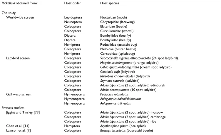
Our screens identified 20 novel strains of arthropod Rick-ettsia from six orders of insects, and these are listed in Table 1. These strains were combined with five previously described arthropod Rickettsia (listed at the bottom of Table 1) to give 25 strains in total. We successfully sequenced all four of our chosen genes from 18 of these strains, and one or more genes from the remaining seven. Rickettsia phylogeny
To obtain a phylogeny of the genus Rickettsia, we com-bined a concatenated alignment of the four genes we sequenced, with data from other Rickettsia strains availa-ble from Genbank (accession number availaavaila-ble in Addi-tional file 1, Accession numbers of genes used in the phylogenetic analysis). For most of the previously described arthropod Rickettsia, only 16S rDNA sequence is available, and so we allowed for missing data in the align-ment where a gene had not been sequenced. Missing data should not decrease phylogenetic resolution for taxa with complete data, and is likely to be a problem for other taxa only when the number of characters is very low [39].
Figure 1a shows that our concatenated alignment with missing data gave a well-resolved tree with strong support for most nodes. Nevertheless, it is important to determine whether there are conflicting signals between the individ-ual genes. Therefore, we used SH tests to compare our con-catenated topology to the maximum likelihood trees inferred from each of the four genes (Table 2). Only the 16S gene tree topology was marginally significantly differ-ent (although this is no longer significant when control-ling for multiple tests by Bonferroni correcting the p-values).
It is also important to investigate the influence of missing data on the phylogeny. Therefore, we constructed a sec-ond tree that included only taxa with complete sequences for the three genes atpA, coxA and gltA (excluding 16S due to its marginally significant SH test). This 'complete data' tree is shown in Figure 1b. Overall, the topologies of the two trees are very similar (Figure 1a and 1b), but most nodes had higher support in the tree with complete sequences. In particular, there is strong bootstrap support for the group largely composed of ladybird symbionts in the complete data tree (Figure 1b) but not on the missing data tree (Figure 1a). An exception is the placement of R. canadensis, which is uncertain in the complete data tree but is well supported on the missing data tree (probably because the missing data tree includes two closely related taxa; Figure 1a). The composition of the transitional group and the placement of Rickettsia prowazekii also differ in the two trees. Rickettsia within the typhus group (R. prowazekii and Rickettsia typhi) are striking in that they reside on longer branches than other Rickettsia in the trees. This is indicative of rate heterogeneity, which can cause a long-branch attraction artefact where the taxa will appear in an incorrect place. In the missing data tree the transi-tional group is monophyletic, while in the complete data tree R. prowazekii groups with Rickettsia akari (Figure 1a and 1b). However, constraining R. akari and the
transi-Table 1: Rickettsia strains sequenced.
Rickettsia obtained from: Host order Host species
This study:
Worldwide screen Lepidoptera Noctuidae (moth)
Neuroptera Chrysopidae (lacewing)
Coleoptera Elateridae (beetle)
Coleoptera Curculionidae (weevil)
Diptera Bombyliidae (bee fly)
Diptera Bombyliidae (bee fly)
Hemiptera Reduviidae (assassin bug)
Coleoptera Meloidae (blister beetle)
Hemiptera Cercopidae (spittlebug)
Ladybird screen Coleoptera Subcoccinella vigintiquattuorpunctata (24 spot ladybird)
Coleoptera Halyzia sedecimguttata (orange ladybird)
Coleoptera Calvia quattuordecimguttata (cream spot ladybird)
Coleoptera Coccidula rufa (ladybird)
Coleoptera Rhizobius chrysomeloides (ladybird)
Coleoptera Scymnus suturalis (ladybird)
Coleoptera Adalia bipunctata (2 spot ladybird) edinburgh
Coleoptera Adalia decempuntata (10 spot ladybird)
Gall wasp screen Hymenoptera Pediobius rotundatus
Hymenoptera Aulogymnus balani/skianeuros
Hymenoptera Aulogymnus trilineatus
Previous studies:
Jiggins and Tinsley [79] Coleoptera Adalia bipunctata (2 spot ladybird) moscow
Coleoptera Adalia bipunctata (2 spot ladybird) cambridge
Coleoptera Adalia bipunctata (2 spot ladybird) ribe
Chen et al. [14] Hemiptera Acyrthosiphon pisum (pea aphid)
Lawson et al. [7] Coleoptera Brachys tessellatus (buprestid beetle)
Table 2: Likelihood values of SH tests. Likelihood of tree topology
Dataset Unconstrained Concatenated lnL p
16S 1486.10 1502.03 31.85 0.045
AtpA 2129.98 2140.90 21.85 0.161
CoxA 3484.47 3490.98 13.02 0.201
GltA 3931.44 3942.56 22.24 0.069
Comparison of the tree topologies obtained from the four genes against the topology of the concatenated dataset using four SH tests. Each dataset was forced to adopt the topology from the concatenated dataset and the log likelihood of this tree was compared with the log likelihood of the unconstrained tree. The taxa used in this analysis are shown in Figure 1b.
tional group to be monophyletic in the complete data tree only causes a marginally significant drop in the likelihood (SH test; lnL = 20.003 p = 0.066).
Together, these phylogenetic analyses reveal five distinct and well-supported major clades of Rickettsia (Figure 1), one (designated the hydra group) containing protist-asso-ciated Rickettsia and a number with unknown host associ-ations from sequences amplified from environmental samples, a second clade (torix) containing Rickettsia from amoeba, leeches and arthropods, a third (rhizobius) con-tains three beetle Rickettsia, a fourth (melloidae) contain-ing a scontain-ingle beetle Rickettsia, a fifth (bellii) containcontain-ing 11
strains of arthropod Rickettsia and a sixth clade of diverse bacteria containing both arthropod and vertebrate Rickett-sia. This final clade can be further subdivided into the fol-lowing groups: onychiurus, adalia, canadensis, spotted fever group, typhus group and transitional group, although bootstrap support for some of these groupings is less strong (all groups are also summarized in Figure 2). Host shifts
By mapping host species onto our phylogeny, we are able to make inferences about patterns of host-switching in the genus. It is clear from Figure 1 that Rickettsia bacteria have an extremely diverse host range, occurring in arthropods, Phylogeny of Rickettsia
Figure 1
Phylogeny of Rickettsia. The name of the host prefixed by (s) is given where the bacterium does not have a species name, as
well as names for each Rickettsia group, life history and host order. (a) Bayesian phylogeny using concatenated sequences of atpA, coxA, gltA, 16S. Posterior support for each node is shown. (b) Maximum likelihood phylogeny based on complete sequences of atpA, coxA and gltA. Bootstrap support is given as a percentage above the node, and posterior support from a Bayesian tree is given as a decimal below the node. aPreviously characterised groups. bOnly circumstantial evidence connects
the trait to the strain.
v 0.04 substitutions per site 1.00 0.99 0.59 1.00 0.96 1.00 0.75 0.74 1.00 0.60 0.57 1.00 0.55 1.00 0.90 1.00 1.00 0.77 0.92 0.94 0.96 0.52 1.00 0.97 0.78 0.71 0.86 0.51 1.00 1.00 0.60 1.00 0.89 0.89 0.89 0.94 0.78 1.00 1.00 0.97 0.95 1.00 0.850.99 0.55 1.00 1.00 0.89 1.00 1.00 1.00 1.00 0.89 1.00 1.00 0.91 0.95 1.00 1.00 1.00 0.68 1.00 1.00 0.82 1.00 1.00 1.00 1.00 1.00 1.00 1.00 1.00 100 100 100 100 100 100 100 100 99 63 64 86 96 94 95 69 92 62 82 88 77 0.07 substitutions per site
(a)
(b)
Name Group Life history Host class Host order
Orientia tsutsugamushi 1) ORIENTIA vertebrate pathogen Acarina Parasitengona (chigger mites)
(s)Deep sea octacoral
(s)Haplosporidium sp. Haplosporea Haplosporidia
(s)Melted red snow (s)Mountain snow
(s)Hydra oligactis Hydrozoa Hydroida (medusae)
(s)Acid impacted lake 2) HYDRA (s)Pasture water
(s)Rice roots (s)Forested wetland (s)Kalahari water
(s)Diophrys appendiculata Spirotrichea Euplotida (cilliate)
(s)Artic tundra 3) TUNDRA
(s)Torix tagoi Clitellata Rhynchobdellida (proboscis leeches)
(s)Limonia chorea Insecta Diptera (true flies)
(s)Indoor dust 4) TORIX
(s)Cerobasis guestfalica Insecta Psocoptera (booklice)
(s)Lutzomyia apache Insecta Diptera (true flies)
(s)Nuclearia pattersoni Tubulinida Amoebozoa (amoeba)
(s)Kytorhinus sharpianus Insecta Coleoptera (weevils)
(s)Curculionidae 5) RHIZOBIUS Insecta Coleoptera (weevils)
(s)Rhizobius chrysomeloides Insecta Coleoptera (beetles)
(s)Meloidae 6) MELOIDAE Insecta Coleoptera (beetles)
(s)Bemisia tabaci Insecta Hemiptera (true bugs)
(s)Empoasca papayae plant pathogen Insecta Hemiptera (true bugs)
(s)Tetranychus urticae Acarina Eleutherengona (mite-like mites)
(s)Bombyliidae Insecta Diptera (true flies)
(s)Acyrthosiphon pisum Insecta Hemiptera (true bugs)
(s)Bombyliidae Insecta Diptera (true flies)
(s)Brachys tessellatus 7) BELLII male-killing Insecta Coleoptera (beetles)
(s)Reduviidae Insecta Hemiptera (true bugs)
(s)Chrysopidae Insecta Neuroptera (lacewing)
R. bellii vertebrate pathogen Acarina Ixodida (ticks)
R. bellii Acarina Ixodida (ticks)
(s)Elateridae Insecta Coleoptera (beetles)
(s)Noctuidae Insecta Lepidoptera (moths)
(s)Onychiurus sinensis 8) ONYCHIURUS Entognatha Collembola (springtails)
(s)Subcoccinella vigintiquattuorpunctata Insecta Coleoptera (beetles)
(s)Scymnus suturalis Insecta Coleoptera (beetles)
(s)Adalia bipunctata Insecta Coleoptera (beetles)
(s)Adalia bipunctata male-killing Insecta Coleoptera (beetles)
(s)Adalia bipunctata 9) ADALIA Insecta Coleoptera (beetles)
(s)Halyzia sedecimguttata Insecta Coleoptera (beetles)
(s)Calvia quattuordecimguttata Insecta Coleoptera (beetles)
(s)Adalia bipunctata Insecta Coleoptera (beetles)
(s)Adalia decempuntata Insecta Coleoptera (beetles)
(s)Coccotrypes dactyliperda Insecta Coleoptera (beetles) R. canadensis 10) CANADENSIS vertebrate pathogen Acarina Ixodida (ticks)
R. tarasevichiae Acarina Ixodida (ticks)
R. helvetica Acarina Ixodida (ticks)
(s)Ixodes scapularis Acarina Ixodida (ticks)
R. montanensis Acarina Ixodida (ticks)
R. massiliae Acarina Ixodida (ticks)
R. japonica 11) SPOTTED FEVER Acarina Ixodida (ticks)
R. peacockii Acarina Ixodida (ticks)
R. rickettsii Acarina Ixodida (ticks)
R. conorii Acarina Ixodida (ticks)
R. sibirica Acarina Ixodida (ticks)
R. typhi 12) TYPHUS vertebrate pathogen Insecta Siphonaptera/Phthiraptera (flea/lice)
R. prowazekii Insecta Psocoptera (lice)
R. australis Acarina Ixodida (ticks)
R. akari Acarina Holothyrida (holothryan mites)
(s)Cercopidae Insecta Hemiptera (true bugs)
(s)Aulogymnus trilineatus Insecta Hymenoptera (wasps)
(s)Aulogymnus balani/skianeuros 13) TRANSITIONAL Insecta Hymenoptera (wasps)
R. felis vertebrate pathogen Insecta Siphonaptera (fleas)
(s)Liposcelis bostrychophila Insecta Psocoptera (booklice)
(s)Liposcelis bostrychophila Insecta Psocoptera (booklice)
(s)Pediobius rotundatus Insecta Hymenoptera (wasps)
(s)Neochrysocharis formosa parthenogenesis inducing Insecta Hymenoptera (wasps)
Ath ro p o d s y m b io n ts a a a a b b b b b ertebrate pathogen v
Host class Host order
Acarina Parasitengona (chigger mites)
Haplosporea Haplosporidia
Hydrozoa Hydroida (medusae)
Spirotrichea Euplotida (cilliate)
Clitellata Rhynchobdellida (proboscis leeches) Insecta Diptera (true flies)
Insecta Psocoptera (booklice) Insecta Diptera (true flies) Tubulinida Amoebozoa (amoeba) Insecta Coleoptera (weevils) Insecta Coleoptera (weevils) Insecta Coleoptera (beetles) Insecta Coleoptera (beetles) Insecta Hemiptera (true bugs) Insecta Hemiptera (true bugs) Acarina Eleutherengona (mite-like mites) Insecta Diptera (true flies) Insecta Hemiptera (true bugs) Insecta Diptera (true flies) Insecta Coleoptera (beetles) Insecta Hemiptera (true bugs) Insecta Neuroptera (lacewing) Acarina Ixodida (ticks) Acarina Ixodida (ticks) Insecta Coleoptera (beetles) Insecta Lepidoptera (moths) Entognatha Collembola (springtails) Insecta Coleoptera (beetles) Insecta Coleoptera (beetles) Insecta Coleoptera (beetles) Insecta Coleoptera (beetles) Insecta Coleoptera (beetles) Insecta Coleoptera (beetles) Insecta Coleoptera (beetles) Insecta Coleoptera (beetles) Insecta Coleoptera (beetles) Insecta Coleoptera (beetles) Acarina Ixodida (ticks) Acarina Ixodida (ticks) Acarina Ixodida (ticks) Acarina Ixodida (ticks) Acarina Ixodida (ticks) Acarina Ixodida (ticks) Acarina Ixodida (ticks) Acarina Ixodida (ticks) Acarina Ixodida (ticks) Acarina Ixodida (ticks) Acarina Ixodida (ticks)
Insecta Siphonaptera/Phthiraptera (flea/lice) Insecta Psocoptera (lice)
Acarina Ixodida (ticks) Acarina Holothyrida (holothryan mites) Insecta Hemiptera (true bugs) Insecta Hymenoptera (wasps) Insecta Hymenoptera (wasps) Insecta Siphonaptera (fleas) Insecta Psocoptera (booklice) Insecta Psocoptera (booklice) Insecta Hymenoptera (wasps) Insecta Hymenoptera (wasps)
vertebrates, plants, amoebas, ciliates, annelids and hydro-zoa, and that there have been numerous shifts between these hosts. The earliest shift splits the genus into two major divisions: the hydra and torix groups and all other arthropod Rickettsia. As mentioned, the hydra group are symbionts of protists and undetermined hosts. Although one member of this group was found in the marine ciliate Diophrys from brackish water [20], and another from a deep sea octocoral, all others are from freshwater environ-ments or damp terrestrial environenviron-ments. In general it appears that marine Rickettsia are rare. Indeed, from over 13 billion open reading frames compiled from marine metagenomic datasets [40] we detected no homologues of greater than 91% identity to the 16S gene of hydra group Rickettsia. The next split in the tree separates all the
remaining Rickettsia from the torix group (Figure 1) which contains symbionts of leeches (phylum Annelida), an amoeba [36] and arthropods (a sandfly, a cranefly, a bit-ing midge and a booklouse). In the torix group, as with the hydra group, the vast majority of the hosts are aquatic at some stage in their life cycle (the sole exception being the booklouse).
The remainder of the arthropod Rickettsia, including all strains sequenced in the present study, form a mono-phyletic group (Figure 1). Parsimony suggests that the ancestral state of this clade is to infect arthropods, with one or more lineages subsequently evolving to also infect vertebrates. In addition, there have been multiple transi-tions between blood feeding and non blood feeding Relationships and approximate dates of divergence of the major clades within the order Rickettsiales
Figure 2
Relationships and approximate dates of divergence of the major clades within the order Rickettsiales. The 16S
rDNA phylogeny was reconstructed using one member of each of the groups shown with a molecular clock enforced (enforc-ing the clock did not reduce the likelihood of the tree: likelihood ratio test lnL = 13.84, df = 12 p = 0.311).
Orientia
hydra
torix
rhizobius
Pelagibacter
Holospora
Midichloria
bellii
adalia
canadensis
onychiurus
meloidae
spotted fever
transitional
Rickettsia
groups
transition to arthropods 75 150 present transition to intracellular 225 300 375 450 525 600millions of years ago
0.03
substitutions
per site
675 750
insects. Perlman et al. [36] demonstrated that forcing R. bellii to group with other blood feeders gives a signifi-cantly worse tree. SH tests of our phylogeny showed that forcing R. canadensis and Rickettsia felis to group with other blood feeders similarly gives a significantly worse fit (SH tests on all groups: p < 0.001).
Our results therefore show clearly that there have been numerous host shifts, sometimes between taxonomically distant hosts. However, it is equally clear that related Rick-ettsia tend to share related hosts. Multiple different strains were detected within ladybird beetles, ticks, lice, parasitic wasps and bee-flies, and in all cases, two or more of these strains cluster together. Nevertheless, this pattern does not seem to be explained by ancestral infection followed by co-speciation of parasite and host. From Figure 1a, the three different strains of Rickettsia found in Adalia bipunc-tata do not appear to be monophyletic as one of the A. bipunctata strains groups with Adalia decempunctata with high posterior support. Unfortunately only four parasi-toid individuals from the oak gall wasp screen were infected, not allowing us to test the influence of host relat-edness, host interaction frequency and geographic isola-tion on frequency of horizontal transfer events.
In addition to clustering according to host type, Figure 1 also demonstrates phylogenetic clustering by ecology (although it is often difficult to separate these effects). For example, the two major groups of vertebrate Rickettsia, the spotted fever or typhus groups, consist solely of vertebrate Rickettsia, containing no arthropod Rickettsia. However, the transitional group differs from this pattern containing both vertebrate Rickettsia and Rickettsia infecting non-blood feeding arthropods (Figure 1). A second ecological adaptation to increase transmission is to skew the sex ratio of the host towards females, which are the sex that most efficiently transmits the infection to offspring for verti-cally transmitted Rickettsia. Some of these Rickettsia are known or suspected to kill male hosts early in their devel-opment, and there appears to be two separate origins of this adaptation on the tree (once within a buprestid beetle in the bellii group and once within ladybirds in the adalia group). There are 11 strains of Rickettsia that infect lady-bird beetles, and nine of these cluster in a single mono-phyletic group. The ones that cluster elsewhere are probably not male-killers (male ladybird beetles are also infected at high prevalence [6]). A third possible source of ecological clustering relates to herbivorous hosts. Such clustering may reflect ecology in two possible ways. Firstly, many symbionts are known to supplement their hosts with amino acids that are rare in phloem sap (although a mutualistic role for Rickettsia has never been demonstrated). Secondly, Rickettsia may be transmitted horizontally through plants (one case is already known). It has previously been asserted that the bellii group con-sists mainly of herbivorous arthropod symbionts [36].
Four Rickettsia in this group are indeed known to infect sap sucking arthropods (a whitefly, a leaf hopper, an aphid and a red spider mite), and three of these group sep-arately from the other members of the bellii group (Figure 1). However, we have uncovered a large number of pred-atory insect hosts in this group, and sap sucking insects in other groups (a spittlebug symbiont is in the transitional group). Therefore, the view that members of the bellii group are mainly associated with herbivorous arthropods is not supported by these new data. However, it is possible that the DNA signal could have come from the guts of these insects, as abdomens were sometimes extracted where there was not enough ovary tissue (although the signal would not be expected to be strong).
Recombination
Recombination events complicate the inference of species trees, and so it is important to investigate the extent of recombination in the Rickettsia genus. We found one clear instance of recent recombination between different Rick-ettsia groups (this taxon was excluded from the analyses above). In the phylogenetic trees of the four individual genes (Additional file 2, Phylogenetic trees of each of the individual genes used in the study), the symbiont of the ladybird Coccidula rufa (sC. rufa) appears in the transi-tional group on the 16S and gltA trees, and in the bellii group on the atpA and coxA trees. An alignment of the pol-ymorphic sites and a hybridisation network indicates that sC. rufa is a chimera of sequences from these two groups (Figure 3). To verify that the recombination pattern for sC. rufa was not the result of contamination, this result was confirmed by sequencing three strains from different indi-viduals of C. rufa. This appears to be the only case of recombination between the four genes because when sC. rufa is excluded from analyses, there is little evidence of topological differences between the datasets (see SH tests above).
We did, however, detect some evidence of recombination events within two of the four genes. The maximum χ2 test
and phi test identified multiple recombination break-points in the gltA and coxA genes. In coxA, the breakpoint pattern indicted that there had been some recombination between an ancestor of the adalia group and of the rhizo-bius group (maximum χ2 test χ2 = 42.79 p < 0.001; phi test
p < 0.001). In gltA, there was evidence of recombination between R. akari of the transitional group and the adalia group (maximum χ2 test χ2 = 46.78 p < 0.001; phi test p =
0.021). In contrast, no recombination was detected within the 16S and atpA genes (maximum χ2 test χ2 = 8.92
p = 0.783; phi test p = 0.960; maximum χ2 test χ2 = 12.13
p = 0.57; phi test p = 0.759 (respectively)).
Split networks were constructed for each of the four genes to identify possible sources of conflicting signal and recombination in the data (Additional file 3, Split
net-works for each of the individual genes used in the study). This method has an advantage over tree-based methods as posterior support and bootstrap values measure robust-ness solely with respect to sampling error (as opposed to systematic bias), and with large sample size robustness
will generally be high as noise in the data is filtered out. The split network constructed for the 16S gene was tree-like (containing no significant splits). In contrast the other three genes showed a small amount of phylogenetic conflict, with statistical support for two different trees. In Sequence alignment and hybridisation network showing the symbiont of Coccidula rufa to be a recombinant
Figure 3
Sequence alignment and hybridisation network showing the symbiont of Coccidula rufa to be a recombinant.
(a) Alignment of concatenated genes atpA, coxA, gltA, 16S showing just polymorphic sites. Nucleotides that are identical to the C. rufa sequence are shown as a dot. The (s)C. rufa sequence of atpA and coxA (shaded) are most similar to (s)Elateridae in the bellii group, while the gltA and 16S sequences (unshaded) are most similar to (s)Pediobius rotundus in the transitional group. (b) A hybridisation network of the concatenated sequences of atpA, coxA, gltA and 16S. A neighbour-net split network was gener-ated and splits were then filtered by weight to include only the (s)C. rufa split. A hybridisation network was then performed on the split network to provide an explicit example of descent from the two different groups.
(s) Rhizobius chrysomeloides (s) Meloidae (blister beetle)
(s) Brachys tessellatus (buprestid beetle)
(s) Bombyliidae (bee fly) 150801 (s) Bombyliidae (bee fly) 223009 (s) Acyrthosiphon pisum (pea aphid)
(s) Pediobius rotundatus (wasp) (s) Aulogymnus balani/skianeuros (wasp) (s) Aulogymnus trilineatus (wasp) 107 (s) Aulogymnus trilineatus (wasp) 109
Rickettsia felis
Rickettsia akari
Rickettsia canadensis Rickettsia prowazekii
Rickettsia bellii RML Rickettsia bellii OSU
spotted fever group
adalia group
0.01 substitutions
per site (s) Brachys tessellatus (buprestid beetle)
(s) Bombyliidae (bee fly) 150801 (s) Bombyliidae (bee fly) 223009 (s) Acyrthosiphon pisum (pea aphid)
Rickettsia bellii RML Rickettsia bellii OSU
(s) Pediobius rotundatus (wasp) (s) Aulogymnus balani/skianeuros (wasp) (s)
( ) ( )
Aulogymnus trilineatusgygy (wasp) 107((
(s) ( ) ( )
Aulogymnus trilineatusgygy (wasp) 109(( p)p)
Rickettsia felis Rickettsia akari
Transitional group
Bellii group
T G A T C A T A T G T G T T G C A T A T C A T A G G A T A C T A T G G C G T G A C A C A C T T T . . . . T . . . C . . . . C A G A . T C T C A C T C C A T T C G C A C C T C T G A C T C G A A A T A C T T T G G G T C A C T C C G A G T A C G A A A G C T T T G T T A G T T C A T T C T C A T T T T A T C T T C A T T A A . . . . C T T A G T C T G C G T G A T A A C T C C G A C A T G C C T A A C G C C C T C T C C T G C A T G T A T C C A A C T C G C A C C C T C A A A T C C A A C T A C T T G T C T C G A C C T C T G T A C . . . A . . . C T C A T G C A C T A T C T T A C A G G G C T A G G A A T T A C A C T A T A G T T A A C . C C T G C G C G T G C G A C G C T A G T G T C T C A C C C T G C C T A A G A T G T C A G A C A T C T T . . A T A . . T A G T A A G G T . A C G G G G T T A A A T T C T G T G C T C T C A G A G C T A C A T A T . C T . . . . A . . . . C A . . . T . . . G . . . . G C G C C C C G A A G C A G C G A T C T T C T T T T A G C C A T G A C T C C A A A A A G T C C G A T A T T A G A C T T A G A T A G G A C G T A A C G G A A A T C A . T . G T G G G G G A A T A . . . . T . . T . . . A . . . C T C G . . . A . . A C C T G A A A T G C G T G G T C A C T T A C A T G G C . T T C . . G T G T C . . . A . . C A A G . . . . (s)Elateridae(a)
(b)
T G AA T C AA T AA T G T G T T G C AA T AA T C AA T AA G G AA T AA C T AA T G G C G T G AA C AA C AA C T T T . . . . T . . . C . . . . C AA G AA . T C T C AA C T C C AA T T C G C AA C C T C T G AA C T C G AA AA AA T AA C T T T G G G T C AA C T C C G AA G T AA C G AA AA AA G C T T T G T T AA G T T C AA T T C T C AA T T T T AA T C T T C AA T T AA AA . . . . C T T AA G T C T G C G T G AA T AA AA C T C C G AA C AA T G C C T AA AA C G C C C T C T C C T G C AA T G T AA T C C AA AA C T C G C AA C C C T C AA AA AA T C C AA AA C T AA C T T G T C T C G AA C C T C T G T AA C . . . AA . . . C T C AA T G C AA C T AA T C T T AA C AA G G G C T AA G G AA AA T T AA C AA C T AA T AA G T T AA AA C . C C T T C C T A C G T C C G C G C . . AA T A T AA T G A A (s)Coccidula rufa (s)Pediobius rotundatus (s)Elateridae (s)Coccidula rufa (s)Pediobius rotundatus (s)Elateridae (s)Coccidula rufa (s)Pediobius rotundatus (s)Elateridae (s)Coccidula rufa (s)Pediobius rotundatus (s)Elateridae (s)Coccidula rufa (s)Pediobius rotundatus (s)Elateridae (s)Coccidula rufa (s)Pediobius rotundatus (s)Coccidula rufaall cases, one of these trees corresponded to that shown in Figure 1, suggesting that this tree accurately reflects the evolutionary history of most of the genome. The discrep-ancies were as follows. The atpA split network showed additional support for a tree where R. prowazekii is basal to the other vertebrate groups. This pattern is consistent with a tree based on protein alignments of the ten Rickettsia genomes [41]. The coxA split network supported a closer relationship between Rickettsia chrysomeloides symbiont and the adalia group, which is consistent with the recom-bination pattern for this gene. The gltA split network also supported this same relationship although this was not reflected in the recombination breakpoint pattern.
Discussion
We have identified a large number of new strains of Rick-ettsia, including several new groups, and shown that arthropod Rickettsia are both common and diverse. We have also constructed the largest and most robust phylo-genetic analysis of the genus to date. Importantly, we used a multiple locus approach, as using single genes to build species phylogenies can seriously confound the true rela-tionship between strains, especially with loci that are prone to recombination [42].
The evolutionary history of Rickettsia
It is useful to view our results in the context of the evolu-tion of the whole order Rickettsiales. To do this, we have used a molecular clock to date the divergence of different groups, and this is shown in Figure 2. The common ances-tor was presumably free-living, as the earliest diverging genus of the order is Pelagibacter. Pelagibacter species account for 26% of the bacterial rDNA sequences from sea water [43] and have the smallest genomes of free-living bacteria. About 525–775 million years ago there was a transition to living within cells, followed by a split into endosymbionts of protists (Holospora) [44,45] and a clade that primarily infects arthropods. Holospora species infect the nuclei of paramecium and are generally considered pathogenic to their hosts; for example, Holospora undulata can sterilise their hosts, reduce the rate of asexual division and increase host mortality [46]. The most parsimonious interpretation of the tree, therefore, is that the transition to infecting arthropods occurred approximately 425–525 million years ago in this lineage (Figure 2), which can be compared to the first appearance of most metazoan phyla in the Cambrian explosion (approximately 540 million years ago).
All other genera in the order Rickettsiales are associated with arthropods although many have other diverse hosts. The genus Midichloria has only been found in Ixodidae ticks, and resides inside mitochondria. Bacteria in the genus Neorickettsia are primarily associated with helminths, where they can be transmitted to vertebrates
[47]. Wolbachia have been described in only arthropods and nematodes, and most are thought to be vertically transmitted (reviewed in [48]). Ehrlichia and Anaplasma are horizontally transmitted in arthropods and vertebrates [49,50] and Orientia are vertically transmitted in mites and can be horizontally transferred to humans [51,52]. The genus Rickettsia is approximately 150 million years old (Figure 2). Parsimony would suggest that the com-mon ancestor of Rickettsia infected arthropods, and that species in the hydra and torix groups then switched to infect other eukaryotes such as protists, leeches and numerous unidentified hosts (many of which may be pro-tists) (Figures 1 and 3). However, care should be taken with this interpretation, as symbionts of arthropods are more thoroughly sampled than those of other animals. In addition, two patterns call into question the interpreta-tion that the ancestral state was arthropod infecinterpreta-tion. First, the genome sequence of R. bellii includes many genes that are more related to other amoeba symbionts than to other Rickettsia [53]. This is compatible with an ancestor of R. bellii infecting amoebas and exchanging genes with other amoebal symbionts. Second, of the arthropod hosts within the torix group (three Diptera and a booklouse), all of the Dipteran hosts have larval stages that feed on aquatic microbiota, with the other hosts within the group also being aquatic. Although host switching could occur in either direction, transmission from protist to arthropod is more intuitive given that the related genus Neorickettsia is transmitted between hosts through ingestion [47]. Fur-ther sampling of oFur-ther eukaryotic hosts may resolve the question of the ancestral state.
Regardless of this, we have shown that the remaining clade of Rickettsia (i.e. those not in the hydra or torix groups) all have associations with arthropods; either as the only known host or in conjunction with a vertebrate or plant host (Figure 1). The rhizobius and meloidae groups, which all infect beetles, diverged from the other taxa early in the evolution of this clade. There was then a rapid radiation about 50 million years ago that led to most of the strains we know of. This includes the bellii group, which is probably the largest group of arthropod Rickettsia as it contains all but three strains from the worldwide sample. This sample includes both a diverse array of arthropods (it rarely includes the same host genus twice), and it will tend to pick up high prevalence infec-tions (only a single specimen of each host species was tested).
Our results show clearly that switching between arthro-pod hosts has been a common feature of Rickettsia evolu-tion. Within the genus, closely related bacteria sometimes infect different host phyla and classes (Figure 1), but the genus arose long after the major arthropod orders
diverged [54] (Figure 2). However, the host phylogeny is not entirely unrelated to the bacterial phylogeny, and there are many cases of related Rickettsia strains infecting related hosts. In the case of many mutualistic symbionts, the bacterial phylogeny precisely mirrors the host phylog-eny, indicating that the bacteria and host have co-speci-ated [55]. However, this is not the case in the Rickettsia. Even in the adalia group, where a group of related bacteria all infect related hosts, the host and bacterial phylogenies are different. Therefore, Rickettsia symbioses are short-lived on an evolutionary scale, which is consistent with most of these infections being parasitic.
Our analysis has also allowed us to reconstruct the changes in the ecology of the genus. Rickettsia are almost entirely restricted to terrestrial and freshwater habitats (Figure 1). Within the genus, there have been three major transitions in life history: becoming sex ratio distorters, arthropod vectored vertebrate pathogens and, in one case, an arthropod vectored plant pathogen. Based on current data, infecting plants and parthenogenesis induction in the arthropod host has arisen only once, and male-killing twice. Until the effect of R. bellii on vertebrates in the field has been properly defined, we cannot say for sure how many times vertebrate pathogenesis has evolved.
Recombination
The recent discovery of plasmids in the genus Rickettsia opens up the possibility that horizontal gene transfer may be common between strains [56-59]. Furthermore, there have been reports of recombination between Rickettsia strains [60,61]. This has important implications for the evolution of Rickettsia, as genes can sweep through differ-ent genetic backgrounds of bacterial strains, thereby potentially increasing the spread of genes altering bacte-rial pathogenicity. Recombination can also complicate the inference of relationships between strains, as recombi-nation violates the assumption that a strain has one evo-lutionary history.
It is clear from our data that these different genes have very similar phylogenetic histories and recombination must therefore be infrequent (although it is possible that the exchange of plasmids may be common). However, we detected one clear-cut case of recombination between dif-ferent groups of Rickettsia. In the symbiont of the ladybird beetle C. rufa (Figure 3) the sequences of atpA and coxA place (s)C. rufa within the bellii group, whereas gltA and 16S place it within the transitional group (Additional file 2, Phylogenetic trees of each of the individual genes used in the study). In the R. felis genome (from the transitional group), the gene sequences of atpA and coxA are approxi-mately 670 kb apart. If this represents one recombination event and the genes are syntenic with the R. felis genome, it will have included approximately 45% of the genome.
The biggest known recombination event in Rickettsia, which occurred in Rickettsia massiliae, is a 54 kb segment containing many genes that facilitate conjugal DNA trans-fer. Intriguingly, although R. massiliae is in the spotted fever group, this region of DNA was also thought to orig-inate from the bellii group [58]. As well as this, Gillespie et al. [34] found that many of the genes on the R. felis plas-mid have a closer relationship to the bellii group. This evi-dence suggests that conjugation with the bellii group Rickettsia may have an important role in the evolution of the groups containing vertebrate pathogens.
We also detected recombination within the coxA and gltA genes. This is particularly surprising given that the individ-ual gene topologies did not seem to conflict in any way (Table 2). This can only be explained if the recombination event is ancient, and indeed the breakpoint patterns affected all members in particular groups, suggesting that the events pre-dated the divergence of the different groups. Even though recombination machinery has been detected in Rickettsia genomes [62], this is the first evi-dence that housekeeping genes recombine, and could have implications for the inference of relationships, espe-cially since housekeeping genes (in particular gltA in Rick-ettsia) are often used to build phylogenies. Therefore recombination should be investigated more fully, espe-cially when using single genes to build phylogenies. These ancient recombination events involve the adalia group and the rhizobius group, as well as the transitional group. This would seem to indicate that recombination is not unique to the bellii and vertebrate groups, and may be widespread throughout all arthropod Rickettsia and possi-bly the other basal groups. However, the recombination signal is different from the above cases, as it is intragenic and over a small area.
Transmission and population dynamics
It is clear from our data that Rickettsia are common and diverse bacteria. However, the basic biology of most of these strains is entirely unknown and it is therefore unclear how these have spread through populations. As Rickettsia are primarily intracellular, they cannot survive for long in the external environment (but see [63] for cell-free persistence of related Wolbachia). For this reason, they are most readily maintained either by vertical transmis-sion (mother to offspring) in their arthropod hosts or, in the case of blood-sucking arthropods, by horizontal trans-mission through an infected vertebrate (one case is also known of transmission through a plant [10]). Because infectious transmission between arthropod hosts is thought to be rare, the general view is that exclusively arthropod Rickettsia are maintained within a host species primarily by transovarial transmission, and therefore must enhance the fitness of infected females [64]. Some Rickettsia raise infected female fitness in an indirect way by
manipulating host reproduction towards infected daugh-ters at the expense of sons, either by killing male offspring as embryos (male-killing) or by inducing parthenogenesis [13,65]. The closely related bacterium O. tsutsugamushi also causes a female biased sex ratio in its mite host [66]. Theoretically, arthropod Rickettsia could also be main-tained by directly providing a fitness benefit to infected females as shown for other bacterial groups [67-71], eg by providing essential nutrients or protection from other infective agents. Although, Rickettsia are required for egg production in the booklouse Liposcelis bostrychophila, and are therefore obligatory, in most cases where the arthro-pod relationship has been studied in depth, Rickettsia are pathogenic [8,72-74] or have no observable effect [75,76], making a mutualistic role for Rickettsia in those hosts unlikely.
For those Rickettsia that are vertebrate pathogens but vec-tored by arthropods, the effects of the bacteria on their arthropod hosts are generally poorly understood [72].Rickettsia prowazekii is clearly pathogenic to infected lice, and transmission through humans is essential to the maintenance of the bacteria in arthropod populations. In every other case, human infections are accidental, but transmission through other vertebrates may allow the bac-teria to persist in populations. Many of the bacbac-teria that can infect vertebrates are also transmitted vertically by the arthropod host [76]. In these cases, even very occasional horizontal transmission through the vertebrate host can enhance the maintenance of bacteria in arthropod popu-lations.
Our data also have implications for transmission. We have shown that R. felis (transitional group),R. canadensis (canadensis group) and R. bellii (bellii group) are more closely related to Rickettsia in non-blood feeding hosts than to those found in other blood feeding hosts. There-fore, are these strains even transmitted horizontally? As far as we are aware, even in cases where the bacteria can infect vertebrates (as is the case with R. felis), there has been no recorded instance of transmission back to arthropods (i.e. ectoparasites can not pick up the infection from verte-brates). Therefore, while there are multiple origins of infecting blood-feeding arthropods, the ability to be trans-mitted from vertebrates back into the arthropod host may have arisen once only, and subsequently been lost in the transitional group after the divergence of R. akari and Rick-ettsia australis.
We still do not have a complete understanding of how Rickettsia are maintained within host populations or how they move horizontally between host species. A better understanding of these dynamic processes can be achieved by detailed studies of representatives from the different groups described here.
Conclusion
We have identified 20 new arthropod Rickettsia and described the major transitions and life-history strategies throughout the phylogeny. This raises many questions about how these bacteria are maintained and spread throughout populations of arthropods and vertebrates. Rickettsia are known to distort the sex ratio of their hosts by male-killing and inducing parthenogenesis, and are also horizontally transmitted through vertebrates and plants. However, these phenotypes are probably not man-ifest in the majority of strains discovered and so there may be other ways in which Rickettsia are maintained in host populations. For example, there seem to be intriguing links to host oogenesis in some strains and a possible case of a beneficial effect in the torix group [77]. Exploring the biology of these new strains is essential if we are to learn more about the genus.
Methods
Bacterial strains
We obtained most of the Rickettsia strains we sequenced from three PCR screens of insects collected in the wild (Table 1). These used primers that amplify the 16S rDNA of Rickettsia [15]. The first screen tested 2149 ladybirds from 21 different species collected from the UK, Germany, Spain and New Zealand for the presence of Rickettsia [6]. We sequenced a Rickettsia from a single individual from each of the eight species shown to be infected. The second screen tested 1458 individuals of Hymenoptera associated with galls induced by oak gall wasps (Hymenoptera: Cynipidae, Cynipini; [78]), comprising nine species of oak gall wasp, 26 species of associated chalcid parasitoid, and ten species of oak gall wasp inquiline (Hymenoptera: Cynipidae, Synergini) (A Aebi and G Stone, unpublished data). We sequenced a Rickettsia from single individuals from three of the four species that were infected. The third study screened 847 individuals, each of which was a dif-ferent species of arthropod from the classes Arachnida, Entognatha, Malacostraca and Insecta. The individuals from Arachinida comprised six of the order Araneae and one Holothyrida. The five Entognatha were all Collem-bola and the individual from Malacostraca was from the order Isopoda. The individuals from the Insecta com-prised 240 of the order Hymenoptera, 218 Diptera, 206 Coleoptera, 86 Hemiptera, 28 Lepidoptera, nine Orthop-tera, nine ThysanopOrthop-tera, eight Odonata, eight Heterop-tera, five HomopHeterop-tera, five Blattodea, four NeuropHeterop-tera, three Dermaptera, and one individual each of Mantodea, Pscoptera, Siphonaptera, Strepsiptera, and Trichoptera (L Weinert and J Werren, unpublished data). The insects were collected from worldwide locations. All nine Rickett-sia isolates from this screen were sequenced. More detailed information on infected and uninfected species from unpublished data can be found in the supplemen-tary information (Additional file 4, The distribution of
Rickettsia among arthropods). We also included a Rickett-sia from the pea aphid Acyrthosiphon pisum [8], a male-kill-ing Rickettsia from the buprestid beetle Brachys tessellatus [7] and three Rickettsia strains from the ladybird beetle A. bipunctata, each of which has been shown to be genetically distinct [73,79].
PCR and sequencing
To obtain estimates of phylogeny from different portions of the genome, we sequenced four different genes, which are at least 200 kbps apart in the R. bellii genome. Of the genes used in a previous study to produce a multi-gene vertebrate Rickettsia phylogeny [33], we sequenced 16S rDNA and atpA, which are the only ones that have homo-logues conserved enough to produce alignments in O. tsutsugamushi. We also targeted the coxA gene as it is used in Wolbachia multilocus strain type analysis [80] and is found in Orientia and all Rickettsia genomes except for R. typhi. We also used the gltA gene, which is commonly sequenced from Rickettsia strains [30] and, although it is absent from the O. tsutsugamushi genome, it is conserved throughout all other Rickettsiales [38]. This provides four genes for our multi-gene analysis. The primers used to amplify the four different genes are given in Table 3[81]. The PCR products were incubated at 37°C for 40 minutes with shrimp alkaline phosphatase (Promega, Southamp-ton, UK) to digest unincorporated dNTPs and exonuclease I (NEB, Hertfordshire, UK) to digest the PCR primers. They were then sequenced using Big Dye technology (Applied Biosystems, CA) in both directions using the PCR primers and run on a 3730 capillary sequencer (Applied Biosystems, CA).
Phylogenetic analysis
Nucleotide sequences were edited and assembled using Sequencher 4.1 (GeneCodes, MI), and aligned using the ClustalW application within Bioedit v.7.0.1. All sequences within alignments were checked to ensure they encoded
functional proteins (with the exception of the 16S gene). The model of sequence evolution used for each gene was selected by including only parameters that significantly improved the fit of the model to our data. These parame-ters were identified by comparing alternative models using hierarchical likelihood ratio tests in the program MODELTEST v.3.7 [82]. The evolutionary models used were as follows: 16S – HKY+G, gltA – K81uf+I+G, coxA – GTR+G and atpA – GTR+G.
Phylogenetic hypotheses were inferred using maximum likelihood in PAUP v.4.b10 and using the Bayesian MC3
approach implemented in MrBayes v3.1 [83]. We com-bined our data with published sequences from all the known non-vertebrate Rickettsia strains, and all the Rick-ettsia from the ancestral, typhus and transitional groups, as well as Rickettsia helvetica, Rickettsia montanaensis, R. massiliae, Rickettsia japonica, Rickettsia conorii, Rickettsia peacockii and Rickettsia rickettsii from the spotted fever group (Figure 1a). We also included O. tsutsugamushi as an outgroup (we checked that this species is a genuine out-group by reconstructing a 16S rDNA tree rooted with Wol-bachia pipientis; data not shown). All accession numbers are given in Additional file 1, Accession numbers of genes used in the phylogenetic analysis. Maximum parsimony trees were created using the tree-bisection reconnection branch swapping method, and these were then used both to estimate model parameters and as a starting tree for the maximum likelihood analysis. The maximum likelihood trees were then found using the nearest-neighbour-inter-changes branch swapping method. The robustness of the tree topologies was assessed by repeating the analysis using 1000 bootstrapped datasets. The GTR+I+G model of evolution was used for the concatenated dataset of the three genes.
The Bayesian analysis incorporated four Markov chains (three heated and one cold chain), consisting of 1,000,000 generations with sampling every 100
genera-Table 3: Primers used for PCR amplification and sequencing.
Gene Description Primer name Primer sequence (5'-3') Reference
16S 16S rDNA 27f AGAGTTTGATCCTGGCTCAG [81]
rssur GAAAGCATCTCTGCGATCCG [15]
atpA ATP synthase F1 alpha subunit atpAf2 ATCAAGCGTTGCACAGATAG this study
Vitr CGACTTACCGAAATACCGAC [33]
atpA536r GGAAGTGCCGTAAGTGAACC this study
gltA citrate synthase rcit133f GGTTTTATGTCTACTGCTTCKTG [10]
rcit1197r CATTTCTTTCCATTGTGCCATC [10]
coxA subunit I of cytochrome C oxidase coxAf2 ACAGCCGTTGATATGGCTA this study
coxA1413r CATATTCCAACCGGCAAAAG this study
coxA322f GGTGCTCCTGATATGGCATT this study
coxAr1 CATATTCCAGCCGGCAAAAG this study
For AtpA, the primers used in tandem are atpAf2 as a forward primer and either VitR or atpA536r as reverse primers. For amplification of coxA, the primers used are coxAF2 with coxA1413r or coxA322f with coxAr1.
tions. Two simultaneous runs with different random start trees were performed, and the first 25% of samples were discarded as burn-in. For the Bayesian analysis including missing data, the data were partitioned for the four differ-ent genes and assigned the appropriate evolutionary model (given above), then unlinked so that the parame-ters were estimated separately and allowed to have a dif-ferent evolutionary rate. The MCMC analysis was then run for 6,000,000 generations, after which the standard devi-ation of split frequencies (a measure of the similarity of the two independent trees in the run) fell below a pro-posed threshold for model convergence of 0.01 [83]. For the phylogeny that contains missing data, we used only the Bayesian approach.
Split networks for each of the four genes were constructed using the neighbour-net method in SplitsTree4 [84,85]. Networks represent multiple trees simultaneously, and they can therefore identify conflicting signals in the data. These may arise from either genetic exchange between bacterial strains, or from systematic error in the underly-ing model of evolution. The neighbour-net method com-putes a matrix of distances (much like the neighbour joining method) and produces a network with weights assigned to each split that are proportional to the number of sites that support the split. We used non-parametric bootstrapping to identify splits supported with > 95% confidence, and only included these statistically signifi-cant splits in our network (otherwise representing the data as a bifurcating tree) [85].
Phylogenetic tests
We tested whether there were significant topological dif-ferences between the maximum likelihood trees of the four genes and a tree produced from the concatenated sequences of all four genes using the Shimodaira-Haseg-awa test [86]. The test statistic for a given gene is generated by comparing the maximised likelihood score for that gene with topology unconstrained, with the likelihood obtained when topology was fixed at the maximum like-lihood topology obtained from the concatenated dataset. The null distribution of the test statistic for a gene is gen-erated from 1000 nonparametric bootstrapped datasets, although to reduce the computational burden, nuisance parameters were fixed at values estimated from the origi-nal dataset (RELL method). This test was applied to each of the genes with the Rickettsia strain from C. rufa removed for reason of recombination (see Results).
We tested for recombination between Rickettsia strains in two ways. First, we used the maximum χ2 test [87]
imple-mented in RDP v3b22 [88]. This test takes all possible tri-plets of sequences, removes any gaps, and makes an alignment of just the polymorphic sites. A window is then slid along this alignment in single nucleotide steps. At
each position a χ2 statistic is calculated as a measure of the
likelihood that recombination has occurred between these sequences. The size of the window was set at approx-imately 3/4 the numbers of polymorphic sites present for each triplet. To correct for the large number of multiple tests performed, we obtained an analysis-wide signifi-cance threshold of χ2 by repeating the analysis on 1000
datasets that were simulated without recombination (sim-ulations performed using Seq-Gen [89]). The maximum χ2 test of recombination is one of the most powerful tests
of recombination [90] but it can occasionally falsely infer the presence of recombination under some conditions, such as in regions that contain mutational hot-spots [91]. Therefore we also used the pairwise homoplasy index (PHI) test of recombination [91] implemented in SplitsTree4. The test exploits the fact that when recombi-nation has occurred, sites that are physically close in the sequence should yield compatible phylogenies more often than distant sites. The phi statistic (Φw) quantifies the degree of congruence between parsimonious trees at closely-linked sites up to 100 bp (w = 100). A p-value can then be obtained by comparing this statistic with a distri-bution of values obtained when the position of sites along the sequence is determined at random. To speed compu-tation, this null distribution can be approximated by a normal distribution, whose mean and variance are calcu-lated analytically from the data.
To date key transitions in the order Rickettsiales, we cali-brated a 16S rDNA phylogeny of the order using the sub-stitution rate of this gene estimated for the endosymbiont Buchnera [55]. This tree was reconstructed with a molecu-lar clock enforced. We checked that enforcing a clock did not significantly reduce the likelihood of the tree by com-paring the likelihoods of a tree with and without a clock enforced using a likelihood ratio test.
Authors' contributions
LAW and FMJ conceived the study. LAW, JHW, AA, GNS and FMJ designed the study. LAW and AA collected the data. JHW, GNS and FMJ provided reagents and equip-ment. LAW and FMJ analysed the data. LAW, JHW, AA, GNS and FMJ interpreted the data. LAW and FMJ drafted the manuscript and JHW, AA and GNS commented on the draft.
Additional material
Additional file 1
Table S1. Accession numbers of genes used in the phylogenetic analysis. Click here for file
[http://www.biomedcentral.com/content/supplementary/1741-7007-7-6-S1.doc]
Acknowledgements
We would like to thank Remy Ware and Mike Majerus for providing sam-ples, Crystal Allen for help with DNA screening, and John Welch for com-ments on the draft. LAW is supported by post graduate NERC funding. FMJ is supported by a Royal Society University Research Fellowship. GNS, FMJ and AA were supported by NERC grants NE/D007178/1, NER/B/504406/1 and NE/E014453/1. The screening for Rickettsia in the arthropod collection at U. Rochester was supported by US NSF EF-0328363 to JHW.
References
1. Williams KP, Sobral BW, Dickerman AW: A robust species tree
for the Alphaproteobacteria. Journal of Bacteriology 2007, 189(13):4578-4586.
2. Hotopp JC, Lin M, Madupu R, Crabtree J, Angiuoli SV, Eisen JA, Ses-hadri R, Ren Q, Wu M, Utterback TR, Smith S, Lewis M, Khouri H, Zhang C, Niu H, Lin Q, Ohashi N, Zhi N, Nelson W, Brinkac LM, Dodson RJ, Rosovitz MJ, Sundaram J, Daugherty SC, Davidsen T, Dur-kin AS, Gwinn M, Haft DH, Selengut JD, Sullivan SA, Zafar N, Zhou L, Benahmed F, Forberger H, Halpin R, Mulligan S, Robinson J, White O, Rikihisa Y, Tettelin H: Comparative Genomics of emerging
human ehrlichiosis agents. PLoS Genet 2006, 2(2):e21.
3. Gross L: How Charles Nicolle of the Pasteur Institute
discov-ered that epidemic typhus is transmitted by lice: Reminis-cences from my years at the Pasteur Institute in Paris. Proceedings of the National Academy of Sciences of the United States of America 1996, 93(20):10539-10540.
4. Gottlieb Y, Ghanim M, Chiel E, Gerling D, Portnoy V, Steinberg S, Tzuri G, Horowitz AR, Belausov E, Mozes-Daube N, Kontsedalov S, Gershon M, Gal S, Katzir N, Zchori-Fein E: Identification and
localization of a Rickettsia sp in Bemisia tabaci (Homoptera : Aleyrodidae). Applied and Environmental Microbiology 2006, 72(5):3646-3652.
5. Fukatsu T, Shimada M: Molecular characterization of Rickettsia
sp in a bruchid beetle, Kytorhinus sharpianus (Coleoptera : Bruchidae). Applied Entomology and Zoology 1999, 34(3):391-397.
6. Weinert LA, Tinsley MC, Temperley M, Jiggins FM: Are we
under-estimating the diversity and incidence of insect bacterial
symbionts? A case study in ladybird beetles. Biology Letters
2007, 3:678-681.
7. Lawson ET, Mousseau TA, Klaper R, Hunter MD, Werren JH: Rick-ettsia associated with male-killing in a buprestid beetle.
Heredity 2001, 86:497-505.
8. Sakurai M, Koga R, Tsuchida T, Meng XY, Fukatsu T: Rickettsia
sym-biont in the pea aphid Acyrthosiphon pisum: Novel cellular tropism, effect on host fitness, and interaction with the essential symbiont Buchnera. Applied and Environmental Microbi-ology 2005, 71(7):4069-4075.
9. Zchori-Fein E, Borad C, Harari AR: Oogenesis in the date stone
beetle, Coccotrypes dactyliperda, depends on symbiotic bac-teria. Physiological Entomology 2006, 31(2):164-169.
10. Davis MJ, Ying ZT, Brunner BR, Pantoja A, Ferwerda FH: Rickettsial
relative associated with papaya bunchy top disease. Current Microbiology 1998, 36(2):80-84.
11. Schulenburg JHG Van der, Hurst GDD, Huigens TME, van Meer MMM, Jiggins FM, Majerus MEN: Molecular evolution and
phylo-genetic utility of Wolbachia ftsZ and wsp gene sequences with special reference to the origin of male-killing. Mol Biol Evol
2000, 17(4):584-600.
12. Perotti MA, Clarke HK, Turner BD, Braig HR: Rickettsia as
obli-gate and mycetomic bacteria. Faseb Journal 2006, 20:2372-2374.
13. Hagimori T, Abe Y, Date S, Miura K: The first finding of a Rickett-sia bacterium associated with parthenogenesis induction
among insects. Current Microbiology 2006, 52(2):97-101.
14. Chen DQ, Campbell BC, Purcell AH: A new Rickettsia from a
her-bivorous insect, the pea aphid Acyrthosiphon pisum (Harris). Current Microbiology 1996, 33(2):123-128.
15. Schulenburg JHG von der, Habig M, Sloggett JJ, Webberley KM, Ber-trand D, Hurst GDD, Majerus MEN: Incidence of male-killing Rickettsia spp. (alpha – proteobacteria) in the ten-spot
lady-bird beetle Adalia decempunctata L. (Coleoptera : Coccinell-idae). Applied and Environmental Microbiology 2001, 67(1):270-277.
16. Werren JH, Hurst GDD, Zhang W, Breeuwer JAJ, Stouthamer R, Majerus MEN: Rickettsial Relative Associated with Male Killing
in the Ladybird Beetle (Adalia-Bipunctata). Journal of Bacteriol-ogy 1994, 176(2):388-394.
17. Rintala H, Pitkaranta M, Toivola M, Paulin L, Nevalainen A: Diversity
and seasonal dynamics of bacterial community in indoor environment. BMC Microbiol 2008, 8:56.
18. Kikuchi Y, Sameshima S, Kitade O, Kojima J, Fukatsu T: Novel clade
of Rickettsia spp. from leeches. Applied and Environmental Microbi-ology 2002, 68(2):999-1004.
19. Dykova I, Veverkova M, Fiala I, Machackova B, Peckova H: Nuclearia pattersoni sp n. (Filosea), a new species of amphizoic amoeba
isolated from gills of roach (Rutilus rutilus), and its rickettsial endosymbiont. Folia Parasitologica 2003, 50(3):161-170.
20. Vannini C, Petroni G, Verni F, Rosati G: A bacterium belonging to
the Rickettsiaceae family inhabits the cytoplasm of the marine ciliate Diophrys appendiculata (Ciliophora, Hypot-richia). Microbial Ecology 2005, 49(3):434-442.
21. Percent SF, Frischer ME, Vescio PA, Duffy EB, Milano V, McLellan M, Stevens BM, Boylen CW, Nierzwicki-Bauer SA: Bacterial
commu-nity structure of acid-impacted lakes: What controls diver-sity? Applied and Environmental Microbiology 2008, 74(6):1856-1868.
22. Gihring TM, Moser DP, Lin LH, Davidson M, Onstott TC, Morgan L, Milleson M, Kieft TL, Trimarco E, Balkwill DL, Dollhopf ME: The
dis-tribution of microbial taxa in the subsurface water of the Kalahari Shield, South Africa. Geomicrobiology Journal 2006, 23(6):415-430.
23. Lu YH, Rosencrantz D, Liesack W, Conrad R: Structure and
activ-ity of bacterial communactiv-ity inhabiting rice roots and the rhizosphere. Environmental Microbiology 2006, 8(8):1351-1360.
24. Hine PM, Wakefield S, Diggles BK, Webb VL, Maas EW:
Ultrastruc-ture of a haplosporidian containing Rickettsiae, associated with mortalities among cultured paua Haliotis iris. Diseases of Aquatic Organisms 2002, 49(3):207-219.
25. Fraune S, Bosch TCG: Long-term maintenance of
species-spe-cific bacterial microbiota in the basal metazoan Hydra. Pro-ceedings of the National Academy of Sciences of the United States of America 2007, 104(32):13146-13151.
26. Rohmer L, Fong C, Abmayr S, Wasnick M, Larson Freeman TJ, Radey M, Guina T, Svensson K, Hayden HS, Jacobs M, Gallagher LA, Manoil C, Ernst RK, Drees B, Buckley D, Haugen E, Bovee D, Zhou Y, Chang J, Levy R, Lim R, Gillett W, Guenthener D, Kang A, Shaffer SA, Taylor
Additional file 2
Figure S1 Phylogenetic trees of each of the individual genes used in the study. Posterior probabilities are given above the node and maximum
likelihood values are given below. Branch lengths are indicated by the scale bar of substitutions per site at the bottom left of each gene tree. Click here for file
[http://www.biomedcentral.com/content/supplementary/1741-7007-7-6-S2.jpeg]
Additional file 3
Figure S2 Split networks for each of the individual genes used in the study. A test of tree-likeness was carried out on each of the individual gene
and only the 95% confidence network is shown, indicating only the sta-tistically significant splits. Branch lengths are indicated by the scale bar of substitutions per site at the bottom left of each gene tree.
Click here for file
[http://www.biomedcentral.com/content/supplementary/1741-7007-7-6-S3.jpeg]
Additional file 4
Table S2. The distribution of Rickettsia among arthropods. Incidence
data is given for the unpublished wasp and worldwide screen. Click here for file
[http://www.biomedcentral.com/content/supplementary/1741-7007-7-6-S4.doc]