HAL Id: hal-02396661
https://hal.umontpellier.fr/hal-02396661
Submitted on 6 Dec 2019
HAL is a multi-disciplinary open access archive for the deposit and dissemination of sci-entific research documents, whether they are pub-lished or not. The documents may come from teaching and research institutions in France or abroad, or from public or private research centers.
L’archive ouverte pluridisciplinaire HAL, est destinée au dépôt et à la diffusion de documents scientifiques de niveau recherche, publiés ou non, émanant des établissements d’enseignement et de recherche français ou étrangers, des laboratoires publics ou privés.
dengue virus serotypes in Southern Thailand
Rodolphe Hamel, Pornapat Surasombatpattana, Sineewanlaya Wichit,
Alexandra Dauvé, Celeste Donato, Julien Pompon, Dhanasekaran
Vijaykrishna, Florian Liegeois, Ronald Morales Vargas, Natthanej Luplertlop,
et al.
To cite this version:
Rodolphe Hamel, Pornapat Surasombatpattana, Sineewanlaya Wichit, Alexandra Dauvé, Celeste Do-nato, et al.. Phylogenetic analysis revealed the co- circulation of four dengue virus serotypes in Southern Thailand. PLoS ONE, Public Library of Science, 2019, 14 (8), pp.e0221179. �10.1371/jour-nal.pone.0221179�. �hal-02396661�
Phylogenetic analysis revealed the
co-circulation of four dengue virus serotypes in
Southern Thailand
Rodolphe Hamel1,2, Pornapat Surasombatpattana3, Sineewanlaya Wichit4,
Alexandra Dauve´5, Celeste Donato6, Julien PomponID1,7, Dhanasekaran Vijaykrishna6,7, Florian Liegeois1, Ronald Morales VargasID5, Natthanej Luplertlop2, Dorothe´e Misse´ID1*
1 MIVEGEC UMR 224, Universite´ de Montpellier, IRD, CNRS, Montpellier, France, 2 Department of Microbiology and Immunology, Faculty of Tropical Medicine, Mahidol University, Bangkok, Thailand, 3 Pathology Department, Prince of Songkla University, Hat Yai, Thailand, 4 Faculty of Medical Technology, Mahidol University, Bangkok, Thailand, 5 Department of Entomology, Faculty of Tropical Medicine, Mahidol University, Bangkok, Thailand, 6 Department of Microbiology, Biomedicine Discovery Institute, Monash University, Clayton, Australia, 7 Programme in Emerging Infectious Diseases, Duke-NUS Medical School, Singapore, Singapore
*dorothee.misse@ird.fr
Abstract
Dengue fever is caused by dengue viruses (DENV) from the Flavivirus genus and is the most prevalent arboviral disease. DENV exists in four immunogenically distinct and geneti-cally-related serotypes (DENV-1 to 4), each subdivided in genotypes. Despite the endemic-ity of all four DENV serotypes in Thailand, no prior study has characterized the circulation of DENV in the southern provinces of the country. To determine the genetic diversity of DENV circulating in Southern Thailand in 2015 and 2016, we investigated 46 viruses from 182 patients’ sera confirmed positive for DENV by serological and Nested RT-PCR tests. Our dataset included 2 DENV-1, 20 DENV-2, 9 DENV-3 and 15 DENV-4. Phylogenetic analysis was performed on viral envelop sequences. This revealed that part of the identified geno-types from DENV-1 and DENV-4 had been predominant in Asia (genotype I for both sero-types), while genotype II for DENV-4 and the Cosmopolitan genotype DENV-2 were also circulating. Whereas DENV-3 genotype II had been predominantly detected in South East Asia during the previous decades, we found genotype III and genotype I in Southern Thai-land. All DENV genotype identified in this study were closely related to contemporary strains circulating in Southeast Asian countries, emphasizing the regional circulation of DENV. These results provide new insights into the co-circulation of all four DENV serotypes in Southern Thailand, confirming the hyperendemicity of DENV in the region. These findings also suggest a new trend of dissemination for some DENV serotypes with a possible shift in genotype distribution; as recently observed in other Asian countries.
Introduction
Dengue viruses (DENV) have become the most prevalent arboviruses in the world [1]. DENV pose a significant public health and economic burden worldwide with more than 2.5 billion
a1111111111 a1111111111 a1111111111 a1111111111 a1111111111 OPEN ACCESS
Citation: Hamel R, Surasombatpattana P, Wichit S,
Dauve´ A, Donato C, Pompon J, et al. (2019) Phylogenetic analysis revealed the co-circulation of four dengue virus serotypes in Southern Thailand. PLoS ONE 14(8): e0221179.https://doi.org/ 10.1371/journal.pone.0221179
Editor: Pierre Roques, CEA, FRANCE Received: May 6, 2019
Accepted: July 31, 2019 Published: August 15, 2019
Copyright:© 2019 Hamel et al. This is an open access article distributed under the terms of the
Creative Commons Attribution License, which permits unrestricted use, distribution, and reproduction in any medium, provided the original author and source are credited.
Data Availability Statement: All sequences from
our study have been deposited in GenBank database and their accession numbers are shown inTable 1of the manuscript.
Funding: This work was supported by grants from
the French Institute for Sustainable Development (IRD, JEAI DENCHICTHAI) and Thailand One Health University Network (THOHUN) through the United States Agency for International Development (agreement no. 017/2016), Mahidol University. This work was also publicly funded through the French National Research Agency under the
people living in regions of high-risk of infection, particularly in high population density urban centers in inter-tropical countries [2]. Symptoms range from fever, characterized by a flu-like febrile illness with or without warning signs, to severe dengue with plasma leakage, bleeding or organ impairment [3]. Despite the availability of a licensed vaccine against DENV in several countries, the poor efficacy of the vaccine has prevented a global vaccination program. Addi-tionally, there are no specific antiviral treatment against DENV infection [4].
DENV belong to the genusFlavivirus of the Flaviviridae family. There are four
immuno-genically and genetically distinct DENV serotypes (DENV-1 to 4). Each of the DENV sero-types are further classified into 3–5 genosero-types based on genetic divergence [5]. While all four serotypes are commonly observed in DENV endemic regions around the world, genotypes within each of the DENV serotypes are usually separated geographically.
In Thailand, all four DENV serotypes are endemic and more than 80,000 cases were reported in 2014 [6]. The first cases of dengue in the Kingdom of Thailand were reported in 1949 [7]. Major outbreaks of Dengue have been reported since the 1960’s associated with dif-ferent serotypes. Dengue continues to cause regular epidemics and still remains a public health priority for Thailand’s authorities [8]. The southern region of Thailand is located in the heart of Southeast Asia at the crossroads of several countries where dengue is endemic. The entan-glement of rural and urban territories and the movement of inhabitants are conducive to the maintenance of DENV and to the emergence of outbreaks in the human community.
Previous studies showed that all DENVs circulated continuously in Thailand with different trends for each serotype [9,10]. This situation is not unique as DENV serotypes are endemic in many countries, resulting in so called hyperendemicity [11–16]. However, even though it was proposed that the hyperendemicity could aggravate the frequency of dengue severe symp-toms, the relationship between concurrent infection and disease severity remain contradictory, with variable conclusions depending on the studies [17]. The co-circulation and the replace-ment of genotype are also associated with virulence and have already been reported [18–21]. In Thailand, genotype replacement for DENV-2 and DENV-3 was reported during the 1980’s and the 1990’s, respectively [22,23].
Despite the presence of DENV in the four regions of Thailand, Limkittikulet al showed the
heterogeneity of the geographical patterns of dengue desease from 2000 to 2011 [9]. This het-erogeneity included serotype distribution variation in time and place, but also a periodic higher incidence rate reported in the south of Thailand compared to the other 3 regions. Southern Thailand is an important area for trade and tourism. The southern provinces of Nar-athiwat, Satun, Songkhla and Yala are land and air gateways into Thailand and are involved in the North-South terrestrial communication between continental Asian countries on the one hand and the large insular region of Southeast Asia (SEA) on the other hand.
Phylogenetic analysis of DENV sequences collected through surveillance allows monitoring of the spatiotemporal dissemination of DENV, which can show the potential rise of a specific genotype in an area or highlight a shift of genotype, as well as changes in virulence, vector competence, geographical adaptation and major epidemic episodes as previously reported [20,
24,25]. In a DENV-ridden country, monitoring the circulation of the different strains should provide a better understanding of the dynamic of the virus and give clues on the impact of spe-cific serotype/genotype in local epidemics.
Here we used a phylogenetic approach to investigate DENV circulation in the southern provinces of Thailand during 2015–2016 and determine the origins and routes of introduction of the DENV strains. While previous studies focused on a single serotype [26,27] or were restricted in certain areas in Thailand [28,29], our objective is to explore, for the first time, the local genetic diversity of the four DENV serotypes and determine whether new strains are emerging or if a switch of genotype is ongoing in Southern Thailand.
"Investissements d’avenir" programme with the reference ANR-16-IDEX-0006.
Competing interests: The authors have declared
Material and methods
Study area, sample collection and characterization
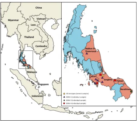
Serum samples were collected from 182 dengue patients in the acute-phase of infection origi-nated from five provinces in Southern Thailand; namely Nakhon Si Thammarat, Narathiwat, Pattani, Satun and Songkhla (Fig 1and SupplementaryS1 Table). Only patients exhibiting febrile illness (temperature >38˚C), and who were suspected of arbovirus infection, according to the World Health Organization (WHO) guideline for dengue diagnosis were included in the study [30]. Serological testing was performed on patient blood samples using a rapid DENV non-structural protein 1 (NS1) antibody test (SD BIOLINE Dengue Duo rapid test; Abbot Lab-oratories, Bangkok, Thailand). Description of study population is recorded inS1 Table.
RT-PCR amplification, serotyping and DENV envelope gene sequencing
Serotyping was conducted on all NS1 positive samples using a nested RT-PCR as previously described [31]. Viral RNA was extracted from all NS1 positive samples using the QIAamp Viral RNA mini kit (Qiagen, Bangkok, Thailand). 140μL of serum samples were used for extraction following the manufacturer’s protocol. Purified viral RNAs were stored at -80˚C until the serotyping and sequencing was performed. Nested RT-PCR was performed using the SensiFast kit (Bioline, Menphis, TN, USA). The first PCR was performed using the primer pair D1F and D1R with the following cycling conditions: activation at 95˚C for 10 min followed by 35 amplification cycles of 95˚C for 5s, 57˚C for 20s and 72˚C for 1min. The nested PCR was performed on the first PCR amplicon using forward primer D1F and serotype specific reverse primer (RTS1, RTS2, RTS3 and RTS4) with the following program: activation at 95˚C for
Fig 1. Map of Thailand showing surveillance region and DENV types detected. Participating provinces are
highlighted in orange. Location from which the patients originated are marked on the map along with DENV serotype: Purple triangles, DENV-2; green diamond, DENV-3; red square, DENV-4; and yellow circle multiple serotypes. The map was annotated using QGIS software.
10min followed by 30 amplification cycles of 95˚C for 15s, 58˚C for 20min and 72˚C for 1min. PCR products were visualized on a 1.8% agarose gel. Primers used to determine the serotype of the virus are provided inS2 Table.
To avoid working on viruses passaged and isolated from cell culture, we decided to directly sequence samples from viral RNA previously extracted from serum as mentioned above. Then, isolated RNA were retro-transcribed in cDNA using random hexamer and MMLV reverse transcription kit (Promega, Charbonnière, France), following the manufacturer’s protocol. Then, PCR was performed to amplify the full Envelope (Env) gene sequence using KAPA2G Fast Ready Mix (KapaBiosystem, Wilmington, MA, USA) following manufacturer’s instruc-tions. The 1st-forward and 4th-reverse primers for each serotype were used to amplify Env gene sequences (S2 Table). Amplification was performed using the following cycling condi-tions: activation at 95˚C for 10 min followed by 40 amplification cycles of 95˚C for 15s, 60˚C for 15s and 72˚C for 30s. PCR products were visualized on a 1.5% agarose gel and specific amplicons were purified from gel using QIAquick PCR Purification kit (QIAGEN, Bangkok, Thailand) following manufacturer’s instructions and stored at -20˚C until sequencing was per-formed. The entire envelope gene was successfully amplified for 46 samples. Purified ampli-cons were directly sequenced in both 5’ and 3’ directions using cycle sequencing and dye terminator methodologies by Macrogen company (Seoul, Korean). A set of eight overlapping primers for each serotype were used for sequencing (S2 Table) in order to cover all the entire gene and avoid discrepancies.
Phylogenetic analysis
To characterize the DENV isolated from clinical cases in Southern Thailand, the sequences encoding the envelope protein E were subject to robust phylogenetic analyses, along with rep-resentative DENV sequences obtained from the NCBI GenBank database (S3 Table).
Sequences were selected to cover a broad geographical area and includes sequences from coun-tries where DENV circulation were reported from several decades. All sequences were refer-enced into the tree in a format consisting of “accession number_country_year of isolation”.
Overlapping reads were assembled into contiguous sequences using CAP3 Sequence Assembly Program [32]. Multiple sequence alignments were conducted using MEGA 7 [33]. Sequences were edited and sites that could not be unambiguously aligned were excluded from subsequent analyses. Maximum likelihood (ML) trees were constructed using PhyML software [34] with the best-fit nucleotide substitution model (GTR+G)identified by Akaike’s Informa-tion criterion (AIC) using Topali v.2 [35]. Bootstrap method was used to measure the robust-ness of nodes with 1000 iterations. Phylogenetic trees were edited with FigTree v1.4.3 software (http://tree.bio.ed.ac.uk/software/figtree/). DENV serotypes 1, 2, 3 and 4 were classified fol-lowing Goncalvezet al, Twiddy et al, Lanciotti et al and Lanciotti et al, respectively [36–39]. All sequences from our study have been deposited in GenBank database and their accession numbers are shown inTable 1.
Ethics statement
This study was approved by the ethical committee and the samples were carried out following the rules and laws that govern the use of human material in Thailand. The study protocol was approved by the Ethical Committees of the Faculty of Tropical Medicine, Mahidol University (Bangkok, Thailand) (approval No. MUTM 2016-002-01), the Faculty of Medicine of Prince of Songkhla University and Hat Yai Hospital (Songkhla, Thailand) (approval No. 59-159-05-2). Informed consent was obtained from study participants and for minors from parents or a legal representative.
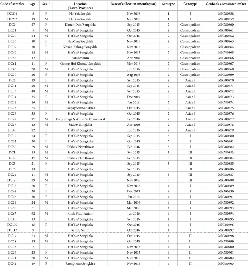
Table 1. Information on dengue-infected patients from whom DENV E protein gene were sequenced. Code of samples Age� Sex� � Location
(Town/Province)
Date of collection (month/year) Serotype Genotype GenBank accession number
DC201 8 F HatYai/Songkla Nov 2016 1 I MK780858
DC202 19 M HatYai/Songkla Nov 2016 1 I MK780859
DC9 27 F Khuan Don/Songkhla Sep 2015 2 Cosmopolitan MK780860
DC21 5 M HatYai/ Songkhla Oct 2015 2 Cosmopolitan MK780861
DC30 24 M HatYai/ Songkhla Oct 2015 2 Cosmopolitan MK780862
DC35 10 F Na Mon/Songkhla Nov 2015 2 Cosmopolitan MK780863
DC39 30 F Khuan Kalong/Songkhla Nov 2015 2 Cosmopolitan MK780864
DC40 12 M HatYai/ Songkhla Nov 2015 2 Cosmopolitan MK780865
DC58 12 F Satun/Satun Apr 2016 2 Cosmopolitan MK780866
DC61 11 F Khlong Hoi Khong/ Songkhla May 2016 2 Cosmopolitan MK780867
DC66 28 M HatYai/ Songkhla Jun 2016 2 Cosmopolitan MK780868
DC70 20 F HatYai/ Songkhla Aug 2016 2 Cosmopolitan MK780869
DC4 19 F HatYai/ Songkhla Sep 2015 2 Asian I MK780870
DC11 20 M HatYai/ Songkhla Sep 2015 2 Asian I MK780871
DC13 48 M HatYai/ Songkhla Sep 2015 2 Asian I MK780872
DC14 35 F HatYai/ Songkhla Dec 2015 2 Asian I MK780873
DC16 16 M HatYai/ Songkhla Jan 2016 2 Asian I MK780874
DC23 25 F Pakpayoon/Songkhla Oct 2015 2 Asian I MK780875
DC26 33 F HatYai/ Songkhla Oct 2015 2 Asian I MK780876
DC49 27 M Tung Song/ Nakhon Si Thammarat Feb 2016 2 Asian I MK780877
DC57 13 M Sadao/ Songkhla Apr 2016 2 Asian I MK780878
DC65 22 F HatYai/ Songkhla Jun 2016 2 Asian I MK780879
DC12 54 F HatYai/ Songkhla Sep 2015 3 I MK780880
DC32 20 F HatYai/ Songkhla Oct 2015 3 I MK780881
DC50 29 M Takbai/ Narathiwat Feb 2016 3 I MK780882
DC1 13 M HatYai/ Songkhla Sep 2015 3 III MK780883
DC2 47 M Takbai/ Narathiwat Sep 2015 3 III MK780884
DC3 21 F HatYai/ Songkhla Sep 2015 3 III MK780885
DC6 13 F HatYai/ Songkhla Sep 2015 3 III MK780886
DC24 11 M HatYai/ Songkhla Sep 2015 3 III MK780887
DC125 29 M HatYai/ Songkhla Nov 2016 3 III MK780888
DC38 20 F HatYai/ Songkhla Nov 2015 4 I MK780889
DC44 20 F HatYai/ Songkhla Dec 2015 4 I MK780890
DC46 30 F HatYai/ Songkhla Jan 2016 4 I MK780891
DC54 24 M HatYai/ Songkhla Mar 2016 4 I MK780892
DC56 7 F HatYai/ Songkhla Mar 2016 4 I MK780893
DC67 62 M Khok Pho/ Pattani Jun 2016 4 I MK780894
DC85 13 F HatYai/ Songkhla Sep 2016 4 I MK780895
DC100 15 F HatYai/ Songkhla Oct 2016 4 I MK780896
DC113 9 F Satun/ Satun Oct 2016 4 I MK780897
DC19 23 M HatYai/ Songkhla Oct 2015 4 II MK780898
DC28 15 M HatYai/ Songkhla Oct 2015 4 II MK780899
DC33 2 F HatYai/ Songkhla Nov 2015 4 II MK780900
DC36 28 F HatYai/ Songkhla Nov 2015 4 II MK780901
DC41 28 M HatYai/ Songkhla Nov 2015 4 II MK780902
DC42 19 F Rattaphum/Songkhla Nov 2015 4 II MK780903
Origins of dengue strains used in the study �Age of patients in years.
��F: female, M: male
Results
Of the 182 serum samples collected in the southern province of Thailand during 2015–2016, from patients presenting with dengue-like symptoms and confirmed positive for dengue by both NS1 serological test and RT-PCR, the envelope gene of 46 were successfully sequenced. The serotype was determined by nested RT-PCR, revealing the co-circulation of all four DENV serotypes in Southern Thailand (Table 1; DENV1, 2; DENV2, 20; DENV3, 9; DENV4, 15). Overall, we detected the four serotypes in overlapping and distinct regions, revealing a complex hyperendemicity dynamic in the region (Fig 1).
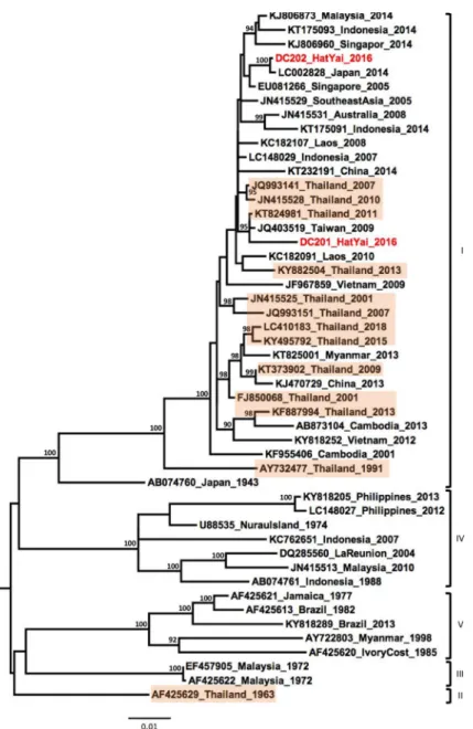
Phylogenetic analysis of the two DENV-1 samples showed their affiliations to the genotype I and that they grouped with contemporary strains isolated in Southeast Asia between 2005– 2014 (Fig 2).
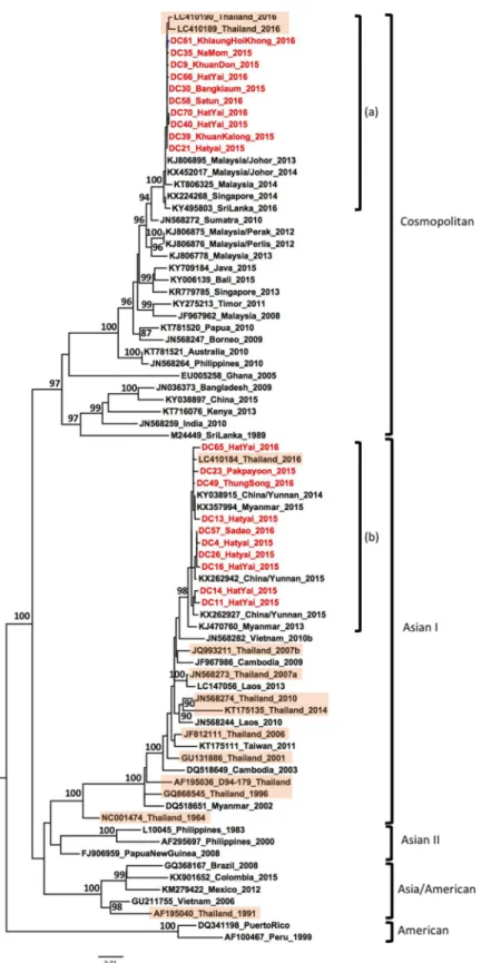
Of the twenty DENV-2 samples, ten belonged to the Cosmopolitan genotype. These formed a monophyletic lineage that was closely related to strains isolated in Malaysia, Singapore, Sri Lanka since 2013, as well as strains isolated from north and central parts of Thailand in 2016 [29], suggesting endemicity of the virus in the region (Fig 3, label a). The remaining ten strains belonged to the genotype Asian I and formed a monophyletic lineage comprising strains iso-lated in the province of Yunnan, China, in 2015 [40], in Myanmar in 2015 [41] and central province in Thailand [29] (Fig 3, label b). The phylogenetic association of the Cosmopolitan genotypes with Southeast Asian countries and the Asian I genotype with China and Myanmar indicates spatial segregation of the DENV-2 genotypes within Asia, however both these geno-types overlapped in Southern Thailand.
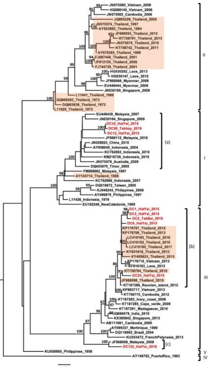
Out of the nine DENV-3 samples, three were identified as genotype I (Fig 4). These grouped together and clustered with strains collected over the past decade from Southeast Asian coun-tries (Indonesia, Singapore, Malaysia) and Australia (Fig 4, label a). The six remaining DENV-3 strains belonged to the genotype III, although they were derived from multiple lineages cir-culating over the last decade in Southeast Asian countries, including Laos, Vietnam, Malaysia and Thailand (Fig 4, labels b and c).
Similar to other serotypes, DENV-4 samples collected from southern Thailand belonged to multiple genotypes. Nine samples belonged to the genotype I and clustered with strains col-lected in Central Thailand in 2016–2017, in Singapore in 2014 and in China in 2013, forming a monophyletic lineage (Fig 5, label a). The six remaining samples grouped in a well-supported lineage (Fig 5, label b) belonging to the genotype II, within clade II-B along with strains from Southeast Asia and Australia between 2000–2016 (Fig 5).
Discussion
Screening of dengue samples collected from patients presenting with dengue-like symptoms to hospitals in southern Thailand during 2015–2016 identified all four DENV serotypes,
highlighting the hyperendemicity in Southern Thailand. To determine the genotypes of DENV and the relationships with other DENV strains, particularly from Thailand and South-east Asia, we generated sequences of DENV envelope gene directly from patient serum sam-ples and conducted a comprehensive phylogenetic analysis.
DENV-1 viruses were identified as genotype I and showed a close genetic link with strains previously reported from several countries in Southeast Asia and the Pacific region. Recently, a DENV-1 strain (LC410183_Thailand_2016) was isolated from the province of Chang Mai in Northern Thailand [29]. Interestingly, the DENV-1 strains from our study were not grouped in the same clade with LC410183_Thailand_2016, suggesting intra-genotype diversity of geno-type I strains circulating within Thailand.
Interestingly, DENV-2 samples belonged to two different genotypes. Ten samples belonged to the Asian I genotype, which has been commonly reported in several countries in Asia such as in Myanmar [42], Vietnam [43], Thailand [28,29], Laos PDR [44] and Taiwan [45]. These 10 strains do not show a wide genetic diversity and cluster into a monophyletic lineage with DENV-2 strains isolated during a large outbreak in China in 2014 and 2015 (Province of Yun-nan) and Myanmar 2015 [40,41]. Furthermore, 10 other DENV-2 strains were classified as Cosmopolitan genotype and formed a specific clade into a single lineage with strains from Malaysia, Singapore, Sri Lanka as well as strains recently isolated from the Central and North-ern region of Thailand. These strains were also related to viruses isolated from 2009 to 2016 in the insular region of Southeast Asia and Australia, where the Cosmopolitan genotype was the
Fig 2. Phylogenetic analysis of DENV-1. Maximum-likelihood tree of dengue virus serotype 1 envelope gene
sequences, generated using the GTR+G substitution model. Bootstrap values are shown on branch nodes. Samples collected in this study are in red colour font and strains from Thailand are highlighted in light orange. Annotation on the right denote DENV genotype.
Fig 3. Phylogenetic analysis of DENV-2. Maximum-likelihood tree of dengue virus serotype 2 envelope gene
sequences, generated using the GTR+G substitution model. Bootstrap values are shown on branch nodes. Samples collected in this study are in red colour font and strains from Thailand are highlighted in light orange. Annotation on the right denote DENV genotype.
predominant genotype (Fig 3). As reported by Twiddyet al, the Cosmopolitan genotype is
spreading worldwide, including throughout Southeast Asia [46]. Our analysis suggested that this genotype has also spread to Thailand, corroborating recent findings in the Central and Northern region of Thailand [29]. The Cosmopolitan genotype strains of our study are geneti-cally close to strains from the Central and Northern regions of Thailand collected in 2016 [29].
Fig 4. Phylogenetic analysis of DENV-3. Maximum-likelihood tree of dengue virus serotype 3 envelope gene
sequences, generated using the GTR+G substitution model. Bootstrap values are shown on branch nodes. Samples collected in this study are in red colour font and strains from Thailand are highlighted in light orange. Annotation on the right denote DENV genotype.
Thus, our findings may suggest a route of introduction of DENV-2 into Thailand from the Malaysian peninsula or from other southern countries of Thailand, considering that our sam-ples were collected in 2015 and 2016. Furthermore, these strains were genetically-related to the DENV-2 strains associated with outbreaks in Singapore and Malaysia in 2013 and 2014 [13,
Fig 5. Phylogenetic analysis of DENV-4. Maximum-likelihood tree of dengue virus serotype 1 envelope gene
sequences, generated using the GTR+G substitution model. Bootstrap values are shown on branch nodes. Samples collected in this study are in red colour font and strains from Thailand are highlighted in light orange. Annotation on the right denote DENV genotype.
47] (Fig 3). Further investigation is needed to assess whether several routes of introduction could be possible from countries to the north or south of Thailand. Both the genotype Asia I and Cosmopolitan genotype were reported to be regionally endemic [37,48], suggesting that there is a frequent crossover between countries in Southeast Asia–highlighting the importance of continuously monitoring DENV in Southern Thailand. While the Asian/American geno-type was displaced by the Asian-I genogeno-type in Vietnam and Cambodia [20], our results suggest a shift towards the Cosmopolitan genotype in Thailand. However, to date, reports of the pres-ence of the Cosmopolitan genotype are seldom [29,49], highlighting the importance of perma-nent surveillance in Thailand.
In the past few years, several studies have shown the replacement of DENV-3 genotype II by the genotype III in Southeast Asia [18,29]. Genotype III is now described as a widespread worldwide “Cosmopolitan genotype” with a high rate of dissemination upon introduction in a new area and has been linked to several outbreaks [18,50]. Our phylogenetic analysis of six DENV-3 samples resulted in their classification within genotype III, suggesting that this geno-type replacement occurred in Thailand between 2012–2013 (Fig 4). Our strains showed a genetic diversity with two distinct lineages classified within genotype III. Indeed, five samples were closely grouped in a monophyletic lineage with strains isolated from Thailand, Laos and Vietnam between 2012 to 2017, while the single strain DC-125 clustered separately with a strain isolated in Malaysia in 2008. This separation in two lineages suggests different routes of introduction or different modes of circulation in Thailand as proposed by Tanet al in Malaysia
[18]. The strain DC125, closely related with the virus from Malaysia 2008, could have been introduced a few years ago and may have been circulating at a low level after its introduction in Southern Thailand. Furthermore, our observation in Southern Thailand corroborates the recent observations in the Central and Northern regions that also showed the establishment of genotype III [29]. Further investigation could confirm whether this is a general trend.
A significant result of our phylogenetic analysis of DENV-3 was that three samples were distributed into the genotype I. To the best of our knowledge, this is the first time since 1988 that the presence of genotype I has been reported in Thailand [23]. Our genotype I samples were grouped into a single clade closely related with viruses mostly circulating in Indonesia, Malaysia and Singapore, which were either isolated from travellers returning from this region [51,52] or from natives during regional surveys and outbreaks [53,54]. Our samples were col-lected from the district of HatYai in Songkhla province and the district of Takbai, in Narathi-wat province, which shares a border with the Kelatan province in Malaysia. Previous studies reported a wide circulation of the genotype I in Malaysia and Indonesia [55]. These findings may suggest a spread of the DENV-3 genotype I from insular Southeast Asia into Southern Thailand and could lead to a replacement of the dominant genotype. However, future research is needed to determine whether genotype I is increasing in Thailand or whether our finding resulted from a time specific event in which genotype I was circulating in the region of our study.
Following the same pattern as the DENV-3 strains, the DENV-4 strains were grouped into two genotypes: lineage (a) which groups nine samples in genotype I and the lineage (b) which groups six strains in genotype II clade B (Fig 5). This observation shows that lineage (a) is closely related with strains isolated during outbreaks in Singapore in 2014 [13], in China from an imported case from Thailand (2013), Bali (2016) [56] and also recently isolated strains from Northern Thailand [29]. Genotype I is commonly found in Thailand and continental countries of Southeast Asia. However, the observation of lineage (b) suggests a possible an introduction that did spread and not established in introduction of a new genotype into Thailand. Indeed, six samples were grouped together into a single lineage belonging to genotype II. This clade formed a monophyletic lineage related to the Southeast Asian and Australian strains. The last
detection of this genotype in Thailand was reported in 2000 in Bangkok [27]. It should be noted that all these samples were collected in 2015 from the province of Songkhla and had a low phylogenetic diversity. This observation may suggest a singular introduction in time and place and not necessarily the emergence of a new DENV-4 genotype in Southern Thailand. However, further investigation would be necessary in order to follow the DENV-4 phyloge-netic dynamic.
In conclusion, our study describes, for the first time, the genetic diversity of all four sero-types of DENV in Southern Thailand, providing important insights into their distributions in the region. We demonstrated that DENV is hyperendemic in Southern Thailand, rendering coinfection between serotypes and genotypes possible. We observed the co-circulation of mul-tiple genotypes within DENV-2 to DENV 4: DENV-2 Cosmopolitan genotype co-circulated with Asian-I; DENV-3 genotype I with genotype III; and DENV-4 genotype I was prevalent while genotype II-B was detected after decades of absence. The observed re-emergence of sev-eral genotypes suggests constant movement and introduction of DENV strains in Southern Thailand. Our study urges furtherin vivo characterization of the local mosquito population to
assess the possible gain ofin vivo advantage for the new genotypes in Southern Thailand. Gain
in vector competence may partly explain the difference in genotype distribution between the North and the South of Thailand. Our data highlights the high incidence of DENV serotype and genotype co-circulation, particularly in urban area, such as the city of Hat Yai and the area along the Malaysian border. It would be informative to evaluate how this hyperendemicity influences disease severity.
Supporting information
S1 Table. Information on dengue positive study population.
(PDF)
S2 Table. Primers used in this study.
(PDF)
S3 Table. Dengue virus sequences used in present study.
(PDF)
Author Contributions
Conceptualization: Dorothe´e Misse´.
Formal analysis: Rodolphe Hamel, Alexandra Dauve´, Celeste Donato, Dhanasekaran
Vijaykrishna.
Funding acquisition: Pornapat Surasombatpattana, Ronald Morales Vargas, Natthanej
Luplertlop, Dorothe´e Misse´.
Investigation: Rodolphe Hamel, Pornapat Surasombatpattana, Sineewanlaya Wichit,
Alexan-dra Dauve´, Celeste Donato.
Methodology: Rodolphe Hamel.
Project administration: Dorothe´e Misse´.
Resources: Pornapat Surasombatpattana, Ronald Morales Vargas, Natthanej Luplertlop. Writing – original draft: Rodolphe Hamel.
Writing – review & editing: Julien Pompon, Dhanasekaran Vijaykrishna, Florian Liegeois,
Dorothe´e Misse´.
References
1. WHO. Dengue and severe dengue 2018. Available from:http://www.who.int/news-room/fact-sheets/ detail/dengue-and-severe-dengue.
2. Lee JS, Mogasale V, Lim JK, Carabali M, Lee KS, Sirivichayakul C, et al. A multi-country study of the economic burden of dengue fever: Vietnam, Thailand, and Colombia. PLoS Negl Trop Dis. 2017; 11 (10):e0006037. Epub 2017/10/30.https://doi.org/10.1371/journal.pntd.0006037PMID:29084220; PubMed Central PMCID: PMC5679658.
3. Muller DA, Depelsenaire AC, Young PR. Clinical and Laboratory Diagnosis of Dengue Virus Infection. J Infect Dis. 2017; 215(suppl_2):S89–S95.https://doi.org/10.1093/infdis/jiw649PMID:28403441. 4. Flasche S, Jit M, Rodrı´guez-Barraquer I, Coudeville L, Recker M, Koelle K, et al. The Long-Term Safety,
Public Health Impact, and Cost-Effectiveness of Routine Vaccination with a Recombinant, Live-Attenu-ated Dengue Vaccine (Dengvaxia): A Model Comparison Study. PLoS Med. 2016; 13(11):e1002181. Epub 2016/11/29.https://doi.org/10.1371/journal.pmed.1002181PMID:27898668; PubMed Central PMCID: PMC5127514.
5. Holmes EC, Burch SS. The causes and consequences of genetic variation in dengue virus. Trends Microbiol. 2000; 8(2):74–7. PMID:10664600.
6. Health MoP. Bureau of Epidemiology, Dengue fever. Nonthaburi: Department of Disease Control Minis-try of Public Health. 2016, Accessed 22 Feb 2016.https://doi.org/10.1186/s12889-016-3382-5
7. Hammon WM. Dengue hemorrhagic fever—do we know its cause? Am J Trop Med Hyg. 1973; 22 (1):82–91.https://doi.org/10.4269/ajtmh.1973.22.82PMID:4567852.
8. Ooi EE, Gubler DJ. Dengue in Southeast Asia: epidemiological characteristics and strategic challenges in disease prevention. Cad Saude Publica. 2009; 25 Suppl 1:S115–24. PMID:19287856.
9. Limkittikul K, Brett J, L’Azou M. Epidemiological trends of dengue disease in Thailand (2000–2011): a systematic literature review. PLoS Negl Trop Dis. 2014; 8(11):e3241. Epub 2014/11/06.https://doi.org/ 10.1371/journal.pntd.0003241PMID:25375766; PubMed Central PMCID: PMC4222696.
10. Nisalak A, Clapham HE, Kalayanarooj S, Klungthong C, Thaisomboonsuk B, Fernandez S, et al. Forty Years of Dengue Surveillance at a Tertiary Pediatric Hospital in Bangkok, Thailand, 1973–2012. Am J Trop Med Hyg. 2016; 94(6):1342–7. Epub 2016/03/28.https://doi.org/10.4269/ajtmh.15-0337PMID:
27022151; PubMed Central PMCID: PMC4889755.
11. Gintarong TJ, Emran A, Sherin A, Thein TT, Aung TS. Circulation of all dengue virus serotypes during dengue outbreak in Sandakan, Sabah, Malaysia (2016). J Vector Borne Dis. 2018; 55(2):168–71.
https://doi.org/10.4103/0972-9062.242566PMID:30280717.
12. Koo C, Nasir A, Hapuarachchi HC, Lee KS, Hasan Z, Ng LC, et al. Evolution and heterogeneity of multi-ple serotypes of Dengue virus in Pakistan, 2006–2011. Virol J. 2013; 10:275. Epub 2013/09/04.https:// doi.org/10.1186/1743-422X-10-275PMID:24007412; PubMed Central PMCID: PMC3844417. 13. Hapuarachchi HC, Koo C, Rajarethinam J, Chong CS, Lin C, Yap G, et al. Epidemic resurgence of
den-gue fever in Singapore in 2013–2014: A virological and entomological perspective. BMC Infect Dis. 2016; 16:300. Epub 2016/06/17.https://doi.org/10.1186/s12879-016-1606-zPMID:27316694; PubMed Central PMCID: PMC4912763.
14. Lardo S, Utami Y, Yohan B, Tarigan SM, Santoso WD, Nainggolan L, et al. Concurrent infections of dengue viruses serotype 2 and 3 in patient with severe dengue from Jakarta, Indonesia. Asian Pac J Trop Med. 2016; 9(2):134–40. Epub 2016/01/13.https://doi.org/10.1016/j.apjtm.2016.01.013PMID:
26919942.
15. Anoop M, Issac A, Mathew T, Philip S, Kareem NA, Unnikrishnan R, et al. Genetic characterization of dengue virus serotypes causing concurrent infection in an outbreak in Ernakulam, Kerala, South India. Indian J Exp Biol. 2010; 48(8):849–57. PMID:21341545.
16. Vinodkumar CS, Kalapannavar NK, Basavarajappa KG, Sanjay D, Gowli C, Nadig NG, et al. Episode of coexisting infections with multiple dengue virus serotypes in central Karnataka, India. J Infect Public Health. 2013; 6(4):302–6. Epub 2013/04/03.https://doi.org/10.1016/j.jiph.2013.01.004PMID:23806706. 17. Cucunawangsih Lugito NPH. Trends of Dengue Disease Epidemiology. Virology (Auckl). 2017;
8:1178122X17695836. Epub 2017/03/15.https://doi.org/10.1177/1178122X17695836PMID:
28579763; PubMed Central PMCID: PMC5428083.
18. Tan KK, Zulkifle NI, Sulaiman S, Pang SP, NorAmdan N, MatRahim N, et al. Emergence of the Asian lineage dengue virus type 3 genotype III in Malaysia. BMC Evol Biol. 2018; 18(1):58. Epub 2018/04/24.
19. Rico-Hesse R, Harrison LM, Salas RA, Tovar D, Nisalak A, Ramos C, et al. Origins of dengue type 2 viruses associated with increased pathogenicity in the Americas. Virology. 1997; 230(2):244–51.
https://doi.org/10.1006/viro.1997.8504PMID:9143280.
20. Vu TT, Holmes EC, Duong V, Nguyen TQ, Tran TH, Quail M, et al. Emergence of the Asian 1 genotype of dengue virus serotype 2 in viet nam: in vivo fitness advantage and lineage replacement in South-East Asia. PLoS Negl Trop Dis. 2010; 4(7):e757. Epub 2010/07/20.https://doi.org/10.1371/journal.pntd. 0000757PMID:20651932; PubMed Central PMCID: PMC2907417.
21. McElroy KL, Santiago GA, Lennon NJ, Birren BW, Henn MR, Muñoz-Jorda´n JL. Endurance, refuge, and reemergence of dengue virus type 2, Puerto Rico, 1986–2007. Emerg Infect Dis. 2011; 17(1):64– 71.https://doi.org/10.3201/eid1701.100961PMID:21192856; PubMed Central PMCID: PMC3204641. 22. Sittisombut N, Sistayanarain A, Cardosa MJ, Salminen M, Damrongdachakul S, Kalayanarooj S, et al.
Possible occurrence of a genetic bottleneck in dengue serotype 2 viruses between the 1980 and 1987 epidemic seasons in Bangkok, Thailand. Am J Trop Med Hyg. 1997; 57(1):100–8.https://doi.org/10. 4269/ajtmh.1997.57.100PMID:9242328.
23. Wittke V, Robb TE, Thu HM, Nisalak A, Nimmannitya S, Kalayanrooj S, et al. Extinction and rapid emer-gence of strains of dengue 3 virus during an interepidemic period. Virology. 2002; 301(1):148–56.
https://doi.org/10.1006/viro.2002.1549PMID:12359455.
24. Ito M, Takasaki T, Kotaki A, Tajima S, Yuwono D, Rimal HS, et al. Molecular and virological analyses of dengue virus responsible for dengue outbreak in East Timor in 2005. Jpn J Infect Dis. 2010; 63(3):181– 4. PMID:20495269.
25. Drumond BP, Fagundes LG, Rocha RP, Fumagalli MJ, Araki CS, Colombo TE, et al. Phylogenetic anal-ysis of Dengue virus 1 isolated from South Minas Gerais, Brazil. Braz J Microbiol. 2016; 47(1):251–8. Epub 2016/01/27.https://doi.org/10.1016/j.bjm.2015.11.016PMID:26887252; PubMed Central PMCID: PMC4827697.
26. Kurosu T, Chaichana P, Phanthanawiboon S, Khamlert C, Yamashita A, A-nuegoonpipat A, et al. Sequence variation of dengue type 2 virus isolated from clinical cases in Thailand. Jpn J Infect Dis. 2014; 67(2):132–4. PMID:24647259.
27. Klungthong C, Zhang C, Mammen MP, Ubol S, Holmes EC. The molecular epidemiology of dengue virus serotype 4 in Bangkok, Thailand. Virology. 2004; 329(1):168–79.https://doi.org/10.1016/j.virol. 2004.08.003PMID:15476884.
28. Rabaa MA, Klungthong C, Yoon IK, Holmes EC, Chinnawirotpisan P, Thaisomboonsuk B, et al. Fre-quent in-migration and highly focal transmission of dengue viruses among children in Kamphaeng Phet, Thailand. PLoS Negl Trop Dis. 2013; 7(1):e1990. Epub 2013/01/17.https://doi.org/10.1371/journal. pntd.0001990PMID:23350000; PubMed Central PMCID: PMC3547850.
29. Phadungsombat J, Lin MY, Srimark N, Yamanaka A, Nakayama EE, Moolasart V, et al. Emergence of genotype Cosmopolitan of dengue virus type 2 and genotype III of dengue virus type 3 in Thailand. PLoS One. 2018; 13(11):e0207220. Epub 2018/11/12.https://doi.org/10.1371/journal.pone.0207220
PMID:30419004; PubMed Central PMCID: PMC6231660.
30. WHO. Dengue and severe dengue, Fact sheet n˚117: World Health Organization; 2009. Available from:http://www.who.int/mediacentre/factsheets/fs117/en/.
31. Lanciotti RS, Calisher CH, Gubler DJ, Chang GJ, Vorndam AV. Rapid detection and typing of dengue viruses from clinical samples by using reverse transcriptase-polymerase chain reaction. J Clin Micro-biol. 1992; 30(3):545–51. PMID:1372617; PubMed Central PMCID: PMC265106.
32. Huang X, Madan A. CAP3: A DNA sequence assembly program. Genome Res. 1999; 9(9):868–77.
https://doi.org/10.1101/gr.9.9.868PMID:10508846; PubMed Central PMCID: PMC310812.
33. Kumar S, Stecher G, Tamura K. MEGA7: Molecular Evolutionary Genetics Analysis Version 7.0 for Big-ger Datasets. Mol Biol Evol. 2016; 33(7):1870–4. Epub 2016/03/22.https://doi.org/10.1093/molbev/ msw054PMID:27004904.
34. Guindon S, Dufayard JF, Lefort V, Anisimova M, Hordijk W, Gascuel O. New algorithms and methods to estimate maximum-likelihood phylogenies: assessing the performance of PhyML 3.0. Syst Biol. 2010; 59(3):307–21. Epub 2010/03/29.https://doi.org/10.1093/sysbio/syq010PMID:20525638.
35. Milne I, Wright F, Rowe G, Marshall DF, Husmeier D, McGuire G. TOPALi: software for automatic iden-tification of recombinant sequences within DNA multiple alignments. Bioinformatics. 2004; 20
(11):1806–7.https://doi.org/10.1093/bioinformatics/bth155PMID:14988107.
36. Goncalvez AP, Escalante AA, Pujol FH, Ludert JE, Tovar D, Salas RA, et al. Diversity and evolution of the envelope gene of dengue virus type 1. Virology. 2002; 303(1):110–9.https://doi.org/10.1006/viro. 2002.1686PMID:12482662.
37. Twiddy SS, Farrar JJ, Vinh Chau N, Wills B, Gould EA, Gritsun T, et al. Phylogenetic relationships and differential selection pressures among genotypes of dengue-2 virus. Virology. 2002; 298(1):63–72.
38. Lanciotti RS, Lewis JG, Gubler DJ, Trent DW. Molecular evolution and epidemiology of dengue-3 viruses. J Gen Virol. 1994; 75 (Pt 1):65–75.https://doi.org/10.1099/0022-1317-75-1-65PMID:
8113741.
39. Lanciotti RS, Gubler DJ, Trent DW. Molecular evolution and phylogeny of dengue-4 viruses. J Gen Virol. 1997; 78 (Pt 9):2279–84.https://doi.org/10.1099/0022-1317-78-9-2279PMID:9292015. 40. Hu TS, Zhang HL, Feng Y, Fan JH, Tang T, Liu YH, et al. Epidemiological and molecular characteristics
of emergent dengue virus in Yunnan Province near the China-Myanmar-Laos border, 2013–2015. BMC Infect Dis. 2017; 17(1):331. Epub 2017/05/08.https://doi.org/10.1186/s12879-017-2401-1PMID:
28482813; PubMed Central PMCID: PMC5422898.
41. Kyaw AK, Ngwe Tun MM, Moi ML, Nabeshima T, Soe KT, Thwe SM, et al. Clinical, virological and epi-demiological characterization of dengue outbreak in Myanmar, 2015. Epidemiol Infect. 2017; 145 (9):1886–97. Epub 2017/04/17.https://doi.org/10.1017/S0950268817000735PMID:28414004. 42. Huang JH, Liao TL, Chang SF, Su CL, Chien LJ, Kuo YC, et al. Laboratory-based dengue surveillance
in Taiwan, 2005: a molecular epidemiologic study. Am J Trop Med Hyg. 2007; 77(5):903–9. PMID:
17984351.
43. Warrilow D, Northill JA, Pyke AT. Sources of dengue viruses imported into Queensland, australia, 2002–2010. Emerg Infect Dis. 2012; 18(11):1850–7.https://doi.org/10.3201/eid1811.120014PMID:
23092682; PubMed Central PMCID: PMC3559152.
44. Phommanivong V, Kanda S, Shimono T, Lamaningao P, Darcy AW, Mishima N, et al. Co-circulation of the dengue with chikungunya virus during the 2013 outbreak in the southern part of Lao PDR. Trop Med Health. 2016; 44:24. Epub 2016/08/04.https://doi.org/10.1186/s41182-016-0020-yPMID:27524929; PubMed Central PMCID: PMC4973078.
45. Chang SF, Yang CF, Hsu TC, Su CL, Lin CC, Shu PY. Laboratory-Based Surveillance and Molecular Characterization of Dengue Viruses in Taiwan, 2014. Am J Trop Med Hyg. 2016; 94(4):804–11. Epub 2016/02/15.https://doi.org/10.4269/ajtmh.15-0534PMID:26880779; PubMed Central PMCID: PMC4824222.
46. Twiddy SS, Pybus OG, Holmes EC. Comparative population dynamics of mosquito-borne flaviviruses. Infect Genet Evol. 2003; 3(2):87–95. PMID:12809802.
47. Ng LC, Chem YK, Koo C, Mudin RN, Amin FM, Lee KS, et al. 2013 dengue outbreaks in Singapore and Malaysia caused by different viral strains. Am J Trop Med Hyg. 2015; 92(6):1150–5. Epub 2015/04/06.
https://doi.org/10.4269/ajtmh.14-0588PMID:25846296; PubMed Central PMCID: PMC4458818. 48. Weaver SC, Vasilakis N. Molecular evolution of dengue viruses: contributions of phylogenetics to
understanding the history and epidemiology of the preeminent arboviral disease. Infect Genet Evol. 2009; 9(4):523–40. Epub 2009/02/13.https://doi.org/10.1016/j.meegid.2009.02.003PMID:19460319; PubMed Central PMCID: PMC3609037.
49. Shihada S, Emmerich P, Thome´ -Bolduan C, Jansen S, Gu¨nther S, Frank C, et al. Genetic Diversity and New Lineages of Dengue Virus Serotypes 3 and 4 in Returning Travelers, Germany, 2006–2015. Emerg Infect Dis. 2017; 23(2):272–5.https://doi.org/10.3201/eid2302.160751PMID:28098525; PubMed Central PMCID: PMC5324807.
50. Messer WB, Gubler DJ, Harris E, Sivananthan K, de Silva AM. Emergence and global spread of a den-gue serotype 3, subtype III virus. Emerg Infect Dis. 2003; 9(7):800–9.https://doi.org/10.3201/eid0907. 030038PMID:12899133; PubMed Central PMCID: PMC3023445.
51. Ernst T, McCarthy S, Chidlow G, Luang-Suarkia D, Holmes EC, Smith DW, et al. Emergence of a new lineage of dengue virus type 2 identified in travelers entering Western Australia from Indonesia, 2010– 2012. PLoS Negl Trop Dis. 2015; 9(1):e0003442. Epub 2015/01/30.https://doi.org/10.1371/journal. pntd.0003442PMID:25635775; PubMed Central PMCID: PMC4311992.
52. Shu PY, Su CL, Liao TL, Yang CF, Chang SF, Lin CC, et al. Molecular characterization of dengue viruses imported into Taiwan during 2003–2007: geographic distribution and genotype shift. Am J Trop Med Hyg. 2009; 80(6):1039–46. PMID:19478273.
53. Ong SH, Yip JT, Chen YL, Liu W, Harun S, Lystiyaningsih E, et al. Periodic re-emergence of endemic strains with strong epidemic potential-a proposed explanation for the 2004 Indonesian dengue epi-demic. Infect Genet Evol. 2008; 8(2):191–204. Epub 2007/12/25.https://doi.org/10.1016/j.meegid. 2007.12.005PMID:18243816.
54. Sasmono RT, Wahid I, Trimarsanto H, Yohan B, Wahyuni S, Hertanto M, et al. Genomic analysis and growth characteristic of dengue viruses from Makassar, Indonesia. Infect Genet Evol. 2015; 32:165–77. Epub 2015/03/14.https://doi.org/10.1016/j.meegid.2015.03.006PMID:25784569.
55. Sucipto TH, Kotaki T, Mulyatno KC, Churrotin S, Labiqah A, Soegijanto S, et al. Phylogenetic Analysis of Dengue Virus in Bangkalan, Madura Island, East Java Province, Indonesia. J Trop Med. 2018; 2018:8127093. Epub 2018/02/15.https://doi.org/10.1155/2018/8127093PMID:29666658; PubMed Central PMCID: PMC5832172.
56. Moore PR, van den Hurk AF, Mackenzie JS, Pyke AT. Dengue viruses in Papua New Guinea: evidence of endemicity and phylogenetic variation, including the evolution of new genetic lineages. Emerg Microbes Infect. 2017; 6(12):e114. Epub 2017/12/20.https://doi.org/10.1038/emi.2017.103PMID: