HAL Id: hal-01368643
https://hal-amu.archives-ouvertes.fr/hal-01368643
Submitted on 12 Oct 2016
HAL is a multi-disciplinary open access
archive for the deposit and dissemination of
sci-entific research documents, whether they are
pub-lished or not. The documents may come from
teaching and research institutions in France or
abroad, or from public or private research centers.
L’archive ouverte pluridisciplinaire HAL, est
destinée au dépôt et à la diffusion de documents
scientifiques de niveau recherche, publiés ou non,
émanant des établissements d’enseignement et de
recherche français ou étrangers, des laboratoires
publics ou privés.
Distributed under a Creative Commons Attribution - NonCommercial - NoDerivatives| 4.0
International License
Embryonal tumors with multilayered rosettes in
children: the SFCE experience
Meryl Horwitz, Christelle Dufour, Pierre Leblond, Franck Bourdeaut, Cécile
Faure-Conter, Anne-Isabelle Bertozzi, Bernadette Delisle, Gilles Palenzuela,
Anne Jouvet, Didier Scavarda, et al.
To cite this version:
Meryl Horwitz, Christelle Dufour, Pierre Leblond, Franck Bourdeaut, Cécile Faure-Conter, et al..
Embryonal tumors with multilayered rosettes in children: the SFCE experience. Child’s Nervous
System, Springer Verlag, 2016, 32 (2), pp.299-305. �10.1007/s00381-015-2920-2�. �hal-01368643�
ORIGINAL PAPER
Embryonal tumors with multilayered rosettes in children:
the SFCE experience
Meryl Horwitz1&Christelle Dufour2&Pierre Leblond3&Franck Bourdeaut4,5&
Cécile Faure-Conter6&Anne-Isabelle Bertozzi7&Marie Bernadette Delisle8&
Gilles Palenzuela9&Anne Jouvet10&Didier Scavarda11&Matthieu Vinchon12&
Laetitia Padovani13&Jean Gaudart14,15&Dominique Figarella Branger16&
Nicolas Andre1,17,18
Received: 22 June 2015 / Accepted: 23 September 2015 / Published online: 5 October 2015 # Springer-Verlag Berlin Heidelberg 2015
Abstract
Purposes The purpose of this study was to retrospectively study embryonal tumors with multilayered rosettes (ETMR), a rare new entity that gathers ETAN-TR (embryonal tumor with abundant neuropil and true rosettes), ependymoblastomas, and
medulloepitheliomas, in order to improve their descriptions and try to better define therapeutic modalities.
Methods Patients with ETMR, ETAN-TR, ependymoblastoma, and medulloepithelioma treated in SFCE centres (Société Française de lutte contre les Cancers et les leucémies de l'Enfant
* Nicolas Andre [email protected] Meryl Horwitz [email protected] Christelle Dufour [email protected] Pierre Leblond [email protected] Franck Bourdeaut [email protected] Cécile Faure-Conter [email protected] Anne-Isabelle Bertozzi [email protected] Marie Bernadette Delisle [email protected] Gilles Palenzuela [email protected] Anne Jouvet [email protected] Didier Scavarda [email protected] Matthieu Vinchon [email protected] Laetitia Padovani [email protected] Jean Gaudart [email protected] Dominique Figarella Branger [email protected]
1
Department of Pediatric Hematology-Oncology, APHM, La Timone Hospital, 264 rue Saint-Pierre, 13005, Marseille, France
2
Department of Pediatric and Adolescent Oncology, Gustave Roussy Campus, 114, rue Edouard-Vaillant, 94805 Villejuif Cedex, France 3
Department of Pediatric Oncology, Oscar Lambret, 3 rue Frederic Combemale, BP 307, 59020 Lille Cedex, France
4
Department of Pediatric Oncology, Institut Curie, 26, rue d’Ulm, 75005 Paris, France
5 INSERM U830, Laboratory of Genetics and Cancer Biology, Institut Curie, Paris, France
6
Department of Pediatrics, Centre Léon Bérard, 28 Prom. Léa et Napoléon Bullukian, 69008 Lyon, France
7
Department of Hematology-Oncology, Children University Hospital, 330 Avenue de Grande Bretagne, 31300 Toulouse, France 8 Department of Neuropathology, Toulouse Rangueil University
Hospital, 1, avenue du Pr Jean Poulhès, 31400 Toulouse, France 9
Department of Pediatric Hematology-Oncology, University Hospital of Montpellier, 191, avenue du doyen Gaston Giraud,
34295 Montpellier cedex, France 10
Department of Pathology and neuropathology,BPierre Werthmeier^ Hospital, Boulevard Pinel, 69677 Bron, France
et de l'adolescent) since 2000 were collected. Data were retrieved from clinical charts.
Results Thirty-eight patients were included in the analysis. Seventeen had an ETAN-TR, 13 had a medulloepithelioma, and 8 had an ETMR. No ependymoblastoma was included. The median age at diagnosis was 31 months (range, 2.8– 141 months). The predominant tumor location was supratentorial (66 %); 18.4 % patients had metastatic lesion.
LIN28A expression was positive in 11/11 patients. Amplifi-cation of the locus 19q13.42 was positive in 10/12 patients. Thirty patients were treated according to the primitive neuroectodermal tumors of high risk (PNET-HR) protocol. The median time of follow-up was 0.9 years (range 0.1 to 15.3 years). The 1-year event-free survival (EFS) and overall survival (OS) were, respectively, 36 % CI 95 % (23–55) and 45 % CI 95 % (31–64). On multivariate analysis, complete surgical resection, radiotherapy, and high-dose chemotherapy were associated with a better overall survival with a relative risk of, respectively, 7.9 CI 95 % (2.6–23.5) p < 0.0002, 41.8 CI 95 % (9.4–186) p < 0.0001, and 3.5 CI 95 % (1.3–9.5) p = 0.012.
Conclusion Prognosis of ETMR remains dismal despite mul-timodal therapy. LIN28A immunostaining and 19q13.42 am-plification should be systematically done to secure the diag-nosis. Complete surgical resection, radiotherapy, and high-dose chemotherapy are associated with better outcome.
Keywords Brain tumors . Children . Radiotherapy . Surgery . Chemotherapy . ETMR
Introduction
Embryonal tumors with neuropil and true rosettes abundant (ETAN-TR), ependymoblastomas, and medulloepitheliomas are three distinct pathologies which actually represent
different manifestations of the same histological and biologi-cal entity [1,2]. The term embryonal tumors with multilayered rosettes (ETMR) was proposed in 2010 as a unifying entity by Paulus and Kleihues [3] as the histological central element found in these tumors was the presence of multilayered ro-settes. Moreover, ETMR seem to have a specific molecular signature with a positivity of LIN28A on immuno-histochemistry and amplification of the locus 19q13.42 in FISH analysis [4,5]. ETMR belongs to the group of the prim-itive neuroectodermal tumors of the central nervous system (PNET) according to the World Health Organization (WHO) classification [6].
ETMR are quite rare entities; their prevalence is not accu-rately estimated. They are likely under-diagnosed with only a few cases reported in the literature since 2000 [2,3,7,8]. They occur mostly in children aged less than 2 years old. In the literature, the localization of the majority of the primitive tu-mor is supratentorial with signs of increased intracranial pres-sure. Treatment of ETMR usually starts with maximal surgical resection. The role of adjuvant treatment with either chemo-therapy and/or radiochemo-therapy has not yet been well established for this disease.
To improve our knowledge about ETMR, we collected cases treated in SFCE centers (Société Française des Cancers et leucémies de l’Enfant et de l’adolescent) from January 2000 to May 2014 in order to retrospectively study clinical, radio-logical, bioradio-logical, and prognostic descriptions to improve their descriptions and try to better define therapeutic modalities.
Patients and methods
Study design, patients, data collection
Patients aged less than 18 years with newly diagnosed ETMR who were treated in France between January 2000 and 1 May 2014 were identified retrospectively by consulting local oncologists’ and pathologists’ databases in SFCE centers using the following diagnosis: ETMR, ETAN-TR, ependymoblastoma, and medulloepithelioma. A standardized data sheet was sent to pediatric oncologists who were mem-bers of the SFCE to collect clinical data. The questionnaire included information about sex, age at time of presentation, duration of the symptoms at time of presentation, location of tumor, extent of resection (complete resection, partial resec-tion, biopsy), pathological biological and molecular diagnosis, adjuvant therapies, and follow-up. At diagnosis, stage of dis-ease was evaluated by initial cranial and spinal MRI and CSF cytology. The extent of initial resection was assessed by MRI or computed tomography performed within 72h after surgery. Metastatic disease was assessed using the Chang staging sys-tem. Tumors were subjected to a central histopathological
300 Childs Nerv Syst (2016) 32:299–305
11
Department of Neurosurgery, APHM, La Timone Hospital, 264 rue Saint-Pierre, 13005 Marseille, France
12 Department of Pediatric Neurosurgery, Lille University Hospital, 2 Avenue Oscar Lambret, 59000 Lille, France
13
Department of Radiation Oncology and Pediatrics, APHM, La Timone Hospital, 264 rue Saint-Pierre, 13005 Marseille, France 14
BIOSTIC, Pôle de Santé Publique, AP-HM, Marseille, 264 rue Saint-Pierre, 13005 Marseille, France
15 UMR912 SESSTIM, Aix-Marseille University, Marseille, France 16
Department of Pathology, APHM, La Timone Hospital, Université Aix-Marseille, 264 rue Saint-Pierre, 13005 Marseille, France 17
INSERM UMR 911, Centre de Recherche en Oncologie biologique et en Oncopharmacologie, Aix-Marseille University, 264 rue Saint-Pierre, 13005 Marseille, France
18 Department of Pediatric Hematology and Oncology, Children Hospital of La Timone, 264, rue Saint-Pierre,
review by members of the national pediatric brain tumors boa rd (GENOP). Whe n it was fea sible, immuno-histochemistry analysis applying a LIN28A polyclonal anti-body and array-based comparative genomic hybridization (CGHarray) was performed. The data were coded to ensure confidentiality. This study has been approved by the National Ethical Committee (CCTIRS).
Statistical analysis:
Relapses and deaths were considered as events. Event-free sur-vival (EFS), overall sursur-vival (OS), and univariate analysis were estimated by the Kaplan-Meier method, and differences be-tween groups were assessed by the log-rank test. Survival esti-mates referred to 1 and 3 years from diagnosis and the related 95 % confidence intervals (95 % CI) were calculated. Cox model was used to calculate the multivariate analysis by a step-wise selection. Descriptive statistics were reported as absolute frequencies and percentages for qualitative data, while median and range were used for continuous variables. When performing univariate analysis, those variables were used: age ≤4 years or >4 years, age <4 years or 4 to 5 years or 5 to 8 years or 8 to 11 years, histological diagnosis (ETMR, ETAN-TR, medulloepithelioma, ependymoblastoma), surgery, chemother-apy, radiotherchemother-apy, high-dose chemotherchemother-apy, and primitive neuroectodermal tumors of high risk (PNET-HR) (yes or no). Positive variables identified with univariate analysis were used in the multivaried analysis. All statistical tests were two sided and aBp^ value <0.05 was considered significant. Statistical analysis was performed using R version 3.0.2.
Results:
Patients’ characteristics
Forty-two patients with an ETMR were identified in the SFCE centers, but 4 patients were excluded because data could not be retrieved or the diagnosis was uncertain. Thirty-eight pa-tients were therefore analyzed. The median age at diagnosis was 31.1 months (range 2.8–141 months) and the sex ratio was 0.4 (male/female). Main clinical signs were a sided weak-ness (34 %) and increased intracranial pressure (53 %). Con-fusion, seizures, and cerebellar syndrome occurred in 26, 21, and 21 % of children, respectively. Torticollis occurred in 8 % of cases and visual impairment was found in 11 % of patients. The predominant tumor location was supratentorial (66 %). Tumors were infratentorial in the others (26 %). One patient had a medullar lesion. Eighteen percent of patients had meta-static disease at initial presentation. In all cases, the metastasis was evidenced by MRI while metastatic cells were never found in the cerebrospinal fluid at the time of diagnosis.
Histopathological characteristics
A histopathological review was performed in 100 % of the 38 patients. Noteworthy, 6 children had an uncorrect diagnosis (1 ependymoma, 5 PNET). Seventeen patients had ETAN-TR, 13 had medulloepithelioma, 8 had ETMR, and 0 had ependymoblastoma (Table 1). LIN28A expression was searched for 11 patients and was positive in 100 % of the cases. The amplification of the locus 19q13.42, searched for in 12 children, was present in 83 % of the patients. The gain of the chromosome 2 and other chromosome gains were searched for in 11 patients and were positive, respectively, in 27 and 54 % of the cases. Data are summarized in Table2.
Treatment strategy and outcome
All children but 8 were treated according to the PNET-HR protocol. The strategy relies on the combination of surgery, conventional chemotherapy (2 courses of etoposide-carboplatin), followed by sequential high-dose chemother-apy (melphalan/cisplatinum/melphalan/cisplatinum/thiote-pa) each course being followed by peripheral blood stem cell transplantation and reduced dose radiation therapy [9]. Details of all the 38 patients are provided in Table3.
All children received adjuvant chemotherapy, after a com-plete resection for 12 patients, or a partial resection/biopsy for 18 patients. Out of the 30 patients who received etoposide/ carboplatinum, 4 experienced disease progression (1 who had a complete resection and 3 who had a partial resection). In patients who underwent complete resection, 6 children re-ceived the complete PNET-HR protocol and 3 are alive with a follow-up of, respectively, 12, 8.6, and 3.6 years. Out of the patients who had a post-surgery residual disease (18 patients), only two completed the whole protocol PNET-HR. The re-maining patients presented tumor progression and all patients but 2 died.
Among the patients who were not treated according the the PNET-HR protocol, one child with a localized ETMR underwent a complete removal of the tumor after 2 surgeries. CSF became positive after the second surgery. She was treated with temozolomide-irinotecan chemotherapy, followed by craniospinal radiotherapy and by a 1-year metronomic temozolomide-irinotecan maintenance. This child is alive and in CR with a follow-up of more than a year, without receiving high-dose chemotherapy. Three other children received chemo-therapy with etoposide/carboplatin after a surgical resection (2 partial and 1 total) and radiotherapy after thiotepa. One of them received a maintenance treatment with temozolomide. Those children are alive with a follow-up of respectively 1, 1.3, and 5.8 years.
Of note, 6 tumors out of the 16 tumors of who received radiotherapy progressed and 10 children are alive. All patients
alive and in complete remission underwent complete surgical resection and received radiotherapy.
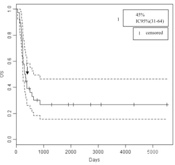
The median time of follow-up for the whole population was 0.9 years (range 0.1 to 15.3 years). The 1-year event-free survival (EFS) and overall survival (OS) rate were 36 % CI 95 % (23–55) and 45 % CI 95 % (31–64), re-spectively (Figs.1 and2). The median EFS and OS rate were 5.4 and 10.7 months, respectively. The median time to first progression/relapsed was 4.3 months (range, 0.9– 44.8 months). Twenty-seven relapses were observed, 74 % were localized, 22 % were localized and metastatic, and 4 % were only metastatic.
Prognostic factors
The univariate analysis indicates that complete surgical resection, radiotherapy, and high-dose chemotherapy were significantly associated with a better OS (respectively, p < 0.003, p < 0.0001, and p < 0.012). Furthermore,
Table 1 Patient characteristics
Total ETANTR MedulloeP ETMR EP
Number (%) 38 (100 %) 17(45 %) 13(34 %) 8(21 %) 0
Median age (months) (min-max) 31 (2.8–141) 32.9 (15.5–141) 31.1 (2.9–143.4) 27.2 (12.5–43.8) Mean age (months) (σ) 40.8 [6] 33.4 (9.5) 60.3 (53.5) 27.3 (10.5) Sex
% Male 26 70.6 84.6 62.5
% Female 72 29.4 15.4 37.5
Sex ratio 0.4 0.4 0.2 0.4
Median time to diagnosis (days) (min-max) 22 (1–203) 32 (1–172) 28 (4–203) 23.5 (15–615) Clinical signs, %* Sided weakness 34 53 7.7 37.5 Confusion 26 23.5 30.8 25 Increased IP 53 53 69.2 25 Seizures 21 23.5 15.4 0 Cerebellar syndrome 21 23.5 15.4 25 Torticolis 8 0 15.4 12.5 Visual impairment 11 11.8 15.4 0 Others 3 5.9 7.7 12.5 MV 3 5.9 0 0 Location, % Supratentorial 66 76.4 53.8 62.5 Infratentorial 26.1 6 46.2 37.5 Other 7.9 17.6 0 0 Metastatic CNS % Yes 18 23.5 23.1 0 No 79 76.5 69.2 0 MV 2.6 0 7.7 0
σ range, M male, F female, MedulloeP medulloepithelioma, EP ependymoblastoma, IP intracranial pressure, CNS central nervous system, MV missing values
*Total can be over 100 % as one patient can present several clinical signs
Table 2 Histopathological and biomolecular characteristics Histopathological review
Number at initial diagnosis (%) 38 (100 %) Number of changes in diagnosis (%) 6 (16 %) LIN28
Number of patients analyzed (%) 11 (29 %) Number of positive staining (%) 11 (100 %) ampl_19q13.42
Number of patients analyzed (%) 12 (32 %) Number of positive (%) 10 (83 %) Gain of chromosome 2
Number of patients analyzed (%) 11 (29 %) Number of positive (%) 3 (27 %) Other chromosomics gains
Number of patients analyzed (%) 11 (29 %) Number of positive (%) 6 (54 %)
according to the multivariate analysis, radiotherapy with a relative risk (RR) of 41.8 CI 95 % (9.4–186) p < 0.0001, surgical complete resection with a 7.9 CI 95 % (2.6–23.5) p < 0.0002, and high-dose chemotherapy with a RR 3.5 CI 95 % (1.3–9.5) p = 0.012 were prognostic factors associ-ated with a better OS.
Discussion
ETMR is a new entity, which encompasses ETAN-TR, ependymoblastomas, medulloepitheliomas. Indeed, those tu-mors seem to present clinical, histopathological, and molecu-lar simimolecu-larities [2–4]. As those tumors are rare, have been only
Table 3 Treatment strategy and outcome
Number Sex Age (year) Location CNS Meta Surgery Chemotherapy High-dose chemotherapy
RX Outcome Last news (m)
1 F 1.3 Infra Yes Total VP-Carbo No No Death 1.6
2 F 2.6 Supra No Total VP-carbo X2 No No Death 11.3
3 F 11.5 Supra No Total No No Yes Alive, CR 183.9
4 F 3.6 Supra No Partial TEMIRI X3/maintenance No Yes Alive, CR 13.4
5 F 11.8 Infra No Total VP-carbo X2 Thio Yes Alive CR 12.2
6 M 3.6 Infra No Partial VP-carbo X2 No No Death 4.2
7 F 2.3 Supra No Partial VP-carbo X2 No No Death 4.3
8 M 1.5 Infra No Partial VP-carbo X2 Thio X2/TMZ Yes Alive, PR 15.9
9 F 2.2 Supra No Partial VP-Carbo X2 No No Death 7.3
10 F 2.6 Supra No Partial VP-carbo X2 Mel/Cis No Death 6.3
11 F 2.7 Supra No Total VP-carbo X2 Mel-Cis X2 No Death 7.6
12 M 2.9 Spinal cord Yes Total VP-carbo X2 Mel-Cis X2/Thio Yes Alive, CR 43.2 13 F 2.2 Supra No Total VP-carbo/carbo Mel-Cis X2/Thio Yes Alive, CR 143.6 14 F 3.3 Supra No Total VP-carbo X2 Mel/Cis/Mel/Thio No Death 10.6
15 M 1.6 Supra Yes Partial VP-carbo X2 Mel/Cis No Death 7.8
16 F 1.3 Supra No Partial VP-carbo X2 Mel/Thio No Death 7.8
17 F 3 Supra No Partial VP-carbo X2 Mel/Cis/Thio X2 Yes Death 16.9
18 M 8.1 Infra No Partial VP-carbo X2 Mel/Cis No Death 9.6
19 F 3.2 Supra No Partial VP-carbo X2 Mel/Cis/Thio Yes Alive, PR 90.9 20 F 6.8 Supra No Total VP-carbo X2 Mel-Cis X2 Yes Alive, CR 63.8 21 F 2 Supra No Partial VP-carbo X2 Mel X2/Thio X2 Yes Alive, CR 143.4
22 F 3.5 Supra No Partial VP-carbo X2 Mel/Cis No Death 4.5
23 F 2.5 Supra Yes Partial VP-carbo X2 Mel/Thio X2 No Death 8
24 M 1.3 Supra + Infra Yes Partial VP-carbo X2 Mel/Cis/Mel No Death 10.1
25 M 2.6 Infra No Biopsy VP-carbo X2 Mel No Death 6.5
26 M 1.4 Infra No Total VP-carbo X2 Mel-Cis X2/Thio Yes Death 21.2
27 F 3.2 Supra No Partial VP-carbo X2 Mel/Cis No Death 8.3
28 F 2.6 Infra No Total VP-carbo X2 Mel-Cis X2/Thio Yes Alive CR 103.3 29 F 1.8 Supra No Total VP-carbo X2 Mel-Cis X2/Thio No Death 13
30 F 4.3 Supra No Partial VP-carbo X2 Mel/Cis Yes Death 10
31 F 3.3 Supra No Total VP-carbo X2 Mel/Cis No Death 19.2
32 F 0.2 Supra MV Total BBFSOP No No Death 9.2
33 M 2 Supra + Infra Yes Biopsy VP-carbo X2 Mel/Cis No Death 5.3 34 F 11.6 Infra Yes Total VP-carbo X2 Thio X2/TMZ Yes Alive CR 70.5 35 F 1.8 Supra No Total VP-Carbo X2 Mel-Cis X2/Thio Yes Death 29
36 F 3.3 Supra No Partial MV MV Yes Death 11.9
37 F 1 Infra No Partial VP-Carbo X2 Mel No Death 6
38 M 2.3 Supra No Total VP-Carbo X2 Mel/Cis No Death 13
N number, F female, M male, Supra supratentorial, Infra infratentorial, Meta metastasis, MV missing values, VP etoposide, carbo carboplatin, TEMIRI temozolomide-irinotecan, TMZ temozolomide, BBSFOP 2 cycles of Paraplatine + Natulan then Vepeside + Cisplatinum then vincristine + cyclophos-phamide, Mel Melphalan, Cis cisplatinum, Thio thiotepa, Rx radiotherapy, CR complete remission, PR partial remission, y years, m months
recently described and diagnosis difficult to establish; there is only a limited number of publications concerning ETMR [2,
3,7,8]. The series we report here represents the second largest series.
The patients’ characteristics we report here appear similar to those reported in previous studies [2,10]. ETMR mainly occurs in young children [1,2,7], with a median of age at diagnosis of 31 months found in our cohort. The sex ratio in our series is 0.4. This ratio is in line with the ratio reported by Picard et al. [10], where a sex ratio of 0.6 were found in the 29 included patients. As previously reported, the most frequent clinical sign is an increased intracranial pressure, which is commonly found in cerebral tumor in children [11–13]. Clin-ical signs at diagnosis are similar for all ETMR histologClin-ical
variants [2]. Our results (Table1) are consistent with these findings. Nevertheless, it should be noted that there is no ependymoblastoma in our series, probably due to the fact that the number of patients included in the cohort we report here is limited. A supratentorial location was found in the majority of the cases (66 %), as previously published [2]. We also report one patient with an ETMR localized to the spinal cord. This had never been reported before. Interestingly, metastasis was present at diagnosis in 18.9 % of the children as reported by Korshunov et al. [2]. Metastasis were only evidenced using MRI but were never found on CSF pathological analysis at diagnosis. Of note, one case had a CSF positivity after second surgery.
The diagnosis of ETMR appears to be challenging, as il-lustrated by the fact that among the histopathological review we report here, there was a change in diagnosis in 16 % cases (Table2). The molecular diagnosis with LIN28A and the re-search of the gain of the chromosome 2 was performed in 11 patients. The amplification of the locus 19q13.42 was per-formed in 12 patients. LIN28A staining was positive in 100 % of children, and 83 % of children presented an ampli-fication of the locus 19q13.42. Those results are in line with the data available in the literature and confirm that ETMR has a specific molecular signature. Therefore, immuno-histochemistry for LIN28A and CGHarray for amplification of the locus 19q13.42 should be done routinely to secure the diagnosis.
One of the interests of the series we report here relies on the fact that the majority of the patients were treated homogenously according to the PNET-HR protocol [9]. De-spite a multimodal therapy given in 30 patients, progressions were observed in all patients with a residual tumor during induction standard therapy or sequential high-dose therapy, and 26 of them died. Noteworthy, most of the patients present-ed either a local or both a local and distant relapse highlighting the aggressiveness of ETMR.
In the literature, radiotherapy was the only prognostic fac-tor in univariate analysis [11]. This is confirmed by our find-ings which show that using both univariate and multivariate analyses, radiotherapy is associated with a better outcome. To the best of our knowledge, there is no other data on the prog-nostic factors available in the medical literature. Noteworthy, radiotherapy is given at the end of the treatment after chemo-therapy which might select patient and introduce a bias. In the present series, we also found that a complete surgery and high-dose chemotherapy were favorable prognostic factors. Indeed, out the 11 survivors, all but 2 children received high-dose chemotherapy. This is consistent with the experience of Dufour et al. [9] who reported the feasibility and effectiveness of high-dose chemotherapy (2 cycles of thiotepa) followed by conventional craniospinal radiotherapy with newly diagnosed high-risk medulloblastoma or supratentorial PNET. In their study, the 5-year overall survival was 85 % for the whole
45% IC95%(31-64) 1
1
censored
Fig. 1 Overall survival
36% IC95%(23-55) censored 1 30% IC95%(18-49) 2 1 2
Fig. 2 Event-free survival
cohort. Unfortunately, the impact of high-dose chemotherapy on long-term survival is difficult to extrapolate from the liter-ature on ETMR due to small patient numbers. But, such ag-gressive chemotherapy, as in other embryonal central nervous system, could compensate for the avoidance or dose reduction in prophylactic craniospinal irradiation [14]. Such a strategy might lead to acceptable outcomes both in terms of toxicity and survival [15].
Patients with ETMR have a poor prognosis with an EFS and OS of, respectively, 36 % CI 95 % (23–55) and 45 % CI 95 % (31–64). In comparison, Korshunov et al. found a worst OS (14 %). This may be due to their treatment strategy which relies on chemotherapy and high-dose chemotherapy after surgery [4] but not on radiotherapy. Radiotherapy may there-fore be recommended in children with ETMR. However, as radiotherapy potentially impairs neurocognitive function, es-pecially in young children [16], and as children with ETMR are for the most of all less than 4 years old, benefits and long-term toxicities have to be balanced.
Conclusion
ETMR are rare cerebral tumors of the young child. A biologic diagnosis with LIN28A immuno-histochemistry and the search for amplification of the locus 19q13.42 should be sys-tematically done to ensure diagnosis. Prognosis remains dis-mal, but complete surgical removal seems to be an important part of the treatment. Radiotherapy may be another crucial point of the treatment but has to be balanced with neurocognitive toxicity that can be caused in young children. Chemotherapy and maintenance chemotherapy may be useful, but their respective roles remain to be determined.
Conflict of interest The authors declare no conflict of interest.
References
1. Ceccom J, Bourdeaut F, Loukh N, Rigau V, Milin S, Takin R, et al. (2014) Embryonal tumor with multilayered rosettes: diagnostic tools update and review of the literature. Clin Neuropathol 33(1): 15–22
2. Korshunov A, Sturm D, Ryzhova M, Hovestadt V, Gessi M, Jones DTW, et al. (2014) Embryonal tumor with abundant neuropil and t r u e r o s e t t e s ( E TA N T R ) , e p e n d y m o b l a s t o m a , a n d medulloepithelioma share molecular similarity and comprise a sin-gle clinicopathological entity. Acta Neuropathol 128(2):279–289
3. Paulus W, Kleihues P (2010) Genetic profiling of CNS tumors extends histological classification. Acta Neuropathol 120(2):269– 270
4. Korshunov A, Remke M, Gessi M, Ryzhova M, Hielscher T, Witt H, et al. (2010) Focal genomic amplification at 19q13.42 comprises a powerful diagnostic marker for embryonal tumors with ependymoblastic rosettes. Acta Neuropathol 120(2):253–260 5. Korshunov A, Ryzhova M, Jones DTW, Northcott PA, van Sluis P,
Volckmann R, et al. (2012) LIN28A immunoreactivity is a potent diagnostic marker of embryonal tumor with multilayered rosettes (ETMR). Acta Neuropathol 124(6):875–881
6. Louis DN, Ohgaki H, Wiestler OD, Cavenee WK, Burger PC, Jouvet A, et al. (2007) The 2007 WHO classification of tumours of the central nervous system. Acta Neuropathol 114(2):97–109 7. Eberhart CG, Brat DJ, Cohen KJ, Burger PC (2000) Pediatric
neuroblastic brain tumors containing abundant neuropil and true rosettes. Pediatr Dev Pathol 3(4):346–352
8. Gessi M, Giangaspero F, Lauriola L, Gardiman M, Scheithauer BW, Halliday W, et al. (2009) Embryonal tumors with abundant neuropil and true rosettes: a distinctive CNS primitive neuroectodermal tumor. Am J Surg Pathol 33(2):211–217 9. Dufour C, Delisle M-B, Geoffray A, Laplanche A, Frappaz D, Icher
C, et al. (2014) CT-003. Tandem high-dose chemotherapy with stem cell rescue followed by risk-adapted radiation in children with high-risk cerebral primitive neuroectodermal tumor: results of the prospective sfce-trial pnet hr + 5. Neuro-Oncology 16(suppl 1):i10– i13
10. Picard D, Miller S, Hawkins CE, Bouffet E, Rogers HA, Chan TSY, et al. (2012) Markers of survival and metastatic potential in child-hood CNS primitive neuro-ectodermal brain tumours: an integra-tive genomic analysis. Lancet Oncol 13(8):838–848
11. Alexiou GA, Stefanaki K, Vartholomatos G, Sfakianos G, Prodromou N, Moschovi M (2013) Embryonal tumor with abun-dant neuropil and true rosettes: a systematic literature review and report of 2 new cases. J Child Neurol 28(12):1709–1715 12. Buccoliero AM, Castiglione F, Rossi Degl’Innocenti D, Franchi A,
Paglierani M, Sanzo M, et al. (2010) Embryonal tumor with abun-dant neuropil and true rosettes: morphological, immunohistochem-ical, ultrastructural and molecular study of a case showing features of medulloepithelioma and areas of mesenchymal and epithelial differentiation. Neuropathology 30(1):84–91
13. Lafay-Cousin L, Hader W, Wei XC, Nordal R, Strother D, Hawkins C, et al. (2014) Post-chemotherapy maturation in supratentorial primitive neuroectodermal tumors. Brain Pathol 24(2):166–172 14. Odagiri K, Omura M, Hata M, Aida N, Niwa T, Goto H, et al.
(2014) Treatment outcomes and late toxicities in patients with em-bryonal central nervous system tumors. Radiat Oncol 9:201 15. Kim SY, Sung KW, Hah JO, Yoo KH, Koo HH, Kang HJ, et al.
(2010) Reduced-dose craniospinal radiotherapy followed by high-dose chemotherapy and autologous stem cell rescue for children with newly diagnosed high-risk medulloblastoma or supratentorial primitive neuroectodermal tumor. Korean J Hematol 45(2):120– 126
16. Padovani L, André N, Constine LS, Muracciole X (2012) Neurocognitive function after radiotherapy for paediatric brain tu-mours. Nat Rev Neurol 8(10):578–588