Distribution of Endosymbiotic Reproductive
Manipulators Reflects Invasion Process and Not
Reproductive System Polymorphism in the Little Fire Ant
Wasmannia auropunctata
Olivier Rey1*, Arnaud Estoup1, Benoit Facon1, Anne Loiseau1, Alexandre Aebi2, Olivier Duron3, Fabrice Vavre4, Julien Foucaud1
1 CBGP UMR1062, INRA, Montpellier, France, 2 Laboratoire de Biologie du Sol, Universite´ de Neuchaˆtel, Neuchaˆtel, Switzerland, 3 Institut des Sciences de l’Evolution UMR5554, CNRS, Universite´ Montpellier 2, Montpellier, France,4 Laboratoire de Biometrie et Biologie Evolutive UMR5558, CNRS, Universite´ Lyon 1, Villeurbanne, France
Abstract
Endosymbiotic reproductive manipulators may have drastic effects on the ecological and evolutionary dynamics of their hosts. The prevalence of these endosymbionts reflects both their ability to manipulate their hosts and the history of the host populations. The little fire ant Wasmannia auropunctata displays a polymorphism in both its reproductive system (sexual versus clonal populations) and the invasive status of its populations (associated to a habitat shift). We first screened for the presence of a diverse array of reproductive parasites in sexual and clonal populations of W. auropunctata, as a means to investigate the role of endosymbionts in reproductive phenotypes. Wolbachia was the only symbiont found and we then focused on its worldwide distribution and diversity in natural populations of W. auropunctata. Using a multilocus scheme, we further characterized the Wolbachia strains present in these populations. We found that almost all the native sexual populations and only a few clonal populations are infected by Wolbachia. The presence of similar Wolbachia strains in both sexual and clonal populations indicates that they are probably not the cause of the reproductive system polymorphism. The observed pattern seems rather associated to the invasion process of W. auropunctata. In particular, the observed loss of Wolbachia in clonal populations, that recurrently emerged from sexual populations, likely resulted from natural heat treatment and/or relaxed selection during the shift in habitat associated to the invasion process.
Citation: Rey O, Estoup A, Facon B, Loiseau A, Aebi A, et al. (2013) Distribution of Endosymbiotic Reproductive Manipulators Reflects Invasion Process and Not Reproductive System Polymorphism in the Little Fire Ant Wasmannia auropunctata. PLoS ONE 8(3): e58467. doi:10.1371/journal.pone.0058467
Editor: Richard Cordaux, University of Poitiers, France
Funding: This work was supported by grants from the French Ministe`re de l’Ecologie et du De´veloppement Durable (ECOTROP programme) awarded to AE and from the French Agropolis foundation (RTRA- Montpellier, BIOFIS project 1001–001) to AE and BF. The funders had no role in study design, data collection and analysis, decision to publish, or preparation of the manuscript.
Competing Interests: The authors have declared that no competing interests exist. * E-mail: [email protected]
Introduction
Maternally inherited symbionts are extremely common in arthropods and constitute an important force in evolution, because their transmission success is tightly linked to host fitness. These symbionts may have drastic effects on the ecological and evolutionary dynamics of their hosts [1–3]. Infections can both results in fitness costs [4–5] and/or benefits, such as defense against natural enemies [6–9]. The fitness cost-benefit ratio, eventually driving the prevalence of endosymbionts in natural host populations, is likely to be influenced by environmental param-eters, such as abiotic conditions [10] or parasitism pressure [11– 12]. In habitats where interspecific interactions are frequent and parasitism elevated, the benefits of an endosymbiotic-driven defense may lead to high infection prevalence, while in habitats where parasite pressure is low, incurred costs may purge populations from infection [12–13]. This process can have important consequences in the context of an exotic habitat change (e.g. during an introduction event), in which introduced popula-tions are likely to face different, sometimes lower, parasite
pressures (i.e., the enemy release hypothesis; [14]). In particular, in the case of a trade-off between immunological and demographic traits (e.g., defensive endosymbiotic bacteria may reduce fecundi-ty), purge of endosymbiotic bacteria following an enemy release is expected to promote the invasion process [15–17]. Accordingly, loss of parasites is frequent in a variety of invasive species [16–18– 20]. It is thus important to investigate possible gains or losses of endosymbiotic bacteria and their effects in the context of invasive events [16].
Apart from general effects over fitness, some maternally inherited symbionts are also able to manipulate the reproduction of their host [21–22]. These latter parasites alter the reproductive system of their host so that the number of infected daughters produced by an infected female exceeds the average production of daughters per uninfected female. Studies that have investigated reproductive parasitism have long been biased towards Wolbachia. This is logical, to some extent, given that it is the most widespread endosymbiotic bacterium within arthropods [23]. To date, this bacterium has been found to induce four manipulation pheno-types: cytoplasmic incompatibility, feminization of genetic males,
male killing and induction of thelytokous parthenogenesis [22–24]. In those cases, the haplodiploid genetic system seems to be a strong predisposition [22]. Wolbachia is, however, just one of the known reproductive parasites, and other endosymbiotic bacteria are now receiving increased attention [22–25–26]. Cardinium has been shown to induce cytoplasmic incompatibility, feminization and induction of parthenogenesis, Rickettsia can cause parthenogenesis and male killing and finally Arsenophonus as well as Spiroplasma can induce male killing within their hosts. All of these reproductive parasites lead to biases in reproductive schemes, which are beneficial for infected matrilines and, therefore, may have drastic effects on the evolutionary dynamics of their hosts [21–22]. It is therefore crucial to investigate their presence and their potential roles in organisms that present peculiar reproductive systems.
In the little fire ant, Wasmannia auropunctata, a small myrmicine species originating from South America and introduced world-wide, infection pattern of endosymbiotic bacteria could reflect both the invasion process and/or a reproductive manipulation. Indeed, this species displays polymorphisms both in the invasive status of its populations (associated to a habitat shift; [27–28]), and reproductive system (sexual vs. clonal populations; [29–30]). Within its native range, one can distinguish two types of W. auropunctata populations regarding invasive status. First, ancestral populations are confined within the primary forest and are characterized by low nest and worker densities [27–28]. Second, more recent invasive populations repeatedly colonized human modified habitats within the native range (e.g. road sides, plantations; [28]). This change of habitat is associated with a major ecological shift, with high worker and nest densities [28]. By contrast, only the invasive type of populations (high worker and nest densities) is found within the introduced range, in human modified habitats [27]. Similarly to other invasive ant species [18– 20], one could expect that the habitat shift of invasive populations within and outside the native range of W. auropunctata is associated with the loss of their endosymbiotic bacteria.
Apart from the possible effects on host fitness, endosymbiotic bacteria could also play a role in the peculiar reproductive system polymorphism displayed by W. auropunctata [29–30]. In some populations (hereafter called ‘‘sexual populations’’), queens and males reproduce following a classical haplo-diploid scheme where diploid females (i.e. queens and sterile workers) are produced sexually and haploid males develop from haploid eggs through arrhenotoky. Some populations (hereafter called ‘‘clonal popula-tions’’) emerged recurrently from these sexual populations, in which reproductives (i.e. queens and males) display an uncommon reproductive system: queens use thelytokous parthenogenesis and sexual reproduction in a conditional manner to produce gynes (i.e. unfertilized queens) and (sterile) workers, respectively [29–30]. A recent study revealed that gynes are produced via automictic parthenogenesis with central fusion [31]. Moreover, contrary to other species in which queens reproduce by thelytoky, unmated queens are unable to lay viable eggs [29]. The production of parthenogenetic eggs seems therefore strictly dependent on the fertilization process. Finally, female parthenogenesis is tightly associated to male clonality [29–30]. The genome of males is transmitted clonally via maternal eggs through a mechanism yet unresolved. Interestingly, a similar reproductive system was described in two unrelated ant species, Vollenhovia emeryi [32] and Paratrechina longicornis [33]. Current knowledge in Hymenoptera does, however, not plead for the involvement of endosymbiotic manipulators in this reproductive system. Indeed, in all cases of symbiont-induced parthenogenesis examined so far, restoration of diploidy is achieved through a process of gamete duplication, making daughters completely homozygous and precluding this
kind of effect in species exhibiting complementary sex determina-tion, such as ants. However, other mechanisms may exist, as in two mite species where Wolbachia-induced parthenogenesis is achieved through a mechanism that is functionally apomictic, as daughters keep the heterozygosity of their mothers [34]. While some evidence would suggest that this peculiar reproductive system might be under genetic determinism [30], no cause has been demonstrated so far, and endosymbiotic manipulation still stands as a possible explanation.
Interestingly, the reproductive system polymorphism is almost always associated to the invasive status of populations [27–35]. While native non-invasive populations are mainly sexual, both native and introduced invasive populations are clonal. This leads to opposite expectations regarding the pattern of endosymbiontic bacterial infection in populations of W. auropunctata, under the hypotheses of an endosymbiotic role in either the invasion process or the reproductive system determinism. If endosymbionts are involved in the determinism of the reproductive system, then the infection pattern would display an excess of infection in clonal compared to sexual populations, and/or clonal and sexual populations would not display similar strains of endosymbionts. On the contrary, from an invasion biology perspective, the expectation of infection pattern would be that invasive clonal populations, within and outside the native range, are free of endosymbionts and native sexual populations are infected.
To distinguish between these hypotheses, we conducted an extensive screening of W. auropunctata native sexual populations and clonal populations from both the native and the invasive range, for five known reproductive parasites, Wolbachia, Arsenopho-nus, Cardinium, Rickettsia and Spiroplasma ixodetis. Wolbachia was the only detected endosymbiont and we then analyzed its distribution among worldwide W. auropunctata populations from both types (i.e. sexual and clonal) in the native and introduced range to identify major correlates and patterns that could indicate the driving force of infection patterns in this species (i.e. reproductive system versus invasion process). We additionally examined the Wolbachia diversity through W. auropunctata population by sequencing a combination of six bacterial genes to better refine our under-standing of the history of infection in this species.
Materials and Methods
Screening for Endosymbiotic Bacteria
We used specific PCR-amplification to investigate for the presence of Wolbachia, Cardinium, Arsenophonus, Rickettsia and Spiroplasma ixodetis on 84 individuals collected in 42 nests from 42 populations (one nest per population) covering most of the distribution of W. auropunctata, including its native and introduced range (Figure 1). Sampling was conducted in public locations that did not required specific authorization. In the native range nine sexual and 12 clonal populations were available from a previous study [27]. In the introduced range, 21 clonal populations were available [35]. When possible (35 nests), a queen and a worker from the same nest were screened. In seven nests no queens were sampled and two workers were thus screened for infection.
The total genomic DNA was extracted from all individuals following a standard CTAB protocol. We performed PCR-based screening using a protocol modified from Shoemaker et al. [19] for Wolbachia detection, and following the protocols described in Duron et al. [25] for the four other endosymbionts. To avoid false negatives, we used an arthropod specific DNA fragment of the W. auropunctata EF1a gene to serve as an internal control of DNA extractions and PCR quality, using the universal primer set trs4F/ trs9R [36]. Positive controls infecting Culex pipiens (Diptera:
Culicidae), Holocnemus pluchei (Araneae: Pholcidae), Hippobosca equina (Diptera: Hippoboscidae), Bemisia tabaci (Hemiptera: Aleyr-odidae) and Cicadella viridis (Hemiptera: Cicadellidae) were used in each PCR for the screening of Wolbachia, Cardinium, Arsenophonus, Rickettsia and Spiroplasma ixodetis respectively. Negative controls (i.e. ultrapure water) were used in each PCR. PCR products were electrophoresed in 1.5% agarose gels and visualized under UV illumination after staining in an ethidium bromide solution.
Wolbachia-specific Additional Screening
Among the five endosymbionts screened, Wolbachia was the only symbiont detected in some W. auropunctata populations (see Results section). We extended our screening of Wolbachia on a larger dataset to more accurately assess the distribution of this endosymbiont among W. auropunctata populations. Together with individuals previously examined, a total of 205 queens and 376 workers were screened following the protocol detailed in the previous section. Altogether, these individuals screened for Wolbachia infection originated from 174 and 248 nests for queens and workers, respectively, from 56 worldwide populations (Table 1 and 2; Figure 1).
Based on this additional screening, we estimated the prevalence of Wolbachia within populations in the queen and worker castes using the ratio of the number of infected individuals to the total number of screened individuals of the same caste. To indirectly test whether Wolbachia could be involved in the reproductive system polymorphism of W. auropunctata, we tested for a statistical association between the reproductive system within nests (i.e. sexual or clonal) and the infection status (i.e. presence or absence of Wolbachia) of individuals within the nests. Nests with at least one individual infected were considered as infected, and nests in which all individuals were Wolbachia-free were considered as non-infected. The association was tested using a Fisher’s exact test and the strength of this association was assessed using a Cramer’s V statistic.
In ants, the prevalence of Wolbachia in the sterile worker caste, which corresponds to a dead-end host, may vary from one nest to another [37]. We therefore specifically estimated the prevalence of Wolbachia within nests by screening 160 additional workers originating from eight distinct nests from both the native and the introduced range (i.e. 20 workers per nests). Half these nests were known to be sexual and the other half to be clonal. The four sexual nests originated from two populations established in the primary forest within the native range: one from French Guiana (M7) and one from Brazil (UNA). The four clonal nests originated from four genetically distinct populations: two from the native range (P2 and IP, from French Guiana and Brazil, respectively), and two from the introduced range (CA and PL, from Australia and New Caledonia, respectively).
Genetic Characterization of the Wolbachia Strains
A phylogenetic analysis was performed to characterize the strains of Wolbachia detected in W. auropunctata populations. To this aim, we sequenced a fragment of the wsp gene from 74 individuals from the 17 identified infected populations from both the native
and introduced range of the species, using the previously described PCR amplification protocol. PCR products were purified and sequenced on an ABI 3730 DNA sequencer (Applied Biosystems). Electrophoregrams were checked for possible errors using the Seqscape software (Applied Biosystems). Because multiple peaks were observed at some base positions in some individual electrophoregrams, infection by multiple strains was suspected in these cases. The amplification products were hence purified (QIAquick PCR Purification Kit, QIAGEN) and ligated into a plasmid vector (pGEM-T easy vector system, Promega) and transformed into JM109 competent cells (Promega). Positively transformed cells were boiled, amplified using T7 and SP6 (primers of plasmid) and sequenced with the same primers. The plasmid DNA of six clones per individual was purified and sequenced.
Our phylogenetic analysis was performed on unique wsp haplotypes (five haplotypes). We also included 33 additional wsp sequences of Wolbachia strains belonging to the supergroups A and B and isolated from other Formicidae available at the Wolbachia MLST website (http://pubmlst.org/perl/mlstdbnet/mlstdbnet. pl?page = query&file = wo_isolates.xml). The tree was rooted using a wsp sequence from a Wolbachia strain belonging to the supergroup F according to the classification of the MLST database (i.e. infecting the wasp Apoica pallens). All sequences were aligned using clustalW [38]. The data set was analyzed using the neighbour-joining method with Kimura two-parameter distance measure in MEGA v. 4 [39]. Bootstrap analysis was performed with 1,000 replicates.
Because the wsp gene is known to be under diversifying selection and to undergo recombination both within and between strains [40], we also conducted additional analyses on a subset of 17 individuals from the same 17 identified infected populations (one individual per population) that were analysed at the wsp phylogenetic analysis (see Figure 2), using the five genes used in the multilocus sequence typing approach (MLST: gatB, fbpA, coxA, ftsZ and hcpA). We amplified fragments of these five genes following Baldo et al. [41]. The PCR products were cloned and the plasmid DNA of six clones per individual were then purified and sequenced. A total of 102 sequences were thus obtained for each gene. Because of the occurrence of multiple infections, we could not conduct phylogenetic analyses on a concatenated set of MLST genes. We hence conducted five independent phylogenetic analyses following the same method as for the wsp gene. All haplotypes from both wsp and MLST genes were archived under Genbank (accession numbers JX499039–JX499070).
Results
Screening of Endosymbiotic Bacteria
Among the five main endosymbiotic bacteria screened in the present study (i.e. Wolbachia, Cardinium, Arsenophonus, Rickettsia and Spiroplasma ixodetis), Wolbachia was the only one found, being present in 16 of the 42 W. auropunctata populations primarily screened. Together with the Wolbachia-specific additional screen-ing, 18 of the 56 W. auropunctata populations screened were found to be infected by Wolbachia. We found a strong association between
Figure 1. Map of the sites sampled to assess the presence of endosymbionts inW. auropunctatapopulations. Note: The presence of five endosymbionts was investigated at 42 sites worldwide, and Wolbachia was investigated at 56 sites (listed in Table 2). The native range of W. auropunctata is indicated by green shading. Clonal and sexual populations are indicated in orange and red, respectively. Wolbachia-infected and non-infected populations are indicated with stars and circles, respectively. When all populations of a given country displayed the same infection status and reproductive system, a single point was added to the map. On the contrary, all populations from a single country are indicated in a dedicated window when populations showed a polymorphism in infection status and/or reproductive system, i.e. for all native (Brazil, Costa Rica, French Guiana) and one introduced populations (Florida).
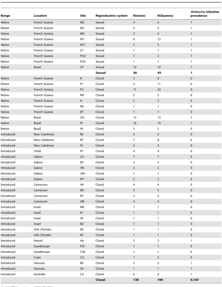
Table 1. Prevalence of Wolbachia infection in W. auropunctata queens from nests sampled in the native and introduced range.
Range Location Site Reproductive system N(nests) N(Queens)
Wolbachia infection prevalence
Native French Guiana M2 Sexual 4 4 1
Native French Guiana M3 Sexual 5 5 1
Native French Guiana M6 Sexual 2 6 1
Native French Guiana M7 Sexual 9 13 1
Native French Guiana M11 Sexual 3 3 1
Native French Guiana Z7 Sexual 1 1 1
Native French Guiana Pi32 Sexual 1 2 1
Native French Guiana Pi52 Sexual 1 1 1
Native Brazil CP Sexual 10 10 1
Sexual 36 45 1
Native French Guiana K Clonal 3 8 0
Native French Guiana P1 Clonal 3 11 0
Native French Guiana P2 Clonal 17 24 0
Native French Guiana RN Clonal 5 5 0
Native French Guiana A Clonal 2 2 0
Native French Guiana ML Clonal 1 1 0
Native French Guiana EP Clonal 1 1 0
Native Brazil CN Clonal 13 13 1
Native Brazil IP Clonal 10 10 1
Native Brazil IN Clonal 5 5 0
Introduced New Caledonia BS Clonal 4 4 0
Introduced New Caledonia BT Clonal 7 8 0
Introduced New Caledonia PL Clonal 4 4 0
Introduced Tahiti PF Clonal 4 4 0
Introduced Gabon LO Clonal 7 7 0
Introduced Gabon EK Clonal 5 5 0
Introduced Gabon OK Clonal 2 2 0
Introduced Gabon LBV Clonal 1 1 0
Introduced Gabon NT Clonal 2 2 0
Introduced Cameroon AK Clonal 4 4 0
Introduced Cameroon BD Clonal 5 5 0
Introduced Cameroon BY Clonal 5 5 0
Introduced Cameroon SM Clonal 4 4 0
Introduced Israel ME Clonal 1 1 0
Introduced Israel KI Clonal 1 1 0
Introduced Israel AF Clonal 1 1 0
Introduced Israel BZ Clonal 1 1 0
Introduced USA (Florida) RS Clonal 1 1 0
Introduced USA (Florida) RC Clonal 1 1 0
Introduced Hawaii Ha Clonal 3 3 1
Introduced Guadeloupe FDI Clonal 1 1 0
Introduced Guadeloupe CAE Clonal 1 1 0
Introduced Cuba CU Clonal 1 2 0
Introduced Vanuatu BK Clonal 5 5 1
Introduced Vanuatu SA Clonal 1 1 1
Introduced Australia CA Clonal 6 6 1
Clonal 138 160 0,167
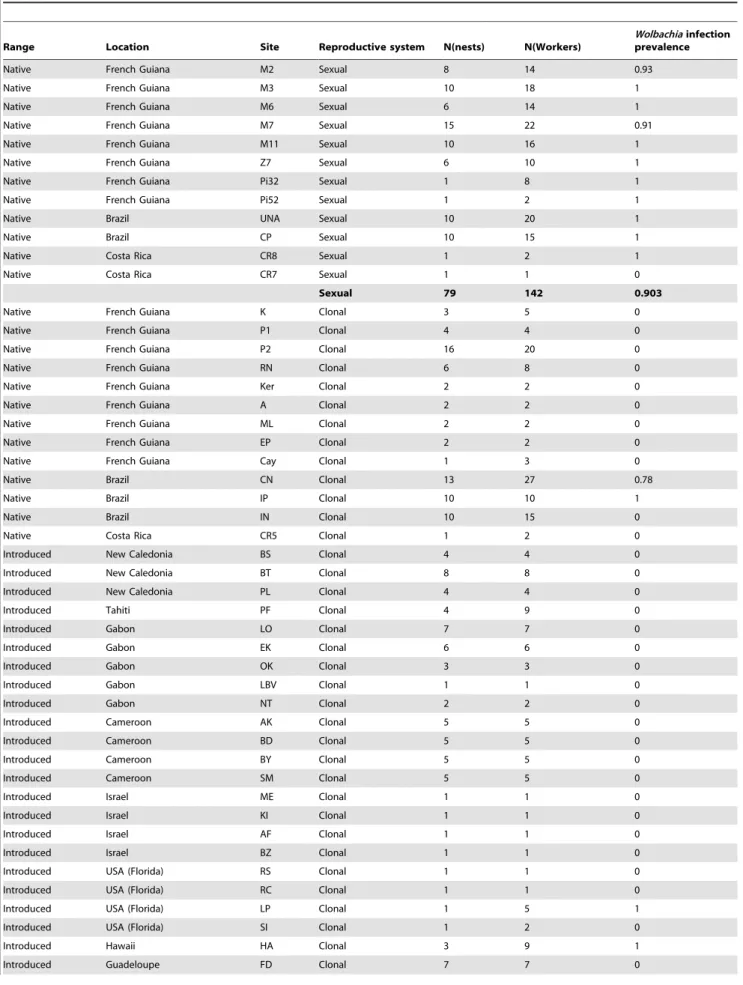
Table 2. Prevalence of Wolbachia infection in W. auropunctata workers from nests sampled in the native and introduced range.
Range Location Site Reproductive system N(nests) N(Workers)
Wolbachia infection prevalence
Native French Guiana M2 Sexual 8 14 0.93
Native French Guiana M3 Sexual 10 18 1
Native French Guiana M6 Sexual 6 14 1
Native French Guiana M7 Sexual 15 22 0.91
Native French Guiana M11 Sexual 10 16 1
Native French Guiana Z7 Sexual 6 10 1
Native French Guiana Pi32 Sexual 1 8 1
Native French Guiana Pi52 Sexual 1 2 1
Native Brazil UNA Sexual 10 20 1
Native Brazil CP Sexual 10 15 1
Native Costa Rica CR8 Sexual 1 2 1
Native Costa Rica CR7 Sexual 1 1 0
Sexual 79 142 0.903
Native French Guiana K Clonal 3 5 0
Native French Guiana P1 Clonal 4 4 0
Native French Guiana P2 Clonal 16 20 0
Native French Guiana RN Clonal 6 8 0
Native French Guiana Ker Clonal 2 2 0
Native French Guiana A Clonal 2 2 0
Native French Guiana ML Clonal 2 2 0
Native French Guiana EP Clonal 2 2 0
Native French Guiana Cay Clonal 1 3 0
Native Brazil CN Clonal 13 27 0.78
Native Brazil IP Clonal 10 10 1
Native Brazil IN Clonal 10 15 0
Native Costa Rica CR5 Clonal 1 2 0
Introduced New Caledonia BS Clonal 4 4 0
Introduced New Caledonia BT Clonal 8 8 0
Introduced New Caledonia PL Clonal 4 4 0
Introduced Tahiti PF Clonal 4 9 0
Introduced Gabon LO Clonal 7 7 0
Introduced Gabon EK Clonal 6 6 0
Introduced Gabon OK Clonal 3 3 0
Introduced Gabon LBV Clonal 1 1 0
Introduced Gabon NT Clonal 2 2 0
Introduced Cameroon AK Clonal 5 5 0
Introduced Cameroon BD Clonal 5 5 0
Introduced Cameroon BY Clonal 5 5 0
Introduced Cameroon SM Clonal 5 5 0
Introduced Israel ME Clonal 1 1 0
Introduced Israel KI Clonal 1 1 0
Introduced Israel AF Clonal 1 1 0
Introduced Israel BZ Clonal 1 1 0
Introduced USA (Florida) RS Clonal 1 1 0
Introduced USA (Florida) RC Clonal 1 1 0
Introduced USA (Florida) LP Clonal 1 5 1
Introduced USA (Florida) SI Clonal 1 2 0
Introduced Hawaii HA Clonal 3 9 1
the reproductive system within W. auropunctata’s nests and the infection status, with a higher prevalence of Wolbachia infection in sexual than in clonal populations (x2= 143.24; p – value ,0.001; Cramer’s V = 0.74). The mean prevalence within sexual popula-tions was 1 and 0.90 within the queen and worker castes respectively (Table 1 and 2). In the 37 sexual nests in which queens were sampled, all 45 sexually reproducing queens as well as 138 on 142 tested workers were infected by Wolbachia. We found only four workers originating from two sexual nests that were Wolbachia-free. These workers belonged to two queenless nests from populations of French Guiana (M2, M7; Table 2). In clonal populations, the mean prevalence within populations was 0.17 and 0.15 within the queen and worker castes respectively (Table 1 and 2). We found that 122 of the 160 clonal queens (i.e. 76.25%) from the 138 sampled clonal nests were Wolbachia-free, irrespective to their origin (i.e. from the native or introduced range). All workers from these clonal nests were also non-infected. A total of 38 infected clonal queens were found within 23 nests of the native range in two Brazilian populations (CN and IP) and within 15 nests of the introduced range (in Hawaii, Vanuatu Islands and Australia; Table 1, Figure 1). Workers from these nests were also all infected (Table 2). Finally, five workers from a unique queenless nest originating from Florida (U.S.A) were all infected by Wolbachia (Table 2, Figure 1).
The prevalence of Wolbachia infection within nests showed a binary pattern. The 20 workers sampled from infected sexual and clonal nests were all infected. On the contrary, workers from the two Wolbachia-free clonal nests were all non-infected.
Genetic Characterization of Wolbachia Strains
The Wolbachia strains identified in W. auropunctata populations all belong to the supergroup A according to the MLST classification. Among the 74 surveyed ants, four (5.4%, all originating from the same sexual population UNA in Brazil) were found to be co-infected by two distinct Wolbachia strains based on their wsp haplotypes. The co-infection of theses ants was also detected at the MLST genes. All of the other 70 ants were found to be infected by a single wsp haplotype. Among the subset of 17 individuals analysed using the MLST genes, six ants, all originating from different sexual populations (M2, Pi32, PI52, M2 in French Guiana and UNA and CP in Brazil) were found to be co-infected by different Wolbachia strains differing in at least one MLST gene. All of the analysed ants from clonal populations were found to be infected by a single Wolbachia strain as revealed by the genetic analyses based on both the wsp and MLST genes.
Examination of the wsp and the five MLST genes revealed the presence of 5 distinct Wolbachia strains, clustering in three different clades (Figure 2; Figures S1, S2, S3, S4, S5). There is no clear partitioning of this diversity between sexual and clonal popula-tions: the most common strain is shared by both sexual and clonal populations, three other strains were specific to sexual populations, and the last strain to a clonal population. In the native range, the two infected Brazilian clonal populations (CN and IP) host a Wolbachia strain closely related to the one infecting the Brazilian sexual population (CP; Figure 2; Figures S1, S2, S3, S4, S5). Additionally, all infected clonal populations from the introduced range (Hawaiian, Australian, Vanuatu and some Floridian populations) harbor the same strain infecting the native French Guianese sexual populations (Figure 2; Figures S1, S2, S3, S4, S5). This last Wolbachia strain is identical to the one previously found to infect other New World ants [37], including S. invicta (Figure 2).
Discussion
Among the five studied endosymbiotic bacteria (i.e. Wolbachia, Rickettsia, Cardinium, Arsenophonus and Spiroplasma ixodetis), only Wolbachia was detected in W. auropunctata. This result is not surprising given that Wolbachia is estimated to infect more than one third of ant species [37], while the four other endosymbionts were sporadically found in few arthropods orders [25] and seldom in ants (but see [42–43]). Wolbachia is not present in individuals (queens and workers) from most of the clonal populations, but when found, highly similar Wolbachia strains were also found to infect sexual populations. These results strongly suggest that the peculiar reproductive system of reproductives from W. auropunctata clonal populations (i.e. queen thelytokous parthenogenesis associ-ated to male clonality) is not induced by Wolbachia, nor by any of the four other endosymbiotic bacteria screened in the present study.
At first glance, our study might seem to indicate an association between the reproductive system of W. auropunctata and the occurrence of Wolbachia. While individuals from clonal populations are mostly Wolbachia-free, all sampled queens and most of workers from sexual populations are infected by the bacterium. This pattern would suggest that Wolbachia infection is well established, if not favored, in sexual populations. However, identical (or almost identical) Wolbachia strains were found in sexual and clonal populations (Figure 2; Figures S1, S2, S3, S4, S5), a pattern in strong opposition to any direct relationship between reproductive systems and Wolbachia phenotypic effects. Additionally, none of the Table 2. Cont.
Range Location Site Reproductive system N(nests) N(Workers)
Wolbachia infection prevalence
Introduced Guadeloupe CAE Clonal 2 2 0
Introduced Guadeloupe PDM Clonal 1 1 0
Introduced Cuba CU Clonal 2 7 0
Introduced Vanuatu BK Clonal 5 10 1
Introduced Vanuatu SA Clonal 1 2 1
Introduced Australia CA Clonal 6 8 1
Introduced Dominica CO Clonal 2 6 0
Introduced Dom. Rep DR Clonal 1 3 0
Clonal 169 234 0.154
described mechanisms by which Wolbachia alters reproductive systems to enhance its own transmission (i.e. male killing, feminization, cytoplasmic incompatibility and obligate for oogen-esis), can explain the maintenance of the bacterium in sexual populations better than in clonal populations. First, under the male killing and the feminization processes, a strong reduction of infected males is expected. Yet, we found some infected males in both native and introduced populations of W. auropunctata (data not shown). Additionally, feminization through known Wolbachia mediated-mechanisms would produce sterile haploid females, rendering this phenotype unlikely within the ants [21–44]. Second, one could argue that Wolbachia could be obligatory for oogenesis in W. auropunctata, as was found in the hymenopteran genus Asobara [45]. However, in the case of W. auropunctata, Wolbachia-free clonal queens use sexual reproduction to produce workers and Wolbachia-infected clonal queens produce parthenogenetic daughters, two features arguing against a role of Wolbachia in oogenesis in this species. Finally, cytoplasmic incompatibility, the most prevalent phenotypic effect induced by Wolbachia [22], is unlikely to be differentially maintained in sexual vs. clonal in W. auropunctata because in both types of populations workers are produced sexually. Parthenogenetic queens like queens from sexual popu-lations may hence suffer from CI through their workers produced sexually.
The Wolbachia infection pattern seems rather associated to the invasive status of W. auropunctata populations, even if this pattern is mainly driven by French Guianese samples due to the sampling scheme. The loss of endosymbiotic bacteria in invasive populations is common in ant species [18–20]. Two main hypotheses were proposed to explain this loss: (i) Wolbachia can be eliminated through drift during introduction if all founders were uninfected [18], or (ii) Wolbachia can be lost in invasive populations after the introduction through drift or selection [18–19]. In the case of W. auropunctata, drift seems unlikely to account for the results because clonal populations have recurrently emerged from sexual popu-lations [30] and the transmission of Wolbachia in sexual popupopu-lations was found to be nearly perfect. It is therefore unlikely that Wolbachia could have been lost in multiple invasive clonal populations through the sole means of drift. Interestingly, the emergence of invasiveness in W. auropunctata follows an important habitat change [27]. Two non-exclusive alternative hypotheses based on ecological features relative to this habitat change might hence explain the infection pattern observed in W. auropunctata populations.
First, clonal populations may have passively lost Wolbachia by natural heat treatment. While non-invasive sexual populations are established in primary forests with low temperature variation below 30uC, clonal populations settle in human-modified areas characterized by hotter and drier microclimates (reaching as far as 40uC; [28]). Additional work by our group demonstrated that workers from clonal populations tolerate temperatures as high as 36uC, while the mortality rate in workers from sexual populations reach 40% at this temperature (unpublished data). Wolbachia is known to be sensitive to such temperatures and heat treatments are commonly used to remove it from hosts for experimental purposes [46]. Classically, these treatments require rearing hosts’ larvae at 33uC to 35uC for few days to several generations. It is
therefore possible that the abiotic conditions of human-modified areas lead to a loss of Wolbachia infection in these zones, i.e. in introduced and native clonal populations of W. auropunctata.
A second hypothesis is that Wolbachia has been actively lost in clonal populations from human-modified habitats through relaxed selection and/or counter-selection against infected individuals. Wolbachia has been shown to induce both costs (e.g. reduction of fecundity, adult survival and locomotor performance; [4–5]) and benefits (e.g., protection from RNA viruses, upregulation of immunity-gene expression against Plasmodium and filarial nema-todes; [7–47]) for infected individuals. In the case of W. auropunctata, sexual populations established in primary forests are likely to face more important biotic pressures than clonal populations established in human-modified areas, notably through higher levels of interspecific interactions with other ant species [28]. The fitness cost-benefit ratio might hence favor the loss of Wolbachia in human-modified areas, and its maintenance in primary forests.
Both hypotheses could explain the unique case of infected native clonal populations (i.e., CN and IP in Brazil; Table 1, 2). The habitats of these particular populations correspond to recently abandoned shaded cocoa plantations where there has been no human activity for at least 10 years. In these exploitations (i.e. shaded cocoa plantations), the vegetation structure and stratifica-tion are considered to be similar to, albeit less complex than, that of natural forests. The ant species richness in these plots roughly corresponds to that of a native forest of low diversity [48]. During exploitation both biotic and abiotic environmental conditions, hence, were different from those of typical habitat where clonal populations are found. Furthermore, since the cessation of human activities, vegetation has grown back so as to change the environmental conditions at the ground level, in particular in reducing the daily and seasonal thermal amplitudes. Under these conditions that enable complex biotic interactions (including higher levels of parasitism), Wolbachia could have been maintained in W. auropunctata clonal populations established on these sites, due to a possible beneficial role against pathogens. This result strengthens the apparent close association between environmental parameters and Wolbachia infection in W. auropunctata.
Finally, the occurrence of Wolbachia in clonal invasive popula-tions outside the native range, in Florida, Hawaii, Australia, and in the Vanuatu Islands, cannot be explained by the two above hypotheses based on ecological features. Interestingly however, the infection of these populations is consistent with their invasion history. Populations established in Florida, Hawaii, Australia, and in the Vanuatu Islands were found to be infected by the same Wolbachia strain. These populations have previously been shown to share a unique mitochondrial haplotype and to display closely related clonal queens genotypes at microsatellite markers [35–49]. The distribution and identity of the Wolbachia strain uncovered in the infected introduced populations are therefore consistent with previous studies. Consequently, these invasive populations are likely to originate from the same ancestral clonal native population and the infection by Wolbachia most probably occurred once in this ancestral population before long dispersal events. This ancestral population remains, however, unknown.
Figure 2. NJ tree based on the wsp nucleotide alignment of the differentWolbachia strains infecting native and introduced populations ofW. auropunctata. Note: Each Wolbachia sequence is labeled with the name of its host species and the allelic number of the wsp sequence according to the MLST database in bold. The country of origin of each identified strain is indicated in parenthesis. Only bootstrap values (computed from 1,000 replicates) of nodes are figured for values .50%. Wolbachia strains found in sexual and clonal populations of W. auropunctata are highlighted in red and orange, respectively. Location of origin of the samples is indicated between parentheses.
In conclusion, this study revealed that, except Wolbachia, none of the reproductive parasite screened in the present study infect wild populations of W. auropunctata. Furthermore, presence or absence of Wolbachia infection is unlikely to explain the reproductive system polymorphism found in W. auropunctata. The infection pattern of Wolbachia in W. auropunctata rather echoes with previous studies illustrating a loss of Wolbachia in invasive populations [18–20]. The most likely explanation is that this loss resulted from natural heat treatment and/or relaxed selection during a shift in habitat in invasive populations. Putative immunological benefits and/or physiological costs induced by Wolbachia should be experimentally tested in the future to distinguish between these hypotheses.
Supporting Information
Figure S1 NJ tree based on thegatB nucleotide align-ment of the different Wolbachia strains infecting native and introduced populations of W. auropunctata. Note: Wolbachia strains found in sexual and clonal populations of W. auropunctata are highlighted in red and orange, respectively. Each other Wolbachia sequence is labeled with the name of its host species and its respective GenBank Accession number in bold. Location of origin of the samples is indicated between parentheses. Only bootstrap values (computed from 1,000 replicates) of nodes are figured for values .50%.
(DOC)
Figure S2 NJ tree based on the fbpA nucleotide align-ment of the different Wolbachia strains infecting native and introduced populations of W. auropunctata. Note: Wolbachia strains found in sexual and clonal populations of W. auropunctata are highlighted in red and orange, respectively. Each other Wolbachia sequence is labeled with the name of its host species and its respective GenBank Accession number in bold. Location of origin of the samples is indicated between parentheses. Only bootstrap values (computed from 1,000 replicates) of nodes are figured for values .50%.
(DOC)
Figure S3 NJ tree based on the CoxA nucleotide alignment of the different Wolbachia strains infecting native and introduced populations ofW. auropunctata. Note: Wolbachia strains found in sexual and clonal populations of W. auropunctata are highlighted in red and orange, respectively. Each other Wolbachia sequence is labeled with the name of its host species and its respective GenBank Accession number in bold.
Location of origin of the samples is indicated between parentheses. Only bootstrap values (computed from 1,000 replicates) of nodes are figured for values .50%.
(DOC)
Figure S4 NJ tree based on the ftsZ nucleotide align-ment of the different Wolbachia strains infecting native and introduced populations of W. auropunctata. Note: Wolbachia strains found in sexual and clonal populations of W. auropunctata are highlighted in red and orange, respectively. Each other Wolbachia sequence is labeled with the name of its host species and its respective GenBank Accession number in bold. Location of origin of the samples is indicated between parentheses. Only bootstrap values (computed from 1,000 replicates) of nodes are figured for values .50%.
(DOC)
Figure S5 NJ tree based on the hcpA nucleotide alignment of the different Wolbachia strains infecting native and introduced populations ofW. auropunctata. Note: Wolbachia strains found in sexual and clonal populations of W. auropunctata are highlighted in red and orange, respectively. Each other Wolbachia sequence is labeled with the name of its host species and its respective GenBank Accession number in bold. Location of origin of the samples is indicated between parentheses. Only bootstrap values (computed from 1,000 replicates) of nodes are figured for values .50%.
(DOC)
Acknowledgments
JF would like to thank A. Cubier and C. Charlut for support. The authors want to thank M. Weill for providing Wolbachia-infected Culex pipiens samples. Some of the data analyzed here, including the genetic data in particular, were generated at the molecular genetic analysis technical facilities of the Environment and Biodiversity IFR 119 at Montpellier (France).
Data Archiving
Sequence data have been submitted to GenBank: accession numbers JX499039–JX499070.
Author Contributions
Conceived and designed the experiments: OR AE BF AA JF. Performed the experiments: OR AE AL JF. Analyzed the data: OR AE BF OD FV JF. Contributed reagents/materials/analysis tools: AE AL AA OD FV. Wrote the paper: OR AE BF JF.
References
1. Feldhaar H (2011) Bacterial symbionts as mediators of ecologically important traits of insect hosts. Ecol Entomol 36(5): 533–543.
2. Moran NA, McCutcheon JP, Nakabachi A (2008) Genomics and evolution of heritable bacterial symbionts. Annu Rev Genet. Palo Alto: Annual Reviews. 165–190.
3. Oliver KM, Degnan PH, Burke GR, Moran NA (2010) Facultative symbionts in aphids and the horizontal transfer of ecologically important traits. Annual Review of Entomology. Palo Alto: Annual Reviews. pp. 247–266.
4. Fleury F, Vavre F, Ris N, Fouillet P, Bouletreau M (2000) Physiological cost induced by the maternally-transmitted endosymbiont Wolbachia in the Drosophila parasitoid Leptopilina heterotoma. Parasitology 121: 493–500.
5. Wenseleers T, Sundstrom L, Billen J (2002) Deleterious Wolbachia in the ant Formica truncorum. P Roy Soc Lond B Bio 269(1491): 623–629.
6. Brownlie JC, Johnson KN (2009) Symbiont-mediated protection in insect hosts. Trends Microbiol 17(8): 348–354.
7. Haine ER (2008) Symbiont-mediated protection. P Roy Soc Lond B Bio 275(1633): 353–361.
8. Hedges LM, Brownlie JC, O’Neill SL, Johnson KN (2008) Wolbachia and virus protection in insects. Science 322(5902): 702–702.
9. Jaenike J (2012) Population genetics of beneficial heritable symbionts. Trends Ecol Evol 27(4): 226–232.
10. Russell JA, Moran NA (2006) Costs and benefits of symbiont infection in aphids: variation among symbionts and across temperatures. P Roy Soc Lond B Bio 273(1586): 603–610.
11. Jaenike J, Brekke TD (2011) Defensive endosymbionts: a cryptic trophic level in community ecology. Ecol Lett 14(2): 150–155.
12. Oliver KM, Campos J, Moran NA, Hunter MS (2008) Population dynamics of defensive symbionts in aphids. P Roy Soc Lond B Bio 275(1632): 293–299. 13. Vorburger C, Gouskov A (2011) Only helpful when required: a longevity cost of
harbouring defensive symbionts. J Evolution Biol 24(7): 1611–1617. 14. Keane RM, Crawley MJ (2002) Exotic plant invasions and the enemy release
hypothesis. Trends Ecol Evol 17(4): 164–170.
15. White TA, Perkins SE (2012) The ecoimmunology of invasive species. Funct Ecol 26(6): 1313–1323.
16. Torchin ME, Lafferty KD, Dobson AP, McKenzie VJ, Kuris AM (2003) Introduced species and their missing parasites. Nature 421(6923): 628–630. 17. Marzal A, Ricklefs RE, Valkiunas G, Albayrak T, Arriero E, et al. (2011)
Diversity, Loss, and Gain of Malaria Parasites in a Globally Invasive Bird. Plos One 6(7).
18. Reuter M, Pedersen JS, Keller L (2005) Loss of Wolbachia infection during colonisation in the invasive Argentine ant Linepithema humile. Heredity 94(3): 364– 369.
19. Shoemaker DD, Ross KG, Keller L, Vargo EL, Werren JH (2000) Wolbachia infections in native and introduced populations of fire ants (Solenopsis spp.). Insect Mol Biol 9(6): 661–673.
20. Yang C-C, Yu YC, Valles SM, Oi DH, Chen YC, et al. (2010) Loss of microbial (pathogen) infections associated with recent invasions of the red imported fire ant Solenopsis invicta. Biol Invasions 12(9): 3307–3318.
21. Cordaux R, Bouchon D, Greve P (2011) The impact of endosymbionts on the evolution of host sex-determination mechanisms. Trends Genet 27(8): 332–341. 22. Engelstadter J, Hurst GDD (2009) The ecology and evolution of microbes that
manipulate host reproduction. Annu Rev Ecol Evol S 40: 127–149. 23. Hilgenboecker K, Hammerstein P, Schlattmann P, Telschow A, Werren JH
(2008) How many species are infected with Wolbachia? - a statistical analysis of current data. FEMS Microbiol Lett 281(2): 215–220.
24. Werren JH, Baldo L, Clark ME (2008) Wolbachia: master manipulators of invertebrate biology. Nat Rev Microbiol 6(10): 741–751.
25. Duron O, Bouchon D, Boutin S, Bellamy L, Zhou L, et al. (2008) The diversity of reproductive parasites among arthropods: Wolbachia do not walk alone. BMC Biol 6(27).
26. Moran NA, McCutcheon JP, Nakabachi A (2008) Genomics and evolution of heritable bacterial symbionts. Annu Rev Genet 42: 165–190.
27. Foucaud J, Orivel J, Fournier D, Delabie JHC, Loiseau A, et al. (2009) Reproductive system, social organization, human disturbance and ecological dominance in native populations of the little fire ant, Wasmannia auropunctata. Mol Ecol 18(24): 5059–5073.
28. Orivel J, Grangier J, Foucaud J, Le Breton J, Andres FX, et al. (2009) Ecologically heterogeneous populations of the invasive ant Wasmannia auropunc-tata within its native and introduced ranges. Ecol Entomol 34(4): 504–512. 29. Foucaud J, Estoup A, Loiseau A, Rey O, Orivel J (2010) Thelytokous
parthenogenesis, male clonality and genetic caste determination in the little fire ant: new evidence and insights from the lab. Heredity.
30. Foucaud J, Fournier D, Orivel J, Delabie JHC, Loiseau A, et al. (2007) Sex and clonality in the little fire ant. Mol Biol Evol 24(11): 2465–2473.
31. Rey O, Loiseau A, Facon B, Foucaud J, Orivel J, et al. (2011) Meiotic recombination dramatically decreased in thelytokous queens of the little fire ant and their sexually produced workers. Mol Biol Evol 28(9): 2591–2601. 32. Ohkawara K, Nakayama M, Satoh A, Trindl A, Heinze J (2006) Clonal
reproduction and genetic caste differences in a queen-polymorphic ant, Vollenhovia emeryi. Biol Lett 2(3): 359–363.
33. Pearcy M, Goodisman MAD, Keller L (2011) Sib mating without inbreeding in the longhorn crazy ant. P Roy Soc Lond B Bio 278(1718): 2677–2681. 34. Weeks AR, Breeuwer JAJ (2001) Wolbachia-induced parthenogenesis in a genus
of phytophagous mites. P Roy Soc Lond B Bio 268(1482): 2245–2251.
35. Foucaud J, Orivel J, Loiseau A, Delabie JHC, Jourdan H, et al. (2010) Worldwide invasion by the little fire ant: routes of introduction and eco-evolutionary pathways. Evol Appl 3(4): 363–374.
36. Ward PS, Brady SG, FIsher BL, Schultz TR (2005) Assembling the ant ‘‘Tree of life’’ (Hymenoptera: Formicidae). Myrm news 7: 87–90.
37. Russell JA (2011) The ants (Hymenoptera: Formicidae) are unique and enigmatic hosts of prevalent Wolbachia (Alphaproteobacteria) symbionts. Myrm news 16: 7–23.
38. Thompson JD, Higgins DG, Gibson TJ (1994) Clustal-W - Improving the sensitivity of progressive multiple sequence alignment through sequence weighting, position-specific gap penalties and weight matrix choice. Nucleic Acids Res 22(22): 4673–4680.
39. Tamura K, Dudley J, Nei M, Kumar S (2007) MEGA4: Molecular evolutionary genetics analysis (MEGA) software version 4.0. Mol Biol Evol 24(8): 1596–1599. 40. Baldo L, Werren JH (2007) Revisiting Wolbachia supergroup typing based on wsp: spurious lineages and discordance with MLST. Curr Microbiol 55(1): 81–87. 41. Baldo L, Hotopp JCD, Jolley KA, Bordenstein SR, Biber SA, et al. (2006)
Multilocus sequence typing system for the endosymbiont Wolbachia pipientis. Appl Environ Microbiol 72(11): 7098–7110.
42. Sebastien A, Gruber MAM, Lester PJ (2012) Prevalence and genetic diversity of three bacterial endosymbionts (Wolbachia, Arsenophonus, and Rhizobiales) associated with the invasive yellow crazy ant (Anoplolepis gracilipes). Insect Soc 59(1): 33–40. 43. Sirvio A, Pamilo P (2010) Multiple endosymbionts in populations of the ant
Formica cinerea. BMC Evol Biol 10(335).
44. Cordaux R, Michel-Salzat A, Frelon-Raimond M, Rigaud T, Bouchon D (2004) Evidence for a new feminizing Wolbachia strain in the isopod Armadillidium vulgare: evolutionary implications. Heredity 93(1): 78–84.
45. Dedeine F, Vavre F, Fleury F, Loppin B, Hochberg ME, et al. (2001) Removing symbiotic Wolbachia bacteria specifically inhibits oogenesis in a parasitic wasp. Proc Natl Acad Sci USA 98(11): 6247–6252.
46. Timmermans M, Ellers J (2009) Wolbachia endosymbiont is essential for egg hatching in a parthenogenetic arthropod. Evol Ecol 23(6): 931–942. 47. Kambris Z, Cook PE, Phuc HK, Sinkins SP (2009) Immune activation by
life-shortening Wolbachia and reduced filarial competence in mosquitoes. Science 326(5949): 134–136.
48. Delabie JHC, Jahyny B, do Nascimento IC, Mariano CSF, Lacau S, et al. (2007) Contribution of cocoa plantations to the conservation of native ants (Insecta: Hymenoptera: Formicidae) with a special emphasis on the Atlantic Forest fauna of southern Bahia, Brazil. Biodivers Conserv 16(8): 2359–2384.
49. Rey O, Estoup A, Vonshak M, Loiseau A, Blanchet S, et al. (2012) Where do adaptive shifts occur during invasion? A multidisciplinary approach to unravelling cold adaptation in a tropical ant species invading the Mediterranean area. Ecol Lett 15(11): 1266–1275.