HAL Id: hal-02128478
https://hal.archives-ouvertes.fr/hal-02128478
Submitted on 7 May 2021
HAL is a multi-disciplinary open access
archive for the deposit and dissemination of
sci-entific research documents, whether they are
pub-lished or not. The documents may come from
teaching and research institutions in France or
abroad, or from public or private research centers.
L’archive ouverte pluridisciplinaire HAL, est
destinée au dépôt et à la diffusion de documents
scientifiques de niveau recherche, publiés ou non,
émanant des établissements d’enseignement et de
recherche français ou étrangers, des laboratoires
publics ou privés.
inflammation by inducing lytic cell death
Carlos Silvestre-Roig, Quinte Braster, Kanin Wichapong, Ernest Lee,
Jean-Marie Teulon, Nihel Berrebeh, Janine Winter, José Adrover, Giancarlo
Santiago Santos, Alexander Froese, et al.
To cite this version:
Carlos Silvestre-Roig, Quinte Braster, Kanin Wichapong, Ernest Lee, Jean-Marie Teulon, et al..
Ex-ternalized histone H4 orchestrates chronic inflammation by inducing lytic cell death. Nature, Nature
Publishing Group, 2019, 569 (7755), pp.236-240. �10.1038/s41586-019-1167-6�. �hal-02128478�
HAL Id: hal-02776180
https://hal.univ-grenoble-alpes.fr/hal-02776180
Submitted on 3 Feb 2021
HAL is a multi-disciplinary open access
archive for the deposit and dissemination of
sci-entific research documents, whether they are
pub-lished or not. The documents may come from
teaching and research institutions in France or
abroad, or from public or private research centers.
L’archive ouverte pluridisciplinaire HAL, est
destinée au dépôt et à la diffusion de documents
scientifiques de niveau recherche, publiés ou non,
émanant des établissements d’enseignement et de
recherche français ou étrangers, des laboratoires
publics ou privés.
inflammation by inducing lytic cell death
Carlos Silvestre-Roig, Quinte Braster, Kanin Wichapong, Ernest Lee, Jean
Teulon, Nihel Berrebeh, Janine Winter, José Adrover, Giancarlo Santos,
Alexander Froese, et al.
To cite this version:
Carlos Silvestre-Roig, Quinte Braster, Kanin Wichapong, Ernest Lee, Jean Teulon, et al.. Externalized
histone H4 orchestrates chronic inflammation by inducing lytic cell death. Nature, Nature Publishing
Group, 2019, 569 (7755), pp.236-240. �10.1038/s41586-019-1167-6�. �hal-02776180�
Letter
https://doi.org/10.1038/s41586-019-1167-6Externalized histone H4 orchestrates chronic
inflammation by inducing lytic cell death
Carlos Silvestre-roig1,2,3,20*, Quinte Braster1,2,3,20, Kanin Wichapong4, ernest Y. Lee5, Jean Marie teulon6, Nihel Berrebeh6,
Janine Winter1, José M. Adrover7, Giancarlo Santiago Santos5, Alexander Froese8,9, Patricia Lemnitzer1,
Almudena Ortega-Gómez1,3, raphael Chevre1, Julian Marschner10, Ariane Schumski1,3, Carla Winter1,3, Laura Perez-Olivares1,
Chang Pan1, Nicole Paulin1, tom Schoufour2, Helene Hartwig1,2, Silvia González-ramos1, Frits Kamp11, remco t. A. Megens1,12,
Kerri A. Mowen13,21, Matthias Gunzer14, Lars Maegdefessel3,15,16, tilman Hackeng4, esther Lutgens1,17, Mat Daemen2,
Julia von Blume18, Hans-Joachim Anders10, Viacheslav O. Nikolaev8,9, Jean-Luc Pellequer6, Christian Weber1,3,4,
Andrés Hidalgo1,7, Gerry A. F. Nicolaes4, Gerard C. L. Wong5 & Oliver Soehnlein1,2,3,16,19*
The perpetuation of inflammation is an important patho physiological contributor to the global medical burden. Chronic inflammation
is promoted by non-programmed cell death1,2; however, how
inflammation is instigated, its cellular and molecular mediators, and its therapeutic value are poorly defined. Here we use mouse models of atherosclerosis—a major underlying cause of mortality worldwide—to demonstrate that extracellular histone H4-mediated membrane lysis of smooth muscle cells (SMCs) triggers arterial tissue damage and inflammation. We show that activated lesional SMCs attract neutrophils, triggering the ejection of neutrophil extracellular traps that contain nuclear proteins. Among them, histone H4 binds to and lyses SMCs, leading to the destabilization of plaques; conversely, the neutralization of histone H4 prevents cell death of SMCs and stabilizes atherosclerotic lesions. Our data identify a form of cell death found at the core of chronic vascular disease that is instigated by leukocytes and can be targeted therapeutically.
Neutrophils are readily available as part of the antimicrobial immune response and are irreplaceable during host defence, yet the same neutrophil-borne mediators can promote tissue injury and uphold inflammation. However, the mechanism by which neutrophils orches-trate collateral damage in nearby tissue is not well understood. Injury-triggered non-programmed cell death is a defining feature of chronic inflammation. Because excessive cell death is a hallmark of plaque destabilization, as exemplified by the importance of deceased SMCs3,
here we studied the effect of lesional neutrophils on SMC survival. We generated advanced atherosclerotic lesions with features of instability in hypercholesterolemic mice4,5 (Extended Data Fig. 1a–f). Lesional
neu-trophils inversely correlated with SMA+ (smooth muscle actin) SMCs
and fibrous cap thickness, while positively correlating with necrotic core area, lesion size and overall vulnerability (Fig. 1a–d, Extended Data Fig. 1g, h). Notably, no association was found between lesional neutrophils and collagen content (Extended Data Fig. 1i), lesional macrophages (Fig. 1b), endothelial cells and the activation status of macrophages and endothelial cells (Extended Data Fig. 1j–o). To estab-lish causality between lesional neutrophil infiltration, SMC death and plaque stability, we induced sustained neutropenia by repeated injec-tion of neutrophil-depleting antibodies or by genetic depleinjec-tion of a neutrophil survival factor (Apoe−/−Ly6gcreMcl1flox/flox). Alternatively, we
generated systemic neutrophilia by pharmacological inhibition of CXCR4
or by deletion of Cxcr4 in myeloid cells (Apoe−/−Lyz2creCxcr4flox/flox,
Extended Data Fig. 2a). Neutropenia and neutrophilia diminished or enhanced, respectively, both circulating and lesional neutrophil counts (Extended Data Table 1, Extended Data Fig. 2b, c). Functionally, neu-tropenia increased SMC content and fibrous cap thickness—which resulted in lower overall plaque vulnerability—whereas the opposite was found in neutrophilic mice (Fig. 1e–h, Extended Data Fig. 2d–s). Alterations in the number of SMCs were associated with increased SMC death (Fig. 1i), but not with changes in cell proliferation (Extended Data Fig. 2t, u). Notably, human lesions that were infiltrated with higher numbers of neutrophils exhibited lower lesional SMC area and increased overall vulnerability (Extended Data Fig. 2v, w). Consistent with the observed neutrophil-based modulation of lesion-resident SMC numbers, intimal neutrophils were found in closer proximity to SMCs than were macrophages (Fig. 1j, k), which suggests a direct interaction between these two cell types.
Phenotypic transition of arterial SMCs towards a pro-inflammatory, secretory phenotype mediates leukocyte infiltration and atheroscle-rosis6. Because neutrophils predominantly located in proximity to
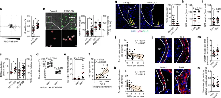
lesional SMCs, we investigated whether activated SMCs guide neu-trophils towards them. Supernatants obtained from platelet-derived growth factor-BB (PDGF-BB)-activated SMCs evoked chemotactic attraction (Fig. 2a, Extended Data Fig. 3a, b), followed by enhanced neutrophil–SMC interaction and neutrophil polarization (Fig. 2b). Because chemokine signalling is a prerequisite for neutrophil acti-vation and neutrophil extracellular trap (NET) release (NETosis)7,
we investigated whether secretory products of activated SMCs trig-ger neutrophils to undergo NETosis. Neutrophils incubated with the supernatant of PDGF-BB-treated SMCs produced increased amounts of reactive oxygen species and released NETs (Fig. 2c). These super-natants were enriched in the CCR2 ligands CCL2 and CCL7 (Fig. 2d, Extended Data Fig. 3c, d). Notably, only recombinant CCL7 evoked NET release (Fig. 2e). Furthermore, intimal CCL7 positively correlated with lesional NETs (Fig. 2f) but not with lesional neutrophil numbers (Extended Data Fig. 3e), and its blockade resulted in reduced numbers of intimal NETs (Fig. 2g). Consistent with the idea that activated SMCs promote NET release within the atherosclerotic lesion, NET-releasing neutrophils in mouse and human atherosclerotic lesions were predom-inantly found in the SMC-rich fibrous cap (Fig. 2h, i). This observation
1Institute for Cardiovascular Prevention (IPEK), LMU München, Munich, Germany. 2Department of Pathology, AMC, Amsterdam, The Netherlands. 3German Center for Cardiovascular Research (DZHK), Partner Site Munich Heart Alliance, Munich, Germany. 4Department of Biochemistry, CARIM, University Maastricht, Maastricht, The Netherlands. 5Department of Bioengineering, University of California, Los Angeles, Los Angeles, CA, USA. 6Université Grenoble Alpes, CEA, CNRS, IBS, Grenoble, France. 7Area of Developmental and Cell Biology, Fundación Centro Nacional de Investigaciones Cardiovasculares Carlos III (CNIC), Madrid, Spain. 8Institute of Experimental Cardiovascular Research, University Medical Center Hamburg-Eppendorf, Hamburg, Germany. 9German Center for Cardiovascular Research (DZHK), Partner Site Hamburg/Kiel/Lübeck, Hamburg, Germany. 10Medizinische Klinik und Poliklinik IV, LMU München, Munich, Germany. 11BMC, Metabolic Biochemistry, LMU München, Munich, Germany. 12Department of Biomedical Engineering, CARIM, University Maastricht, Maastricht, The Netherlands. 13The Scripps Research Institute, La Jolla, CA, USA. 14Institute for Experimental Immunology and Imaging, University Hospital Essen, Essen, Germany. 15Department of Vascular and Endovascular Surgery, Technical University Munich, Munich, Germany. 16Department of Medicine Solna, Karolinska Institute, Stockholm, Sweden. 17Department of Medical Biochemistry, AMC, Amsterdam, The Netherlands. 18Max Planck Institute of Biochemistry, Martinsried, Germany. 19Department of Physiology and Pharmacology (FyFa), Karolinska Institutet, Stockholm, Sweden. 20These authors contributed equally: Carlos Silvestre-Roig, Quinte Braster. 21Deceased: Kerri A. Mowen. *e-mail: [email protected]; [email protected]
raised the question of whether intimal NETosis might exert cytotoxic effects that account for the inverse relationship between the num-bers of lesional neutrophils and SMCs. Consistent with this, intimal NETs in mouse (Fig. 2j) and human (Fig. 2k) atherosclerotic lesions inversely correlated with SMC content and resided in close proximity to dead SMCs (Extended Data Fig. 3f, g). Notably, such trap–DNA structures were found to be primarily of neutrophil origin (Extended Data Fig. 3h–j). To investigate the cytotoxic effect of NETs on SMCs, SMCs were incubated with phorbol myristate acetate (PMA)- or CCL7- induced NETs (Extended Data Fig. 3k, l), which resulted in reduced cell viability. Pharmacological (Cl-amidine treatment) or genetic (mice that lacked Pad4) blockade of NET release resulted in increased lesional SMC content and decreased overall plaque vulnerability (Fig. 2l–n, Extended Data Fig. 4, Extended Data Table 1).
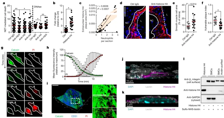
NETs are decorated with granule-derived and nuclear proteins that determine their functionality8. Antibody- and inhibitor-based blocking
of abundant NET-resident granule proteins did not affect NET-evoked cytotoxicity (Extended Data Fig. 5a, b), whereas the neutralization of histone H2A and H4 significantly rescued SMC viability (Fig. 3a). Notably, NET structures disrupted by DNase treatment retained cyto-toxicity capacity. However, under these conditions, the neutralization of H2A failed to block NET-mediated cytotoxicity, whereas the effect of histone H4 blockade was preserved (Fig. 3a). In addition, recombinant histone H4 potently induced cell death (Fig. 3b), which suggests that NET-bound and free histone H4 exhibit strong cytotoxicity (Extended Data Fig. 5c). As histone H4 was abundant in intimal NETs (Extended Data Fig. 5d) and because extranuclear histone H4 positively corre-lated with the number of intimal neutrophils (Fig. 3c, Extended Data Fig. 5e), we investigated the effect of histone H4 on the destabilization of atherosclerotic plaques. Antibody blockade of histone H4 in hyper-cholesterolemic mice with established lesions generated plaques with reduced vulnerability and increased lesional SMC content (Fig. 3d–f, Extended Data Fig. 6, Extended Data Table 1).
NET-derived histones can induce endothelial cell cytotoxicity through the activation of Toll-like receptors (TLRs)9. However,
inhi-bition of TLR signalling did not affect NET-mediated SMC death (Extended Data Fig. 7a). Instead, histone H4 induced the ultra-rapid release of cytoplasmic calcein and uptake of propidium iodide in SMCs (Fig. 3g, h, Supplementary Video 1) and whole-mount atherosclerotic lesions (Fig. 3i), suggesting a non-programmed cell-death process. Guided by previous reports that suggested the membrane activity of histone H4 in bacteria10, we proposed that histone H4 interacts with
the SMC plasma membrane to induce cell lysis. High-resolution imag-ing of SMCs with either NETimag-ing neutrophils (Fig. 3j, Extended Data Fig. 7b) or recombinant histone H4 (Fig. 3k, Extended Data Fig. 7c, Supplementary Video 2) revealed a direct interaction of histone H4 with the plasma membrane, a finding that was corroborated by biotin- assisted pull-down of membrane-bound proteins of SMCs (Fig. 3l). In accordance with a cell lysis mechanism, NETs or histone H4 induced SMC swelling and the release of ATP (Extended Data Fig. 7d–g). Given the cationic properties of histone H4 (ζ potential 26.64 ± 2.82 mV) and the anionic nature of the plasma membrane, we tested whether histone H4–membrane interaction was mediated by electrostatic forces. Manipulation of the SMC surface charge using oleylamine or choles-terol sulfate increased or decreased cationicity, respectively (Extended Data Fig. 7h), thus inhibiting or promoting histone H4–membrane anchorage (Extended Data Fig. 7i) and consequent cell death (Extended Data Fig. 7j).
The apparent disruption of the integrity of the SMC plasma mem-brane and the rapid induction of death by histone H4 prompted us to investigate the biophysical basis of histone H4-mediated membrane permeation. Atomic force microscopy on artificially reconstituted membrane bilayers incubated with histone H4 showed the appearance of pores (Fig. 4a). Similarly, when SMCs were exposed to histone H4, alterations in the membrane were observed (Extended Data Fig. 8a) as a result of dynamic bending and pore formation (Fig. 4b).
SMC P = 0.006 60 40 20 0 Distance to proximate neutrophil ( μ m) Macrophage a b c d e f g h k 10 μm 10 μm 10 μm i j 0 1 2 3 4 5 6 7 8 9 0 5 10 15 Neutrophils per section Neutrophils per section Neutrophils per section Neutrophils per section r = –0.386 P = 0.043 0 1 2 3 4 5 6 7 8 9 0 20 40 60
Macrophage area (% of intima)
r = 0.230 P = 0.239 0 1 2 3 4 5 6 7 8 9 0 20 40 60
Necrotic core area
(% of intima)
Necrotic core area
(relative to respective control)
r = 0.442 P = 0.019 0 1 2 3 4 5 6 7 8 9 0 1 2 3
Vulnerable plaque index
Vulnerable plaque index
(relative to respective control)
r = 0.429 P = 0.023 Anti-Ly6GMcl1 ΔN AMD3100Cxcr4 ΔN Anti-Ly6 G Mcl1 ΔN AMD3100Cxcr4 ΔN Anti-Ly6GMcl1 ΔN AMD3100Cxcr4 ΔN Anti-Ly6GMcl1 ΔN AMD3100Cxcr4 ΔN Anti-Ly6GMcl1 ΔN AMD3100Cxcr4 ΔN 0 1 2 3 4
Smooth muscle cell area
(relative to respective control)
SMC area (% of intima) * * * ** 0 1 2 3 Macrophage area
(relative to respective control)
* 0 1 2 3 4 * * 0 1 2 3 4 5 * * ** * 0 5 10 15 20 TUNEL
+ smooth muscle cells
(relative to respective control)
* * * ** 0.00 5 10 15 20 25 30 35 40 45 50 55 60 65 70 1.0 Distance (μm) Distance ( μm) Grey valu e Macrophage SMC Neutrophil 05 10 15 20 25 30 35 40 45 50 55 0. 0 1.0 Grey value SM CS MC Neutrophil SM C
Fig. 1 | Neutrophils dictate plaque stability. a–d, Advanced lesions were generated by insertion of a shear stress modifier around the carotid artery. Pearson correlation between lesional neutrophils and SMC area
(SMA+, a), macrophage area (CD68+, b), necrotic core area (c), and overall
vulnerability (d). n = 28 mice. Dotted line represents 95% confidence interval. e–i, Neutropenia (anti-Ly6G) or neutrophilia (AMD3100) were induced during the last 4 weeks of the experiment. Genetically neutropenic
Apoe−/− mice (Ly6gcreMcl1flox/flox, Mcl1ΔN) were fed a high-fat diet for a
total of 11 weeks and advanced atherosclerotic lesions were analysed in
aortic roots. Genetic neutrophilia was established in Apoe−/− mice that
were lethally irradiated and reconstituted with bone marrow from Lyz2cre
mice or from Lyz2creCxcr4flox/flox (Cxcr4ΔN) mice. Antibody-induced
neutropenic (anti-Ly6G, n = 10 mice (e–i), genetic neutropenic (Mcl1ΔN),
n = 16 mice (e–h), n = 10 mice (i)), pharmacological neutrophilic
(AMD3100, n = 15 mice (e–h), n = 7 mice (i)) and genetic neutrophilic
(Cxcr4ΔN, n = 13 mice (e–h), n = 11 mice (i)) are compared with respective
controls (isotype IgG, n = 10 mice (e–i), Ly6gcre, n = 18 mice (e–h), n = 10
mice (i), vehicle (n = 15 mice (e–h), n = 7 mice (i)), or Lyz2cre (n = 11
mice (e–h), n = 9 mice (i))), respectively, dashed line. Displayed is the
quantification of the SMC (SMA+) area (e), macrophage area (CD68+, f),
necrotic core area (g), and overall vulnerability (h). i, Dead SMCs were
quantified as TUNEL+SMA+ cells. For the AMD3100 condition, a
two-sided Mann–Whitney test was used. j, Representative immunofluorescence
micrograph showing lesional neutrophils (Ly6G+, grey), SMCs (SMA+, red),
macrophages (CD68+, magenta) and nuclei (DAPI, blue). Dotted
lines indicate cross-section views. The diagonal cross-section is shown at the top (xyz) and the vertical cross-section is shown on the right (yz). Intensity profiles of the indicated emission wavelengths are shown. k, Violin plot showing the distance of intimal neutrophils to macrophages
(CD68+) (n = 148 cells) and SMCs (SMA+) (n = 171 cells). The median is
represented by the horizontal line within the white box, and the boundaries of the box indicate the interquartile range. Two-sided unpaired t-tests were used unless otherwise stated; *P < 0.05; **P < 0.01; ***P < 0.001. Data are mean ± s.d.
A machine-learning classifier that was trained to identify α-helical membrane-active sequences11 revealed high σ scores within the histone
H4 N terminus (Extended Data Fig. 8b). Such scores correlate with the ability to generate negative Gaussian curvature in membranes required for pore formation, as shown for cell-penetrating polypeptides12,13. To
validate this prediction we performed small-angle X-ray scattering on small unilamellar vesicles incubated with the histone H4 N terminus, and observed strong, dose-dependent membrane deformation rich in negative Gaussian curvature (Pn3m cubic phases). Gaussian curva-ture (⟨ ⟩K ) values were comparable to those obtained from other membrane-remodelling proteins12; such remodelling is facilitated in
cholesterol-containing membranes (Fig. 4c, Extended Data Fig. 8c). Consistent with this, the histone H4 N terminus interacted with SMC membranes (Extended Data Fig. 8d) and induced cell death (Extended Data Fig. 8e).
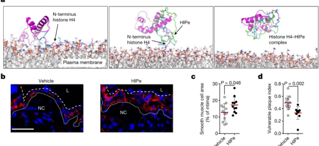
Owing to the importance of SMC death in the regulation of plaque vulnerability, we explored therapeutic strategies to prevent histone H4-driven cytotoxicity. Molecular dynamic simulation of histone H4 in the presence of membrane structures confirmed that the N-terminal region is responsible for the H4–membrane interaction (Fig. 5a). Consequently, we screened for peptides that were predicted to form stable complexes with the H4 N terminus. The cyclical peptide HIPe (histone inhibitory peptide) showed a potential ability to efficiently disturb histone H4–membrane interactions (Fig. 5a). Consistent with this prediction, HIPe prevented histone H4 from interacting with
(Extended Data Fig. 8f) and altering (Extended Data Fig. 8g, h) mem-branes, and from inducing SMC death (Extended Data Fig. 8i). We next investigated the therapeutic benefits of HIPe on atherosclerotic plaque vulnerability. The continuous administration of HIPe to mice with pre-existing atherosclerotic lesions resulted in increased lesional SMC content, and generated lesions with increased stability traits both in carotid (Fig. 5b–d, Extended Data Fig. 9a–q, Extended Data Table 1) and brachiocephalic (Extended Data Fig. 9r–x) lesions without altering neutrophil recruitment (Extended Data Fig. 9y, z).
The identification of new therapeutic strategies to inhibit cell death associated with chronic inflammation, as exemplified by cardiovascular diseases, is a major challenge in modern medicine. Here we identify a form of histone-induced death as a source of chronic inflammation within arterial walls that increases instability traits in atherosclerotic plaques. Histone H4 is released from neutrophils which are attracted and rendered prone to releasing histone-containing NETs by SMCs (Extended Data Fig. 9aa, Supplementary Video 3).
Upon danger, neutrophils rapidly infiltrate the inflammatory site and cause severe tissue injury as a collateral effect of their function. At the vascular site, rapid release of cytotoxic NETs exerts severe tissue damage, thereby reducing structural signs of stability of the atherosclerotic plaque. Luminal14 or abluminal deposition of
NET-derived products may lead to death of endothelial cells or SMCs, respectively, and hence dictate diverse aetiologies of cardiovascular pathologies. – CCL2CCL7 0 20 40 60 80
NETs per field (fold change
) P = 0.0024 Ctrl IgG Anti-CC L7 0 2 4 6 8 10 P = 0.027 a PDGF-BB SPN Ctrl PDGF-BB Ctrl PDGF-BB Ctrl PDGF-BB Ctrl PDGF-BB Ctrl PDGF-BB 0.0 0.1 0.2 0.3 0.4 Speed ( μ m s –1) * 0 100 200 300 0 50 100 150 Adherent neutrophil s Polarized neutrophils (% of total ) * **
NETs per field (relative to ctrl)
0.5 1.0 1.5 2.0 0.5 1.0 1.5 2.0 ROS productio n (relative to ctrl ) * ** Control PDGF-BB CCL2 CCL7 0 1 2 3 4 5 50 100 150 200 Chemokine (ng ml –1) P = 0.0004 P = 0.013 Ctrl PDGF-BB
CoreCap CoreCap
0 50 100 150
P = 0.011
NET location (% of total
)
NET location (% of total
) 0 50 100 150 P = 0.043 b c d e f h i j k m 0 1 2 3 4 * * 0.0 0.5 1.0 1.5 * * n g Ctrl IgG Anti-CCL7
DAPI Ly6G Cit H3
l
Pad4+/+ Pad4–/– Pad4
–/– L NC NC PBS Cl-a Cl-a Pad4 –/– Cl-a L NC L NC 0 50 100 0 2 4 6 8 10 12 CCL7 (integrated intensity)
NETs per sectio
n
NETs per sectio
n r = 0.608 P = 0.0006 00 1 2 3 6 12 18 24
NETs per section
r = –0.277 P = 0.037 0 2 4 6 8 0 2 4 6 8
NETs per section
r = –0.377 P = 0.019
L
Smooth muscle cell are
a
(% of intima)
Smooth muscle cell are
a
(% of intima)
Smooth muscle cell are
a
(relative to respective control)
Vulnerable plaque index
(relative to respective control)
Fig. 2 | Activated smooth muscle cells trigger NETosis promoting atherosclerotic plaque vulnerability. a, Tracks (left) and speed (right) of neutrophil migration towards the supernatant of PDGF-BB-activated SMCs. n = 20 ctrl; n = 19 PDGF-BB neutrophils. *P = 0.00001. b, Number of adherent neutrophils to SMCs (Ly6G, green; n = 39 ctrl,
n = 35 PDGF-BB) and their CD62L polarization frequency (red; n = 26
ctrl, n = 28 PDGF-BB). Scale bar, 50 µm. *P = 0.0002; **P = 0.009. c, Reactive oxygen species (ROS) production (left, n = 9 ctrl; n = 10 PDGF-BB) and NET release (right, n = 22 ctrl; n = 25 PDGF-BB) of neutrophils exposed to supernatants of resting (ctrl) or PDGF-BB-activated SMCs. *P = 0.004; **P = 0.008. d, CCL2 and CCL7 levels in supernatants from PDGF-BB-activated SMCs. n = 10. Two-sided paired t-test. e, NET release by VCAM-1-bound neutrophils treated with vehicle, CCL2 or CCL7, compared to non-coated control. n = 10 fields. One-way ANOVA with Holm–Sidak correction. f, Pearson correlation between NETs and intimal CCL7 in advanced atherosclerotic lesions in
mice. n = 28 sections. g, Apoe−/− mice with advanced atherosclerotic
lesions were treated with anti-CCL7 or isotype IgG and lesional NETs were quantified. Representative micrographs are shown. Ly6G, magenta;
citrullinated histone H3, green; DNA (DAPI), blue. Dashed line delineates intima. Scale bar, 100 µm. n = 44 sections IgG; n = 27 sections anti-CCL7. h, i, Percentage of NETing neutrophils present in the indicated compartments of mouse (h) or human (i) atherosclerotic lesions. n = 33 sections in h and n = 13 core and n = 16 cap in i. j, k, Pearson correlation
between NETs and SMC (SMA+) area in mouse (j, n = 57 mice) and
human (k, n = 38 sections) atherosclerotic lesions. l–n, Advanced lesions were generated by insertion of a shear stress modifier around the carotid artery. Pharmacologic (Cl-amidine, Cl-a, during last 4 weeks, n = 9 mice)
or genetic (Apoe−/−Pad4−/−, n = 8 mice) inhibition of NET release and
respective controls (PBS treatment, n = 8 mice, or Apoe−/−Pad4+/+,
n = 6 mice). Representative immunostaining images (l) show SMC
(SMA, red) and nuclei (DAPI, blue). The dotted line delineates SMCs from the necrotic core, the dashed line outlines the lumen. Scale bar, 50
µm. Quantification of SMC area (SMA+, m) and vulnerable plaque index
(n). Tested against respective control. *P < 0.05. All data are presented as mean (red line) ± s.d. L, lumen; NC, necrotic core; SPN, supernatant. Dotted lines in f, j and k represent 95% confidence interval. Two-sided unpaired t-test; n indicates biological samples unless otherwise stated.
As well as the architectural function of histones in chromatin organ-ization, histones released upon cell damage are noxious and induce tissue damage15,16. Our data indicate that histones—and in particular
histone H4—that originate from intimal NETs and possibly necrotic
cells induce rapid, receptor-independent cell death in SMCs via the formation of membrane pores, thus contributing to vascular tissue damage. Owing to the controversial antimicrobial effector function of NETs17, peptide-based histone H4 blockade could have potential as a
a 1.15 μm 0.0 μm 0.0 μm 2.8 μm 3.7 μm0.0 μm 1.25 μm 0.0 μm 0.0 μm 3.2 μm 4.3 μm0.0 μm 20 min 0 min b c 12.0 nm 8.0 4.0 0.0 Ctrl 15.0 nm 12.0 0.0 4.0 Histone H4 3.5 3.0 2.5 2.0 1.5 1.0 0.5 0.0 Average NGC (10 –2 nm –2)
Peptide/lipid charge ratio 0.0 0.5 1.0 1.5 2.0 8.0 DOPS/DOPE/ chol 20/80/0 DOPS/DOPE/ chol 20/75/5 DOPS/DOPE/ chol 20/70/10 DOPS/DOPE/ chol 20/60/20
Fig. 4 | Membrane-pore-forming activity of histone H4. a, Atomic force microscopy images of reconstituted membrane lipid bilayers incubated with recombinant histone H4. Scale bar, 1 µm. b, Live scanning ion conductance microscopy of SMCs. Images represent the plasma membrane before (top) and after (bottom) incubation with histone H4. c, Small angle X-ray scattering data demonstrate that the N-terminal domain of histone H4 induces negative Gaussian curvature (NGC) in small unilamellar
vesicles, with membrane compositions as indicated at increasing peptide:lipid ratios. The peptide induced Pn3m cubic phases, which are rich in NGC, and are indicative of membrane permeation. The plot shows the absolute value of NGC induced by histone H4 N terminus as a function of the protein:lipid charge ratio. Chol, cholesterol; DOPE,
phosphoethanolamine; DOPS,
1,2-dioleoyl-sn-glycero-3-phospho-l-serine. Ctrl IgG Ctrl Ig G Ctrl Ctrl Ig G Anti-H2AAnti-H3Anti-H Anti-H2A
4 Anti-H4 0 5 10 15 20
NET-mediated cell death
* ** ** * DNAse a Histone H4 Anti-hist one H4 Ctrl Ig G Anti-hist one H4 0 3 6 9 12
Fold change (relative to ctrl) * 0.0 0.5 1.0 1.5 * 0 10 20 30 40 P = 0.014 Calcein 0 25 50 75 100 125 Time (min)
Mean fluorescence intensity
(relative to maximum) 0 5 10 PI Calcein CD31 PI 8.5 min 17 min 0 min Anti-histone H4 Anti-GAPDH (cytosol) Anti-β1 integrin (cell surface) SMC s SMC s Avidin-purified Sulfo-NHS-biotin – + – + Histone H4 + + + + Histone H4 – + Time (min) Calcein PI 00:00 04:43 08:13 10:43 b c d e f g h i k j l Ctrl IgG Anti-histone H4 L NC L NC 10 μm S NET S
DAPI Lectin Histone H4
DAPI Lectin Histone H4
2 μm 0 1 2 3 4 0.000 0.005 0.010 0.015 0.020 0.025 Neutrophils per section Extranuclear histone H4/ plaque area (mm 2) r = 0.6936 P = 0.0007
Smooth muscle cell are
a
(% of intima
)
Vulnerable plaque index
Fig. 3 | NET-derived histone H4 induces SMC lysis and exacerbates plaque instability. a, SMCs were treated with NETs pre-incubated with antibodies to histone isoforms and cell death was measured as propidium
iodide positive (PI+) cells. n = 57 IgG; n = 58 anti-H2A; n = 59 anti-H3;
n = 58 anti-H4 fields. One-way ANOVA with Tukey’s correction;
*P = 0.014; **P = 0.007. Where indicated, NETs were treated with DNase before incubation with indicated antibodies. n = 24 fields. Two-sided unpaired t-test; *P = 0.044; **P = 0.007. b, Histone H4-induced SMC death quantified by PI uptake. n = 24 fields. Two-sided unpaired t-test,
*P = 1.27 × 10⁻⁹. c, Pearson correlation between extranuclear histone
H4 and neutrophils in intimas. n = 20 mice. Dotted line represents 95%
confidence interval. d–f, Apoe−/− mice with advanced lesions were treated
with isotype IgG (n = 14 mice) or anti-histone H4 (n = 15 mice) during the last 4 weeks of the experiment. d, Representative immunostaining for SMCs (SMA, red) and nuclei (DAPI, blue). The dotted line delineates SMCs from the necrotic core, the dashed line outlines the lumen. Scale
bar, 50 µm. e, f, Quantification of SMC area (SMA+, e) and plaque
vulnerability (f). Two-sided unpaired t-test. *P = 0.0009. g, h, Time-lapse microscopy of SMCs treated with histone H4 showing PI entry and calcein dispersion. g, Representative micrographs. Scale bar, 10 µm. h, Quantification over time. n = 15 cells. i, Time-lapse two-photon microscopy of whole-mount lesions stained with calcein (green) and anti-CD31 (blue) recorded in the presence of PI and histone H4. Scale bar, 100 µm. j, k, Visualization of cell membrane (lectin, white), histone H4 (magenta) and DNA (DAPI, cyan) in a SMC (S) and neutrophil co-culture (j) or in SMCs treated with recombinant histone H4 (k). White arrows indicate NET-derived histone H4 SMC plasma membrane interactions. l, SMCs treated with recombinant histone H4 were labelled with sulfo-NHS-SS-biotin. Western blot of non-purified lysates (SMCs) and plasma membrane fractions (SMCs avidin-purified) with indicated antibodies (for gel source data, see Supplementary Fig. 1). Data are mean ± s.d.
therapeutic strategy for the prevention of damage to vascular as well as other types of tissue, without substantially compromising NETosis-mediated host defence.
Online content
Any methods, additional references, Nature Research reporting summaries, source data, statements of data availability and associated accession codes are available at https://doi.org/10.1038/s41586-019-1167-6.
Received: 28 November 2017; Accepted: 2 April 2019; Published online 1 May 2019.
1. Kolb, J. P., Oguin, T. H., III, Oberst, A. & Martinez, J. Programmed cell death and inflammation: winter is coming. Trends Immunol. 38, 705–718 (2017). 2. Tabas, I. Macrophage death and defective inflammation resolution in
atherosclerosis. Nat. Rev. Immunol. 10, 36–46 (2010).
3. Clarke, M. C. H. et al. Apoptosis of vascular smooth muscle cells induces features of plaque vulnerability in atherosclerosis. Nat. Med. 12, 1075–1080 (2006). 4. Hartwig, H. et al. Atherosclerotic plaque destabilization in mice: a comparative
study. PLoS ONE 10, e0141019 (2015).
5. Silvestre-Roig, C. et al. Atherosclerotic plaque destabilization: mechanisms, models, and therapeutic strategies. Circ. Res. 114, 214–226 (2014). 6. Bennett, M. R., Sinha, S. & Owens, G. K. Vascular smooth muscle cells in
atherosclerosis. Circ. Res. 118, 692–702 (2016).
7. Rossaint, J. et al. Synchronized integrin engagement and chemokine activation is crucial in neutrophil extracellular trap-mediated sterile inflammation. Blood
123, 2573–2584 (2014).
8. Papayannopoulos, V. Neutrophil extracellular traps in immunity and disease.
Nat. Rev. Immunol. 18, 134–147 (2018).
9. Kumar, S. V. et al. Neutrophil extracellular trap-related extracellular histones cause vascular necrosis in severe GN. J. Am. Soc. Nephrol. 26, 2399–2413 (2015). 10. Tagai, C., Morita, S., Shiraishi, T., Miyaji, K. & Iwamuro, S. Antimicrobial
properties of arginine- and lysine-rich histones and involvement of bacterial outer membrane protease T in their differential mode of actions. Peptides 32, 2003–2009 (2011).
11. Lee, E. Y., Fulan, B. M., Wong, G. C. & Ferguson, A. L. Mapping membrane activity in undiscovered peptide sequence space using machine learning. Proc. Natl
Acad. Sci. USA 113, 13588–13593 (2016).
12. Schmidt, N. W. et al. Criterion for amino acid composition of defensins and antimicrobial peptides based on geometry of membrane destabilization. J. Am.
Chem. Soc. 133, 6720–6727 (2011).
13. Schmidt, N. W. & Wong, G. C. Antimicrobial peptides and induced membrane curvature: geometry, coordination chemistry, and molecular engineering. Curr.
Opin. Solid State Mater. Sci. 17, 151–163 (2013).
14. Franck, G. et al. Roles of PAD4 and NETosis in experimental atherosclerosis and arterial injury: implications for superficial erosion. Circ. Res. 123, 33–42 (2018).
15. Saffarzadeh, M. et al. Neutrophil extracellular traps directly induce epithelial and endothelial cell death: a predominant role of histones. PLoS ONE 7, e32366 (2012).
16. Xu, J. et al. Extracellular histones are major mediators of death in sepsis. Nat.
Med. 15, 1318–1321 (2009).
17. Menegazzi, R., Decleva, E. & Dri, P. Killing by neutrophil extracellular traps: fact or folklore? Blood 119, 1214–1216 (2012).
Acknowledgements The study was supported by the DFG (SFB914 TP B8,
SFB1123 TP A6, B5, Z1, SO876/6-1, SO876/11-1, AN372/14-3, AN372/24-1, INST409/97-1FUGG and INST409/150-1FUGG), the EKFS (2016_A118, 2017_A13), the NWO (VIDI project 91712303), the Leducq foundation, and the Vetenskapradet. Funding was also provided by grants SAF2015-65607-R to A.H., BES-2013–065550 to J.M.A., and Severo Ochoa Center of Excellence (award SEV-2015-0505) to CNIC, all from the Ministerio de Ciencia, Innovacion y Universidades. We thank O. Schengel for mouse genotyping and V. Lavilla for generating video animations. This work used the platforms of the Grenoble Instruct-ERIC Center (ISBG: UMS 3518 CNRS-CEA-UGA-EMBL) with support from FRISBI (ANR-10 INSB-05-02) and GRAL (ANR-10-LABX-49-01). E.Y.L. acknowledges support from the Systems and Integrative Biology Training Program (T32GM008185), the Medical Scientist Training Program (T32GM008042), and the Dermatology Scientist Training Program (T32AR071307) at UCLA. E.Y.L. and G.C.L.W. acknowledge an Early Career Research Grant and a Discovery Grant, respectively, from the National Psoriasis Foundation. G.C.L.W. also acknowledges support from NIH R56AI125429-01A1.
Reviewer information Nature thanks Peter Libby and the other anonymous
reviewer(s) for their contribution to the peer review of this work.
Author contributions C.S.-R. and Q.B. designed and performed experiments,
analysed data, interpreted data and wrote the manuscript. K.W., E.Y.L., J.M.T., N.B., J.W., J.M.A., G.S.S., A.F., A.O.-G., J.M., P.L., R.C., A.S., C. Winter, L.P.-O., C.P., T.S., H.H., J.v.B. and S.G.-R. contributed to data acquisition and analysis; N.P. supervised animal experimentation; F.K., R.T.A.M., H.-J.A., V.O.N., A.H., J.-L.P., G.C.L.W. and G.A.F.N. supervised specific data acquisition and analysis and provided funding; C. Weber provided scientific infrastructure, access to
Lyz2creCxcr4flox mice, and contributed to the funding of K.W.; K.A.M. provided Pad4 mice; M.G. provided Ly6gcre mice; T.H. synthetized peptides; and L.M.,
E.L. and M.D. provided human samples and contributed to data analyses. O.S. conceived and supervised the study, designed experiments and interpreted data, provided funding and wrote the paper. A.H., G.C.L.W. and G.A.F.N. contributed equally to this study.
Competing interests The authors declare no competing interests. Additional information
Extended data is available for this paper at
https://doi.org/10.1038/s41586-019-1167-6.
Supplementary information is available for this paper at https://doi.org/
10.1038/s41586-019-1167-6.
Reprints and permissions information is available at http://www.nature.com/
reprints.
Correspondence and requests for materials should be addressed to O.S.
or C.S.
Publisher’s note: Springer Nature remains neutral with regard to jurisdictional
claims in published maps and institutional affiliations.
© The Author(s), under exclusive licence to Springer Nature Limited 2019
Vehicl e
HIPe VehicleHIPe
0 10 20 30 P = 0.046 0.0 0.2 0.4 0.6 0.8 P = 0.002 a b c d Vehicle L NC HIPe L NC N-terminus histone H4 N-terminus histone H4 Plasma membrane Histone H4–HIPe complex HIPe
Vulnerable plaque index
Smooth muscle cell are
a
(% of intima)
Fig. 5 | Therapeutic disruption of the histone H4–plasma membrane interaction stabilizes atherosclerotic lesions. a, Molecular dynamics simulations of the interaction between histone H4 (magenta) and the plasma membrane. The N-terminal domain of histone H4 (cyan) exhibits membrane activity (left). Histone inhibitory peptide (HIPe, green) binds to the N terminus of histone H4 (middle) and disrupts the interaction with the cell membrane (right). b–d, Hypercholesterolemic mice with established atherosclerotic lesions were implanted with osmotic
minipumps delivering vehicle or HIPe for the last four weeks of the experiment. b, Representative immunostaining images for SMC (SMA, red) and nuclei (DAPI, blue) of atherosclerotic lesions. The dotted line delineates SMCs from the necrotic core, the dashed line outlines the
lumen. Scale bar, 50 µm. c, d, Quantification of SMC area (SMA+, c) and
overall lesion vulnerability (d). Two-sided unpaired t-test, n = 12 mice (vehicle) or n = 11 mice (HIPe).
MEthodS
Ethics statement. All mouse experiments were performed according to European guidelines for the Care and Use of Laboratory Animals. Protocols were approved by the Committee on the Ethics of Animal Experiments of the Academic Medical Center (DBC102939), Amsterdam and Regierung von Oberbayern (55.2-1-54-2532-159-2014).
Mouse procedures. For mouse experiments, statistical power calculations were performed as described at http://www.stat.uiowa.edu/~rlenth/Power/ to deter-mine sample size. Mice were assigned to groups randomly and data collection and analysis were performed blinded. Mice were housed according to institutional regulations with ad libitum access to food and water. All mice used were females
in the C57BL/6J background. Apolipoprotein E-deficient mice (Apoe−/−) were
purchased from The Jackson Laboratory. Ly6gcre mice (ref. 18) (obtained from
M.G., Institute for Experimental Immunology and Imaging, University Hospital,
University Duisburg-Essen) were intercrossed with Apoe−/− and Mcl1flox/flox mice
(ref. 18) (obtained from Y.-W. He, Department of Immunology, Duke University
Medical Center) to generate triple-mutant mice. Pad4−/− mice (obtained from
K.A.M., The Scripps Research Institute) were intercrossed with Apoe−/− mice to
generate double-mutant mice. Apoe−/− recipient mice were lethally irradiated and
reconstituted with bone marrow obtained from Apoe−/− Lyz2creCxcr4flox/flox mice or
Apoe−/− mice to generate mouse chimaeras. Vulnerable atherosclerotic lesions were
induced as described in ref. 4. In brief, eight-week-old mice were fed a high-fat diet
(HFD; 21% fat and 0.15% cholesterol) for 11 weeks. Two weeks after the initiation of the HFD, a cast was placed around the left common carotid artery. To induce neutropenia, mice received anti-Ly6G (clone 1A8) or control IgG (2A3, 50 µg, every other day, BioXcell) intraperitoneally (i.p.). Neutrophilia was induced by
daily subcutaneous injection of AMD3100 (5 mg kg−1, Tocris) or PBS as control.
Pharmacological inhibition of NET release was performed by daily subcutaneous
administration of Cl-amidine (10 mg kg−1, Essen Scientific). To block histone H4,
mice were treated with intraperitoneal injection of anti-histone H4 (20 µg per day, Biorbyt, orb225483) or control IgG (Dianova). Administration of peptide HIPe or scrambled HIPe (sHIPe) was performed by using osmotic minipumps (4 mg per kg per day, Alzet, model 2004). All treatments were performed during the last 4 weeks of the experiment to therapeutically treat pre-established atherosclerotic
plaques. For the model of spontaneous atheroprogresion, Apoe−/− mice were fed
a HFD for 16 weeks. For in vivo blockade of CCL7, 18–24 week-old Apoe−/− mice
were injected with anti-CCL7 or isotype IgG (5 µg i.p., 3 times) over the last two days before euthanasia. To assess the effect of HIPe on neutrophil recruitment and mobilization, mice receiving either vehicle control or HIPe (100 µg, i.v.) were sub-jected to peritonitis by administration of TNF (100 ng, i.p., 4 h). Neutrophil counts in peritoneum lavage, bone marrow and blood were quantified by flow cytometry. Human carotid endarterectomy specimens. Carotid endarterectomy (CEA) specimens were obtained from the vascular surgery department of the Academic Medical Center in Amsterdam. Immediately after removal, specimens were transferred to the Department of Pathology, fixed in 10% formalin and processed for paraffin embedding. Carotid atherosclerotic plaques were collected from all carotid endarterectomy surgeries performed at the Academic Medical Center in Amsterdam between 2008 and 2010. Specimens were collected according to the Code for Proper Secondary Use of Human Tissue in the Netherlands. This means that all personal information on age, use of medication, all underlying diseases is not available, and can indeed cause a potential bias. Informed consent as well as ethical approval are not required, as this is considered ‘waste material’ and can be used for science according to the Code for Proper Secondary Use of Human Tissue in the Netherlands.
Tissue processing. Mice were euthanized by ketamine/xylazine overdose, the blood was collected by heart puncture after which the mice were flushed with 20 ml of ice-cold PBS-EDTA (5 mM EDTA). Subsequently, the left common carotid artery was embedded in Tissue Tek O.C.T. compound (Sakura Finetek) for anal-ysis. Aortic arches and hearts were isolated, fixed with 4% PFA and embedded in paraffin and in Tissue Tek O.C.T. compound (Sakura Finetek), respectively. Flow cytometry. Blood was incubated with red blood cell lysis buffer (150 mM
NH4Cl, 10 mM KHCO3, 0.1 mM Na2EDTA) for 5 min at room temperature.
Leukocytes were stained with antibodies to CD45 (BioLegend, clone: 30-F11), CD11b (BioLegend, clone: M1/70), Ly6G (BioLegend, clone: 1A8), Ly6C (BioLegend, clone: Hk1.4), CD115 (eBioscience, clone: AFS98), Ly6B.2 (BIORAD, clone: 7/4) in staining buffer (20 min, 4 °C). Flow cytometry was performed using the LSR Fortessa (Beckton Dickinson) and data was analysed using FlowJo software (Beckton Dickinson). Haematologic counts were determined with the ScilVet ABC Plus analyser (scil animal care company GmbH).
Histology and immunofluorescence. Carotid (7 µm) and aortic root (4 µm) cryosections or aortic arch paraffin sections (4 µm) were histologically stained with haematoxylin and eosin (H&E) in 70, 40, or 40 µm intervals, respectively. Total collagen content was assessed by Pricrosirius Red staining in consecutive sections. For immunofluorescence staining, cryosections were fixed with cold
acetone followed by antigen blockade using 5% goat serum/phosphate buffered saline. Paraffin sections underwent antigen retrieval with citrate buffer (10 mM, pH 6.0) before antigen blockade using 5% goat serum/phosphate buffered saline. Next, sections were incubated overnight at 4 °C with the following primary anti-bodies: rabbit anti-mouse CD68 (Abcam, 1:200), rat anti-mouse Ly6G (BD, 1:200), mouse anti-mouse smooth muscle actin (SMA)-FITC or -Cy3 conjugated (Sigma, 1:500), rabbit anti-mouse histone H4 (Abcam, 1:200), rabbit anti-mouse histone H4 Alexa 488 conjugated (Abcam, 1:200), rabbit anti-mouse citrullinated histone H3 (Abcam, 1:200), rat anti-mouse CD31-Alexa 455 conjugated (BioLegend, 1:50), rat anti-mouse VCAM-1 (R&D, 1:100), Armenian hamster anti-mouse ICAM-1 (BD Biosciences, 1:100), rabbit anti-iNOS (Abcam,1:200),rat anti-mouse CD206-Fitc conjugated (BioLegend, 1:50), rabbit anti-mouse Ki67 (Thermo Fisher, 1:100), rab-bit mouse smooth muscle myosin heavy chain 11 (Abcam, 1:200), rabrab-bit anti-mouse CCL7 (Biorbyt, 1:100). After extensive washing, sections were incubated with secondary antibodies conjugated with Dylight 488, DyLight 550 or DyLight 650 (Thermo Fisher, 1:500). Counterstain to visualize nuclei was performed by
incubating with DAPI (Molecular Probes). Cell death (TUNEL+ cells) was detected
using ApopTag Red in situ Apoptosis Detection Kit (Millipore) following the man-ufacturer’s instructions. For staining of plasma membranes, cells were incubated with Cytopainter Phalloidin Fluor 647 (Abcam, 1:1,000) or lectin-FITC conjugated (Triticum vulgaris, Sigma-Aldrich, 1:1,000). Biotinylated fragments of histone H4 were detected using streptavidin-PE (BioLegend, 1:100). Immunofluorescence sections were imaged using a Leica TCS SP8 (Leica Microsystems) equipped with a UV laser, a freely tunable, pulsed white light laser, hybrid detectors and a 63X1.40 oil objective. Raw pictures were deconvolved with Huygens Professional (v.16.10, Scientific Volume Imaging) and maximum intensity projections of deconvolved data were generated with the Leica Application Suite X (v.3.1 Leica Microsystems). Histological sections were quantified by computer-assisted morphometric analysis using ImageJ software (National Institutes of Health).
For human samples, plaque size and necrotic core area were quantified in 4 µm paraffin sections stained with H&E. Consecutive sections were used for histological analysis of neutrophils, neutrophil extracellular traps (NETs) and smooth muscle cells (SMCs). First, antigen retrieval was performed with Laboratory Vision citrate buffer (Thermo Fisher) and sections were blocked using Laboratory Vision Ultra V-Block (Thermo Fisher). Next, sections were incubated with antibodies against CD177 (Abnova, 1:2,000), citrullinated histone H3 (1:4,000) and smooth muscle actin (1:200), followed by secondary antibodies conjugated with alkaline phos-phatase (Immunologic, 1:100) or poly-horseradish peroxidase (Southern Biotech, 1:100). Staining was developed by diaminobenzidine and Vector Blue substrate (Vector Laboratories), and counter-stained with nuclear red. Images were acquired by Philips Scanner (Philips).
To analyse extranuclear histone H4, we performed staining of nuclei (DAPI) and histone H4. Original pictures were converted into binary images (nuclei and histone H4) using ImageJ software. To obtain extranuclear histone H4, the signal from the nucleus was subtracted from the signal from histone H4.
Murine and human plaque analysis. Plaque vulnerability was assessed as
described in ref. 4 (Extended Data Fig. 1a–f). In brief, intima, media and necrotic
core area was analysed in H&E-stained sections. To assess lesion volumes, lesion area of sections was measured every 70 µm. To calculate the lesion volume, the volume of a cylinder with a different radius was calculated as:
∑
× π × π + π × + π + π + = h n n n n 1 3Intimaarea( ) Intimaarea( ) Intimaarea( 1)
Intimaarea( 1) i n 1 2 2
where h is the distance between quantified sections of a mouse and n is the number of quantified sections.
The necrotic core (NC) was defined as the area devoid of nuclei underneath a formed fibrous cap. Collagen content and fibrous cap thickness were measured on Pricosirius Red-stained sections. Fibrous cap (FC) thickness was defined as the average of length measurements in the positions overlapping with the lines of a square-shaped grid. For atherosclerotic lesions from the carotid artery but not from the brachiocephalic artery or the aortic root, averaged FC were corrected by intima size. Vulnerability Plaque Index (VPI) was calculated as VPI = (% NC area + % CD68 area) / (% SMA area + % collagen area). Mice with carotid samples showing absence of lesion formation were excluded from this analysis. To process and analyse
immunofluorescence pictures for SMA, MYH11 and CD68 were segmented by thresholding to create binary images. Binary images were mathematically
com-bined to create cell masks of identified cell populations: SMA+ MYH11+CD68−,
cell mask was created by subtracting CD68 signal from SMA-MYH11 co-localized a
rea; SMA−MYH11+CD68− cell mask was created by subtracting SMA and CD68
signal from MYH11 and CD68+MYH11−SMA− cell mask was created by
subtract-ing SMA and MYH11 signal from CD68 area. NETs were identified by combination of antibodies to Ly6G and CitH3 as well as by DAPI staining. Where stated, MPO and histone H4 were used as complementary markers (see Fig. 2g, Extended Data Figs. 3h, 5d).
For human atherosclerosis, H&E stained sections were scored blinded by two independent, experienced pathologists with little inter- and intra-observer varia-bility. Plaques were classified as early, advanced and complicated lesions regarding
Virmani histopathological classification19. Next, advanced lesions were divided into
regions of FC, shoulders and core using ImageJ software. Lesions with discontin-uous fibrous caps were not included in this study.
Lipid measurements. Plasma cholesterol and triglycerides were determined by CHOD-PAP kit (Roche/Hitachi) and GPO-PAP kit (Roche/Hitachi) respectively, according to the manufacturer’s instructions.
Cell culture and activation. Mouse vascular aorta/smooth muscle cells (MOVAS) (ATCC, CRL-2797) were cultured in complete medium (DMEM, Gibco)
supple-mented with 10% fetal bovine serum (Gibco), 0.2 mg ml−1 G418 (Invitrogen)
and 5 mM sodium pyruvate (Sigma). All cells were maintained in an incubator
at 37 °C, 5% CO2. To activate MOVAS, cells were cultured in complete medium
supplemented with 10 ng ml−1 recombinant murine PDGF-BB (Peprotech) for 6 h.
Subsequently cells were washed to remove the stimulus and maintained with fresh complete medium for 24 h. After this time, supernatants were recollected, centrifuged (300g, 5 min, 4 °C), and frozen until use. Thioglycollate-elicited mac-rophages and SVEC4-10 endothelial cells (ATCC) were cultured in complete medium (DMEM, Gibco) supplemented with 10% fetal bovine serum (Gibco). All cell lines were authenticated by ATCC and tested for mycoplasma contaminations. Multiplex ELISA of smooth muscle cell supernatants. Growth factors, cytokines and chemokines were measured in supernatants from activated SMCs using Luminex Multiplex-assays ELISA (Thermo Fisher) according to the manufactur-er’s instructions.
Neutrophil isolation. Human blood neutrophils were isolated from human blood using Polymorphprep (Axi-Shield) following the manufacturer’s instructions. Mouse bone-marrow-derived neutrophils were isolated from tibias and femurs from C57BL/6J mice by negative selection using the Neutrophil Isolation Kit (Miltenyi) according to the manufacturer’s instructions.
NET isolation. Human blood neutrophils (2 × 10⁶) were seeded for 30 min and
stimulated with 100 nM Phorbol 12-Myristate 13-Acetate (PMA) or 10 ng ml−1
of human recombinant CCL7 (Peprotech) in HBSS (Life Technologies) supple-mented with 5 mM HEPES (Gibco) for 4 h. Next, neutrophils were washed and
incubated for 30 min with DMEM containing 10 U ml−1 of restriction enzyme AluI
(New England BioLabs). Supernatants containing the NET fragments were collected and centrifuged (5 min, 300g) to remove remaining cell debris. Quant-iT PicoGreen dsDNA Assay Kit (Thermo Fisher) was used to measure DNA concen-tration according to the manufacturer’s instructions.
Cell viability assays. SMCs were incubated with indicated amounts of isolated NETs, histone H4 (Biomol), or equimolecular amounts of histone H4 fragments
(Pepscan). Endothelial cells and macrophages were incubated with 50 µg ml−1 of
histone H4 (Biomol). Cell viability was measured based on PI uptake. PI+ cells were
visualized using a climate chamber fluorescence microscope (Leica, DMi8) and quantified by ImageJ software. In some experiments, SMC viability was measured using Vybrant MTT cell proliferation assay (Thermo Fisher) according to the man-ufacturer’s instructions and measured with a plate reader (Tecan, InfiniteF200Pro). For DNase I treatment, isolated NETs were incubated with DNase I (NE Biolabs)
at 10 U ml−1 for 1 h before addition to SMCs.
Live imaging of cell death was performed on SMCs stained with Calcein AM
(Thermo Fisher, 1:1,000). After adding 50 µg ml−1 of histone H4 or phosphate
buffer (control) to the medium, images were acquired every 30 s for 30 min using a climate chamber fluorescence microscope to measure PI and calcein signal. PI influx and calcein efflux was measured using ImageJ software. For live imaging
of cell death on atherosclerotic lesions, aortas of HFD-fed Apoe−/− mice were
opened longitudinally and mounted en face on a silicone gel surface. Aortas were incubated with anti-CD31 conjugated to eFluor 450 (Thermo Fisher, 1:50) in com-bination with Calcein AM (1:1,000) for 1 h. Samples were imaged in z stacks, in intervals of 3 min for a period of 15 min using a LeicaSP5IIMP two-photon laser scanning microscope with a pre-chirped and pulsed Ti:Sapphire Laser (Spectra Physics MaiTai Deepsee) and a 20× NA1.00 (Leica) water dipping objective. Image acquisition and processing were performed using Las software (Leica).
Inhibition of NET or histone H4 cytotoxicity. Before the addition to SMCs, NETs
(500 ng ml−1) were incubated for 1 h with 50 ng ml−1 of the following antibodies:
Ctrl IgG (Santa Cruz), anti-Myeloperoxidase (Merck), anti-LL37 (Santa Cruz), anti-Neutrophil Elastase (Biorbyt), anti-Cathepsin G (Biorbyt), anti-Proteinase 3 (Santa Cruz), anti-Histone H2A (Cell Signaling Technology), anti-Histone H3 (Abcam), or anti-Histone H4 (Cell Signaling Technology). For inhibition of serine protease activity in NETs, isolated NETs were incubated with 100 nM of Myeloperoxidase Inhibitor-1 (Millipore), Elastase Inhibitor IV (Millipore) or secretory leukocyte protease (SLPI) (R&D systems) for 1 h. For Toll-like receptor
inhibition, SMCs were incubated 1 h before NET incubation with 1 µg ml−1 of
TLR1/2, TLR3 and TLR4 inhibitors (Tocris). For inhibition of histone H4
cyto-toxicity, recombinant histone H4 was incubated with 100 µg ml−1 of peptide HIPe
for 1 h before addition to SMCs.
Histone H4-membrane interaction assays. SMCs were incubated for 5 min at
4 °C to avoid internalization with 50 µg ml−1 histone H4 or equimolar amounts
of biotinylated histone H4 fragments. For inhibition with HIPe, histone H4 was
pre-treated with 100 µg ml−1 HIPe for 1 h before incubating with SMCs. Next,
cells were extensively washed and fixed with 2% PFA. Co-cultures of SMCs and neutrophils were stimulated by 100 nM PMA for 2 h. After washing, cells were incubated for another 2 h and then fixed by 2% PFA. Interaction between plasma membranes (lectin) and histone H4 was visualized using confocal microscopy. ATP measurement. SMCs were incubated with 50 µg ml−1 of recombinant histone
H4 for 1 h or 500 ng ml−1 of isolated NETs for 48 h. Supernatants were centrifuged
(5 min, 300g) and extracellular ATP was measured using Cell Titer Glo (Promega) and Tecan plate reader according to the manufacturer’s instructions.
Surface charge alteration and ζ-potential measurement. SMCs were treated for 30 min with 200 µM oleylamine or 200 µM sodium cholesterol sulfate and 1 µM STX64 sulfatase inhibitor (both purchased from Sigma). For cell viability or
membrane interaction assays, cells were washed and incubated with 50 µg ml−1
histone H4 for 1 h at 37 °C or 5 min at 4 °C, respectively. Cell viability or mem-brane interaction was measured as describe above. For ζ-potential analysis, SMCs were resuspended in 10 mM sodium chloride and 270 mM sucrose solution.
Recombinant histone H4 and BSA (control) were diluted to 0.1 mg ml−1 in a 16×
diluted PBS (Gibco) containing 30 mM sucrose. 100 µl of the sample was injected in the bottom of a disposable folded capillary cell (DTS1070, Malvern), which was prefilled with 600 µl 10 mM NaCl solution. The ζ potential was measured at 37 °C using Zetasizer Nano (Malvern).
Neutrophil extracellular trap analysis. Neutrophils (1 × 10⁵) were seeded and incubated with conditioned media obtained from control- or PDGF-BB-treated
SMCs, CCL2 (10 ng ml−1) or CCL7 (10 ng ml−1) for 4 h. In CCL2 and CCL7
exper-iments, wells were coated with human recombinant VCAM-1 (1 µg ml−1, R&D
systems). After washing, neutrophils were fixed with 4% PFA and stained with anti-mouse citrullinated histone H3, anti-anti-mouse Ly6G and DAPI. Immunofluorescence signal was measured with a fluorescent microscope or using a plate reader (Tecan, InfiniteF200Pro).
Avidin–biotin pull-down of plasma membrane proteins. SMCs cells were grown in 10-cm dishes upon 80% confluence. Cells were washed three times with PBS
and subsequently incubated with 50 µg ml−1 H4 (diluted in PBS) or vehicle and
incubated for 5 min at 4 °C. Then cells were washed 10 times with ice-cold PBS and
subsequently labelled with 250 µg ml−1 sulfo-NHS-SS-biotin (Pierce) for 30 min
on ice. The biotin-labelled cells were incubated with 150 mM glycine solution for 20 min to quench unlabelled free biotin followed by an ice-cold PBS wash. Cells were lysed in PBS Triton (1%) including complete protease inhibitor mixture (Roche), and the lysates were affinity-purified using immobilized NeutrAvidin beads (Thermo Scientific) for 3 h at 4 °C. Finally, the beads were washed with PBS Triton (1%) buffer followed by the addition of SDS–PAGE sample loading buffer and analysed by SDS–PAGE and western blotting using β1-integrin (antibody generated in the laboratory of R. Fässler, cell surface), H4 and GAPDH (Thermo Fisher, cytosol) antibodies.
Scanning electron microscopy. SMCs were incubated with 50 µg ml−1 of recom-binant histone H4 or phosphate buffer for 1 h. Next, cells were washed and the samples were fixed using 4% PFA plus 2.5% glutaraldehyde in PBS (2 h, 4 °C). Cells were then dehydrated by serial 5-min incubations in increasing concentrations of ethanol (30%, 50%, 70%, 80%, 90%, 100%). Samples were dried in an automated critical point dryer (Leica EM CDP 300) and then coated with chromium in a rotary-pumped coating system (Quorum Technologies Q150RS). Imaging was performed at 15 kV with a field emission microscope (JEOL 7600F).
Neutrophil (trans-)migration. Migration assays were performed in Zigmond cham-bers according to the manufacturer. In brief, Ly6G-PE (1A8, BioLegend) labelled neutrophils were seeded (30 min) on a collagen-coated coverslip and mounted on a glass slide chamber. A gradient of PDGF-BB activated SMC vs non-activated SMC supernatants was created and images were acquired over 30 min in 30 s intervals using a climate chamber fluorescence microscope (20× dry objective, Leica, DMi8).
Speed and displacement were calculated as previously described20.
To analyse transmigration, neutrophils (2 × 10⁵) were added to the top